Except where otherwise noted, this book is licensed under CC BY-NC-SA 4.0.
Accessing and Using the Organic and Biochemistry Supplement to Enhanced Introductory College Chemistry
1
Welcome to the Organic and Biochemistry Supplement to Enhanced Introductory College Chemistry
This book is designed to be a continuation of Enhanced Introductory College Chemistry Chapters 1-18. Formatting, chapter numbering and content reference refer to this first text.
This textbook is designed to be accessible using standard web browsers, mobile devices, screen readers and other assistive technology. You can access the book in a number of formats. Requirements, tools, and suggestions for navigating and using the book are listed on this page. If you encounter any issues in accessing the book, please connect with your professor.
Never used an Open Educational Resource (OER) before?
Refer back to web book to access links/interactive activities
Do you prefer a printed textbook?
This book is free to access, use, and print in any of the above formats for non-commercial purposes. If you prefer a printed textbook, you are encouraged to print sections/the entire book.
By Chapter PDFs here.
Printing – Recommendations
Check for printing costs at your on-campus print shop (such as Grenville at Georgian College) or a local print shop (Staples, etc)
Consider printing this textbook in black & white (not full colour), and refer to the web-book or PDF where you need to examine the colour diagrams
Printing a large document is often significantly less expensive at a print shop than it is to print on your home printer or at the Library
Ask about binding or 3 hole punching when you order, as this is usually low cost and will make your textbook easier to use
This book uses regular HTML to express simple math equations (such as <sup>, <sub>, italics and special characters) combined with Latex coding (rendered by MathJax) to prepare and display complex mathematical and chemical equations. Users of assistive technology may need to configure their software or download/activate a plugin to properly interpret the math.
Please consult with your Adaptive technologist or Accessibility advisor for assistance if necessary.
MathJax offers numerous accessibility features, including the ability to present equations in different formats, zoom, etc. Please see our Accessibility Statement for more details.
Experiencing navigation issues?
If you encounter navigation issues while accessing this text via a link from your course in Blackboard (or other learning management system), please try accessing the online web book by using the web address in your browser. The bottom left and right corners of the web book allow you to navigate through the book (previous/next) and the top left hand corner of the web book features a drop down table of contents.
Funded by the Government of Ontario.The views expressed in this publication are the views of the author(s) and do not necessarily reflect those of the Government of Ontario or the Ontario Online Learning Consortium.
This open educational resource (OER) textbook was developed collaboratively by Georgian College, and Loyalist College. Each institution generously provided additional support, with contributors noted below.
Contributors
Content Authors
Gregory A. Anderson, HBSc, MSc, PhD – Georgian College
Caryn Fahey, BSc, MEd – Loyalist College
Adrienne Richards, HBSc, MSc – Georgian College
Samantha Sullivan Sauer, HBSc, MASc, BEd – Georgian College
David Wegman, HBSc, PhD – Georgian College
Copyright, AODA & Tech Support
Jen Booth, BA, MISt – Georgian College
Additional Supports
Revathi Mahadevan – Georgian College
Jessica Jones – Georgian College
Original OER Sources
This OER, Organic and Biochemistry Supplement to Enhanced Introductory College Chemistry, is a collection of resources adapted to meet the needs of students in introductory organic chemistry courses. In most sections of this OER, updates have been made to the existing content to improve usability and accessibility, incorporate interactive elements and improve the overall student experience. Except where otherwise noted, images and diagrams are derived from the source attributed at the end of each page.
Much of the OER chemistry content available on the web is derived from a few key sources. In this OER, we’ve attempted to provide clear attribution to the sources we used, while also acknowledging those original sources. Often, the differences in derivatives from those key original sources is minor. We’ve done our best to provide accurate attribution. Ancillary resources for this textbook include an image bank in PPT format, and for this reason, we’ve attempted to attribute the images throughout the text to their original source, since they’ll be used separately from the textbook.
Attribution is complicated for these sources, due to a large amount of remixing and re purposing of chemistry content on various platforms. In some cases, pages within the same book are shared under different licenses, and this chemistry text reflects the original licenses. Individual source content is identified on each page of this text, and has been remixed as permitted by the licenses of the source text to improve reading flow, enhance student understanding, and streamline to meet the needs of Georgian and Loyalist students.
The following key sources (and their derivatives) were reused in this textbook:
Organic and Biochemistry Supplement to Enhanced Introductory College Chemistryis licensed under CC BY-NC-SA 4.0, except where otherwise noted. Individual sections, content, images and activities are marked with their relevant copyright and open licensing information.
This digital text OER has been developed with Universal Design for Learning (UDL) elements in mind. We have made every reasonable effort to ensure accessibility (AODA) and copyright compliance. We welcome your review and feedback. We encourage you to reach out to the project manager or copyright and AODA reviewer with any concerns, suggestions for modifications, and ideas for enhancements.
Please submit feedback to OER[at]georgiancollege.ca
Disclaimer
We have done our best to acknowledge all participants involved in this project. In the event, we have made an error please reach out to the project manager to have this corrected.
Acknowledgements of Land and Diversity
3
Land Acknowledgements
Georgian College
Georgian College acknowledges that all campuses are situated on the traditional land of the Anishnaabeg people. The Anishnaabeg include the Odawa, Ojibwe and Pottawatomi nations, collectively known as the Three Fires Confederacy. Georgian College is dedicated to honouring Indigenous history and culture and committed to moving forward in the spirit of reconciliation and respect with all First Nations, Métis and Inuit people.
Loyalist College is built upon the lands governed by the Dish with One Spoon wampum agreement. We affirm and thank the Haudenosaunee, Anishinaabeg, and Huron-Wendat nations for their continued caretaking of the land. We offer respect to Indigenous people from all nations who call this area home. We honour elders and traditional knowledge keepers, past, present, and future.
Commitment to Equity, Diversity, Inclusion and Indigenization in Chemistry
As with our previous OER, Enhanced Introductory College Chemistry Chapters 1-18, our commitment to equity, diversity, inclusion and indigenization is forefront and continued. In compiling this resource, the contributors have made conscious efforts to include examples of diversity of organic chemistry applications and scholars.
Except where otherwise noted, “Acknowledgement of Land and Diversity” is written by Samantha Sullivan Sauer is licensed under CC BY 4.0.
Accessibility Statement
4
Accessibility features of the web version of this resource
The web version of Organic and Biochemistry Supplement to Enhanced Introductory College Chemistry has been designed with accessibility in mind by incorporating the following features:
It has been optimized for people who use screen-reader technology.
content can be navigated using a keyboard.
links, headings, and tables are formatted to work with screen readers.
All images in this OER are described fully in the text, alt-tag or in an image description section for complex images.
Due to the complex nature of chemistry images and diagrams, alt text description may be longer than anticipated. This allows for full description of the information being conveyed by image/size/colour/labels.
Some images may not have fully descriptive alt-text due to the visual nature of exercises and questions.
If there are errors or concerns, please contact OER[at]georgiancollege.ca
Information is not conveyed by colour, bold or italics alone.
Pressbooks has built in features such as the ability to change font size.
Efforts have been made to improve the user experience in all formats – if you encounter any access issues or barriers, please connect with your professor.
Known accessibility issues and areas for improvement
This book’s adapters have attempted to improve upon existing features from the original sources and improve these materials for all users.
While we strive to ensure that this resource is as accessible and usable as possible, we might not always get it right. Any issues we identify will be listed below. If you encounter issues with this text, please notify your Professor.
List of Known Accessibility Issues
Location of Issue
Need for Improvement
Timeline
Work Around
Chapter Review Pages throughout book
Questions/answers may not be optimized in ordered lists due to limitations of the footnote tool.
unknown
Ordered lists have been used when possible. Some footnote/answers will read as a sentence with answers.
Math operators such as − (minus)
Due to the large variety of sources, there may be inconsistencies in the use of dash – or negative −, and other math operators in HTML.
as found
Replacing standard characters with math operators whenever possible/as identified.
Superscript & Subscript
Some screenreaders may not correctly read out sub or superscript numbers/etc.
unknown
Students should consult their adaptive technologist or accessibility advisor and explore settings to enable their software to identify and read characters / math correctly.
Representation of bonds
use of = to represent double bond and ≡ to represent triple bond, and – to represent single bond
unknown
none at present. Will update as possible when a workaround is found.
Alt-text for infographics
Infographics have been used throughout the book to enhance student learning, but may not be an optimal format for some users.
unknown
Textual equivalents have been created to help describe the content relayed via infographics. Links to more information have been provided where possible. Review & updating of alt-text throughout the book is an on-going project.
Alkanes, Alkenes, Alkynes and Aromatic Hydrocarbons
Alcohols and Ethers
Aldehydes, Ketones, Carboxylic Acids, and Esters
Amines and Amides
Classifying Functional Groups
To better support your learning, you should be familiar with the following concepts before starting this chapter:
The elements hydrogen and carbon.
Figure 19.0a. All organic compounds contain carbon and most are formed by living things, although they are also formed by geological and artificial processes. (credits left: modification of work by Jon Sullivan, PDM; left middle: modification of work by Deb Tremper, PDM; right middle: modification of work by annszyp, CC BY 2.0; right: modification of work by George Shuklin, PDM)
All living things on earth are formed mostly of carbon compounds. The prevalence of carbon compounds in living things has led to the epithet “carbon-based” life. The truth is we know of no other kind of life. Early chemists regarded substances isolated from organisms (plants and animals) as a different type of matter that could not be synthesized artificially, and these substances were thus known as organic compounds. The widespread belief called vitalism held that organic compounds were formed by a vital force present only in living organisms. The German chemist Friedrich Wohler was one of the early chemists to refute this aspect of vitalism, when, in 1828, he reported the synthesis of urea, a component of many body fluids, from nonliving materials. Since then, it has been recognized that organic molecules obey the same natural laws as inorganic substances, and the category of organic compounds has evolved to include both natural and synthetic compounds that contain carbon. Some carbon-containing compounds are not classified as organic, for example, carbonates and cyanides, and simple oxides, such as CO and CO2. Although a single, precise definition has yet to be identified by the chemistry community, most agree that a defining trait of organic molecules is the presence of carbon as the principal element, bonded to hydrogen and other carbon atoms.
Scientists of the 18th and early 19th centuries studied compounds obtained from plants and animals and labeled them organic because they were isolated from “organized” (living) systems. Compounds isolated from nonliving systems, such as rocks and ores, the atmosphere, and the oceans, were labeled inorganic. For many years, scientists thought organic compounds could be made by only living organisms because they possessed a vital force found only in living systems.
The word organic has different meanings. Organic fertilizer, such as cow manure, is organic in the original sense; it is derived from living organisms. Organic foods generally are foods grown without synthetic pesticides or fertilizers. Organic chemistry is the chemistry of compounds of carbon. Refer to Appendix A: Key Element Information for more details about carbon and other elements.
Carbon is unique among the other elements in that its atoms can form stable covalent bonds with each other and with atoms of other elements in a multitude of variations. The resulting molecules can contain from one to millions of carbon atoms. We previously surveyed organic chemistry by dividing its compounds into families based on functional groups. We begin with the simplest members of a family and then move on to molecules that are organic in the original sense—that is, they are made by and found in living organisms. These complex molecules (all containing carbon) determine the forms and functions of living systems and are the subject of biochemistry.
Organic compounds, like inorganic compounds, obey all the natural laws. Often there is no clear distinction in the chemical or physical properties among organic and inorganic molecules. Nevertheless, it is useful to compare typical members of each class, as in Table 19.0a.
Table 19.0a: General Contrasting Properties and Examples of Organic and Inorganic Compounds
Organic
Hexane
Inorganic
NaCl
low melting points
−95°C
high melting points
801°C
low boiling points
69°C
high boiling points
1,413°C
low solubility in water; high solubility in nonpolar solvents
insoluble in water; soluble in gasoline
greater solubility in water; low solubility in nonpolar solvents
Keep in mind, however, that there are exceptions to every category in this table. To further illustrate typical differences among organic and inorganic compounds, Table 19a also lists properties of the inorganic compound sodium chloride (common table salt, NaCl) and the organic compound hexane (C6H14), a solvent that is used to extract soybean oil from soybeans (among other uses). Many compounds can be classified as organic or inorganic by the presence or absence of certain typical properties, as illustrated in Table 19a.
The largest databaseThis is the Beilstein database, now available through the Reaxys site . of organic compounds lists about 10 million substances, which include compounds originating from living organisms and those synthesized by chemists. The number of potential organic compounds has been estimatedPeplow, Mark. “Organic Synthesis: The Robo-Chemist,” Nature 512 (2014): 20–2. at 1060—an astronomically high number. The existence of so many organic molecules is a consequence of the ability of carbon atoms to form up to four strong bonds to other carbon atoms, resulting in chains and rings of many different sizes, shapes, and complexities.
The simplest organic compounds contain only the elements carbon and hydrogen, and are called hydrocarbons. Even though they are composed of only two types of atoms, there is a wide variety of hydrocarbons because they may consist of varying lengths of chains, branched chains, and rings of carbon atoms, or combinations of these structures. In addition, hydrocarbons may differ in the types of carbon-carbon bonds present in their molecules. Many hydrocarbons are found in plants, animals, and their fossils; other hydrocarbons have been prepared in the laboratory. We use hydrocarbons every day, mainly as fuels, such as natural gas, acetylene, propane, butane, and the principal components of gasoline, diesel fuel, and heating oil. The familiar plastics polyethylene, polypropylene, and polystyrene are also hydrocarbons. We can distinguish several types of hydrocarbons by differences in the bonding between carbon atoms. This leads to differences in geometries and in the hybridization of the carbon orbitals.
As mentioned above, hydrocarbons can be man-made (in the laboratory) or naturally present. How do we know which is better for us overall? Well, it depends on the organic compound in question. Both man-made and natural products can be good or bad for us. Infographic 19.0a looks at some common misconceptions about man-made and natural chemicals.
Today, organic compounds are key components of plastics, soaps, perfumes, sweeteners, fabrics, pharmaceuticals, and many other substances that we use every day. The value to us of organic compounds ensures that organic chemistry is an important discipline within the general field of chemistry. In this chapter, we discuss why the element carbon gives rise to a vast number and variety of compounds, how those compounds are classified, and the role of organic compounds in representative biological and industrial settings. Infographic 19.0b looks at one of the roles organic compounds play in our everyday lives such as the beautiful aromas from various flowers.
Spotlight on Everyday Chemistry: The Scent of Flowers
The infographic 19.0b below demonstrates one of the many interesting characteristics of organic compounds. The specific aroma of some common flowers is due to the organic compound that makes up the flower.
19.1 Alkanes, Alkenes, Alkynes and Aromatic Hydrocarbons
1
Learning Objectives
By the end of this section, you will be able to:
Classify saturated and unsaturated hydrocarbons, and molecules derived from them
Identify alkanes, alkenes, alkynes and aromatic hydrocarbons
Alkanes
Alkanes, or saturated hydrocarbons, contain only single covalent bonds between carbon atoms. Each of the carbon atoms in an alkane has sp3 hybrid orbitals and is bonded to four other atoms, each of which is either carbon or hydrogen. The Lewis structures and models of methane, ethane, and pentane are illustrated in Figure 19.1a. Carbon chains are usually drawn as straight lines in Lewis structures, but one has to remember that Lewis structures are not intended to indicate the geometry of molecules. Notice that the carbon atoms in the structural models (the ball-and-stick and space-filling models) of the pentane molecule do not lie in a straight line. Because of the sp3 hybridization, the bond angles in carbon chains are close to 109.5°, giving such chains in an alkane a zigzag shape.
The structures of alkanes and other organic molecules may also be represented in a less detailed manner by condensed structural formulas (or simply, condensed formulas). Instead of the usual format for chemical formulas in which each element symbol appears just once, a condensed formula is written to suggest the bonding in the molecule. These formulas have the appearance of a molecular structure from which most or all of the bond symbols have been removed. Condensed structural formulas for ethane and pentane are shown at the bottom of Figure 19.1a.
Figure 19.1a Pictured are the expanded structural formulas, ball-and-stick models, and space-filling models for molecules of methane, ethane, and pentane. Below the chemical names methane, ethane and pentane represent the condensed structural formulas. (credit: Chemistry (OpenStax), CC BY 4.0).
Alkenes
Organic compounds that contain one or more double or triple bonds between carbon atoms are described as unsaturated. You have likely heard of unsaturated fats. These are complex organic molecules with long chains of carbon atoms, which contain at least one double bond between carbon atoms. Unsaturated hydrocarbon molecules that contain one or more double bonds are called alkenes. Carbon atoms linked by a double bond are bound together by two bonds, one σ bond and one π bond. Double and triple bonds give rise to a different geometry around the carbon atom that participates in them, leading to important differences in molecular shape and properties. The differing geometries are responsible for the different properties of unsaturated versus saturated fats.
Ethene, C2H4, is the simplest alkene. Each carbon atom in ethene, commonly called ethylene, has a trigonal planar structure. The second member of the series is propene (propylene) (Figure 19.1b); the butene structures follow in the series.
Figure 19.1b. The expanded molecular structures, ball-and-stick structures, and space-filling models for the alkenes ethene, propene, and 1-butene are shown (credit: Chemistry (OpenStax), CC BY 4.0).
Ethylene (the common industrial name for ethene) is a basic raw material in the production of polyethylene and other important compounds. Over 135 million tons of ethylene were produced worldwide in 2010 for use in the polymer, petrochemical, and plastic industries. Ethylene is produced industrially in a process called cracking, in which the long hydrocarbon chains in a petroleum mixture are broken into smaller molecules.
Alkynes
Hydrocarbon molecules with one or more triple bonds are called alkynes; they make up another series of unsaturated hydrocarbons. Two carbon atoms joined by a triple bond are bound together by one σ bond and two π bonds. The sp-hybridized carbons involved in the triple bond have bond angles of 180°, giving these types of bonds a linear, rod-like shape. The molecular structure for ethyne, a linear molecule, is:
The simplest member of the alkyne series is ethyne, C2H2, commonly called acetylene is represented in Figure 19.1c.
Aromatic Hydrocarbons
Figure 19.1d. The ring structure of benzene, the simplest aromatic compound. (credit: Chemistry (OpenStax), CC BY 4.0).
Benzene, C6H6, is the simplest member of a large family of hydrocarbons, called aromatic hydrocarbons. Benzene is represented in Figure 19.1d. These compounds contain ring structures and exhibit bonding that must be described using the resonance hybrid concept of valence bond theory or the delocalization concept of molecular orbital theory. (To review these concepts, refer to the earlier chapters on chemical bonding). The resonance structures for benzene, C6H6, are:
Valence bond theory describes the benzene molecule and other planar aromatic hydrocarbon molecules as hexagonal rings of sp2-hybridized carbon atoms with the unhybridized p orbital of each carbon atom perpendicular to the plane of the ring. Three valence electrons in the sp2 hybrid orbitals of each carbon atom and the valence electron of each hydrogen atom form the framework of σ bonds in the benzene molecule. The fourth valence electron of each carbon atom is shared with an adjacent carbon atom in their unhybridized p orbitals to yield the π bonds. Benzene does not, however, exhibit the characteristics typical of an alkene. Each of the six bonds between its carbon atoms is equivalent and exhibits properties that are intermediate between those of a C–C single bond and a double bond. To represent this unique bonding, structural formulas for benzene and its derivatives are typically drawn with single bonds between the carbon atoms and a circle within the ring as shown in Figure 19.1e.
Figure 19.1e. This condensed formula shows the unique bonding structure of benzene (credit: Image by Jynto, PDM).
There are many derivatives of benzene. The hydrogen atoms can be replaced by many different substituents. Aromatic compounds more readily undergo substitution reactions than addition reactions; replacement of one of the hydrogen atoms with another substituent will leave the delocalized double bonds intact.
Toluene and xylene are important solvents and raw materials in the chemical industry. Styrene is used to produce the polymer polystyrene. These molecules are shown in Figure 19.1f.
Figure 19.1f. Toluene represents a typical example of substituted benzene derivative (credit: Image by Jarozwj, PDM)
Incorporation of an oxygen atom into carbon- and hydrogen-containing molecules leads to new functional groups and new families of compounds. When the oxygen atom is attached by single bonds, the molecule is either an alcohol or ether.
Alcohols are derivatives of hydrocarbons in which an –OH group has replaced a hydrogen atom. Although all alcohols have one or more hydroxyl (–OH) functional groups, they do not behave like bases such as NaOH and KOH. NaOH and KOH are ionic compounds that contain OH– ions. Alcohols are covalent molecules; the –OH group in an alcohol molecule is attached to a carbon atom by a covalent bond.
Figure 19.2a. The formation of ethanol derived by the fermentation of yeast and sugars (credit: Chemistry (OpenStax), CC BY 4.0).
Ethanol, CH3CH2OH, also called ethyl alcohol, is a particularly important alcohol for human use. Ethanol is the alcohol produced by some species of yeast that is found in wine, beer, and distilled drinks. It has long been prepared by humans harnessing the metabolic efforts of yeasts in fermenting various sugars as illustrated in Figure 19.2a.
Figure 19.2b. Ethanol produced by the addition of water with ethylene in the presence of an acid (credit: Chemistry (OpenStax), CC BY 4.0).
Alternatively, large quantities of ethanol are synthesized from the addition reaction of water with ethylene using an acid as a catalyst as shown in Figure 19.2b.
Figure 19.2c. Alcohols containing more than one hydroxyl group such as 1,2-ethanediol and 1,2,3-propanetriol (credit: Chemistry (OpenStax), CC BY 4.0).
Alcohols containing two or more hydroxyl groups can be made. Examples include 1,2-ethanediol (ethylene glycol, used in antifreeze) and 1,2,3-propanetriol (glycerine, used as a solvent for cosmetics and medicines) as shown in Figure 19.2c.
The name of an alcohol comes from the hydrocarbon from which it was derived. The final -e in the name of the hydrocarbon is replaced by -ol, and the carbon atom to which the –OH group is bonded is indicated by a number placed before the name.
Ethers are compounds that contain the functional group –O–. Ethers do not have a designated suffix like the other types of molecules we have named so far. In the IUPAC system, the oxygen atom and the smaller carbon branch are named as an alkoxy substituent and the remainder of the molecule as the base chain, as in alkanes. As shown in the following compound, the red symbols represent the smaller alkyl group and the oxygen atom, which would be named “methoxy.” The larger carbon branch would be ethane, making the molecule methoxyethane. Many ethers are referred to with common names instead of the IUPAC system names. For common names, the two branches connected to the oxygen atom are named separately and followed by “ether.” The common name for the compound shown in Figure 19.2d is ethylmethyl ether:
Figure 19.2e. The production of diethyl ether from two molecules of ethanol (credit: Chemistry (OpenStax), CC BY 4.0).
Ethers can be obtained from alcohols by the elimination of a molecule of water from two molecules of the alcohol as illustrated in Figure 19.2e. For example, when ethanol is treated with a limited amount of sulfuric acid and heated to 140 °C, diethyl ether and water are formed as demonstrated in Figure 19.2e.
In the general formula for ethers, R—O—R, the hydrocarbon groups (R) may be the same or different. Diethyl ether, the most widely used compound of this class, is a colourless, volatile liquid that is highly flammable. It was first used in 1846 as an anesthetic, but better anesthetics have now largely taken its place. Diethyl ether and other ethers are presently used primarily as solvents for gums, fats, waxes, and resins. Tertiary-butyl methyl ether, C4H9OCH3 (abbreviated MTBE—italicized portions of names are not counted when ranking the groups alphabetically—so butyl comes before methyl in the common name), is used as an additive for gasoline. MTBE belongs to a group of chemicals known as oxygenates due to their capacity to increase the oxygen content of gasoline.
19.3 Aldehydes, Ketones, Carboxylic Acids, and Esters
3
Learning Objectives
By the end of this section, you will be able to:
Classify aldehydes, ketones, carboxylic acids and esters
The Carbonyl Group
Another class of organic molecules contains a carbon atom connected to an oxygen atom by a double bond, commonly called a carbonyl group as shown in Figure 19.3a. The trigonal planar carbon in the carbonyl group can attach to two other substituents leading to several subfamilies (aldehydes, ketones, carboxylic acids and esters) described in this section.
Both aldehydes and ketones contain a carbonyl group, a functional group with a carbon-oxygen double bond as shown in Figure 19.3a. The names for aldehyde and ketone compounds are derived using similar nomenclature rules as for alkanes and alcohols, and include the class-identifying suffixes -al and -one, respectively.
In an aldehyde, the carbonyl group is bonded to at least one hydrogen atom. In a ketone, the carbonyl group is bonded to two carbon atoms as shown in Figure 19.3b.
Figure 19.3c. The aldehyde is represented as –CHO whereas a ketone is represented as –C(O)– or –CO– (credit: Chemistry (OpenStax), CC BY 4.0).
As text, an aldehyde group is represented as –CHO; a ketone is represented as –C(O)– or –CO– as shown in the examples within Figure 19.3c.
In both aldehydes and ketones, the geometry around the carbon atom in the carbonyl group is trigonal planar; the carbon atom exhibits sp2 hybridization. Two of the sp2 orbitals on the carbon atom in the carbonyl group are used to form σ bonds to the other carbon or hydrogen atoms in a molecule. The remaining sp2 hybrid orbital forms a σ bond to the oxygen atom. The unhybridized p orbital on the carbon atom in the carbonyl group overlaps a p orbital on the oxygen atom to form the π bond in the double bond.
Like the bond in carbon dioxide, the bond of a carbonyl group is polar (recall that oxygen is significantly more electronegative than carbon, and the shared electrons are pulled toward the oxygen atom and away from the carbon atom). Many of the reactions of aldehydes and ketones start with the reaction between a Lewis base and the carbon atom at the positive end of the polar bond to yield an unstable intermediate that subsequently undergoes one or more structural rearrangements to form the final product (Figure 19.3d).
Figure 19.3d. The carbonyl group is polar, and the geometry of the bonds around the central carbon is trigonal planar (credit: Chemistry (OpenStax), CC BY 4.0).
The importance of molecular structure in the reactivity of organic compounds is illustrated by the reactions that produce aldehydes and ketones. We can prepare a carbonyl group by oxidation of an alcohol—for organic molecules, oxidation of a carbon atom is said to occur when a carbon-hydrogen bond is replaced by a carbon-oxygen bond as shown in Figure 19.3e.
Aldehydes are commonly prepared by the oxidation of alcohols (Figure 19.3f), whose –OH functional group is located on the carbon atom at the end of the chain of carbon atoms in the alcohol:
Figure 19.3f. The formation of an aldehyde from the oxidation of an alcohol (credit: Chemistry (OpenStax), CC BY 4.0).
Alcohols that have their –OH groups in the middle of the chain are necessary to synthesize a ketone (Figure 19.3g), which requires the carbonyl group to be bonded to two other carbon atoms:
An alcohol with its –OH group bonded to a carbon atom that is bonded to no or one other carbon atom will form an aldehyde. An alcohol with its –OH group attached to two other carbon atoms will form a ketone. If three carbons are attached to the carbon bonded to the –OH, the molecule will not have a C–H bond to be replaced, so it will not be susceptible to oxidation.
Formaldehyde, an aldehyde with the formula HCHO, is a colourless gas with a pungent and irritating odour. It is sold in an aqueous solution called formalin, which contains about 37% formaldehyde by weight. Formaldehyde causes coagulation of proteins, so it kills bacteria (and any other living organism) and stops many of the biological processes that cause tissue to decay. Thus, formaldehyde is used for preserving tissue specimens and embalming bodies. It is also used to sterilize soil or other materials. Formaldehyde is used in the manufacture of Bakelite, a hard plastic having high chemical and electrical resistance.
Dimethyl ketone, CH3COCH3, commonly called acetone, is the simplest ketone. It is made commercially by fermenting corn or molasses, or by oxidation of 2-propanol. Acetone is a colourless liquid. Among its many uses are as a solvent for lacquer (including fingernail polish), cellulose acetate, cellulose nitrate, acetylene, plastics, and varnishes; as a paint and varnish remover; and as a solvent in the manufacture of pharmaceuticals and chemicals.
Carboxylic Acids and Esters
The odour of vinegar is caused by the presence of acetic acid, a carboxylic acid, in the vinegar. The odour of ripe bananas and many other fruits is due to the presence of esters, compounds that can be prepared by the reaction of a carboxylic acid with an alcohol. Because esters do not have hydrogen bonds between molecules, they have lower vapor pressures than the alcohols and carboxylic acids from which they are derived (see Figure 19.3h).
Figure 19.3h. Esters are responsible for the odours associated with various plants and their fruits (credit: Chemistry (OpenStax), CC BY 4.0).
Both carboxylic acids and esters contain a carbonyl group with a second oxygen atom bonded to the carbon atom in the carbonyl group by a single bond (Figure 19.3i). In a carboxylic acid, the second oxygen atom also bonds to a hydrogen atom. In an ester, the second oxygen atom bonds to another carbon atom. The names for carboxylic acids and esters include prefixes that denote the lengths of the carbon chains in the molecules and are derived following nomenclature rules similar to those for inorganic acids and salts (see Figure 19.3i).
Figure 19.3i. The functional groups for an acid and for an ester are shown in red in these formulas (credit: Chemistry (OpenStax), CC BY 4.0).
Carboxylic acids are weak acids, meaning they are not 100% ionized in water. Generally only about 1% of the molecules of a carboxylic acid dissolved in water are ionized at any given time. The remaining molecules are undissociated in solution.
The simplest carboxylic acid is formic acid, HCO2H, known since 1670. Its name comes from the Latin word formicus, which means “ant”; it was first isolated by the distillation of red ants. It is partially responsible for the pain and irritation of ant and wasp stings, and is responsible for a characteristic odour of ants that can be sometimes detected in their nests.
Acetic acid, CH3CO2H, constitutes 3–6% vinegar. Cider vinegar is produced by allowing apple juice to ferment without oxygen present. Yeast cells present in the juice carry out the fermentation reactions. The fermentation reactions change the sugar present in the juice to ethanol, then to acetic acid. Pure acetic acid has a penetrating odour and produces painful burns. It is an excellent solvent for many organic and some inorganic compounds, and it is essential in the production of cellulose acetate, a component of many synthetic fibers such as rayon.
The distinctive and attractive odours and flavours of many flowers, perfumes, and ripe fruits are due to the presence of one or more esters. Among the most important of the natural esters are fats (such as lard, tallow, and butter) and oils (such as linseed, cottonseed, and olive oils), which are esters of the trihydroxyl alcohol glycerine, C3H5(OH)3, with large carboxylic acids, such as palmitic acid, CH3(CH2)14CO2H, stearic acid, CH3(CH2)16CO2H, and oleic acid, CH3(CH2)7CH=CH(CH2)7CO2H. Oleic acid is an unsaturated acid; it contains a C=C double bond. Palmitic and stearic acids are saturated acids that contain no double or triple bonds.
Amines are molecules that contain carbon-nitrogen bonds. The nitrogen atom in an amine has a lone pair of electrons and three bonds to other atoms, either carbon or hydrogen. Various nomenclatures are used to derive names for amines, but all involve the class-identifying suffix –ine as illustrated here for a few simple examples in Figure 19.4a.
Figure 19.4a. The simplest amines: methyl amine, dimethyl amine and trimethyl amine (credit: Chemistry (OpenStax), CC BY 4.0).
In some amines, the nitrogen atom replaces a carbon atom in an aromatic hydrocarbon. Pyridine (Figure 19.4b) is one such heterocyclic amine. A heterocyclic compound contains atoms of two or more different elements in its ring structure.
Figure 19.4b. The illustration shows one of the resonance structures of pyridine (credit: Chemistry (OpenStax), CC BY 4.0).
Like ammonia, amines are weak bases (Figure 19.4c), due to the lone pair of electrons on their nitrogen atoms:
Figure 19.4c. Methyl amine, an example, to demonstrate how amines are weak bases similar to ammonia (credit: Chemistry (OpenStax), CC BY 4.0).
The basicity of an amine’s nitrogen atom plays an important role in much of the compound’s chemistry. Amine functional groups are found in a wide variety of compounds, including natural and synthetic dyes, polymers, vitamins, and medications such as penicillin and codeine. They are also found in many molecules essential to life, such as amino acids, hormones, neurotransmitters, and DNA.
Spotlight on Everyday Chemistry: Addictive Alkaloids
Since ancient times, plants have been used for medicinal purposes. One class of substances, called alkaloids, found in many of these plants has been isolated and found to contain cyclic molecules with an amine functional group. These amines are bases. They can react with H3O+ in a dilute acid to form an ammonium salt, and this property is used to extract them from the plant:
The name alkaloid means “like an alkali.” Thus, an alkaloid reacts with acid. The free compound can be recovered after extraction by reaction with a base:
The structures of many naturally occurring alkaloids have profound physiological and psychotropic effects in humans. Examples of these drugs include nicotine, morphine, codeine, and heroin (Figure 19.4d). The plant produces these substances, collectively called secondary plant compounds, as chemical defenses against the numerous pests that attempt to feed on the plant:
Figure 19.4d. The structural images of drugs include nicotine, morphine, codeine, and heroin (credit: Chemistry (OpenStax), CC BY 4.0).
In these diagrams, as is common in representing structures of large organic compounds, carbon atoms in the rings and the hydrogen atoms bonded to them have been omitted for clarity. The solid wedges indicate bonds that extend out of the page. The dashed wedges indicate bonds that extend into the page. Notice that small changes to a part of the molecule change the properties of morphine, codeine, and heroin. Morphine, a strong narcotic used to relieve pain, contains two hydroxyl functional groups, located at the bottom of the molecule in this structural formula. Changing one of these hydroxyl groups to a methyl ether group forms codeine, a less potent drug used as a local anesthetic. If both hydroxyl groups are converted to esters of acetic acid, the powerfully addictive drug heroin results (Figure 19.4e).
Figure 19.4e. Poppies can be used in the production of opium, a plant latex that contains morphine from which other opiates, such as heroin, can be synthesized (credit: Karen Roe, Chemistry (Open Stax), CC BY 4.0).
Furthermore, in some foods that contain poppy seeds, trace amounts of codeine and morphine are present up to 48 hours in urine. In Hungary, there is a sweet roll filled with poppy seeds along with a walnut or chestnut paste, known as Bejgli often served at Christmas. For more information on the Bejgli dessert, see Compound Chemistry Advent 2023.
Amides
Amides are molecules that contain nitrogen atoms connected to the carbon atom of a carbonyl group (Figure 19.4f). Like amines, various nomenclature rules may be used to name amides, but all include use of the class-specific suffix -amide:
Figure 19.4f. Examples of amides acetamide (bottom left) and hexanamide (bottom right) with the top structure representing the amide functional group (credit: Chemistry (Open Stax), CC BY 4.0).
Amides can be produced when carboxylic acids react with amines or ammonia in a process called amidation (Figure 19.4g). A water molecule is eliminated from the reaction, and the amide is formed from the remaining pieces of the carboxylic acid and the amine.
Figure 19.4g. The reaction between a carboxylic acid and an amine produces an amide and water (credit: Chemistry (Open Stax), CC BY 4.0).
19.5 Families of Organic Molecules - Functional Groups
5
Learning Objectives
By the end of this section, you will be able to:
Identify and describe functional groups in organic molecules.
Organic molecules can be classified into families based on structural similarities. Within a family, molecules have similar physical behavior and often have predictable chemical reactivity. The structural components differentiating different organic families involve specific arrangements of atoms or bonds, called functional groups. If you understand the behavior of a particular functional group, you can describe the general properties of that class of compounds.
The simplest organic compounds are in the alkane family and contain only carbon–carbon and carbon–hydrogen single bonds but do not have any specific functional group. Hydrocarbons containing at least one carbon–carbon double bond, (denoted C=C), are in the alkene family. Alkynes have at least one carbon–carbon triple bond (C≡C). Both carbon–carbon double bonds and triple bonds chemically react in specific ways that differ from reactions of alkanes and each other, making these specific functional groups.
In the next few chapters, we will learn more about additional functional groups that are made up of atoms or groups of atoms attached to hydrocarbons. Being able to recognize different functional groups will help to understand and describe common medications and biomolecules such as amino acids, carbohydrates, and fats. Table 19.5a. below list several of the functional groups to become familiar with as you learn about organic chemistry.
The table here summarizes the structures discussed in this chapter:
Table 19.5a. Summary of the Classification of Organic Compounds.
Source: Exercise 19.5b by Samantha Sullivan Sauer is licensed under CC BY-NC 4.0
Spotlight on Everyday Chemistry: Doping in Sports
Doping in sports continues to make headlines as some athletes have turned to various drugs to enhance their performance against their opponents. Regulating bodies of professional sports monitor doping closely. The infographic looks at the major drugs used in doping.
Describe the reactions characteristic of saturated and unsaturated hydrocarbons
Common Chemical Reactions in Organic Chemistry
There are multiple types of organic chemical reactions. Some of the general organic reactions are additions, eliminations, substitutions, rearrangements and oxidation-reduction.
Addition reactions
Addition reactions occur when two reactants add together to form a single product with no atoms “left over.” An example that we’ll be studying soon is the reaction of an alkene, such as ethylene, with HBr to yield an alkyl bromide as shown in Figure 19.6a.
Chemically, the alkynes are similar to the alkenes. Since the functional group has two π bonds, alkynes typically react even more readily, and react with twice as much reagent in addition reactions. The reaction of acetylene with bromine is a typical example that is demonstrated in Figure 19.6b.
Elimination reactions are, in a sense, the opposite of addition reactions. They occur when a single reactant splits into two products, often with the formation of a small molecule such as water or HBr. An example is the acid-catalyzed reaction of an alcohol to yield water and an alkene as shown in Figure 19.6c.
Substitution reactionsoccur when two reactants exchange parts to give two new products. An example is the reaction of an ester such as methyl acetate with water to yield a carboxylic acid plus an alcohol as shown in Figure 19.6d. Similar reactions occur in many biological pathways, including the metabolism of dietary fats.
Another typical substitution reaction is one that involves alkanes, where one or more of the alkane’s hydrogen atoms is replaced with a different atom or group of atoms. No carbon-carbon bonds are broken in these reactions, and the hybridization of the carbon atoms does not change. For example, the reaction between ethane and molecular chlorine depicted in Figure 19.6e. is a substitution reaction:
Figure 19.6e. Substitution reaction between ethane and chlorine to yield chloroethane and HCl. (credit: Chemistry (OpenStax), CC BY 4.0).
Rearrangement reactions
Rearrangement reactions occur when a single reactant undergoes a reorganization of bonds and atoms to yield an isomeric product. An example as demonstrated in Figure 19.6f. is the conversion of dihydroxyacetone phosphate into its constitutional isomer glyceraldehyde 3-phosphate, a step in the glycolysis pathway by which carbohydrates are metabolized.
Figure 19.6f. Rearrangement reaction where dihydroxyacetone phosphate is converted to its constitutional isomer glyceraldehyde 3-phosphate (credit: Organic Chemistry (OpenStax), CC BY NC SA 4.0).
Oxidation-reduction reactions
Oxidation–reduction reactions, which are common in organic chemistry, can often be identified by changes in the number of oxygen atoms at a particular position in the hydrocarbon skeleton or in the number of bonds between carbon and oxygen at that position (Figure 19.6g.). An increase in either corresponds to an oxidation, whereas a decrease corresponds to a reduction (Figure 19.6h.). Conversely, an increase in the number of hydrogen atoms in a hydrocarbon is often an indication of a reduction. In Figure 19.6g, ethane is oxidized to carbon dioxide. The number of oxygen atoms and this the number of bonds between carbon and oxygen increase through the reaction so it is an oxidation of carbon reaction. In Figure 19.6h., carbon dioxide is reduced to ethane. The number of carbon-oxygen bonds is decreased; more carbon-hydrogen bonds are formed so it is a reduction of carbon reaction.
Figure 19.6g: Oxidation of ethane to carbon dioxide. (credit: Samantha Sullivan Sauer / Biovia Draw, CC BY-NC-SA 4.0)Figure 19.6h. Reduction of carbon dioxide to ethane (Chen et al, 2015). (Image credit: Samantha Sullivan Sauer / Biovia Draw, CC BY-NC-SA 4.0.
We can illustrate these points by considering how the oxidation state of the carbon atom changes in the series of compounds, which is shown in part (a) in Figure 19.6i. The number of oxygen atoms or the number of bonds to oxygen changes throughout the series. Hence the conversion of methane to formic acid is an oxidation, whereas the conversion of carbon dioxide to methanol is a reduction. Also, the number of hydrogen atoms increases in going from the most oxidized to least oxidized compound. As expected, as the oxidation state of carbon increases, the carbon becomes a more potent electrophile. Thus the carbon of CO2 is a stronger electrophile (i.e., more susceptible to nucleophilic attack) than the carbon of an alkane such as methane.
Figure 19.6i. The Oxidation State of Carbon in Oxygen- and Nitrogen-Containing Functional Groups (a) In a hydrocarbon, oxidation is indicated by an increase in the number of oxygen atoms or carbon–oxygen bonds or a decrease in the number of hydrogen atoms. (b) In nitrogen-containing compounds, the number of carbon–nitrogen bonds changes with the oxidation state of carbon. (credit: General Chem: Principles, Patterns, and Applications (Averill), CC BY-NC-SA 3.0).
Similarly, in compounds with a carbon–nitrogen bond, the number of bonds between the C and N atoms increases as the oxidation state of the carbon increases (part (b) in Figure 19.6i. In a nitrile, which contains the –C≡N group, the carbon has the same oxidation state (+2) as in a carboxylic acid, characterized by the –CO2H group. We therefore expect the carbon of a nitrile to be a rather strong electrophile.
An example of an oxidation reaction is a combustion reaction involving hydrocarbons such as alkanes. Alkanes are relatively stable molecules, but heat or light will activate reactions that involve the breaking of C–H or C–C single bonds. Combustion is one such reaction:
Alkanes burn in the presence of oxygen, a highly exothermic oxidation-reduction reaction that produces carbon dioxide and water. As a consequence, alkanes are excellent fuels. For example, methane, CH4, is the principal component of natural gas. Butane, C4H10, used in camping stoves and lighters is an alkane. Gasoline is a liquid mixture of continuous- and branched-chain alkanes, each containing from five to nine carbon atoms, plus various additives to improve its performance as a fuel. Kerosene, diesel oil, and fuel oil are primarily mixtures of alkanes with higher molecular masses. The main source of these liquid alkane fuels is crude oil, a complex mixture that is separated by fractional distillation. Fractional distillation takes advantage of differences in the boiling points of the components of the mixture. You may recall that boiling point is a function of intermolecular interactions. Acetylene and the other alkynes also burn readily. An acetylene torch takes advantage of the high heat of combustion for acetylene.
Smudging is an example of a combustion reaction. Learn more about this by looking at the Indigenous Perspective below.
Indigenous Perspectives: Smudging
Combustion reactions occur within indigenous smudging ceremonies through the burning of natural plants such as sage, sweetgrass, cedar and tobacco.
the substitution reaction of potassium cyanide with 1-chloropropane to give CH3CH2CH2CN (butyronitrile)
the addition reaction of HBr with cis-2-butene
Solution
Given: reactants, products, and reaction mechanism
Asked for: equation
Strategy: Use the mechanisms described to show how the indicated products are formed from the reactants.
The CN− ion of KCN can displace the chlorine atom of 1-chloropropane, releasing a chloride ion. Substitution results in the formation of a new C–C bond:
\( CN^{-}+\underset{1-chloropropane}{CH_{3}CH_{2}CH_{2}Cl}\rightarrow \underset{butylnitrile}{CH_{3}CH_{2}CH_{2}CN} + Cl^{-} \)
The reactions described above only cover the common reactions seen in organic chemistry, however, there are plenty more! Infographic 19.6a. provides an overview of the various types of organic chemistry reactions that can occur.
Having looked at the kinds of reactions that take place, let’s now see how they occur. An overall description of how a reaction occurs is called a reaction mechanism. A mechanism describes in detail exactly what takes place at each stage of a chemical transformation—which bonds are broken and in what order, which bonds are formed and in what order, and what the relative rates are for each step. A complete mechanism must also account for all reactants used and all products formed.
Arrows in organic chemistry reactions are important to understand as they differ depending on the type of reaction taking place. Infographic 19.6b. summarizes the various arrows used in organic chemistry reactions.
All chemical reactions involve bond-breaking and bond-making. When two molecules come together, react, and yield products, specific bonds in the reactant molecules are broken and specific bonds in the product molecules are formed. Fundamentally, there are two ways in which a covalent two-electron bond can break. A bond can break in an electronically unsymmetrical way so that both bonding electrons remain with one product fragment, leaving the other with a vacant orbital, or a bond can break in an electronically symmetrical way so that one electron remains with each product fragment. The unsymmetrical cleavage is said to be heterolytic, and the symmetrical cleavage is said to be homolytic.
Generally, the movement of two electrons in the unsymmetrical process is indicated using a full-headed curved arrow (), whereas the movement of one electron in the symmetrical process is indicated using a half-headed, or “fishhook,” arrow () as demonstrated in Figure 19.6k.
Figure 19.6k. The unsymmetrical and symmetrical bond breaking showing the full headed and half-headed arrows respectively. (credit: Organic Chemistry (OpenStax), CC BY NC SA 4.0).
Just as there are two ways in which a bond can break, there are two ways in which a covalent two-electron bond can form. A bond can form in an electronically unsymmetrical way if both bonding electrons are donated to the new bond by one reactant, or in a symmetrical way if one electron is donated by each reactant as demonstrated in Figure 19.6l.
Processes that involve unsymmetrical bond-breaking and bond-making are called polar reactions. Polar reactions involve species that have an even number of electrons and thus have only electron pairs in their orbitals. Polar processes are by far the more common reaction type in both organic and biological chemistry, and a large part of this book is devoted to their description.
Processes that involve symmetrical bond-breaking and bond-making are called radical reactions. A radical, often called a free radical, is a neutral chemical species that contains an odd number of electrons and thus has a single, unpaired electron in one of its orbitals.
In addition to polar and radical reactions, there is a third, less commonly encountered process called a pericyclic reaction.
For a visual explanation of reaction mechanism refer to the video below called Intro to Reaction Mechanisms.
Chen, C. S., Wan, J. H., & Yeo, B. S. (2015). Electrochemical reduction of Carbon Dioxide to Ethane using nanostructured Cu2O-Derived Copper Catalyst and Palladium(II) Chloride. The Journal of Physical Chemistry, 119(48), 26875-26882. DOI: 10.1021/acs.jpcc.5b09144
19.7 Introduction to Green Chemistry
7
Learning Objectives
By the end of this section, you will be able to:
Describe the principles of green chemistry
Outline the general strategy of greening a reaction
Organic chemistry reactions are essential to supporting the world’s economic progress. As a result, the chemical industry is a large user of energy and greenhouse gas emissions. Ethylene, propylene, methanol, and aromatics production account for much of the demands and production. See Compound Interest: The environmental impact of industrial reactions – in C&EN for more details.
What is Green Chemistry?
Green chemistry involves inventing new chemicals, new chemical processes and commercial products that reduce chemical hazards and minimize hazardous effects on human health and the environment. The philosophy is based on 12 principles and are summarized by four key ideas:
Prevent the formation of waste in the first place.
Employ safer reagents or solvents.
Implement selective and efficient transformations (reactions).
Avoid unnecessary transformations.
For more details on the 12 principles, view Infographic 19.7a.
The process of greening a chemical reaction involves a cyclic process that is outlined in Figure 19.7a. It starts by assessing the existing procedure including the reagents, products, by-products, solvents, reaction conditions, and efficacy. Essentially any components that are used to start, run or are produced in a reaction need to be examined.
Figure 19.7a: The process of greening a chemical process.
Examples of Green Chemistry
A search of the internet produces many examples of green chemistry. Much of today’s current research in chemistry is focused on green chemistry.
One example is shown in Infographic 19.7b, finding a new source for the production of rubber in tires.
Doxsee, K. M. & Hutchinson, J. E. (2004). Green Organic Chemistry. Thomson: Brooks-Cole.
Chapter 19 - Summary
8
19.1 Alkanes, Alkenes, Alkynes and Aromatic Hydrocarbons
Strong, stable bonds between carbon atoms produce complex molecules containing chains, branches, and rings. The chemistry of these compounds is called organic chemistry. Hydrocarbons are organic compounds composed of only carbon and hydrogen. The alkanes are saturated hydrocarbons—that is, hydrocarbons that contain only single bonds. Alkenes contain one or more carbon-carbon double bonds. Alkynes contain one or more carbon-carbon triple bonds. Aromatic hydrocarbons contain ring structures with delocalized π electron systems.
19.2 Alcohols and Ethers
Many organic compounds that are not hydrocarbons can be thought of as derivatives of hydrocarbons. A hydrocarbon derivative can be formed by replacing one or more hydrogen atoms of a hydrocarbon by a functional group, which contains at least one atom of an element other than carbon or hydrogen. The properties of hydrocarbon derivatives are determined largely by the functional group. The –OH group is the functional group of an alcohol. The –R–O–R– group is the functional group of an ether.
19.3 Aldehydes, Ethers, Carboxylic Acids and Esters
Functional groups related to the carbonyl group include the –CHO group of an aldehyde, the –CO– group of a ketone, the –CO2H group of a carboxylic acid, and the –CO2R group of an ester. The carbonyl group, a carbon-oxygen double bond, is the key structure in these classes of organic molecules: Aldehydes contain at least one hydrogen atom attached to the carbonyl carbon atom, ketones contain two carbon groups attached to the carbonyl carbon atom, carboxylic acids contain a hydroxyl group attached to the carbonyl carbon atom, and esters contain an oxygen atom attached to another carbon group connected to the carbonyl carbon atom. All of these compounds contain oxidized carbon atoms relative to the carbon atom of an alcohol group.
19.4 Amines and Amides
The addition of nitrogen into an organic framework leads to two families of molecules. Compounds containing a nitrogen atom bonded in a hydrocarbon framework are classified as amines. Compounds that have a nitrogen atom bonded to one side of a carbonyl group are classified as amides. Amines are a basic functional group. Amines and carboxylic acids can combine in a condensation reaction to form amides.
19.5 Families of Organic Molecules – Functional Groups
All chemical reactions, whether in the laboratory or in living organisms, follow the same chemical rules. To understand both organic and biological chemistry, it’s necessary to know not just what occurs but also why and how chemical reactions take place. In this chapter, we’ve taken a brief look at the fundamental kinds of organic reactions, we’ve seen why reactions occur, and we’ve seen how reactions can be described.
There are four common kinds of reactions: addition reactions take place when two reactants add together to give a single product; elimination reactions take place when one reactant splits apart to give two products; substitution reactions take place when two reactants exchange parts to give two new products; and rearrangement reactions take place when one reactant undergoes a reorganization of bonds and atoms to give an isomeric product.
Additionally, there are oxidation reactions that occur. Oxidation reactions are those that involve a reaction between hydrocarbons and oxygen producing carbon dioxide and water. An example of an oxidation reaction is combustion.
Lastly, oxidation–reduction reactions in organic chemistry are identified by the change in the number of oxygens in the hydrocarbon skeleton or the number of bonds between carbon and oxygen or carbon and nitrogen.
19.7 Introduction to Green Chemistry
Green chemistry involves inventing new chemicals, new chemical processes and commercial products that reduce chemical hazards and minimize hazardous effects on human health and the environment. The philosophy is based on 12 principles and are summarized by four key ideas: prevent the formation of waste in the first place, employ safer reagents or solvents, implement selective and efficient transformations (reactions), and avoid unnecessary transformations. Current research has many examples of green chemistry in action.
Attribution & References
Except where otherwise noted, this page is adapted by Adrienne Richards and Samantha Sullivan Sauer from
19.1 – 19.5 Alkanes, Alkenes, Alkynes and Aromatic Hydrocarbons; Alcohols and Ethers; Aldehydes, Ketones, Carboxylic Acids and Esters; Amines and Amides; and Families of Organic Molecules – Functional Groups
Classify each compound as organic or inorganic. Check answersa) organic b) inorganic c) inorganic d) organic
C3H8O
CaCl2
Cr(NH3)3Cl3
C30H48O3N
Which compound is likely organic and which is likely inorganic? Check answersa) organic b) inorganic
a flammable compound that boils at 80°C and is insoluble in water
a compound that does not burn, melts at 630°C, and is soluble in water
Classify each compound as organic or inorganic. Check answersa) organic b) inorganic c) organic
C6H10
CoCl2
C12H22O11
Classify each compound as organic or inorganic.
CH3NH2
NaNH2
Cu(NH3)6Cl2
Which member of each pair has a higher melting point? Check answersa) NaOH b) KCl
CH3OH and NaOH
CH3Cl and KCl
Which member of each pair has a higher melting point?
C2H6 and CoCl2
CH4 and LiH
Locate and identify the functional groups in the following molecules.
Classify each of the following reactions as an addition, elimination, substitution, or rearrangement: Check answersa) CH3OH + KBr b) H2C=CH2 + KBr c) CH3CH3
1. CH3Br + KOH ⟶
2. CH3CH2Br ⟶
3. H2C=CH2 + H2 ⟶
Identify the nucleophile and the electrophile in the nucleophilic substitution reaction of 2-bromobutane with KCN. Check answerCN− is the nucleophile, and C2H5Cδ+HBrCH3 is the electrophile.
Identify the nucleophile and the electrophile in the nucleophilic substitution reaction of 1-chloropentane with sodium methoxide.
Do you expect an elimination reaction to be favoured by a strong or a weak base? Why?
Why do molecules with π bonds behave as nucleophiles when mixed with strong electrophiles?
Sketch the mechanism for the nucleophilic substitution reaction of potassium cyanide with iodoethane.
Sketch the mechanism for the nucleophilic substitution reaction of NaSH with 1-bromopropane.
Sketch the mechanism for the elimination reaction of cyclohexylchloride with potassium ethoxide. Identify the electrophile and the nucleophile in this reaction.
What is the product of the elimination reaction of 1-bromo-2-methylpropane with sodium ethoxide?
Write the structure of the product expected from the electrophilic addition of HBr to cis-3-hexene.
Write the structure of the product expected from the electrophilic addition of 1-methylcyclopentene to HBr. Identify the electrophile and the nucleophile, and then write a mechanism for this reaction.
Write a synthetic scheme for making propene from propane. After synthesizing propene, how would you make 2-bromopropane?
Write a synthetic scheme for making ethylene from ethane. After synthesizing ethylene, how would you make iodoethane?
From the high-temperature reaction of Br2 with 3-methylpentane, how many monobrominated isomers would you expect to be produced? Which isomer is produced from the most stable radical? Check answer four; 3-bromo-3-methylpentane
For the photochemical reaction of Cl2 with 2,4-dimethylpentane, how many different monochlorinated isomers would you expect to be produced? Which isomer is produced from the most stable precursor radical?
How many different radicals can be formed from the photochemical reaction of Cl2 with 3,3,4-trimethylhexane? Check answer seven
How many monobrominated isomers would you expect from the photochemical reaction of Br2 with
isobutene?
2,2,3-trimethylpentane?
Arrange acetone, ethane, carbon dioxide, acetaldehyde, and ethanol in order of increasing oxidation state of carbon.
What product(s) do you expect from the reduction of a ketone? the oxidation of an aldehyde?
What product(s) do you expect from the reduction of formaldehyde? the oxidation of ethanol? Check answer methanol; acetaldehyde, followed by acetic acid and finally CO2
Create your own organic nomenclature quiz to identify functional groups using Organic Nomenclature (orgchem101.com). You can customize the types of questions you receive and get instant feedback.
Attribution & References
Except where otherwise noted, this page is adapted by Adrienne Richards from:
Infographic about natural and man-made chemicals. A common misconception is that all man-made chemicals are harmful and all natural chemicals are good for us. However, many natural chemicals are just as harmful to human health, if not more so, than man-made chemicals.
Toxic effects seen at 1000mg/kg of body weight:
Natural:
Muscimol: found in fly agaric mushrooms
Solanine: found in green potatoes
Amygdalin: found in apple seeds
Man-made:
Ethylene glycol: Used in anti-freeze.
Aspirin: used as a pain-reliving drug.
Sodium thiopental: formerly used for lethal injections.
No toxic effects seen at 1000mg/kg of body weight:
Natural:
Sucrose: also known as table sugar.
Water: essential for life.
Citric acid: found in lemons and limes.
Man-made:
Teflon (PTFE): used in non-stick pans.
Propylene glycol: food additive-solvent, humectant and thickener.
Aspartame: artificial sweetener.
“Everything is poison, there is poison in everything. Only the dose makes a thing not a poison.” Paracelsus, 1493-1541, “The father of toxicology”.
Any substance, if given in large enough amounts, can cause death. Some are lethal after only a few nanograms, whilst others require kilograms to achieve a lethal dose.
Chemical toxicology is a sliding scale, not black and white – and whether a chemical is naturally occurring or man-made tells us nothing about its toxicity.
19.0b The chemical compounds behind the smell of flowers
Infographic on aroma compounds in common flowers. A wide range of compounds contribute to the scent of flowers. The following is a majority of the common flowers and a broad overview of their components, with chemical structure images. Note that the volatile aroma compounds can vary significantly between species.
Doping in sports has been in the news in the run up to the Olympics. What drugs will doping tests at the Olympics be looking for? See below some of the major groups of drugs used in doping, their effects, and why athletes might take them.
Anabolic agents: The largest class of prohibited drugs, and the most commonly detected. Anabolic steroids mimic the hormone testosterone, increasing muscle mass and physical strength. This class also includes some non-steroidal drugs. They have a range of side effects. 74 named banned agents. 48% of positive tests in 2014.
Stimulants: Stimulants are used to improve alertness, attention and energy. Many behave similarly to the hormones adrenaline and noradrenaline. They include amphetamines. Taking them can increase blood pressure and cause cardiac problems. 66 named banned agents. 15% of positive tests in 2014.
Hormones and modulators: A range of drugs which generally interfere with human hormones, including meldonium. They can be used with anabolic steroids, to suppress some of the undesirable effects of these drugs. Some affect oestrogen levels in the body, whereas others affect human metabolism. 20 named banned agents. 5% of positive tests in 2014.
Diuretics and masking agents: This includes furosemide. Diuretics remove fluids from the body and can be used by athletes to regular their body mass, as well as diluting urine so lower levels of banned substances are registered in tests. Masking agents are drugs taken to conceal the presence of illegal drugs in urine samples. 22 named banned agents. 13% positive tests in 2014.
Infographic on a short guide to arrows in chemistry.
Chemical reaction arrows:
Reaction arrow: These arrows point from the reactants to the products of a chemical reaction. Reaction conditions, reagents or catalysts may be written above or below the reaction arrow.
Multiple arrow: Chemists use stacked multiple arrows to indicate that there are several reaction steps between the reagents and the products shown on either side of the arrows.
Broken arrow (an arrow with an ‘x’ through it or an arrow with slanting parallel lines): Chemists use these arrows to indicate chemical reactions that do not take place. The reactants shown cannot be transformed into the products shown.
Reversible reaction arrow (two arrows running parallel pointing opposite directions): Chemists use these arrows to indicate that a reaction is reversible – the reactants react to produce the products, but the products can also react to make the reactants.
Equilibrium arrow ( two parallel half-headed arrows pointing opposite directs): These arrows show that a reversible reaction is at equilibrium: the forward and reverse reactions occur at the same rate. The length of the arrows can be varied to show if reactants or products are favoured.
Retrosynthesis arrow (two parallel lines with an arrow head pointing in one direction): Organic chemists us these arrows to show that the molecule on the left can be made from the stating material on the right, often through several reaction steps.
Electron movement arrows:
Resonance arrow (one double headed arrow pointing opposite directions): Chemists use these arrows to show different resonance forms of the same molecule. The forms differ in electron arrangements; the true structure of the molecule is an average.
Curly arrow (an arched arrow or half headed arched arrow): Curly arrows show electron movement in reaction mechanisms in organic chemistry. A double headed arrow shows the movement of an electron pair, while a single headed arrow shows the movement of a single electron.
19.7a The Twelve Principles of Green Chemistry: What it is, & Why it Matters
Green chemistry is an approach to chemistry that aims to maximize efficiency and minimize hazardous effects on human health and the environment. While no reaction can be perfectly “green”, the overall negative impact of chemistry research and the chemical industry can be reduced by implementing the 12 Principles of Green Chemistry whenever possible.
Waste prevention: Prioritize the prevention of waste, rather than cleaning up and treating waste after it has been created. Plan ahead to minimize waste at every step.
Atom economy: Reduce waste at the molecular level by maximizing the number of atoms from all reagents that are incorporated into the final product. Use atom economy to evaluate reaction efficiency.
Less hazardous chemical synthesis: Design chemical reactions and synthetic routes to be as safe as possible. Consider the hazards of all substances handled during the reaction, including waste.
Designing safer chemicals: Minimize toxicity directly by molecular design. Predict and evaluate aspects such as physical properties, toxicity, and environmental fate throughout the design process.
Safer solvents and auxiliaries: Choose the safest solvent available for any given step. Minimize the total amount of solvents and auxiliary substances used, as these make up a large percentage of the total waste created.
Design for energy efficiency: Choose the least energy-intensive chemical route. Avoid heating and cooling, as well as pressurized and vacuum conditions (i.e. ambient temperature and pressure are optimal).
Use of renewable feedstocks: Use chemicals which are made from renewable (i.e. plant-based) sources, rather than other, equivalent chemicals originating from petrochemical sources.
Reduce derivatives: Minimize the use of temporary derivatives such as protecting groups. Avoid derivatives to reduce reaction steps, resources required, and waste created.
Catalysis: Use catalytic instead of stoichiometric reagents in reactions. Choose catalysts to help increase selectivity, minimize waste, and reduce reaction times and energy demands.
Design for degradation: Design chemicals that degrade and can be discarded easily. Ensure that both chemicals and their degradation products are not toxic, bioaccumulative, or environmentally persistent.
Real-time pollution prevention: Monitor chemical reactions in real-time as they occur to prevent the formation and release of any potentially hazardous and polluting substances.
Safer chemistry for accident prevention: Choose and develop chemical procedures that are safer and inherently minimize the risk of accidents. Know the possible risks and assess them beforehand.
19.7b Dandelion chemistry: Diuretics and the tyres of the future
Dandelions’ Medicinal Uses
The Frech name for dandelion is pissenlit (‘wet the bed’) from their supposed ability to act as a diuretic, increasing the production of urine. Research attributes this to several diuretic compounds, but evidence for the effect is mixed. Dandelions’ high potassium content helps replace potassium lost through urine.
Potassium content of dandelion leaves versus bananas: Dandelion leaves 397 mg per 100 g. Bananas 358 mg per 100 g. (Source: US Department of Agriculture – FoodData Central)
Studies show dandelion extracts or compounds have anti-inflammatory, anti-carcinogenic and anti-oxidative actions. These effects are mostly due to polyphenols and sesquiterpenese, also responsible for the bitter flavour of the leaves.
Taraxinic acid β-D-glucopyranoside – A sesquiterpene lactone in dandelion, also thought to be a contact allergen.
Rubber from Dandelions
The sticky white liquid that seeps out from dandelion stems when they’re picked contains a natural latex, which can be turned into rubber. The roots of Russian dandelions (Taraxacum koksaghzy) contain a particularly high percentage of latex, making them ideal for rubber production.
Percentage of the USSR’s rubber provided by the Russian dandelion during rubber shortages in World War II. 1941: 30%
Main constituent of rubber: cis-1,4-polyisoprene
In the past decade, tyre manufacturers have been developing dandelion rubber tyres. Currently bike tyres made from dandelion rubber are commercially available and tyres for cars and trucks will be available within ten years.
Except where otherwise noted, content on this page has been created as a textual summary of the infographics used within our OER. Please refer to the original website (noted below each description) for further details about the image.
Chapter 20: Alkanes and Alkyl Halides
II
Organic and Biochemistry Supplement to Enhanced Introductory College Chemistry
by Gregory Anderson; Jen Booth; Caryn Fahey; Adrienne Richards; Samantha Sullivan Sauer; and David Wegman
General concepts of organic chemistry (from Chapter 19: Organic Chemistry)
As you just learned, there is a wide variety of organic compounds containing different functional groups. However, all organic compounds are hydrocarbons, they contain hydrogen and carbon. The general rule for hydrocarbons is that any carbon must be bonded to at least one other carbon atom, except in the case of methane which only contains one carbon. The bonded carbons form the backbone of the molecule to which the hydrogen atoms (or other functional groups) are attached. Refer to Appendix A: Key Element Information for more details about carbon.
Hydrocarbons with only carbon-to-carbon single bonds (C–C) are called alkanes (or saturated hydrocarbons). Saturated, in this case, means that each carbon atom is bonded to four other atoms (hydrogen or carbon)—the most possible; there are no double or triple bonds in these molecules.
Saturated fats and oils are organic molecules that do not have carbon-to-carbon double bonds (C=C).
The three simplest alkanes—methane (CH4), ethane (C2H6), and propane (C3H8) shown in Figure 20.0a., are the beginning of a series of compounds in which any two members in a sequence differ by one carbon atom and two hydrogen atoms—namely, a CH2 unit (called methylene). Alkanes follow the general formula: CnH2n+2. Using this formula, we can write a molecular formula for any alkane with a given number of carbon atoms. For example, an alkane with eight carbon atoms has the molecular formula C8H(2 × 8) + 2 = C8H18.
An understanding of the physical properties of the alkanes is important in that petroleum and natural gas and the many products derived from them—gasoline, bottled gas, solvents, plastics, and more—are composed primarily of alkanes. This understanding is also vital because it is the basis for describing the properties of other organic and biological compound families. For example, large portions of the structures of lipids consist of nonpolar alkyl groups as shown in Figure 20.0b. Lipids include the dietary fats and fatlike compounds called phospholipids and sphingolipids that serve as structural components of living tissues. These compounds have both polar and nonpolar groups, enabling them to bridge the gap between water-soluble and water-insoluble phases. This characteristic is essential for the selective permeability of cell membranes.
Figure 20.0b. Tripalmitin (a), a typical fat molecule, has long hydrocarbon chains typical of most lipids. Compare these chains to hexadecane (b), an alkane with 16 carbon atoms (credit: Intro Chem: GOB (V. 1.0). ,CC BY-NC-SA 3.0).
20.1 Characteristics of Alkanes
11
Learning Objectives
By the end of this section, you will be able to:
Understand carbon-hydrogen bonding
Identify and name simple (straight-chain) alkanes given formulas and write formulas for straight-chain alkanes given their names.
Identify the physical properties of alkanes and describe trends in these properties.
Alkanes
We begin our study of organic chemistry with the hydrocarbons, the simplest organic compounds, which are composed of carbon and hydrogen atoms only. As we noted, there are several different kinds of hydrocarbons. They are distinguished by the types of bonding between carbon atoms and the properties that result from that bonding.
The bonding in the hydrogen molecule is fairly straightforward, but the situation is more complicated in organic molecules with tetravalent carbon atoms. Take methane, CH4, for instance. As we’ve seen, carbon has four valence electrons (2s2 2p2) and forms four bonds. Because carbon uses two kinds of orbitals for bonding, 2s and 2p, we might expect methane to have two kinds of C–H bonds. In fact, though, all four C–H bonds in methane are identical and are spatially oriented toward the corners of a regular tetrahedron as shown in Figure 20.1a. How can we explain this?
Figure 20.1a. A representation of van’t Hoff’s tetrahedral carbon atom. The solid lines represent bonds in the plane of the paper, the heavy wedged line represents a bond coming out of the plane of the page toward the viewer, and the dashed line represents a bond going back behind the plane of the page away from the viewer (credit: Organic Chemistry (OpenStax),CC BY-NC-SA 4.0).
An answer was provided in 1931 by Linus Pauling, who showed mathematically how an s orbital and three p orbitals on an atom can combine, or hybridize, to form four equivalent atomic orbitals with tetrahedral orientation. Shown in Figure 20.1b., these tetrahedrally oriented orbitals are called sp3 hybrid orbitals. Note that the superscript 3 in the name sp3 tells how many of each type of atomic orbital combine to form the hybrid, not how many electrons occupy it.
Figure 20.1b. Four sp3 hybrid orbitals, oriented toward the corners of a regular tetrahedron, are formed by the combination of an s orbital and three p orbitals (red/blue). The sp3 hybrids have two lobes and are unsymmetrical about the nucleus, giving them a directionality and allowing them to form strong bonds to other atoms (credit: Organic Chemistry (OpenStax),CC BY-NC-SA 4.0).
The concept of hybridization explains how carbon forms four equivalent tetrahedral bonds but not why it does so. The shape of the hybrid orbital suggests the answer to why. When an s orbital hybridizes with three p orbitals, the resultant sp3 hybrid orbitals are unsymmetrical about the nucleus. One of the two lobes is larger than the other and can therefore overlap more effectively with an orbital from another atom to form a bond. As a result, sp3 hybrid orbitals form stronger bonds than do unhybridized s or p orbitals.
The asymmetry of sp3 orbitals arises because, as noted previously, the two lobes of a p orbital have different algebraic signs, + and –, in the wave function. Thus, when a p orbital hybridizes with an s orbital, the positive p lobe adds to the s orbital but the negative p lobe subtracts from the s orbital. The resultant hybrid orbital is therefore unsymmetrical about the nucleus and is strongly oriented in one direction.
When each of the four identical sp3 hybrid orbitals of a carbon atom overlaps with the 1s orbital of a hydrogen atom, four identical C–H bonds are formed and methane results. Each C–H bond in methane has a strength of 439 kJ/mol (105 kcal/mol) and a length of 109 pm. Because the four bonds have a specific geometry, we also can define a property called the bond angle. The angle formed by each H–C–H is 109.5°, the so-called tetrahedral angle. Methane thus has the structure shown in Figure 20.1c.
The flat representations shown do not accurately portray bond angles or molecular geometry. Methane has a tetrahedral shape that chemists often portray with wedges indicating bonds coming out toward you and dashed lines indicating bonds that go back away from you. An ordinary solid line indicates a bond in the plane of the page. Recall that the VSEPR theory correctly predicts a tetrahedral shape for the methane molecule (Figure 20.1d.).
By using the PhET Interactive Simulation, view the CH4 molecule by selecting the Real Molecules option. From the drop-down menu, select the compound CH4 then select Show Lone Pairs, Bond Angles, Electron Geometry, and Molecular Geometry. Alternatively, you can build your own CH4 molecule by selecting the Model option.
Hydrocarbons with only carbon-to-carbon single bonds (C–C) and existing as a continuous chain of carbon atoms also bonded to hydrogen atoms are called alkanes (or saturated hydrocarbons). Saturated, in this case, means that each carbon atom is bonded to four other atoms (hydrogen or carbon)—the most possible; there are no double or triple bonds in the molecules. The word saturatedhas the same meaning for hydrocarbons as it does for the dietary fats and oils: the molecule has no carbon-to-carbon double bonds (C=C).
The three simplest alkanes—methane (CH4), ethane (C2H6), and propane (C3H8) and they are shown in Figure 20.1e.
Methane (CH4), ethane (C2H6), and propane (C3H8) are the beginning of a series of compounds in which any two members in a sequence differ by one carbon atom and two hydrogen atoms—namely, a CH2 unit. The first 10 members of this series are given in Table 20.1a.
Table 20.1a. The First 10 Straight-Chain Alkane Formulas
Consider the series in Figure 20.1f. The sequence starts with C3H8, and a CH2 unit is added in each step moving up the series. Any family of compounds in which adjacent members differ from each other by a definite factor (here a CH2 group) is called a homologous series. The members of such a series, called homologs, have properties that vary in a regular and predictable manner. The principle of homology gives organization to organic chemistry in much the same way that the periodic table gives organization to inorganic chemistry. Instead of a bewildering array of individual carbon compounds, we can study a few members of a homologous series and from them deduce some of the properties of other compounds in the series.
Figure 20.1f. Members of a Homologous Series. Each succeeding formula incorporates one carbon atom and two hydrogen atoms more than the previous formula(credit: Intro Chemistry: GOB (V. 1.0)., CC BY-NC-SA 4.0).
The principle of homology allows us to write a general formula for alkanes: CnH2n+ 2. Using this formula, we can write a molecular formula for any alkane with a given number of carbon atoms. For example, an alkane with eight carbon atoms has the molecular formula C8H(2 × 8) + 2 = C8H18.
The video What is Organic Chemistry below gives an introduction to alkanes.
Because alkanes have relatively predictable physical properties and undergo relatively few chemical reactions other than combustion, they serve as a basis of comparison for the properties of many other organic compound families. Let’s consider their physical properties first.
Table 20.1b. describes some of the properties of some of the first 10 straight-chain alkanes. Because alkane molecules are nonpolar, they are insoluble in water, which is a polar solvent, but are soluble in nonpolar and slightly polar solvents. Consequently, alkanes themselves are commonly used as solvents for organic substances of low polarity, such as fats, oils, and waxes. Nearly all alkanes have densities less than 1.0 g/mL and are therefore less dense than water (the density of H2O is 1.00 g/mL at 20°C). These properties explain why oil and grease do not mix with water but rather float on its surface as demonstrated in Figure 20.1g.
Table 20.1b. Physical Properties of Some Alkanes
Molecular Name
Formula
Melting Point (°C)
Boiling Point (°C)
Density (20°C)*
Physical State (at 20°C)
methane
CH4
–182
–164
0.668 g/L
gas
ethane
C2H6
–183
–89
1.265 g/L
gas
propane
C3H8
–190
–42
1.867 g/L
gas
butane
C4H10
–138
–1
2.493 g/L
gas
pentane
C5H12
–130
36
0.626 g/mL
liquid
hexane
C6H14
–95
69
0.659 g/mL
liquid
octane
C8H18
–57
125
0.703 g/mL
liquid
decane
C10H22
–30
174
0.730 g/mL
liquid
*Note the change in units going from gases (grams per liter) to liquids (grams per milliliter). Gas densities are at 1 atm pressure.
Table 20.1b. indicates that the first four members of the alkane series are gases at ordinary temperatures. Natural gas is composed chiefly of methane, which has a density of about 0.67 g/L. The density of air is about 1.29 g/L. Because natural gas is less dense than air, it rises. When a natural-gas leak is detected and shut off in a room, the gas can be removed by opening an upper window. On the other hand, bottled gas can be either propane (density 1.88 g/L) or butanes (a mixture of butane and isobutane; density about 2.5 g/L). Both are much heavier than air (density 1.2 g/L). If bottled gas escapes into a building, it collects near the floor. This presents a much more serious fire hazard than a natural-gas leak because it is more difficult to rid the room of the heavier gas.
Also shown in Table 20.1b are the boiling points of the straight-chain alkanes increase with increasing molar mass. This general rule holds true for the straight-chain homologs of all organic compound families. Larger molecules have greater surface areas and consequently interact more strongly; more energy is therefore required to separate them. For a given molar mass, the boiling points of alkanes are relatively low because these nonpolar molecules have only weak dispersion forces to hold them together in the liquid state.
Figure 20.1g. Oil Spills. Crude oil coats the water’s surface in the Gulf of Mexico after the Deepwater Horizon oil rig sank following an explosion. The leak was a mile below the surface, making it difficult to estimate the size of the spill. One liter of oil can create a slick 2.5 hectares (6.3 acres) in size. This and similar spills provide a reminder that hydrocarbons and water don’t mix. (Credit: NASA Goddard / MODIS Rapid Response Team, NASA terms of use).
Spotlight on Everyday Chemistry: Characteristics of Alkanes
Hydrocarbons are the simplest organic compounds, but they have interesting physiological effects. These effects depend on the size of the hydrocarbon molecules and where on or in the body they are applied. Alkanes of low molar mass—those with from 1 to approximately 10 or so carbon atoms—are gases or light liquids that act as anesthetics. Inhaling (“sniffing”) these hydrocarbons in gasoline or aerosol propellants for their intoxicating effect is a major health problem that can lead to liver, kidney, or brain damage or to immediate death by asphyxiation by excluding oxygen.
Swallowed, liquid alkanes do little harm while in the stomach. In the lungs, however, they cause “chemical” pneumonia by dissolving fatlike molecules from cell membranes in the tiny air sacs (alveoli). The lungs become unable to expel fluids, just as in pneumonia caused by bacteria or viruses. People who swallow gasoline or other liquid alkane mixtures should not be made to vomit, as this would increase the chance of getting alkanes into the lungs. (There is no home-treatment antidote for gasoline poisoning; call a poison control center.)
Figure 20.1h. Butane is a hydrocarbon with the formula [latex]ce{C4H10}\) and is a gas at room temperature and atmospheric pressure. Under excess pressures, it can be liquidized like the fuel shown in this butane lighters. (Credit: work by Frank Vincentz, CC BY-SA 3.0)
Liquid alkanes with approximately 5–16 carbon atoms per molecule wash away natural skin oils and cause drying and chapping of the skin, while heavier liquid alkanes (those with approximately 17 or more carbon atoms per molecule) act as emollients (skin softeners). Such alkane mixtures as mineral oil and petroleum jelly can be applied as a protective film. Water and aqueous solutions such as urine will not dissolve such a film, which explains why petroleum jelly protects a baby’s tender skin from diaper rash.
Attribution & References
Except where otherwise noted, this page is adapted by Adrienne Richards from:
Write condensed structural formulas for alkanes given complete structural formulas.
Draw line structures given condensed or structural formulas.
Types of Formulas to Represent Hydrocarbons
We use several kinds of formulas to describe organic compounds. A molecular formula shows only the kinds and numbers of atoms in a molecule. The general formula for alkanes: CnH2n+ 2. Using this formula, we can write a molecular formula for any alkane with a given number of carbon atoms. Table 20.2a. shows the molecular formulas of the first 10 straight-chain alkanes.
Table 20.2a. The First 10 Straight-Chain Alkane Molecular Formulas
For example in Table 20.2a. above, the molecular formula C4H10 tells us there are 4 carbon atoms and 10 hydrogen atoms in a molecule, but it doesn’t distinguish between butane and isobutane. A structural formula shows all the carbon and hydrogen atoms and the bonds attaching them. Thus, structural formulas identify the specific isomers (learned in the next section) by showing the order of attachment of the various atoms.
Unfortunately, structural formulas are difficult to type/write and take up a lot of space. Chemists often use condensed structural formulas to alleviate these problems. The condensed structural formulas for the first 10 alkanes are demonstrated in Table 20.2a. Parentheses in condensed structural formulas indicate that the enclosed grouping of atoms is attached to the adjacent carbon atom. The condensed formulas show hydrogen atoms right next to the carbon atoms to which they are attached, as illustrated in Figure 20.2a. for butane:
The ultimate condensed formula is a line structure formula. In this type of structure, carbon atoms are not symbolized with a C, but represented by each end of a line or bend in a line. Hydrogen atoms are not drawn if they are attached to a carbon. Other atoms besides carbon and hydrogen are represented by their elemental symbols. For example in Figure 20.2b., we can represent pentane (CH3CH2CH2CH2CH3) and isopentane [(CH3)2CHCH2CH3] as follows:
Drawing organic molecules can be challenging. There is software available to assist with drawing molecules. Refer to Appendix B: Organic Chemistry Software for more details and tutorials.
In summary, the following Figure 20.2c. demonstrates the structural (expanded), condensed and line structure formulas.
Figure 20.2c. Structural representations for butane and its isomer, 2-methylpropane. (The colours are used to help identify carbons and do not represent any special properties.) (credit: Fundamentals of GOB ( McMurry et al.),CC BY-NC-SA 3.0)
For a summary chart of the formulae used in organic chemistry, infographic 20.2a. demonstrates the different ways to represent organic compounds. Molecular and empirical formulas are explained as the simplest followed by condensed, structural and skeletal (or line) formulae.
Each carbon atom is converted into the end of a line or the place where lines intersect. All hydrogen atoms attached to the carbon atoms are left out of the structure (although we still need to recognize they are there):
Identify the molecular formula of the molecule represented here:
Solution
There are eight places where lines intersect or end, meaning that there are eight carbon atoms in the molecule. Since we know that carbon atoms tend to make four bonds, each carbon atom will have the number of hydrogen atoms that are required for four bonds. This compound contains 16 hydrogen atoms for a molecular formula of C8H16.
Identify the location of each carbon atom in the DDT structure. DDT is dichlorodiphenyltrichloroethane. It contains two aromatic rings connected by one carbon of an ethane structure. The molecular formula is C14H9Cl5. (Structure is shown in as a line structure.)
Check Your Answer:Carbons are highlighted in yellow in this image. There are 14 carbon atoms. Six in each aromatic ring, one that connects the rings together and one that branches off the middle carbon.
Activity source: “Identifying carbons in line structure” by Samantha Sullivan Sauer, licensed under CC BY-NC 4.0
Links to Enhanced Learning
For an interactive practice link to the interactive activity on Hydrocarbons to determine the number of hydrogens in an organic compound from Jessica Anderson.
Attribution & References
Except where otherwise noted, this page is adapted by Adrienne Richards from
Identify simple alkanes as straight-chain or branched-chain.
Describe and recognize structural and functional group isomers.
Name alkanes by the IUPAC system and write formulas for alkanes given IUPAC names
Isomers
Hydrocarbons with the same formula, including alkanes, can have different structures. For example, two alkanes have the formula C4H10: They are called n-butane and 2-methylpropane (or isobutane), and have the following structural formulas as shown in Figure 20.3a:
Figure 20.3a. Structural and ball-and-stick formulas for butane and 2-methylpropane (credit: General Chemistry 1 & 2 , CC BY 4.0).
The compounds n-butane and 2-methylpropane are structural isomers (the term constitutional isomers is also commonly used). Constitutional isomers have the same molecular formula but different spatial arrangements of the atoms in their molecules. The n-butane molecule contains an unbranched chain, meaning that no carbon atom is bonded to more than two other carbon atoms. We use the term normal, or the prefix n, to refer to a chain of carbon atoms without branching. The compound 2–methylpropane has a branched chain (the carbon atom in the center of the structural formula is bonded to three other carbon atoms).
Recall from section 20.2, Table 20.3a. which shows the molecular formula and condensed structural formulas for the first 10 straight-chain alkanes. Table 20.3a also shows the number of isomers for each alkane. The number of isomers increases rapidly as the number of carbon atoms increases.
Table 20.3a. The First 10 Straight-Chain Alkane Formulas and Isomers
Identifying isomers from structural formulae is not as easy as it looks. Structural formulae that look different may actually represent the same isomers. For example, the three structures in Figure 20.3b all represent the same molecule, n-butane, and hence are not different isomers. They are identical because each contains an unbranched chain of four carbon atoms.
Figure 20.3b. These three representations of the structure of n-butane are not isomers because they all contain the same arrangement of atoms and bonds (credit: General Chemistry 1 & 2 , CC BY 4.0.)
When identifying isomers, it is useful to trace the carbon backbone with your finger or a pencil and count carbons until you need to lift your hand or pencil to get the another carbon. Try this with each of the above arrangements of four carbons above in Figure 20.3b. Butane has a continuous chain of four carbons no matter how the bonds are rotated – you can connect the carbons in a line without lifting your finger from the page. In a later portion of this chapter, you will learn how to systematically name compounds by counting the number of carbons in the longest continuous chain and identifying any functional groups present.
Adding one more carbon to the butane chain gives pentane, which has the formula, C5H12. Pentane and its two branched-chain isomers are shown below in Figure 20.3c. The compound at the far left is pentane because it has all five carbon atoms in a continuous chain. The compound in the middle is isopentane; like isobutane, it has a one CH3 branch off the second carbon atom of the continuous chain. The compound at the far right, discovered after the other two, was named neopentane (from the Greek neos, meaning “new”). Although all three have the same molecular formula, they have different properties, including boiling points: pentane, 36.1°C; isopentane, 27.7°C; and neopentane, 9.5°C. The names isopentane and neopentane are common names for these molecules. As mentioned above, we will learn the systematic rules for naming compounds next.
Looking at Table 20.3a., there are 3 pentanes, 5 hexanes, 9 heptanes, and 18 octanes. It would be difficult to assign unique individual names that we could remember. A systematic way of naming hydrocarbons and other organic compounds has been devised by the International Union of Pure and Applied Chemistry (IUPAC). These rules, used worldwide, are known as the IUPAC System of Nomenclature. A stem name (Table 20.3b.) indicates the number of carbon atoms in the longest continuous chain (LCC). Atoms or groups attached to this carbon chain, called substituents, are then named, with their positions indicated by numbers. For now, we will consider only those substituents called alkyl groups.
Table 20.3b. Stems That Indicate the Number of Carbon Atoms in Organic Molecules
An alkyl group is a group of atoms that results when one hydrogen atom is removed from an alkane. The group is named by replacing the -ane suffix of the parent hydrocarbon with -yl. For example, the -CH3 group derived from methane (CH4) results from subtracting one hydrogen atom and is called a methyl group. The alkyl groups we will use most frequently are listed in Table 20.3c. Alkyl groups are not independent molecules; they are parts of molecules that we consider as a unit to name compounds systematically.
Table 20.3c. Common Alkyl Groups
Parent Alkane
Alkyl Group
Condensed Structural Formula
methane
methyl
CH3–
ethane
ethyl
CH3CH2–
propane
propyl
CH3CH2CH2–
isopropyl
(CH3)2CH–
butane
butyl*
CH3CH2CH2CH2–
Table 20.3c. note: *There are four butyl groups, two derived from butane and two from isobutane. We will introduce the other three where appropriate. (Image credits: Introduction to Chemistry: GOB (V. 1.0). , CC BY-NC-SA 3.0).
Simplified IUPAC rules for naming alkanes are as follows (demonstrated in Example 20.3a.).
Name alkanes according to the LCC (longest continuous chain) of carbon atoms in the molecule (rather than the total number of carbon atoms). This LCC, considered the parent chain, determines the base name, to which we add the suffix –ane to indicate that the molecule is an alkane.
If the hydrocarbon is branched, number the carbon atoms of the LCC. Numbers are assigned in the direction that gives the lowest numbers to the carbon atoms with attached substituents. Hyphens are used to separate numbers from the names of substituents; commas separate numbers from each other. (The LCC need not be written in a straight line; for example, the LCC in the following has five carbon atoms.) Figure 20.3d. An example showing that finding the longest carbon chain does not have to be in a straight line. (Credit: Introduction to Chemistry: GOB (V. 1.0). ,CC BY-NC-SA 3.0.)
Place the names of the substituent groups in alphabetical order before the name of the parent compound. If the same alkyl group appears more than once, the numbers of all the carbon atoms to which it is attached are expressed. If the same group appears more than once on the same carbon atom, the number of that carbon atom is repeated as many times as the group appears. Moreover, the number of identical groups is indicated by the Greek prefixes di-, tri-, tetra-, and so on. These prefixes are not considered in determining the alphabetical order of the substituents. For example, ethyl is listed before dimethyl; the di- is simply ignored. The last alkyl group named is prefixed to the name of the parent alkane to form one word.
When these rules are followed, every unique compound receives its own exclusive name. The rules enable us to not only name a compound from a given structure but also draw a structure from a given name. The best way to learn how to use the IUPAC system is to put it to work, not just memorize the rules. It’s easier than it looks.
Example 20.3a
Name the molecule whose structure is shown here:
Solution
The longest carbon chain runs horizontally across the page and contains six carbon atoms (this makes the base of the name hexane, but we will also need to incorporate the name of the branch). In this case, we want to number from right to left (as shown by the blue numbers) so the branch is connected to carbon 3 (imagine the numbers from left to right—this would put the branch on carbon 4, violating our rules). The branch attached to position 3 of our chain contains two carbon atoms (numbered in red)—so we take our name for two carbons eth- and attach -yl at the end to signify we are describing a branch. Putting all the pieces together, this molecule is 3-ethylhexane.
The LCC has five carbon atoms, and so the parent compound is pentane (rule 1). There is a methyl group (rule 2) attached to the second carbon atom of the pentane chain. The name is therefore 2-methylpentane.
The LCC has six carbon atoms, so the parent compound is hexane (rule 1). Methyl groups (rule 2) are attached to the second and fifth carbon atoms. The name is 2,5-dimethylhexane.
The LCC has eight carbon atoms, so the parent compound is octane (rule 1). There are methyl and ethyl groups (rule 2), both attached to the fourth carbon atom (counting from the right gives this carbon atom a lower number; rule 3). The correct name is thus 4-ethyl-4-methyloctane.
In drawing structures, always start with the parent chain.
The parent chain is butane, indicating four carbon atoms in the LCC.
Then add the groups at their proper positions. You can number the parent chain from either direction as long as you are consistent; just don’t change directions before the structure is done. The name indicates two methyl (CH3) groups, one on the second carbon atom and one on the third.
Finally, fill in all the hydrogen atoms, keeping in mind that each carbon atom must have four bonds.
The parent chain is heptane in this case, indicating seven carbon atoms in the LCC. –C–C–C–C–C–C–C–
Adding the groups at their proper positions gives
Filling in all the hydrogen atoms gives the following condensed structural formulas:
Note that the bonds (dashes) can be shown or not; sometimes they are needed for spacing.
Name cycloalkanes given their formulas and write formulas for these compounds given their names.
Cylcoalkanes
The hydrocarbons we have encountered so far have been composed of molecules with open-ended chains of carbon atoms. When a chain contains three or more carbon atoms, the atoms can join to form ring or cyclic structures. The simplest of these cyclic hydrocarbons has the formula C3H6. Each carbon atom has two hydrogen atoms attached (Figure 20.4a.) and is called cyclopropane.
Spotlight on Everyday Chemistry: Cyclopropane (C3H6) as an Anesthetic
With its boiling point of −33°C, cyclopropane is a gas at room temperature. It is also a potent, quick-acting anesthetic with few undesirable side effects in the body. It is no longer used in surgery, however, because it forms explosive mixtures with air at nearly all concentrations. A line structure of cyclopropane is shown in Figure 20.4b.
The cycloalkanes—cyclic hydrocarbons with only single bonds—are named by adding the prefix cyclo- to the name of the open-chain compound having the same number of carbon atoms as there are in the ring. Thus the name for the cyclic compound C4H8 is cyclobutane. The carbon atoms in cyclic compounds can be represented by line structure formulas that result in regular geometric figures. Keep in mind, however, that each corner of the geometric figure represents a carbon atom plus as many hydrogen atoms as needed to give each carbon atom four bonds. Figure 20.4 demonstrates the line structure formulas of both cyclopropane and cyclohexane.
Some cyclic compounds have substituent groups attached. Example 20.4a interprets the name of a cycloalkane with a single substituent group.
Example 20.4a
Draw the structure for each compound.
cyclopentane
methylcyclobutane
Solution
The name cyclopentane indicates a cyclic (cyclo) alkane with five (pent-) carbon atoms. It can be represented as a pentagon. (Credit: Intro Chem: GOB (V. 1.0). ,CC BY-NC-SA 3.0. )
The name methylcyclobutane indicates a cyclic alkane with four (but-) carbon atoms in the cyclic part. It can be represented as a square with a CH3 group attached. (Credit: Intro Chem: GOB (V. 1.0). ,CC BY-NC-SA 3.0.)
The properties of cyclic hydrocarbons are generally quite similar to those of the corresponding open-chain compounds. So cycloalkanes (with the exception of cyclopropane, which has a highly strained ring) act very much like noncyclic alkanes. Cyclic structures containing five or six carbon atoms, such as cyclopentane and cyclohexane, are particularly stable. We will see later that some carbohydrates (sugars) form five- or six-membered rings in solution.
The cyclopropane ring is strained because the C–C–C angles are 60°, and the preferred (tetrahedral) bond angle is 109.5°. (This strain is readily evident when you try to build a ball-and-stick model of cyclopropane; see Figure 20.4a.) Cyclopentane and cyclohexane rings have little strain because the C–C–C angles are near the preferred angles. Cyclohexane is shown in Figure 20.4b.
Substituted Cycloalkanes
We’ll see numerous instances in future chapters where the chemistry of a given functional group is affected by being in a ring rather than an open chain. Because cyclic molecules are encountered in most pharmaceuticals and in all classes of biomolecules, including proteins, lipids, carbohydrates, and nucleic acids, it’s important to understand the behaviour of cyclic structures.
Although we’ve only discussed open-chain compounds up to now, most organic compounds contain rings of carbon atoms. Chrysanthemic acid in Figure 20.4c, for instance, whose esters occur naturally as the active insecticidal constituents of chrysanthemum flowers, contains a three-membered (cyclopropane) ring.
Prostaglandins, potent hormones that control an extraordinary variety of physiological functions in humans, contain a five-membered (cyclopentane) ring. An example of a prostaglandin is in Figure 20.4d.
Steroids, such as cortisone, contain four rings joined together—three six-membered (cyclohexane) and one five-membered.
Figure 20.4e. The chemical structure shown is Cortisone which shows four substituted rings joined together. There are three cyclohexanes and one cyclopentane (credit: Organic Chemistry (OpenStax), CC BY-NC-SA 4.0).
Substituted cycloalkanes are named by rules similar to those we saw for open-chain alkanes. For most compounds, there are only two steps.
Find the parent. Count the number of carbon atoms in the ring and the number in the largest substituent. If the number of carbon atoms in the ring is equal to or greater than the number in the substituent, the compound is named as an alkyl-substituted cycloalkane. If the number of carbon atoms in the largest substituent is greater than the number in the ring, the compound is named as a cycloalkyl-substituted alkane. Refer to Figure 20.4f. for examples of substituted cycloalkanes. Figure 20.4f. Alkyl substituted cycloalkanes methylcyclopentane and 1-cyclopropyl butane. (credit: Organic Chemistry (OpenStax), CC BY-NC-SA 4.0)
Number the substituents and write the name. For an alkyl- or halo-substituted cycloalkane, choose a point of attachment as carbon 1 and number the substituents on the ring so that the second substituent has as low a number as possible. If ambiguity still exists, number so that the third or fourth substituent has as low a number as possible, until a point of difference is found as shown in the example within Figure 20.4g.
(a) When two or more different alkyl groups are present that could potentially take the same numbers, number them by alphabetical priority, ignoring numerical prefixes such as di- and tri-. An example is demonstrated in Figure 20.4h.
Structures a, b, and c are the same. 1,2-dimethylcyclohexane. Structures d, e, and f are the same. 1,3-dimethylcyclohexane. Ring number start at the location that results in the lowest number for all substituents and can proceed in a clockwise or counterclockwise direction.
Activity source: Exercise 20.4b is created by Samantha Sullivan Sauer, using images from Biovia Draw, licensed under CC BY-NC 4.0
Cis-Trans Isomerism of Cycloalkanes
In many respects, the chemistry of cycloalkanes is like that of open-chain alkanes: both are nonpolar and fairly inert. There are, however, some important differences. One difference is that cycloalkanes are less flexible than open-chain alkanes. In contrast with the relatively free rotation around single bonds in open-chain alkanes, there is much less freedom in cycloalkanes. Cyclopropane, for example, must be a rigid, planar molecule because three points (the carbon atoms) define a plane. No bond rotation can take place around a cyclopropane carbon–carbon bond without breaking open the ring as shown in Figure 20.4k.
Figure 20.4k. Bond rotation in ethane and cyclopropane. (a) Rotation occurs around the carbon–carbon bond in ethane, but (b) no rotation is possible around the carbon–carbon bonds in cyclopropane without breaking open the ring.(credit: Organic Chemistry (OpenStax), CC BY-NC-SA 4.0)
Larger cycloalkanes have increasing rotational freedom, and very large rings (C25 and up) are so floppy that they are nearly indistinguishable from open-chain alkanes. The common ring sizes (C3–C7), however, are severely restricted in their molecular motions.
Because of their cyclic structures, cycloalkanes have two faces when viewed edge-on, a “top” face and a “bottom” face. As a result, isomerism is possible in substituted cycloalkanes. For example, there are two different 1,2-dimethylcyclopropane isomers, one with the two methyl groups on the same face of the ring and one with the methyl groups on opposite faces as shown in Figure 20.4l. Both isomers are stable compounds, and neither can be converted into the other without breaking and reforming chemical bonds.
Figure 20.4l. There are two different 1,2-dimethylcyclopropane isomers, one with the methyl groups on the same face of the ring (cis) and the other with the methyl groups on opposite faces of the ring (trans). The two isomers do not interconvert (credit: Organic Chemistry (OpenStax), CC BY-NC-SA 4.0).
Unlike the constitutional isomers butane and isobutane, which have their atoms connected in a different order, the two 1,2-dimethylcyclopropanes have the same order of connections but differ in the spatial orientation of the atoms. Such compounds, with atoms connected in the same order but differing in three-dimensional orientation, are called stereochemical isomers, or stereoisomers. As we saw previously, the term stereochemistry is used generally to refer to the three-dimensional aspects of structure and reactivity. Figure 20.4m. demonstrates the difference between the types of isomers.
The 1,2-dimethylcyclopropanes are members of a subclass of stereoisomers called cis–trans isomers as shown in Figure 20.4n. The prefixes cis– (Latin “on the same side”) and trans– (Latin “across”) are used to distinguish between them. Cis–trans isomerism is a common occurrence in substituted cycloalkanes and in many cyclic biological molecules.
Figure 20.4n. The cis and trans isomers are shown through cis-1, 3-Dimethylcyclobutane and trans-1-Bromo-3-ethylcyclopentane (credit: Organic Chemistry (OpenStax), CC BY-NC-SA 4.0).
Example 20.4b
Naming Cycloalkanes
Name the following substances, including the cis– or trans– prefix:
In these views, the ring is roughly in the plane of the page, a wedged bond protrudes out of the page, and a dashed bond recedes into the page. Two substituents are cis if they are both out of or both into the page, and they are trans if one is out of and one is into the page.
Solution
(a)trans-1,3-Dimethylcyclopentane
(b)cis-1,2-Dichlorocyclohexane
For a more in-depth look at cycloalkanes, watch the video Cyclohexanes as shown below.
Name halogenated hydrocarbons given formulas and write formulas for these compounds given names.
Halogenated Alkanes
Many organic compounds are closely related to the alkanes. As we noted previously, alkanes react with halogens to produce halogenated hydrocarbons, the simplest of which have a single halogen atom substituted for a hydrogen atom of the alkane. Even more closely related are the cycloalkanes, compounds in which the carbon atoms are joined in a ring, or cyclic fashion.
Halogens are found in column 7A of the periodic table and include Fluorine, Chlorine, Bromine and Iodine. Refer to Appendix A: Key Element Information for more details about halogens.
Figure 20.5a. The gases released during volcanic eruptions contain large amounts of organohalides, including chloromethane, chloroform, dichlorodifluoromethane, and many others. (credit: modification of “Tavurvur volcano” by Taro Taylor edit by Richard Bartz, CC BY 2.0).
Alkyl halides are encountered less frequently than their oxygen-containing relatives and are not often involved in the biochemical pathways of terrestrial organisms, but some of the kinds of reactions they undergo—nucleophilic substitutions and eliminations—are encountered frequently. Thus, alkyl halide chemistry is a relatively simple model for many mechanistically similar but structurally more complex reactions found in biomolecules.
Now that we’ve covered the chemistry of hydrocarbons, it’s time to start looking at more complex substances that contain elements in addition to C and H. We’ll begin by discussing the chemistry of organohalides, compounds that contain one or more halogen atoms.
Halogen-substituted organic compounds are widespread in nature, and more than 5000 organohalides have been found in algae and various other marine organisms. Chloromethane, for instance, is released in large amounts by ocean kelp, as well as by forest fires and volcanoes. Halogen-containing compounds also have an array of industrial applications, including their use as solvents, inhaled anesthetics in medicine, refrigerants, and pesticides as shown in Figure 20.5b.
Figure 20.5b. Examples of halogen containing compounds trichloroethylene (a solvent), halothane (an inhaled anesthetic), tetrafluoropropene (a refrigerant), and bromomethane (a fumigant) (credit: Organic Chemistry (OpenStax), CC BY-NC-SA 4.0).
Still other halo-substituted compounds are used as medicines and food additives. The nonnutritive sweetener sucralose, marketed as Splenda, contains three chlorine atoms, for instance. Sucralose is about 600 times as sweet as sucrose, so only 1 mg is equivalent to an entire teaspoon of table sugar. Refer to Figure 20.5c. for the structural formula of sucralose.
A large variety of organohalides are known. The halogen might be bonded to an alkynyl group (C≡C−X), a vinylic group (C═C–XC═C–X">C═C–X), an aromatic ring (Ar−X), or an alkyl group.
The reactions of alkanes with halogens produce halogenated hydrocarbons, compounds in which one or more hydrogen atoms of a hydrocarbon have been replaced by halogen atoms (F, Cl, Br and I):
The replacement of only one hydrogen atom gives an alkyl halide (or haloalkane) as shown in Figure 20.5d. above.
Indigenous Perspectives: The Impact of Fluorocarbons on Inuit People of Canada
The long-chained fluorocarbon ending with a -SO3H group is known as perfluorooctanesulfonic acid also known as PFOS (Figure 20.5e.). PFOS and their relatives contain a long fluorocarbon backbone that is extremely resistant to decomposition. In addition, the PFOS family of compounds are volatile and can spread throughout the Earth’s surface. Research has shown that through global distillation, these PFOS have accumulated in the Arctic. This is a problematic health concern for the Inuit in the north. Several food sources that are part of the Inuit diet have been tested and contain PFOS. Exposure to PFOS have numerous health implications such as cancer, endocrine delays and others as described in the article below. There is no end in sight from exposure to these harmful fluorocarbons as they are expected to remain in the arctic for hundreds and possibly thousands of years (Anderson & Rayner-Canham, 2022).
Figure 20.5e. The ball-and-stick model of perfluorooctanesulfonic acid. (credit: Image by Jynto, CC0)
Although commonly called alkyl halides, halogen-substituted alkanes are named systematically as haloalkanes, treating the halogen as a substituent on a parent alkane chain. There are three steps:
Find the longest chain, and name it as the parent. If a double or triple bond is present, the parent chain must contain it.
Number the carbons of the parent chain beginning at the end nearer the first substituent, whether alkyl or halo. Assign each substituent a number according to its position on the chain. Figure 20.5f. provides two examples on how to number the carbon chain with either an alkyl or halo first appearing in the chain. Figure 20.5f. Numbering the carbon chain using 5-Bromo-2,4-dimethylheptane and 2-Bromo-4,5-deimethylheptane as examples (credit: Organic Chemistry (OpenStax), CC BY-NC-SA 4.0).
If different halogens are present, number each one and list them in alphabetical order when writing the name as shown in Figure 20.5g. Figure 20.5g. Numbering the carbon chain 1-Bromo-3-chloro-4-methylpentane (credit: Organic Chemistry (OpenStax), CC BY-NC-SA 4.0).
If the parent chain can be properly numbered from either end by step 2, begin at the end nearer the substituent that has alphabetical precedence. Figure 20.5h. shows an example of alphabetical precedence. Figure 20.5h. An example of alphabetical precedence of 2-Bromo-5-methylhexane (credit: Organic Chemistry (OpenStax), CC BY-NC-SA 4.0).
In addition to their systematic names, many simple alkyl halides are also named by identifying first the alkyl group and then the halogen. For example in Figure 20.5i., CH3I can be called either iodomethane or methyl iodide. Such names are well entrenched in the chemical literature and in daily usage, but they won’t be used in this book.
In summary, the common names of alkyl halides consist of two parts: the name of the alkyl group plus the stem of the name of the halogen, with the ending -ide. The IUPAC system uses the name of the parent alkane with a prefix indicating the halogen substituents, preceded by number indicating the substituent’s location. The prefixes are fluoro-, chloro-, bromo-, and iodo-. Thus CH3CH2Cl has the common name ethyl chloride and the IUPAC name chloroethane. Alkyl halides with simple alkyl groups (one to four carbon atoms) are often called by common names. Those with a larger number of carbon atoms are usually given IUPAC names.
Example 20.5a
Name the molecule whose structure is shown here:
Solution
The four-carbon chain is numbered from the end with the chlorine atom. This puts the substituents on positions 1 and 2 (numbering from the other end would put the substituents on positions 3 and 4). Four carbon atoms means that the base name of this compound will be butane. The bromine at position 2 will be described by adding 2-bromo-; this will come at the beginning of the name, since bromo- comes before chloro- alphabetically. The chlorine at position 1 will be described by adding 1-chloro-, resulting in the name of the molecule being 2-bromo-1-chlorobutane.
Give the common and IUPAC names for each compound.
CH3CH2CH2Br
(CH3)2CHCl
Solution
The alkyl group (CH3CH2CH2–) is a propyl group, and the halogen is bromine (Br). The common name is therefore propyl bromide. For the IUPAC name, the prefix for bromine (bromo) is combined with the name for a three-carbon chain (propane), preceded by a number identifying the carbon atom to which the Br atom is attached, so the IUPAC name is 1-bromopropane.
The alkyl group [(CH3)2CH–] has three carbon atoms, with a chlorine (Cl) atom attached to the middle carbon atom. The alkyl group is therefore isopropyl, and the common name of the compound is isopropyl chloride. For the IUPAC name, the Cl atom (prefix chloro-) attached to the middle (second) carbon atom of a propane chain results in 2-chloropropane.
The parent alkane has five carbon atoms in the longest continuous chain; it is pentane. A bromo (Br) group is attached to the second carbon atom of the chain. The IUPAC name is 2-bromopentane.
The parent alkane is hexane. Methyl (CH3) and bromo (Br) groups are attached to the second and fourth carbon atoms, respectively. Listing the substituents in alphabetical order gives the name 4-bromo-2-methylhexane.
A wide variety of interesting and often useful compounds have one or more halogen atoms per molecule. For example, methane (CH4) can react with chlorine (Cl2), replacing one, two, three, or all four hydrogen atoms with Cl atoms. Several halogenated products derived from methane and ethane (CH3CH3) are listed in Table 20.5a., along with some of their uses.
Table 20.5a. Some Halogenated Hydrocarbons
Formula
Derived from
Common Name
IUPAC Name
Some Important Uses
CH3Cl
CH4
methyl chloride
chloromethane
refrigerant; the manufacture of silicones, methyl cellulose, and synthetic rubber
CH2Cl2
CH4
methylene chloride
dichloromethane
laboratory and industrial solvent
CHCl3
CH4
chloroform
trichloromethane
industrial solvent
CCl4
CH4
carbon tetrachloride
tetrachloromethane
dry-cleaning solvent and fire extinguishers (but no longer recommended for use)
CBrF3
CH4
halon-1301
bromotrifluoromethane
fire extinguisher systems
CCl3F
CH4
chlorofluorocarbon-11 (CFC-11)
trichlorofluoromethane
foaming plastics
CCl2F2
CH4
chlorofluorocarbon-12 (CFC-12)
dichlorodifluoromethane
refrigerant
CH3CH2Cl
CH3CH3
ethyl chloride
chloroethane
local anesthetic
ClCH2CH2Cl
CH3CH3
ethylene dichloride
1,2-dichloroethane
solvent for rubber
CCl3CH3
CH3CH3
methylchloroform
1,1,1-trichloroethane
solvent for cleaning computer chips and molds for shaping plastics
Alkyl halides can be both harmful and beneficial to your health. In the next examples, To Your Health and Spotlight in Chemistry you can learn more about these interesting compounds.
Spotlight on Everyday Chemistry: Halogenated Hydrocarbons Risks and Benefits
Once widely used in consumer products, many chlorinated hydrocarbons are suspected carcinogens (cancer-causing substances) and also are known to cause severe liver damage. An example is carbon tetrachloride (CCl4), once used as a dry-cleaning solvent and in fire extinguishers but no longer recommended for either use. Even in small amounts, its vapor can cause serious illness if exposure is prolonged. Moreover, it reacts with water at high temperatures to form deadly phosgene (COCl2) gas, which makes the use of CCl4 in fire extinguishers particularly dangerous.
Ethyl chloride, in contrast, is used as an external local anesthetic. When sprayed on the skin, it evaporates quickly, cooling the area enough to make it insensitive to pain. It can also be used as an emergency general anesthetic.
Bromine-containing compounds are widely used in fire extinguishers and as fire retardants on clothing and other materials. Because they too are toxic and have adverse effects on the environment, scientists are engaged in designing safer substitutes for them, as for many other halogenated compounds.
Halomon (IUPAC name (3S,6R)-6-Bromo-3-(bromomethyl)-2,3,7-trichloro-7-methyloct-1-ene) is a pentahalogenated alkene. Halomon as shown in Figure 20.5j., has been isolated from the red alga Portieria hornemannii and found to have anticancer activity against several human tumor cell lines.
Understand the reactions of alkanes: combustion and substitution.
Reactions of Alkanes
Alkane molecules are nonpolar and therefore generally do not react with ionic compounds such as most laboratory acids, bases, oxidizing agents, or reducing agents. Consider butane as an example in Figure 20.6a.
Figure 20.6a. Butane plus O H superscript negative sign yields no reaction. There is also no reaction of butane with H superscript positive sign and Mn O subscript 4 superscript negative sign. (Credit: Intro Chem: GOB (V. 1.0)., CC BY-NC-SA 4.0.)
Neither positive ions nor negative ions are attracted to a nonpolar molecule. In fact, the alkanes undergo so few reactions that they are sometimes called paraffins, from the Latin parum affinis, meaning “little affinity.”
However, heat or light can initiate the breaking of C–H or C–C single bonds in reactions called combustion and substitution.
Recall that organic functional groups can be converted into other functional groups through reactions. A map of some of the more common reactions to convert functional groups can be found in Section 19.6 – General Reactions of Carbonin Infographic 19.6a.
Combustion
Alkanes are relatively stable molecules, but heat or light will activate reactions that involve the breaking of C–H or C–C single bonds. Combustion is one such reaction:
Alkanes burn in the presence of oxygen, a highly exothermic oxidation-reduction reaction that produces carbon dioxide and water. As a consequence, alkanes are excellent fuels. For example, methane, CH4, is the principal component of natural gas. Butane, C4H10, used in camping stoves and lighters is an alkane. Gasoline is a liquid mixture of continuous- and branched-chain alkanes, each containing from five to nine carbon atoms, plus various additives to improve its performance as a fuel. Kerosene, diesel oil, and fuel oil are primarily mixtures of alkanes with higher molecular masses. The main source of these liquid alkane fuels is crude oil, a complex mixture that is separated by fractional distillation. Fractional distillation takes advantage of differences in the boiling points of the components of the mixture (see Figure 20.6b.). You may recall that boiling point is a function of intermolecular interactions.
Figure 20.6b. In a column for the fractional distillation of crude oil, oil heated to about 425 °C in the furnace vaporizes when it enters the base of the tower. The vapors rise through bubble caps in a series of trays in the tower. As the vapors gradually cool, fractions of higher, then of lower, boiling points condense to liquids and are drawn off. (credit left: modification of work by Luigi Chiesa, CC BY 3.0, right: General Chemistry 1 & 2, CC BY 4.0 )
If the reactants of combustion reactions are adequately mixed, and there is sufficient oxygen, the only products are carbon dioxide (CO2), water (H2O), and energy—heat for cooking foods, heating homes, and drying clothes. Because conditions are rarely ideal, other unwanted by-products are frequently formed. When the oxygen supply is limited, carbon monoxide (CO) is a by-product:
This reaction is responsible for dozens of deaths each year from unventilated or improperly adjusted gas heaters. (Similar reactions with similar results occur with kerosene heaters.)
Spotlight on Everyday Chemistry: Fuel
We use fuel (or petrol in the UK) in our vehicles everyday. Fuel comes from fossil fuels. Read more about how fuel works in Infographic 20.6a.
In a substitution reaction (ex. halogenation), another typical reaction of alkanes, one or more of the alkane’s hydrogen atoms is replaced with a different atom or group of atoms. No carbon-carbon bonds are broken in these reactions, and the hybridization of the carbon atoms does not change. For example in Figure 20.6c., the reaction between ethane and molecular chlorine demonstrates a substitution reaction.
Figure 20.6c. Substitution reaction between ethane and chlorine to produce chloroethane and HCl. (credit: General Chemistry 1 & 2, CC BY 4.0)
The C–Cl portion of the chloroethane molecule is an example of a functional group, the part or moiety of a molecule that imparts a specific chemical reactivity. The types of functional groups present in an organic molecule are major determinants of its chemical properties and are used as a means of classifying organic compounds as detailed in the remaining sections of this chapter.
A wide variety of interesting and often useful compounds have one or more halogen atoms per molecule. For example, methane (CH4) can react with chlorine (Cl2), replacing one, two, three, or all four hydrogen atoms with Cl atoms. With more chlorine, a mixture of products is obtained: CH3Cl, CH2Cl2, CHCl3, and CCl4. Fluorine (F), the lightest halogen, combines explosively with most hydrocarbons. Iodine (I) is relatively unreactive. Fluorinated and iodinated alkanes are produced by indirect methods.
Several halogenated products derived from methane and ethane (CH3CH3) are listed in Table 20.6a., along with some of their uses.
Table 20.6a. Some Halogenated Hydrocarbons
Formula
Common Name
IUPAC Name
Some Important Uses
Derived from CH4
CH3Cl
methyl chloride
chloromethane
refrigerant; the manufacture of silicones, methyl cellulose, and synthetic rubber
CH2Cl2
methylene chloride
dichloromethane
laboratory and industrial solvent
CHCl3
chloroform
trichloromethane
industrial solvent
CCl4
carbon tetrachloride
tetrachloromethane
dry-cleaning solvent and fire extinguishers (but no longer recommended for use)
CBrF3
halon-1301
bromotrifluoromethane
fire extinguisher systems
CCl3F
chlorofluorocarbon-11 (CFC-11)
trichlorofluoromethane
foaming plastics
CCl2F2
chlorofluorocarbon-12 (CFC-12)
dichlorodifluoromethane
refrigerant
Derived from CH3CH3
CH3CH2Cl
ethyl chloride
chloroethane
local anesthetic
ClCH2CH2Cl
ethylene dichloride
1,2-dichloroethane
solvent for rubber
CCl3CH3
methylchloroform
1,1,1-trichloroethane
solvent for cleaning computer chips and molds for shaping plastics
Figure 20.6d. Ozone in the upper atmosphere shields Earth’s surface from UV radiation from the sun, which can cause skin cancer in humans and is also harmful to other animals and to some plants. Ozone “holes” in the upper atmosphere (the gray, pink, and purple areas at the center) are large areas of substantial ozone depletion. They occur mainly over Antarctica from late August through early October and fill in about mid-November. Ozone depletion has also been noted over the Arctic regions. The largest ozone hole ever observed occurred on 24 September 2006. (credit:NASA Ozone Watch, PDM, edited by Anonymous )
Spotlight on Everyday Chemistry: Chlorofluorocarbons (CFC’s), The Ozone Layer and Susan Solomon
Chlorofluorocarbons and the Ozone Layer
Alkanes substituted with both fluorine (F) and chlorine (Cl) atoms have been used as the dispersing gases in aerosol cans, as foaming agents for plastics, and as refrigerants. Two of the best known of these chlorofluorocarbons (CFCs) are listed in Table 20.6a.
Chlorofluorocarbons contribute to the greenhouse effect in the lower atmosphere. They also diffuse into the stratosphere, where they are broken down by ultraviolet (UV) radiation to release Cl atoms. These in turn break down the ozone (O3) molecules that protect Earth from harmful UV radiation as shown in Figure 20.6d. Worldwide action has reduced the use of CFCs and related compounds. The CFCs and other Cl- or bromine (Br)-containing ozone-destroying compounds are being replaced with more benign substances. Hydrofluorocarbons (HFCs), such as CH2FCF3, which have no Cl or Br to form radicals, are one alternative. Another is hydrochlorofluorocarbons (HCFCs), such as CHCl2CF3. HCFC molecules break down more readily in the troposphere, and fewer ozone-destroying molecules reach the stratosphere.
Thanks to Susan Solomon as described in infographic 20.6b, she confirmed that ozone could react with CFC’s in the stratosphere breaking it down..
Ethylene and propylene, the simplest alkenes, are the two most important organic chemicals produced industrially. Approximately 220 million tons of ethylene and 138 million tons of propylene are produced worldwide each year for use in the synthesis of polyethylene, polypropylene, ethylene glycol, acetic acid, acetaldehyde, and a host of other substances (Figure 20.6e.).
Ethylene, propylene, and butene are synthesized from (C2–C8) alkanes by a process called steam cracking at temperatures up to 900 °C. This process is shown in Figure 20.6f. below.
The cracking process is complex, although it undoubtedly involves radical reactions. The high-temperature reaction conditions cause spontaneous breaking of C−C and C−H bonds, with the resultant formation of smaller fragments. We might imagine, for instance, that a molecule of butane splits into two ethyl radicals, each of which then loses a hydrogen atom to generate two molecules of ethylene as demonstrated in Figure 20.6g.
Two additional alkane reactions include dehydrogenation and isomerization. Dehydrogenation is an elimination reaction where a hydrogen is lost from an alkane to create an alkene under high temperatures. The results are a by-product of hydrogen gas and an alkene. This reaction is unpredictable as the location of the carbon-carbon double bond is random. The dehydrogenation processis used in the production of motor fuels and petrochemicals (Hein et al., 2013, p. 464). The dehydrogenation reaction of butane is shown in Figure 20.6h.
Figure 20.6h. Dehydrogenation of butane resulting in a mixture of butene compounds (credit: Image by Hbf878, CC0).
Isomerization occurs when there is a rearrangement of the molecular structure under heat, pressure and exposure to a catalyst. Again, this process is used in the production of motor fuels and petrochemicals (Hein et al., 2013, p. 464). For example, in Figure 20.6i., the isomerization of butane is demonstrated.
Figure 20.6i. The isomerization reaction of butane to produce 2-methylpropane using heat and a catalyst (credit: Image by Smokefoot, CC0).
For more details on reactions involving alkanes refer to themap of some of the more common reactions to convert functional groups in Section 19.6 – General Reactions of Carbonin Infographic 19.6a.
Attribution & References
Except where otherwise noted, this page is adapted by Adrienne Richards from
Hein, M., Pattison, S., Arena, S., & Best, L. (2013). Introduction to general, organic, and biochemistry (11th ed.). John Wiley & Sons, Inc.
Chapter 20 - Summary
17
20.1 Characteristics of Alkanes
Organic chemistry is the chemistry of carbon compounds, and inorganic chemistry is the chemistry of all the other elements. Carbon atoms can form stable covalent bonds with other carbon atoms and with atoms of other elements, and this property allows the formation the tens of millions of organic compounds. Hydrocarbons contain only hydrogen and carbon atoms.
Hydrocarbons in which each carbon atom is bonded to four other atoms are called alkanes or saturated hydrocarbons. They have the general formula CnH2n+ 2. Any given alkane differs from the next one in a series by a CH2 unit. Any family of compounds in which adjacent members differ from each other by a definite factor is called a homologous series.
20.2 Isomers and Nomenclature of Alkanes
Carbon atoms in alkanes can form straight chains or branched chains. Two or more compounds having the same molecular formula but different structural formulas are isomers of each other. There are no isomeric forms for the three smallest alkanes; beginning with C4H10, all other alkanes have isomeric forms.
A structural formula shows all the carbon and hydrogen atoms and how they are attached to one another. A condensed structural formula shows the hydrogen atoms right next to the carbon atoms to which they are attached. A line formula is a formula in which carbon atoms are implied at the corners and ends of lines. Each carbon atom is understood to be attached to enough hydrogen atoms to give each carbon atom four bonds.
The IUPAC System of Nomenclature provides rules for naming organic compounds. An alkyl group is a unit formed by removing one hydrogen atom from an alkane.
The physical properties of alkanes reflect the fact that alkane molecules are nonpolar. Alkanes are insoluble in water and less dense than water.
20.3 Cycloalkanes
Cycloalkanes are hydrocarbons whose molecules are closed rings rather than straight or branched chains. A cyclic hydrocarbon is a hydrocarbon with a ring of carbon atoms.
20.4 Halogenated Alkanes
Alkanes react with halogens by substituting one or more halogen atoms for hydrogen atoms to form halogenated hydrocarbons. An alkyl halide (haloalkane) is a compound resulting from the replacement of a hydrogen atom of an alkane with a halogen atom.
20.5 Reactions with Alkanes
Alkanes are generally unreactive toward laboratory acids, bases, oxidizing agents, and reducing agents. They do burn (undergo combustion reactions) and undergo substitution reactions with halogens to create halogenated hydrocarbons.
Attribution & References
Chapter 20 - Review
18
20.1 Characteristics of Alkanes
Without referring to a table, predict which has a higher boiling point—hexane or octane. Explain. Check answeroctane because of its greater molar mass
If 25 mL of hexane were added to 100 mL of water in a beaker, which of the following would you expect to happen? Explain.
Hexane would dissolve in water.
Hexane would not dissolve in water and would float on top. Check answer b; hexane is insoluble in water and less dense than water.
Hexane would not dissolve in water and would sink to the bottom of the container.
Without referring to a table or other reference, predict which member of each pair has the higher boiling point.
pentane or butane Check answerpentane
heptane or nonane Check answernonane
For which member of each pair is hexane a good solvent?
pentane or water
sodium chloride or soybean oil
Why are alkanes sometimes called paraffins? Check answerAlkanes do not react with many common chemicals. They are sometimes called paraffins, from the Latin parum affinis, meaning “little affinity.”
20.2 Alkane Formulas
Write the condensed structural formula for each structural formula.
Briefly identify the important distinctions between a straight-chain alkane and a branched-chain alkane. Check answerStraight-chain alkanes and branched-chain alkanes have different properties as well as different structures.
How are butane and isobutane related? How do they differ?
Briefly identify the important distinctions between an alkane and an alkyl group. Check answerAn alkane is a molecule; an alkyl group is not an independent molecule but rather a part of a molecule that we consider as a unit.
How many carbon atoms are present in each molecule?
2-methylbutane
3-ethylpentane
How many carbon atoms are present in each molecule?
What is a substituent? How is the location of a substituent indicated in the IUPAC system?
Briefly identify the important distinctions between a common name and an IUPAC name. Check answerCommon names are widely used but not very systematic; IUPAC names identify a parent compound and name other groups as substituents.
Draw the structures for the five isomeric hexanes (C6H14). Name each by the IUPAC system. Check answerCH3CH2CH2CH2CH2CH3; hexane
20.4 Cycloalkanes
What is the molecular formula of cyclooctane? Check answerC8H16
What is the IUPAC name for this compound? Check answerethylcyclopropane
Cycloalkyl groups can be derived from cycloalkanes in the same way that alkyl groups are derived from alkanes. These groups are named as cyclopropyl, cyclobutyl, and so on. Name each cycloalkyl halide.
Halogenated cycloalkanes can be named by the IUPAC system. As with alkyl derivatives, monosubstituted derivatives need no number to indicate the position of the halogen. To name disubstituted derivatives, the carbon atoms are numbered starting at the position of one substituent (C1) and proceeding to the second substituted atom by the shortest route. Name each compound.
What is the IUPAC name for the HFC that has the formula CH2FCF3? (Hint: you must use a number to indicate the location of each substituent F atom.) Check answer1,1,1,2-tetrafluoroethane
What is the IUPAC name for the HCFC that has the formula CHCl2CF3? Check answer1,1,1-trifluoro-2,2-dichloroethane
Write the condensed structural formula for each compound.
methyl chloride Check answerCH3Cl
chloroform Check answerCHCl3
Write the condensed structural formula for each compound.
ethyl bromide
carbon tetrachloride
Write the condensed structural formulas for the two isomers that have the molecular formula C3H7Br. Give the common name and the IUPAC name of each. Check answerCH3CH2CH2Br, propyl bromide, 1-bromopropane; CH3CHBrCH3, isopropyl bromide, 2-bromopropane
Write the condensed structural formulas for the four isomers that have the molecular formula C4H9Br. Give the IUPAC name of each.
What is a CFC? How are CFCs involved in the destruction of the ozone layer? Check answercompounds containing Cl, F, and C; by releasing Cl atoms in the stratosphere
Explain why each compound is less destructive to the ozone layer than are CFCs.
fluorocarbons
HCFCs
20.6 Reactions of Alkanes
Which halogen reacts most readily with alkanes? Which reacts least readily? Check answermost readily: F2; least readily: I2
Why do alkanes usually not react with ionic compounds such as most laboratory acids, bases, oxidizing agents, or reducing agents? Check answerAlkanes are nonpolar; they do not attract ions.
Write an equation for the complete combustion of methane (CH4), the main component of natural gas.
What is the most important reaction of alkanes?
Name some substances other than oxygen that react readily with alkanes.
Write equations for the complete combustion of each compound.
propane (a bottled gas fuel) Check answerC3H8 + 5O2 → 3CO2 + 4H2O
octane (a typical hydrocarbon in gasoline). Check answer2C8H18 + 25O2 → 16CO2 + 18H2O
Links to Enhanced Learning
Create your own organic nomenclature quiz to identify, name and draw alkanes using Organic Nomenclature (orgchem101.com). You can customize the types of questions you receive and get instant feedback.
Attribution & References
Except where otherwise noted, this page (including images in solutions) is adapted by Adrienne Richards from
20.2a A Brief Guide to Types of Organic Chemistry Formulae
Infographic of a guide to formulae in organic chemistry.
Molecular formula: The molecular formula of an organic compound simply shows the number of each type of atom present. It tells you nothing about the bonding within the compound. Example: .
Empirical formula: The empirical formula of an organic compound gives the simplest possible whole number ratio of the different types of atoms within the compound. Example: .
Condensed formula: In condensed formulae, each formulae atom is listed separately, with atoms attached to it following. In cyclic parts of molecules, like benzene, carbons are grouped.
Displayed formula: A displaced formula shows all of the atoms and all of the bonds present in an organic compound. The bonds are represented as lines.
Structural formula: Similar to displayed formula; not all bonds are shown, although all atoms are still indicated using script numbers. Carbon hydrogen bonds are often simplified.
Skeletal formula: In skeletal formulae, most hydrogen atoms are omitted. Line ends or vertices represent carbon. Functional groups and atoms other than carbon or hydrogen are still shown.
20.3a A Brief Guide to Types of Isomerism in Organic Chemistry
Infographic about the types of isomerism in organic chemistry.
A guide to the five main types of isomerism that can be exhibited by organic compounds. An isomer of a molecule is a molecule with the same molecular formula but a different structural or spatial arrangement of atoms. This variation can lead to a difference in physical or chemical properties.
Structural isomerism:
Chain: different arrangement of molecule’s carbon skeleton. The position of the carbon atoms in the molecule can be rearranged to give ‘branched’ carbon chains coming off the main chain. The name of the molecule changes to reflect this, but the molecular formula is still the same. Examples: Butane, 2-Methyl propane.
Position: The differing position of the same functional group in the molecule. The molecular formula remains the same; the type of functional group also remains the same, but its position in the molecule changes. The name of the molecule changes to reflect the new position of the functional group. Examples: But-2-ene, But-1-ene.
Functional: Differing positions of atoms give a different functional group. Also referred to as functional group isomerism, these isomers have the same molecular formula but the atoms are rearranged to five a different functional group. The name of the molecule changes to reflect the new functional group. Examples: But-2-ene, Cyclobutane.
Stereoisomerism:
Geometric: Different substituents around a bond with restricted rotation. Commonly exhibited by alkenes, the presence of two different substituents on both carbon atoms at wither end of the double bond can give rise to two different, non-superimposable isomers due to the restricted rotation of the bond. Examples: (E)-1,2-Dicholorethene , (Z)-1,2-Dichloroethene.
Optical: Non-superimposable mirror images of the same molecule. Optical isomers differ by the placement of different substituents, around one or more atoms in a molecule. Different arrangements of these substituents can be impossible to superimpose – these are optical isomers.
20.3b A Basic Guide to Decoding Organic Compound Names
Infographic of basic guide to decoding organic compound names. The names of organic molecules can be long and look lie a confusing mix of words and numbers. However, they follow a particular set of rules which allows their structure to be decoded from their name.
Organic compound representation: Organic molecules are usually represented using skeletal formula. In these diagrams, the line ends and vertices represent carbon atoms. Hydrogen atoms are ‘implied’ – that is, they are not usually shown, but each carbon must have four bonds, and it’s assumed they have the required number of hydrogens for this to be the case. Atoms other than carbon or hydrogen are always shown, and hydrogen atoms are shown if they are bonded to one of these ‘heteroatoms’.
Functional groups: A molecule’s functional group is the group of atoms that give it its chemical properties and reactivity. It’s usually indicated by a suffix at the end of the name, with a number indicating its position if this is required for clarity. There are many different functional groups. Different functional groups have different suffixes. Examples of funcitonal groups: Alchohols (-ol) e.g. ethanol, aldehydes (-al) e.g. ethanal, ketones (-one) e.g. propane, and amine (-amine) e.g. ethanamine.
Bond types: Carbon atoms can be lined by single bonds, double bonds, or even triple bonds. The name of the molecules reflects the bonds present.
Types of molecule bonds
Phrase present in the name
Type of molecule bond present
-an-
molecule contains only single bonds.
-en-
molecule contains at least 1 double bond.
-yn-
molecule contains at least 1 triple bond.
Double or triple bonds
numbers indicate their position.
Parent chain: Part of the organic molecule’s name denotes how many carbons make up its ‘parent chain’. This is defined as the longest continuously connected chain of carbon atoms including the functional group in the molecule. Carbons not included are dealt with as ‘side chains’. Examples: Butane, Hexane.
Number of carbons denoted by prefix
Prefix
Number of carbons
Meth-
1
Eth-
2
Prop-
3
But-
4
Pent-
5
Hex-
6
Hept-
7
Oct-
8
Non-
9
Dec-
10
Side chains: Molecules can have one or more carbons that aren’t part of the parent chain, referred to as a ‘side chain’. The number of carbons in the side chain is used to name it, in the same way as for the parent chain, but the ending -yl is then added. A number is added to show the location of the side chain on the parent chain. If there is more than one of the same side chain at different points, the prefixes di- (2), tri- (3), or tetra- (4) are used in the name. Examples: 2-Methylbutane, 3-Methylpentane, 2-4-Dimethylpentane, 4-Ethylnonan-1-Ol, 3,5,7-Trimethyldecane.
Stereoisomerism: Chemical names sometimes contain a letter in brackets; for examples, (Z), (E), (R), or (S). These refer to stereoisomerism: when a molecules has the same chemical formula as another, but a different arrangement in 3D space. This can be due to a different arrangement of atoms around a double fond, or when a molecule has two different arrangements of four different groups of atoms around a central carbon which are non-superimposable mirror images.
20.6a The Chemistry of Petrol & The Tetraethyl Lead Story
Petrol and diesel are obtained by fractional distillation of crude oil. Diesel is removed from crude oil at a higher boiling point, and contains a larger amount of energy per litre, meaning more miles can be covered with the same volume of fuel. Petrol: 35-200 degree Celsius, 5-12 carbons, and 33,7 megajoules per litre. Diesel: 250-300 degrees Celsius, 10-15 carbons, 36.9 megajoules per litre.
In the engine, a mixture of air and fuels is compressed and burned. Combustion forces the piston down, then the piston pushes back up to expel exhaust gases and the cycle begins again. In diesel engines, the fuel is injected after the air has been compressed before combustion.
Engine knocking happens when the combustion of the fuel doesn’t occur in sync with the engine cycle, causing lower engine efficiency and engine damage. Octane ratings measure how well fuel avoids this problem; higher values indicate less knocking. Isooctane and n-heptane are refences.
Compounds added to petrol to boost octane rating: Tetraethyl lead, was banned in most countries due to releasing toxic lead fumes. Anti-knocking agents used in unleaded petrol: methyl tertiary-butyl ether (MTBE), ethanol, benzene, and toluene.
20.6b Today in Chemistry History: Susan Solomon, ozone depletion, and CFCs
Susan Solomon was born in 1956. They provided first direct evidence of chlorine compounds breaking down ozone. Solomon’s work confirmed that ozone could react with chlorofluorocarbons on the surface of polar stratospheric clouds. Her work informed the Montreal Protocol, legislation which regulates chemicals that damage the ozone layer.
Ozone and Chlorofluorocarbons: In the stratosphere, CFCs are broken down by UV radiation, releasing highly reactive chlorine radicals. These reacts with and break down ozone molecules. The chlorine radicals are regenerated, so they can go on the to react with thousands of ozone molecules.
Except where otherwise noted, content on this page has been created as a textual summary of the infographics used within our OER. Please refer to the original website (noted below each description) for further details about the image.
Chapter 21: Advanced Theories of Covalent Bonding
III
Organic and Biochemistry Supplement to Enhanced Introductory College Chemistry
by Gregory Anderson; Jen Booth; Caryn Fahey; Adrienne Richards; Samantha Sullivan Sauer; and David Wegman
Figure 21.0a. Oxygen molecules orient randomly most of the time, as shown in the top magnified view. However, when we pour liquid oxygen through a magnet, the molecules line up with the magnetic field, and the attraction allows them to stay suspended between the poles of the magnet where the magnetic field is strongest. Other diatomic molecules (like N2) flow past the magnet. The detailed explanation of bonding described in this chapter allows us to understand this phenomenon. (credit: modification of work by Jefferson Lab)Figure 21.0b. Lewis structure of nitrogen molecule and oxygen molecule showing multiple bonds and lone pairs of electrons (credit: Chemistry (Open Stax), CC BY 4.0).
We have examined the basic ideas of bonding, showing that atoms share electrons to form molecules with stable Lewis structures and that we can predict the shapes of those molecules by valence shell electron pair repulsion (VSEPR) theory. These ideas provide an important starting point for understanding chemical bonding. But these models sometimes fall short in their abilities to predict the behaviour of real substances. How can we reconcile the geometries of s, p, and d atomic orbitals with molecular shapes that show angles like 120° and 109.5°? Furthermore, we know that electrons and magnetic behaviour are related through electromagnetic fields. Both N2 and O2 have fairly similar Lewis structures that contain lone pairs of electrons.
Yet oxygen demonstrates very different magnetic behaviour than nitrogen. We can pour liquid nitrogen through a magnetic field with no visible interactions, while liquid oxygen is attracted to the magnet and floats in the magnetic field. We need to understand the additional concepts of valence bond theory, orbital hybridization, and molecular orbital theory to understand these observations.
Describe the formation of covalent bonds in terms of atomic orbital overlap
Define and give examples of σ and π bonds
As we know, a scientific theory is a strongly supported explanation for observed natural laws or large bodies of experimental data. For a theory to be accepted, it must explain experimental data and be able to predict behavior. For example, VSEPR theory has gained widespread acceptance because it predicts three-dimensional molecular shapes that are consistent with experimental data collected for thousands of different molecules. However, VSEPR theory does not provide an explanation of chemical bonding.
There are successful theories that describe the electronic structure of atoms. We can use quantum mechanics to predict the specific regions around an atom where electrons are likely to be located: A spherical shape for an s orbital, a dumbbell shape for a p orbital, and so forth. However, these predictions only describe the orbitals around free atoms. When atoms bond to form molecules, atomic orbitals are not sufficient to describe the regions where electrons will be located in the molecule. A more complete understanding of electron distributions requires a model that can account for the electronic structure of molecules. One popular theory holds that a covalent bond forms when a pair of electrons is shared by two atoms and is simultaneously attracted by the nuclei of both atoms. In the following sections, we will discuss how such bonds are described by valence bond theory and hybridization.
Valence bond theorydescribes a covalent bond as the overlap of half-filled atomic orbitals (each containing a single electron) that yield a pair of electrons shared between the two bonded atoms. We say that orbitals on two different atoms overlap when a portion of one orbital and a portion of a second orbital occupy the same region of space. According to valence bond theory, a covalent bond results when two conditions are met: (1) an orbital on one atom overlaps an orbital on a second atom and (2) the single electrons in each orbital combine to form an electron pair. The mutual attraction between this negatively charged electron pair and the two atoms’ positively charged nuclei serves to physically link the two atoms through a force we define as a covalent bond. The strength of a covalent bond depends on the extent of overlap of the orbitals involved. Orbitals that overlap extensively form bonds that are stronger than those that have less overlap.
The energy of the system depends on how much the orbitals overlap. Figure 21.1a. illustrates how the sum of the energies of two hydrogen atoms (the coloured curve) changes as they approach each other. When the atoms are far apart there is no overlap, and by convention we set the sum of the energies at zero. As the atoms move together, their orbitals begin to overlap. Each electron begins to feel the attraction of the nucleus in the other atom. In addition, the electrons begin to repel each other, as do the nuclei. While the atoms are still widely separated, the attractions are slightly stronger than the repulsions, and the energy of the system decreases. (A bond begins to form.) As the atoms move closer together, the overlap increases, so the attraction of the nuclei for the electrons continues to increase (as do the repulsions among electrons and between the nuclei). At some specific distance between the atoms, which varies depending on the atoms involved, the energy reaches its lowest (most stable) value. This optimum distance between the two bonded nuclei is the bond distance between the two atoms. The bond is stable because at this point, the attractive and repulsive forces combine to create the lowest possible energy configuration. If the distance between the nuclei were to decrease further, the repulsions between nuclei and the repulsions as electrons are confined in closer proximity to each other would become stronger than the attractive forces. The energy of the system would then rise (making the system destabilized), as shown at the far left of Figure 21.1a.
Figure 21.1a. (a) The interaction of two hydrogen atoms changes as a function of distance. (b) The energy of the system changes as the atoms interact. The lowest (most stable) energy occurs at a distance of 74 pm, which is the bond length observed for the H2 molecule (credit: Chemistry (OpenStax), CC BY 4.0).
The bond energy is the difference between the energy minimum (which occurs at the bond distance) and the energy of the two separated atoms. This is the quantity of energy released when the bond is formed. Conversely, the same amount of energy is required to break the bond. For the H2 molecule shown in Figure 21.1a, at the bond distance of 74 pm the system is 7.24 × 10⁻¹⁹ J lower in energy than the two separated hydrogen atoms. This may seem like a small number. However, we know from our earlier description of thermochemistry that bond energies are often discussed on a per-mole basis. For example, it requires 7.24 × 10⁻¹⁹ J to break one H–H bond, but it takes 4.36 × 10⁵ J to break 1 mole of H–H bonds. A comparison of some bond lengths and energies is shown in Table 21.1a. We can find many of these bonds in a variety of molecules, and this table provides average values. For example, breaking the first C–H bond in CH4 requires 439.3 kJ/mol, while breaking the first C–H bond in H–CH2C6H5 (a common paint thinner) requires 375.5 kJ/mol.
Table 21.1a. Representative Bond Energies and Lengths
In addition to the distance between two orbitals, the orientation of orbitals also affects their overlap (other than for two s orbitals, which are spherically symmetric). Greater overlap is possible when orbitals are oriented such that they overlap on a direct line between the two nuclei. Figure 21.1b. illustrates this for two p orbitals from different atoms; the overlap is greater when the orbitals overlap end to end rather than at an angle.
Figure 21.1b. (a) The overlap of two p orbitals is greatest when the orbitals are directed end to end. (b) Any other arrangement results in less overlap. The dots indicate the locations of the nuclei (credit: Chemistry (OpenStax), CC BY 4.0).
The overlap of two s orbitals (as in H2), the overlap of an s orbital and a p orbital (as in HCl), and the end-to-end overlap of two p orbitals (as in Cl2) all produce sigma bonds (σ bonds), as illustrated in Figure 21.1c. A σ bond is a covalent bond in which the electron density is concentrated in the region along the internuclear axis; that is, a line between the nuclei would pass through the center of the overlap region. Single bonds in Lewis structures are described as σ bonds in valence bond theory.
Figure 21.1c. Sigma (σ) bonds form from the overlap of the following: (a) two s orbitals, (b) an s orbital and a p orbital, and (c) two p orbitals. The dots indicate the locations of the nuclei (credit: Chemistry (OpenStax), CC BY 4.0).
A pi bond (π bond) is a type of covalent bond that results from the side-by-side overlap of two p orbitals, as illustrated in Figure 21.1d. In a π bond, the regions of orbital overlap lie on opposite sides of the internuclear axis. Along the axis itself, there is a node, that is, a plane with no probability of finding an electron.
Figure 21.1d. Pi (π) bonds form from the side-by-side overlap of two p orbitals. The dots indicate the node location of the nuclei (credit: Chemistry (OpenStax), CC BY 4.0).
While all single bonds are σ bonds, multiple bonds consist of both σ and π bonds. As the Lewis structures in suggest, O2 contains a double bond, and N2 contains a triple bond. The double bond consists of one σ bond and one π bond, and the triple bond consists of one σ bond and two π bonds. Between any two atoms, the first bond formed will always be a σ bond, but there can only be one σ bond in any one location. In any multiple bond, there will be one σ bond, and the remaining one or two bonds will be π bonds. These bonds are described in more detail later in this chapter.
Figure 21.1e. Examples of single bond (one sigma bond), double bond (one sigma bond and one pi bond) and triple bond (one sigma bond and two pi bonds) (credit: Chemistry (OpenStax), CC BY 4.0).
As seen in Table 21.1a., an average carbon-carbon single bond is 347 kJ/mol, while in a carbon-carbon double bond, the π bond increases the bond strength by 267 kJ/mol. Adding an additional π bond causes a further increase of 225 kJ/mol. We can see a similar pattern when we compare other σ and π bonds. Thus, each individual π bond is generally weaker than a corresponding σ bond between the same two atoms. In a σ bond, there is a greater degree of orbital overlap than in a π bond.
Butadiene, C6H6, is used to make synthetic rubber. Identify the number of σ and π bonds contained in this molecule.
Solution
There are six σ C–H bonds and one σ C–C bond, for a total of seven from the single bonds. There are two double bonds that each have a π bond in addition to the σ bond. This gives a total nine σ and two π bonds overall.
Exercise 21.1a
Identify each illustration as depicting a σ or π bond:
Explain the concept of atomic orbital hybridization
Determine the hybrid orbitals associated with various molecular geometries
Thinking in terms of overlapping atomic orbitals is one way for us to explain how chemical bonds form in diatomic molecules. However, to understand how molecules with more than two atoms form stable bonds, we require a more detailed model. As an example, let us consider the water molecule, in which we have one oxygen atom bonding to two hydrogen atoms. Oxygen has the electron configuration 1s22s22p4, with two unpaired electrons (one in each of the two 2p orbitals). Valence bond theory would predict that the two O–H bonds form from the overlap of these two 2p orbitals with the 1s orbitals of the hydrogen atoms. If this were the case, the bond angle would be 90°, as shown in Figure 21.2a.Note that orbitals may sometimes be drawn in an elongated “balloon” shape rather than in a more realistic “plump” shape in order to make the geometry easier to visualize., because p orbitals are perpendicular to each other. Experimental evidence shows that the bond angle is 104.5°, not 90°. The prediction of the valence bond theory model does not match the real-world observations of a water molecule; a different model is needed.
Figure 21.2a. The hypothetical overlap of two of the 2p orbitals on an oxygen atom (red) with the 1s orbitals of two hydrogen atoms (blue) would produce a bond angle of 90°. This is not consistent with experimental evidence (credit: Chemistry (OpenStax), CC BY 4.0).
Quantum-mechanical calculations suggest why the observed bond angles in H2O differ from those predicted by the overlap of the 1s orbital of the hydrogen atoms with the 2p orbitals of the oxygen atom. The mathematical expression known as the wave function, ψ, contains information about each orbital and the wavelike properties of electrons in an isolated atom. When atoms are bound together in a molecule, the wave functions combine to produce new mathematical descriptions that have different shapes. This process of combining the wave functions for atomic orbitals is called hybridization and is mathematically accomplished by the linear combination of atomic orbitals, LCAO, (a technique that we will encounter again later). The new orbitals that result are called hybrid orbitals. The valence orbitals in an isolated oxygen atom are a 2s orbital and three 2p orbitals. The valence orbitals in an oxygen atom in a water molecule differ; they consist of four equivalent hybrid orbitals that point approximately toward the corners of a tetrahedron (Figure 21.2b.). Consequently, the overlap of the O and H orbitals should result in a tetrahedral bond angle (109.5°). The observed angle of 104.5° is experimental evidence for which quantum-mechanical calculations give a useful explanation: Valence bond theory must include a hybridization component to give accurate predictions.
Figure 21.2b. (a) A water molecule has four regions of electron density, so VSEPR theory predicts a tetrahedral arrangement of hybrid orbitals. (b) Two of the hybrid orbitals on oxygen contain lone pairs, and the other two overlap with the 1s orbitals of hydrogen atoms to form the O–H bonds in H2O. This description is more consistent with the experimental structure (credit: Chemistry (OpenStax), CC BY 4.0).
The following ideas are important in understanding hybridization:
Hybrid orbitals do not exist in isolated atoms. They are formed only in covalently bonded atoms.
Hybrid orbitals have shapes and orientations that are very different from those of the atomic orbitals in isolated atoms.
A set of hybrid orbitals is generated by combining atomic orbitals. The number of hybrid orbitals in a set is equal to the number of atomic orbitals that were combined to produce the set.
All orbitals in a set of hybrid orbitals are equivalent in shape and energy.
The type of hybrid orbitals formed in a bonded atom depends on its electron-pair geometry as predicted by the VSEPR theory.
Hybrid orbitals overlap to form σ bonds. Unhybridized orbitals overlap to form π bonds.
In the following sections, we shall discuss the common types of hybrid orbitals.
sp Hybridization
The beryllium atom in a gaseous BeCl2 molecule is an example of a central atom with no lone pairs of electrons in a linear arrangement of three atoms. There are two regions of valence electron density in the BeCl2 molecule that correspond to the two covalent Be–Cl bonds. To accommodate these two electron domains, two of the Be atom’s four valence orbitals will mix to yield two hybrid orbitals. This hybridization process involves mixing of the valence s orbital with one of the valence p orbitals to yield two equivalent sp hybrid orbitals that are oriented in a linear geometry (Figure 21.2c.). In this figure, the set of sp orbitals appears similar in shape to the original p orbital, but there is an important difference. The number of atomic orbitals combined always equals the number of hybrid orbitals formed. The p orbital is one orbital that can hold up to two electrons. The sp set is two equivalent orbitals that point 180° from each other. The two electrons that were originally in the s orbital are now distributed to the two sp orbitals, which are half filled. In gaseous BeCl2, these half-filled hybrid orbitals will overlap with orbitals from the chlorine atoms to form two identical σ bonds.
Figure 21.2c. Hybridization of an s orbital (blue) and a p orbital (red) of the same atom produces two sp hybrid orbitals (purple). Each hybrid orbital is oriented primarily in just one direction. Note that each sp orbital contains one lobe that is significantly larger than the other. The set of two sp orbitals are oriented at 180°, which is consistent with the geometry for two domains (credit: Chemistry (OpenStax), CC BY 4.0).
We illustrate the electronic differences in an isolated Be atom and in the bonded Be atom in the orbital energy-level diagram in Figure 21.2d. These diagrams represent each orbital by a horizontal line (indicating its energy) and each electron by an arrow. Energy increases toward the top of the diagram. We use one upward arrow to indicate one electron in an orbital and two arrows (up and down) to indicate two electrons of opposite spin.
Figure 21.2d. This orbital energy-level diagram shows the sp hybridized orbitals on Be in the linear BeCl2 molecule. Each of the two sp hybrid orbitals holds one electron and is thus half filled and available for bonding via overlap with a Cl 3p orbital (credit: Chemistry (OpenStax), CC BY 4.0).
When atomic orbitals hybridize, the valence electrons occupy the newly created orbitals. The Be atom had two valence electrons, so each of the sp orbitals gets one of these electrons. Each of these electrons pairs up with the unpaired electron on a chlorine atom when a hybrid orbital and a chlorine orbital overlap during the formation of the Be–Cl bonds.
Any central atom surrounded by just two regions of valence electron density in a molecule will exhibit sp hybridization. Other examples include the mercury atom in the linear HgCl2 molecule, the zinc atom in Zn(CH3)2, which contains a linear C–Zn–C arrangement, and the carbon atoms in HCCH and CO2.
sp2 Hybridization
The valence orbitals of a central atom surrounded by three regions of electron density consist of a set of three sp2 hybrid orbitals and one unhybridized p orbital. This arrangement results from sp2 hybridization, the mixing of one s orbital and two p orbitals to produce three identical hybrid orbitals oriented in a trigonal planar geometry (Figure 21.2e.).
Figure 21.2e. The hybridization of an s orbital (blue) and two p orbitals (red) produces three equivalent sp2 hybridized orbitals (purple) oriented at 120° with respect to each other. The remaining unhybridized p orbital is not shown here, but is located along the z axis (credit: Chemistry (OpenStax), CC BY 4.0).
Although quantum mechanics yields the “plump” orbital lobes as depicted in Figure 21.2e., sometimes for clarity these orbitals are drawn thinner and without the minor lobes, as in Figure 21.2f., to avoid obscuring other features of a given illustration. We will use these “thinner” representations whenever the true view is too crowded to easily visualize.
Figure 21.2f. This alternate way of drawing the trigonal planar sp2 hybrid orbitals is sometimes used in more crowded figures (credit: Chemistry (OpenStax), CC BY 4.0).
The observed structure of the borane molecule, BH3, suggests sp2 hybridization for boron in this compound. The molecule is trigonal planar, and the boron atom is involved in three bonds to hydrogen atoms (Figure 21.2g.). We can illustrate the comparison of orbitals and electron distribution in an isolated boron atom and in the bonded atom in BH3 as shown in the orbital energy level diagram in Figure 21.2h. We redistribute the three valence electrons of the boron atom in the three sp2 hybrid orbitals, and each boron electron pairs with a hydrogen electron when B–H bonds form.
Figure 21.2g. BH3 is an electron-deficient molecule with a trigonal planar structure (credit: Chemistry (OpenStax), CC BY 4.0).Figure 21.2h. In an isolated B atom, there are one 2s and three 2p valence orbitals. When boron is in a molecule with three regions of electron density, three of the orbitals hybridize and create a set of three sp2 orbitals and one unhybridized 2p orbital. The three half-filled hybrid orbitals each overlap with an orbital from a hydrogen atom to form three σ bonds in BH3 (credit: Chemistry (OpenStax), CC BY 4.0).
Any central atom surrounded by three regions of electron density will exhibit sp2 hybridization. This includes molecules with a lone pair on the central atom, such as ClNO (Figure 21.2i.), or molecules with two single bonds and a double bond connected to the central atom, as in formaldehyde, CH2O, and ethene, H2CCH2.
Figure 21.2i. The central atom(s) in each of the structures shown contain three regions of electron density and are sp2 hybridized. As we know from the discussion of VSEPR theory, a region of electron density contains all of the electrons that point in one direction. A lone pair, an unpaired electron, a single bond, or a multiple bond would each count as one region of electron density (credit: Chemistry (OpenStax), CC BY 4.0).
sp3 Hybridization
The valence orbitals of an atom surrounded by a tetrahedral arrangement of bonding pairs and lone pairs consist of a set of four sp3 hybrid orbitals. The hybrids result from the mixing of one s orbital and all three p orbitals that produces four identical sp3 hybrid orbitals (Figure 21.2j.). Each of these hybrid orbitals points toward a different corner of a tetrahedron.
Figure 21.2j. The hybridization of an s orbital (blue) and three p orbitals (red) produces four equivalent sp3 hybridized orbitals (purple) oriented at 109.5° with respect to each other (credit: Chemistry (OpenStax), CC BY 4.0).
A molecule of methane, CH4, consists of a carbon atom surrounded by four hydrogen atoms at the corners of a tetrahedron. The carbon atom in methane exhibits sp3 hybridization. We illustrate the orbitals and electron distribution in an isolated carbon atom and in the bonded atom in CH4 in Figure 21.2k. The four valence electrons of the carbon atom are distributed equally in the hybrid orbitals, and each carbon electron pairs with a hydrogen electron when the C–H bonds form.
Figure 21.2k. The four valence atomic orbitals from an isolated carbon atom all hybridize when the carbon bonds in a molecule like CH4 with four regions of electron density. This creates four equivalent sp3 hybridized orbitals. Overlap of each of the hybrid orbitals with a hydrogen orbital creates a C–H σ bond (credit: Chemistry (OpenStax), CC BY 4.0).
In a methane molecule, the 1s orbital of each of the four hydrogen atoms overlaps with one of the four sp3 orbitals of the carbon atom to form a sigma (σ) bond. This results in the formation of four strong, equivalent covalent bonds between the carbon atom and each of the hydrogen atoms to produce the methane molecule, CH4.
The structure of ethane, C2H6, is similar to that of methane in that each carbon in ethane has four neighbouring atoms arranged at the corners of a tetrahedron—three hydrogen atoms and one carbon atom (Figure 21.2l.). However, in ethane an sp3 orbital of one carbon atom overlaps end to end with an sp3 orbital of a second carbon atom to form a σ bond between the two carbon atoms. Each of the remaining sp3 hybrid orbitals overlaps with an s orbital of a hydrogen atom to form carbon–hydrogen σ bonds. The structure and overall outline of the bonding orbitals of ethane are shown in Figure 21.2l. The orientation of the two CH3 groups is not fixed relative to each other. Experimental evidence shows that rotation around σ bonds occurs easily.
Figure 21.2l. (a) In the ethane molecule, C2H6, each carbon has four sp3 orbitals. (b) These four orbitals overlap to form seven σ bonds (credit: Chemistry (OpenStax), CC BY 4.0).
An sp3 hybrid orbital can also hold a lone pair of electrons. For example, the nitrogen atom in ammonia is surrounded by three bonding pairs and a lone pair of electrons directed to the four corners of a tetrahedron. The nitrogen atom is sp3 hybridized with one hybrid orbital occupied by the lone pair.
The molecular structure of water is consistent with a tetrahedral arrangement of two lone pairs and two bonding pairs of electrons. Thus we say that the oxygen atom is sp3 hybridized, with two of the hybrid orbitals occupied by lone pairs and two by bonding pairs. Since lone pairs occupy more space than bonding pairs, structures that contain lone pairs have bond angles slightly distorted from the ideal. Perfect tetrahedra have angles of 109.5°, but the observed angles in ammonia (107.3°) and water (104.5°) are slightly smaller. Other examples of sp3 hybridization include CCl4, PCl3, and NCl3.
sp3d and sp3d2 Hybridization
To describe the five bonding orbitals in a trigonal bipyramidal arrangement, we must use five of the valence shell atomic orbitals (the s orbital, the three p orbitals, and one of the d orbitals), which gives five sp3d hybrid orbitals. With an octahedral arrangement of six hybrid orbitals, we must use six valence shell atomic orbitals (the s orbital, the three p orbitals, and two of the d orbitals in its valence shell), which gives six sp3d2 hybrid orbitals. These hybridizations are only possible for atoms that have d orbitals in their valence subshells (that is, not those in the first or second period).
In a molecule of phosphorus pentachloride, PCl5, there are five P–Cl bonds (thus five pairs of valence electrons around the phosphorus atom) directed toward the corners of a trigonal bipyramid. We use the 3s orbital, the three 3p orbitals, and one of the 3d orbitals to form the set of five sp3d hybrid orbitals (Figures 21.2m. and 21.2n.) that are involved in the P–Cl bonds. Other atoms that exhibit sp3d hybridization include the sulfur atom in SF4 and the chlorine atoms in ClF3 and in ClF4+. (The electrons on fluorine atoms are omitted for clarity.)
Figure 21.2m. The three compounds pictured exhibit sp3d hybridization in the central atom and a trigonal bipyramid form. SF4 and ClF4+ have one lone pair of electrons on the central atom, and ClF3 has two lone pairs giving it the T-shape shown (credit: Chemistry (OpenStax), CC BY 4.0).Figure 21.2n. (a) The five regions of electron density around phosphorus in PCl5 require five hybrid sp3d orbitals. (b) These orbitals combine to form a trigonal bipyramidal structure with each large lobe of the hybrid orbital pointing at a vertex. As before, there are also small lobes pointing in the opposite direction for each orbital (not shown for clarity) (credit: Chemistry (OpenStax), CC BY 4.0).
The sulfur atom in sulfur hexafluoride, SF6, exhibits sp3d2 hybridization. A molecule of sulfur hexafluoride has six bonding pairs of electrons connecting six fluorine atoms to a single sulfur atom. There are no lone pairs of electrons on the central atom. To bond six fluorine atoms, the 3s orbital, the three 3p orbitals, and two of the 3d orbitals form six equivalent sp3d2 hybrid orbitals, each directed toward a different corner of an octahedron. Other atoms that exhibit sp3d2 hybridization include the phosphorus atom in PCl6−, the iodine atom in the interhalogens IF6+, IF5, ICl4−, IF4− and the xenon atom in XeF4.
Figure 21.2o. (a) Sulfur hexafluoride, SF6, has an octahedral structure that requires sp3d2 hybridization. (b) The six sp3d2 orbitals form an octahedral structure around sulfur. Again, the minor lobe of each orbital is not shown for clarity (credit: Chemistry (OpenStax), CC BY 4.0).
Assignment of Hybrid Orbitals to Central Atoms
The hybridization of an atom is determined based on the number of regions of electron density that surround it. The geometrical arrangements characteristic of the various sets of hybrid orbitals are shown in Figure 16. These arrangements are identical to those of the electron-pair geometries predicted by VSEPR theory. VSEPR theory predicts the shapes of molecules, and hybrid orbital theory provides an explanation for how those shapes are formed. To find the hybridization of a central atom, we can use the following guidelines:
Determine the Lewis structure of the molecule.
Determine the number of regions of electron density around an atom using VSEPR theory, in which single bonds, multiple bonds, radicals, and lone pairs each count as one region.
Assign the set of hybridized orbitals from Figure 21.2p that corresponds to this geometry.
Figure 21.2p. The shapes of hybridized orbital sets are consistent with the electron-pair geometries. For example, an atom surrounded by three regions of electron density is sp2 hybridized, and the three sp2 orbitals are arranged in a trigonal planar fashion (credit: Chemistry (OpenStax), CC BY 4.0).
Exercise 21.2a
Explore the electron and molecule geometry of various real molecules. Turn on the electron geometry, molecule geometry, lone pairs and bond angles for the molecule being shown. Compare the results to Figure 21.2p. Be sure to look at H2O, CH4, and CO2.
Practice using the following PhET simulation: Molecule Shapes
It is important to remember that hybridization was devised to rationalize experimentally observed molecular geometries. The model works well for molecules containing small central atoms, in which the valence electron pairs are close together in space. However, for larger central atoms, the valence-shell electron pairs are farther from the nucleus, and there are fewer repulsions. Their compounds exhibit structures that are often not consistent with VSEPR theory, and hybridized orbitals are not necessary to explain the observed data. For example (Figure 21.2q), we have discussed the H–O–H bond angle in H2O, 104.5°, which is more consistent with sp3 hybrid orbitals (109.5°) on the central atom than with 2p orbitals (90°). Sulfur is in the same group as oxygen, and H2S has a similar Lewis structure. However, it has a much smaller bond angle (92.1°), which indicates much less hybridization on sulfur than oxygen. Continuing down the group, tellurium is even larger than sulfur, and for H2Te, the observed bond angle (90°) is consistent with overlap of the 5p orbitals, without invoking hybridization. We invoke hybridization where it is necessary to explain the observed structures.
Check Your AnswerThe selenium atom is sp3d hybridized.
Example 21.2b
Assigning Hybridization
Urea, NH2C(O)NH2, is sometimes used as a source of nitrogen in fertilizers. What is the hybridization of each nitrogen and carbon atom in urea?
The nitrogen atoms are surrounded by four regions of electron density, which arrange themselves in a tetrahedral electron-pair geometry. The hybridization in a tetrahedral arrangement is sp3 (Figure 21.2p). This is the hybridization of the nitrogen atoms in urea.
The carbon atom is surrounded by three regions of electron density, positioned in a trigonal planar arrangement. The hybridization in a trigonal planar electron pair geometry is sp2 (Figure 21.2p), which is the hybridization of the carbon atom in urea.
Exercise 21.2c
Acetic acid, H3CC(O)OH, is the molecule that gives vinegar its odour and sour taste. What is the hybridization of the two carbon atoms in acetic acid?
Describe multiple covalent bonding in terms of atomic orbital overlap
Relate the concept of resonance to π-bonding and electron delocalization
The hybrid orbital model appears to account well for the geometry of molecules involving single covalent bonds. Is it also capable of describing molecules containing double and triple bonds? We have already discussed that multiple bonds consist of σ and π bonds. Next we can consider how we visualize these components and how they relate to hybrid orbitals. The Lewis structure of ethene, C2H4, (Figure 21.3a.) shows us that each carbon atom is surrounded by one other carbon atom and two hydrogen atoms.
The three bonding regions form a trigonal planar electron-pair geometry. Thus we expect the σ bonds from each carbon atom are formed using a set of sp2 hybrid orbitals that result from hybridization of two of the 2p orbitals and the 2s orbital (Figure 21.3b.). These orbitals form the C–H single bonds and the σ bond in the C=C double bond (Figure 21.3c.). The π bond in the C=C double bond results from the overlap of the third (remaining) 2p orbital on each carbon atom that is not involved in hybridization. This unhybridized p orbital (lobes shown in red and blue in Figure 21.3c.) is perpendicular to the plane of the sp2 hybrid orbitals. Thus the unhybridized 2p orbitals overlap in a side-by-side fashion, above and below the internuclear axis (Figure 21.3c.) and form a π bond.
Figure 21.3b. In ethene, each carbon atom is sp2 hybridized, and the sp2 orbitals and the p orbital are singly occupied. The hybrid orbitals overlap to form σ bonds, while the p orbitals on each carbon atom overlap to form a π bond (credit: Chemistry (OpenStax), CC BY 4.0).Figure 21.3c. In the ethene molecule, C2H4, there are (a) five σ bonds. One C–C σ bond results from overlap of sp2 hybrid orbitals on the carbon atom with one sp2 hybrid orbital on the other carbon atom. Four C–H bonds result from the overlap between the C atoms’ sp2 orbitals with s orbitals on the hydrogen atoms. (b) The π bond is formed by the side-by-side overlap of the two unhybridized p orbitals in the two carbon atoms. The two lobes of the π bond are above and below the plane of the σ system (credit: Chemistry (OpenStax), CC BY 4.0).
In an ethene molecule, the four hydrogen atoms and the two carbon atoms are all in the same plane. If the two planes of sp2 hybrid orbitals tilted relative to each other, the p orbitals would not be oriented to overlap efficiently to create the π bond. The planar configuration for the ethene molecule occurs because it is the most stable bonding arrangement. This is a significant difference between σ and π bonds; rotation around single (σ) bonds occurs easily because the end-to-end orbital overlap does not depend on the relative orientation of the orbitals on each atom in the bond. In other words, rotation around the internuclear axis does not change the extent to which the σ bonding orbitals overlap because the bonding electron density is symmetric about the axis. Rotation about the internuclear axis is much more difficult for multiple bonds; however, this would drastically alter the off-axis overlap of the π bonding orbitals, essentially breaking the π bond.
In molecules with sp hybrid orbitals, two unhybridized p orbitals remain on the atom (Figure 21.3d.). We find this situation in acetylene, H−C≡C−H, which is a linear molecule. The sp hybrid orbitals of the two carbon atoms overlap end to end to form a σ bond between the carbon atoms (Figure 21.3e.). The remaining sp orbitals form σ bonds with hydrogen atoms. The two unhybridized p orbitals per carbon are positioned such that they overlap side by side and, hence, form two π bonds. The two carbon atoms of acetylene are thus bound together by one σ bond and two π bonds, giving a triple bond.
Figure 21.3d. Diagram of the two linear sp hybrid orbitals of a carbon atom, which lie in a straight line, and the two unhybridized p orbitals at perpendicular angles (credit: Chemistry (OpenStax), CC BY 4.0).Figure 21.3e. (a) In the acetylene molecule, C2H2, there are two C–H σ bonds and a C≡C triple bond involving one C–C σ bond and two C–C π bonds. The dashed lines, each connecting two lobes, indicate the side-by-side overlap of the four unhybridized p orbitals. (b) This shows the overall outline of the bonds in C2H2. The two lobes of each of the π bonds are positioned across from each other around the line of the C–C σ bond (credit: Chemistry (OpenStax), CC BY 4.0).
Hybridization involves only σ bonds, lone pairs of electrons, and single unpaired electrons (radicals). Structures that account for these features describe the correct hybridization of the atoms. However, many structures also include resonance forms. Remember that resonance forms occur when various arrangements of π bonds are possible. Since the arrangement of π bonds involves only the unhybridized orbitals, resonance does not influence the assignment of hybridization.
For example, molecule benzene has two resonance forms (Figure 21.3f.). We can use either of these forms to determine that each of the carbon atoms is bonded to three other atoms with no lone pairs, so the correct hybridization is sp2. The electrons in the unhybridized p orbitals form π bonds. Neither resonance structure completely describes the electrons in the π bonds. They are not located in one position or the other, but in reality are delocalized throughout the ring. Valence bond theory does not easily address delocalization. Bonding in molecules with resonance forms is better described by molecular orbital theory in the next section.
Figure 21.3f. Each carbon atom in benzene, C6H6, is sp2 hybridized, independently of which resonance form is considered. The electrons in the π bonds are not located in one set of p orbitals or the other, but rather delocalized throughout the molecule (credit: Chemistry (OpenStax), CC BY 4.0).
Example 21.3a
Assignment of Hybridization Involving Resonance
Some acid rain results from the reaction of sulfur dioxide with atmospheric water vapor, followed by the formation of sulfuric acid. Sulfur dioxide, SO2, is a major component of volcanic gases as well as a product of the combustion of sulfur-containing coal. What is the hybridization of the S atom in SO2?
The sulfur atom is surrounded by two bonds and one lone pair of electrons in either resonance structure. Therefore, the electron-pair geometry is trigonal planar, and the hybridization of the sulfur atom is sp2.
Exercise 21.3a
Another acid in acid rain is nitric acid, HNO3, which is produced by the reaction of nitrogen dioxide, NO2, with atmospheric water vapor. What is the hybridization of the nitrogen atom in NO2? (Note: the lone electron on nitrogen occupies a hybridized orbital just as a lone pair would.)
Outline the basic quantum-mechanical approach to deriving molecular orbitals from atomic orbitals
Describe traits of bonding and antibonding molecular orbitals
Calculate bond orders based on molecular electron configurations
Write molecular electron configurations for first- and second-row diatomic molecules
Relate these electron configurations to the molecules’ stabilities and magnetic properties
For almost every covalent molecule that exists, we can now draw the Lewis structure, predict the electron-pair geometry, predict the molecular geometry, and come close to predicting bond angles. However, one of the most important molecules we know, the oxygen molecule O2, presents a problem with respect to its Lewis structure as shown in Figure 21.4a. We would write the following Lewis structure for O2:
This electronic structure adheres to all the rules governing Lewis theory. There is an O=O double bond, and each oxygen atom has eight electrons around it. However, this picture is at odds with the magnetic behavior of oxygen. By itself, O2 is not magnetic, but it is attracted to magnetic fields. Thus, when we pour liquid oxygen past a strong magnet, it collects between the poles of the magnet and defies gravity. Such attraction to a magnetic field is called paramagnetism, and it arises in molecules that have unpaired electrons. And yet, the Lewis structure of O2 indicates that all electrons are paired. How do we account for this discrepancy?
Magnetic susceptibility measures the force experienced by a substance in a magnetic field. When we compare the weight of a sample to the weight measured in a magnetic field (Figure 21.4b.), paramagnetic samples that are attracted to the magnet will appear heavier because of the force exerted by the magnetic field. We can calculate the number of unpaired electrons based on the increase in weight.
Figure 21.4b. A Gouy balance compares the mass of a sample in the presence of a magnetic field with the mass with the electromagnet turned off to determine the number of unpaired electrons in a sample (credit: Chemistry (OpenStax), CC BY 4.0).
Experiments show that each O2 molecule has two unpaired electrons. The Lewis-structure model does not predict the presence of these two unpaired electrons. Unlike oxygen, the apparent weight of most molecules decreases slightly in the presence of an inhomogeneous magnetic field. Materials in which all of the electrons are paired are diamagnetic and weakly repel a magnetic field. Paramagnetic and diamagnetic materials do not act as permanent magnets. Only in the presence of an applied magnetic field do they demonstrate attraction or repulsion.
Water, like most molecules, contains all paired electrons. Living things contain a large percentage of water, so they demonstrate diamagnetic behavior. If you place a frog near a sufficiently large magnet, it will levitate.
Molecular orbital theory (MO theory) provides an explanation of chemical bonding that accounts for the paramagnetism of the oxygen molecule. It also explains the bonding in a number of other molecules, such as violations of the octet rule and more molecules with more complicated bonding (beyond the scope of this text) that are difficult to describe with Lewis structures. Additionally, it provides a model for describing the energies of electrons in a molecule and the probable location of these electrons. Unlike valence bond theory, which uses hybrid orbitals that are assigned to one specific atom, MO theory uses the combination of atomic orbitals to yield molecular orbitals that are delocalized over the entire molecule rather than being localized on its constituent atoms. MO theory also helps us understand why some substances are electrical conductors, others are semiconductors, and still others are insulators. Table 21.4a. summarizes the main points of the two complementary bonding theories. Both theories provide different, useful ways of describing molecular structure.
Table 21.4a. Comparison of Bonding Theories
Valence Bond Theory
Molecular Orbital Theory
considers bonds as localized between one pair of atoms
considers electrons delocalized throughout the entire molecule
creates bonds from overlap of atomic orbitals (s, p, d…) and hybrid orbitals (sp, sp2, sp3…)
combines atomic orbitals to form molecular orbitals (σ, σ*, π, π*)
forms σ or π bonds
creates bonding and antibonding interactions based on which orbitals are filled
predicts molecular shape based on the number of regions of electron density
predicts the arrangement of electrons in molecules
Molecular orbital theory describes the distribution of electrons in molecules in much the same way that the distribution of electrons in atoms is described using atomic orbitals. Using quantum mechanics, the behavior of an electron in a molecule is still described by a wave function, Ψ, analogous to the behavior in an atom. Just like electrons around isolated atoms, electrons around atoms in molecules are limited to discrete (quantized) energies. The region of space in which a valence electron in a molecule is likely to be found is called a molecular orbital (Ψ2). Like an atomic orbital, a molecular orbital is full when it contains two electrons with opposite spin.
We will consider the molecular orbitals in molecules composed of two identical atoms (H2 or Cl2, for example). Such molecules are called homonuclear diatomic molecules. In these diatomic molecules, several types of molecular orbitals occur.
The mathematical process of combining atomic orbitals to generate molecular orbitals is called the linear combination of atomic orbitals (LCAO). The wave function describes the wavelike properties of an electron. Molecular orbitals are combinations of atomic orbital wave functions. Combining waves can lead to constructive interference, in which peaks line up with peaks, or destructive interference, in which peaks line up with troughs (Figure 21.4c.). In orbitals, the waves are three dimensional, and they combine with in-phase waves producing regions with a higher probability of electron density and out-of-phase waves producing nodes, or regions of no electron density.
Figure 21.4c. (a) When in-phase waves combine, constructive interference produces a wave with greater amplitude. (b) When out-of-phase waves combine, destructive interference produces a wave with less (or no) amplitude (credit: Chemistry (OpenStax), CC BY 4.0).
There are two types of molecular orbitals that can form from the overlap of two atomic s orbitals on adjacent atoms. The two types are illustrated in Figure 21.4d. The in-phase combination produces a lower energy σs molecular orbital (read as “sigma-s”) in which most of the electron density is directly between the nuclei. The out-of-phase addition (which can also be thought of as subtracting the wave functions) produces a higher energy molecular orbital (read as “sigma-s-star”) molecular orbital in which there is a node between the nuclei. The asterisk signifies that the orbital is an antibonding orbital. Electrons in a σs orbital are attracted by both nuclei at the same time and are more stable (of lower energy) than they would be in the isolated atoms. Adding electrons to these orbitals creates a force that holds the two nuclei together, so we call these orbitals bonding orbitals. Electrons in the orbitals are located well away from the region between the two nuclei. The attractive force between the nuclei and these electrons pulls the two nuclei apart. Hence, these orbitals are called antibonding orbitals. Electrons fill the lower-energy bonding orbital before the higher-energy antibonding orbital, just as they fill lower-energy atomic orbitals before they fill higher-energy atomic orbitals.
Figure 21.4d. Sigma (σ) and sigma-star (σ*) molecular orbitals are formed by the combination of two s atomic orbitals. The plus (+) signs indicate the locations of nuclei (credit: Chemistry (OpenStax), CC BY 4.0).
In p orbitals, the wave function gives rise to two lobes with opposite phases, analogous to how a two-dimensional wave has both parts above and below the average. We indicate the phases by shading the orbital lobes different colours. When orbital lobes of the same phase overlap, constructive wave interference increases the electron density. When regions of opposite phase overlap, the destructive wave interference decreases electron density and creates nodes. When p orbitals overlap end to end, they create σ and σ* orbitals (Figure 21.4e.). If two atoms are located along the x-axis in a Cartesian coordinate system, the two px orbitals overlap end to end and form σpx (bonding) and (antibonding) (read as “sigma-p-x” and “sigma-p-x star,” respectively). Just as with s-orbital overlap, the asterisk indicates the orbital with a node between the nuclei, which is a higher-energy, antibonding orbital.
Figure 21.4e. Combining wave functions of two p atomic orbitals along the internuclear axis creates two molecular orbitals, σp and σ*p. (credit: Chemistry (OpenStax), CC BY 4.0).
The side-by-side overlap of two p orbitals gives rise to a pi (π) bonding molecular orbital and a π* antibonding molecular orbital, as shown in Figure 21.4f. In valence bond theory, we describe π bonds as containing a nodal plane containing the internuclear axis and perpendicular to the lobes of the p orbitals, with electron density on either side of the node. In molecular orbital theory, we describe the π orbital by this same shape, and a π bond exists when this orbital contains electrons. Electrons in this orbital interact with both nuclei and help hold the two atoms together, making it a bonding orbital. For the out-of-phase combination, there are two nodal planes created, one along the internuclear axis and a perpendicular one between the nuclei.
Figure 21.4f. Side-by-side overlap of each two p orbitals results in the formation of two π molecular orbitals. Combining the out-of-phase orbitals results in an antibonding molecular orbital with two nodes. One contains the internuclear axis, and one is perpendicular to the axis. Combining the in-phase orbitals results in a bonding orbital. There is a node (blue) containing the internuclear axis with the two lobes of the orbital located above and below this node (credit: Chemistry (OpenStax), CC BY 4.0).
In the molecular orbitals of diatomic molecules, each atom also has two sets of p orbitals oriented side by side (py and pz), so these four atomic orbitals combine pairwise to create two π orbitals and two π* orbitals. The πpy and orbitals are oriented at right angles to the πpz and orbitals. Except for their orientation, the πpy and πpz orbitals are identical and have the same energy; they are degenerate orbitals. The and antibonding orbitals are also degenerate and identical except for their orientation. A total of six molecular orbitals results from the combination of the six atomic p orbitals in two atoms: σpx and , πpy and , πpz and .
Example 21.4a
Molecular Orbitals
Predict what type (if any) of molecular orbital would result from adding the wave functions so each pair of orbitals shown overlap. The orbitals are all similar in energy.
is an in-phase combination, resulting in a σ3p orbital
will not result in a new orbital because the in-phase component (bottom) and out-of-phase component (top) cancel out. Only orbitals with the correct alignment can combine.
is an out-of-phase combination, resulting in a orbital.
Exercise 21.4a
Label the molecular orbital shown as σ or π, bonding or antibonding and indicate where the node occurs.
The orbital is located along the internuclear axis, so it is a σ orbital. There is a node bisecting the internuclear axis, so it is an antibonding orbital.
Figure 21.4g. Walter Kohn developed methods to describe molecular orbitals. (credit: image courtesy of Walter Kohn, (Chemistry (OpenStax), CC BY 4.0.)
Spotlight on Everyday Chemistry: Scientist Walter Kohn
Walter Kohn (Figure 21.4g.) is a theoretical physicist who studies the electronic structure of solids. His work combines the principles of quantum mechanics with advanced mathematical techniques. This technique, called density functional theory, makes it possible to compute properties of molecular orbitals, including their shape and energies. Kohn and mathematician John Pople were awarded the Nobel Prize in Chemistry in 1998 for their contributions to our understanding of electronic structure. Kohn also made significant contributions to the physics of semiconductors.
Kohn’s biography has been remarkable outside the realm of physical chemistry as well. He was born in Austria, and during World War II he was part of the Kindertransport program that rescued 10,000 children from the Nazi regime. His summer jobs included discovering gold deposits in Canada and helping Polaroid explain how its instant film worked. Although he is now an emeritus professor, he is still actively working on projects involving global warming and renewable energy.
Computational Chemistry in Drug Design
While the descriptions of bonding described in this chapter involve many theoretical concepts, they also have many practical, real-world applications. For example, drug design is an important field that uses our understanding of chemical bonding to develop pharmaceuticals. This interdisciplinary area of study uses biology (understanding diseases and how they operate) to identify specific targets, such as a binding site that is involved in a disease pathway. By modeling the structures of the binding site and potential drugs, computational chemists can predict which structures can fit together and how effectively they will bind (see Figure 21.4h.). Thousands of potential candidates can be narrowed down to a few of the most promising candidates. These candidate molecules are then carefully tested to determine side effects, how effectively they can be transported through the body, and other factors. Dozens of important new pharmaceuticals have been discovered with the aid of computational chemistry, and new research projects are underway.
Figure 21.4h. The molecule shown, HIV-1 protease, is an important target for pharmaceutical research. By designing molecules that bind to this protein, scientists are able to drastically inhibit the progress of the disease (credit: Chemistry (OpenStax), CC BY 4.0).
Molecular Orbital Energy Diagrams
The relative energy levels of atomic and molecular orbitals are typically shown in a molecular orbital diagram (Figure 21.4i.). For a diatomic molecule, the atomic orbitals of one atom are shown on the left, and those of the other atom are shown on the right. Each horizontal line represents one orbital that can hold two electrons. The molecular orbitals formed by the combination of the atomic orbitals are shown in the center. Dashed lines show which of the atomic orbitals combine to form the molecular orbitals. For each pair of atomic orbitals that combine, one lower-energy (bonding) molecular orbital and one higher-energy (antibonding) orbital result. Thus we can see that combining the six 2p atomic orbitals results in three bonding orbitals (one σ and two π) and three antibonding orbitals (one σ* and two π*).
We predict the distribution of electrons in these molecular orbitals by filling the orbitals in the same way that we fill atomic orbitals, by the Aufbau principle. Lower-energy orbitals fill first, electrons spread out among degenerate orbitals before pairing, and each orbital can hold a maximum of two electrons with opposite spins (Figure 21.4i.). Just as we write electron configurations for atoms, we can write the molecular electronic configuration by listing the orbitals with superscripts indicating the number of electrons present. For clarity, we place parentheses around molecular orbitals with the same energy. In this case, each orbital is at a different energy, so parentheses separate each orbital. Thus we would expect a diatomic molecule or ion containing seven electrons (such as Be2+) would have the molecular electron configuration . It is common to omit the core electrons from molecular orbital diagrams and configurations and include only the valence electrons.
Figure 21.4i. This is the molecular orbital diagram for the homonuclear diatomic Be2+, showing the molecular orbitals of the valence shell only. The molecular orbitals are filled in the same manner as atomic orbitals, using the Aufbau principle and Hund’s rule (credit: Chemistry (OpenStax), CC BY 4.0).
Bond Order
The filled molecular orbital diagram shows the number of electrons in both bonding and antibonding molecular orbitals. The net contribution of the electrons to the bond strength of a molecule is identified by determining the bond order that results from the filling of the molecular orbitals by electrons.
When using Lewis structures to describe the distribution of electrons in molecules, we define bond order as the number of bonding pairs of electrons between two atoms. Thus, a single bond has a bond order of 1, a double bond has a bond order of 2, and a triple bond has a bond order of 3. We define bond order differently when we use the molecular orbital description of the distribution of electrons, but the resulting bond order is usually the same. The MO technique is more accurate and can handle cases when the Lewis structure method fails, but both methods describe the same phenomenon.
In the molecular orbital model, an electron contributes to a bonding interaction if it occupies a bonding orbital, and it contributes to an antibonding interaction if it occupies an antibonding orbital. The bond order is calculated by subtracting the destabilizing (antibonding) electrons from the stabilizing (bonding) electrons. Since a bond consists of two electrons, we divide by two to get the bond order. We can determine bond order with the following equation:
The order of a covalent bond is a guide to its strength; a bond between two given atoms becomes stronger as the bond order increases (Table 11.5 a and b in Chapter 11.5 Strengths of Ionic and Covalent Bonds). If the distribution of electrons in the molecular orbitals between two atoms is such that the resulting bond would have a bond order of zero, a stable bond does not form. We next look at some specific examples of MO diagrams and bond orders.
Bonding in Diatomic Molecules
A dihydrogen molecule (H2) forms from two hydrogen atoms. When the atomic orbitals of the two atoms combine, the electrons occupy the molecular orbital of lowest energy, the σ1s bonding orbital. A dihydrogen molecule, H2, readily forms because the energy of a H2 molecule is lower than that of two H atoms. The σ1s orbital that contains both electrons is lower in energy than either of the two 1s atomic orbitals.
A molecular orbital can hold two electrons, so both electrons in the H2 molecule are in the σ1s bonding orbital; the electron configuration is . We represent this configuration by a molecular orbital energy diagram (Figure 21.4j.) in which a single upward arrow indicates one electron in an orbital, and two (upward and downward) arrows indicate two electrons of opposite spin.
Figure 21.4j. The molecular orbital energy diagram predicts that H2 will be a stable molecule with lower energy than the separated atoms (credit: Chemistry (OpenStax), CC BY 4.0).
A dihydrogen molecule contains two bonding electrons and no antibonding electrons so we have
Because the bond order for the H–H bond is equal to 1, the bond is a single bond.
A helium atom has two electrons, both of which are in its 1s orbital. Two helium atoms do not combine to form a dihelium molecule, He2, with four electrons, because the stabilizing effect of the two electrons in the lower-energy bonding orbital would be offset by the destabilizing effect of the two electrons in the higher-energy antibonding molecular orbital. We would write the hypothetical electron configuration of He2 as as in Figure 21.4k. The net energy change would be zero, so there is no driving force for helium atoms to form the diatomic molecule. In fact, helium exists as discrete atoms rather than as diatomic molecules. The bond order in a hypothetical dihelium molecule would be zero.
A bond order of zero indicates that no bond is formed between two atoms.
Figure 21.4k. The molecular orbital energy diagram predicts that He2 will not be a stable molecule, since it has equal numbers of bonding and antibonding electrons (credit: Chemistry (OpenStax), CC BY 4.0).
The Diatomic Molecules of the Second Period
Eight possible homonuclear diatomic molecules might be formed by the atoms of the second period of the periodic table: Li2, Be2, B2, C2, N2, O2, F2, and Ne2. However, we can predict that the Be2 molecule and the Ne2 molecule would not be stable. We can see this by a consideration of the molecular electron configurations (Table 21.4b.).
Table 21.4b. Electron Configuration and Bond Order for Molecular Orbitals in Homonuclear Diatomic Molecules of Period Two Elements
We predict valence molecular orbital electron configurations just as we predict electron configurations of atoms. Valence electrons are assigned to valence molecular orbitals with the lowest possible energies. Consistent with Hund’s rule, whenever there are two or more degenerate molecular orbitals, electrons fill each orbital of that type singly before any pairing of electrons takes place.
As we saw in valence bond theory, σ bonds are generally more stable than π bonds formed from degenerate atomic orbitals. Similarly, in molecular orbital theory, σ orbitals are usually more stable than π orbitals. However, this is not always the case. The MOs for the valence orbitals of the second period are shown in Figure 21.4l. Looking at Ne2 molecular orbitals, we see that the order is consistent with the generic diagram shown in the previous section. However, for atoms with three or fewer electrons in the p orbitals (Li through N) we observe a different pattern, in which the σp orbital is higher in energy than the πp set. Obtain the molecular orbital diagram for a homonuclear diatomic ion by adding or subtracting electrons from the diagram for the neutral molecule.
Figure 21.4l. This shows the MO diagrams for each homonuclear diatomic molecule in the second period. The orbital energies decrease across the period as the effective nuclear charge increases and atomic radius decreases. Between N2 and O2, the order of the orbitals changes (credit: Chemistry (OpenStax), CC BY 4.0).
This switch in orbital ordering occurs because of a phenomenon called s-p mixing. s-p mixing does not create new orbitals; it merely influences the energies of the existing molecular orbitals. The σs wavefunction mathematically combines with the σp wavefunction, with the result that the σs orbital becomes more stable, and the σp orbital becomes less stable (Figure 21.4m.). Similarly, the antibonding orbitals also undergo s-p mixing, with the σs* becoming more stable and the σp* becoming less stable.
Figure 21.4m. Without mixing, the MO pattern occurs as expected, with the σp orbital lower in energy than the σp orbitals. When s-p mixing occurs, the orbitals shift as shown, with the σp orbital higher in energy than the πp orbitals (credit: Chemistry (OpenStax), CC BY 4.0).
s-p mixing occurs when the s and p orbitals have similar energies. When a single p orbital contains a pair of electrons, the act of pairing the electrons raises the energy of the orbital. Thus the 2p orbitals for O, F, and Ne are higher in energy than the 2p orbitals for Li, Be, B, C, and N. Because of this, O2, F2, and N2 only have negligible s-p mixing (not sufficient to change the energy ordering), and their MO diagrams follow the normal pattern, as shown in Figure 21.4n. All of the other period 2 diatomic molecules do have s-p mixing, which leads to the pattern where the σp orbital is raised above the πp set.
Using the MO diagrams shown in Figure 21.4m., we can add in the electrons and determine the molecular electron configuration and bond order for each of the diatomic molecules. As shown in Table 21.4b., Be2 and Ne2 molecules would have a bond order of 0, and these molecules do not exist.
The combination of two lithium atoms to form a lithium molecule, Li2, is analogous to the formation of H2, but the atomic orbitals involved are the valence 2s orbitals. Each of the two lithium atoms has one valence electron. Hence, we have two valence electrons available for the σ2s bonding molecular orbital. Because both valence electrons would be in a bonding orbital, we would predict the Li2 molecule to be stable. The molecule is, in fact, present in appreciable concentration in lithium vapor at temperatures near the boiling point of the element. All of the other molecules in Table 21.4b. with a bond order greater than zero are also known.
The O2 molecule has enough electrons to half fill the (, ) level. We expect the two electrons that occupy these two degenerate orbitals to be unpaired, and this molecular electronic configuration for O2 is in accord with the fact that the oxygen molecule has two unpaired electrons (Figure 21.4n.). The presence of two unpaired electrons has proved to be difficult to explain using Lewis structures, but the molecular orbital theory explains it quite well. In fact, the unpaired electrons of the oxygen molecule provide a strong piece of support for the molecular orbital theory.
Figure 21.4n. The molecular orbital energy diagram for O2 predicts two unpaired electrons (credit: Chemistry (OpenStax), CC BY 4.0).
Band Theory
When two identical atomic orbitals on different atoms combine, two molecular orbitals result (see Figure 21.4d.). The bonding orbital is lower in energy than the original atomic orbitals because the atomic orbitals are in-phase in the molecular orbital. The antibonding orbital is higher in energy than the original atomic orbitals because the atomic orbitals are out-of-phase.
In a solid, similar things happen, but on a much larger scale. Remember that even in a small sample there are a huge number of atoms (typically > 1023 atoms), and therefore a huge number of atomic orbitals that may be combined into molecular orbitals. When N valence atomic orbitals, all of the same energy and each containing one (1) electron, are combined, N/2 (filled) bonding orbitals and N/2 (empty) antibonding orbitals will result. Each bonding orbital will show an energy lowering as the atomic orbitals are mostly in-phase, but each of the bonding orbitals will be a little different and have slightly different energies. The antibonding orbitals will show an increase in energy as the atomic orbitals are mostly out-of-phase, but each of the antibonding orbitals will also be a little different and have slightly different energies. The allowed energy levels for all the bonding orbitals are so close together that they form a band, called the valence band. Likewise, all the antibonding orbitals are very close together and form a band, called the conduction band. Figure 21.4o. shows the bands for three important classes of materials: insulators, semiconductors, and conductors.
Figure 21.4o. Molecular orbitals in solids are so closely spaced that they are described as bands. The valence band is lower in energy and the conduction band is higher in energy. The type of solid is determined by the size of the “band gap” between the valence and conduction bands. Only a very small amount of energy is required to move electrons from the valance band to the conduction band in a conductor, and so they conduct electricity well. In an insulator, the band gap is large, so that very few electrons move, and they are poor conductors of electricity. Semiconductors are in between: they conduct electricity better than insulators, but not as well as conductors (credit: Chemistry (OpenStax), CC BY 4.0).
In order to conduct electricity, electrons must move from the filled valence band to the empty conduction band where they can move throughout the solid. The size of the band gap, or the energy difference between the top of the valence band and the bottom of the conduction band, determines how easy it is to move electrons between the bands. Only a small amount of energy is required in a conductor because the band gap is very small. This small energy difference is “easy” to overcome, so they are good conductors of electricity. In an insulator, the band gap is so “large” that very few electrons move into the conduction band; as a result, insulators are poor conductors of electricity. Semiconductors conduct electricity when “moderate” amounts of energy are provided to move electrons out of the valence band and into the conduction band. Semiconductors, such as silicon, are found in many electronics.
Semiconductors are used in devices such as computers, smartphones, and solar cells. Solar cells produce electricity when light provides the energy to move electrons out of the valence band. The electricity that is generated may then be used to power a light or tool, or it can be stored for later use by charging a battery. As of December 2014, up to 46% of the energy in sunlight could be converted into electricity using solar cells.
Example 21.4b
Molecular Orbital Diagrams, Bond Order, and Number of Unpaired Electrons
Draw the molecular orbital diagram for the oxygen molecule, O2. From this diagram, calculate the bond order for O2. How does this diagram account for the paramagnetism of O2?
Solution
We draw a molecular orbital energy diagram similar to that shown in Figure 21.4m. Each oxygen atom contributes six electrons, so the diagram appears as shown in Figure 21.4n.
We calculate the bond order as
Oxygen’s paramagnetism is explained by the presence of two unpaired electrons in the (π2py, π2pz)* molecular orbitals.
Exercise 21.4b
The main component of air is N2. From the molecular orbital diagram of N2, predict its bond order and whether it is diamagnetic or paramagnetic.
Check Your Learning
N2 has a bond order of 3 and is diamagnetic.
Example 21.4c
Ion Predictions with MO Diagrams
Give the molecular orbital configuration for the valence electrons in C22−. Will this ion be stable?
Solution
Looking at the appropriate MO diagram, we see that the π orbitals are lower in energy than the σp orbital. The valence electron configuration for C2 is . Adding two more electrons to generate the C22− anion will give a valence electron configuration of . Since this has six more bonding electrons than antibonding, the bond order will be 3, and the ion should be stable.
Exercise 21.4c
How many unpaired electrons would be present on a Be22− ion? Would it be paramagnetic or diamagnetic?
Valence bond theory describes bonding as a consequence of the overlap of two separate atomic orbitals on different atoms that creates a region with one pair of electrons shared between the two atoms. When the orbitals overlap along an axis containing the nuclei, they form a σ bond. When they overlap in a fashion that creates a node along this axis, they form a π bond.
21.2 Hybrid Atomic Orbitals
We can use hybrid orbitals, which are mathematical combinations of some or all of the valence atomic orbitals, to describe the electron density around covalently bonded atoms. These hybrid orbitals either form sigma (σ) bonds directed toward other atoms of the molecule or contain lone pairs of electrons. We can determine the type of hybridization around a central atom from the geometry of the regions of electron density about it. Two such regions imply sp hybridization; three, sp2 hybridization; four, sp3 hybridization; five, sp3d hybridization; and six, sp3d2 hybridization. Pi (π) bonds are formed from unhybridized atomic orbitals (p or d orbitals).
21.3 Multiple Bonds
Multiple bonds consist of a σ bond located along the axis between two atoms and one or two π bonds. The σ bonds are usually formed by the overlap of hybridized atomic orbitals, while the π bonds are formed by the side-by-side overlap of unhybridized orbitals. Resonance occurs when there are multiple unhybridized orbitals with the appropriate alignment to overlap, so the placement of π bonds can vary.
21.4 Molecular Orbital Theory
Molecular orbital (MO) theory describes the behavior of electrons in a molecule in terms of combinations of the atomic wave functions. The resulting molecular orbitals may extend over all the atoms in the molecule. Bonding molecular orbitals are formed by in-phase combinations of atomic wave functions, and electrons in these orbitals stabilize a molecule. Antibonding molecular orbitals result from out-of-phase combinations of atomic wave functions and electrons in these orbitals make a molecule less stable. Molecular orbitals located along an internuclear axis are called σ MOs. They can be formed from s orbitals or from p orbitals oriented in an end-to-end fashion. Molecular orbitals formed from p orbitals oriented in a side-by-side fashion have electron density on opposite sides of the internuclear axis and are called π orbitals.
We can describe the electronic structure of diatomic molecules by applying molecular orbital theory to the valence electrons of the atoms. Electrons fill molecular orbitals following the same rules that apply to filling atomic orbitals; Hund’s rule and the Aufbau principle tell us that lower-energy orbitals will fill first, electrons will spread out before they pair up, and each orbital can hold a maximum of two electrons with opposite spins. Materials with unpaired electrons are paramagnetic and attracted to a magnetic field, while those with all-paired electrons are diamagnetic and repelled by a magnetic field. Correctly predicting the magnetic properties of molecules is in advantage of molecular orbital theory over Lewis structures and valence bond theory.
Explain how σ and π bonds are similar and how they are different. Check answerSimilarities: Both types of bonds result from overlap of atomic orbitals on adjacent atoms and contain a maximum of two electrons. Differences: σ bonds are stronger and result from end-to-end overlap and all single bonds are σ bonds; π bonds between the same two atoms are weaker because they result from side-by-side overlap, and multiple bonds contain one or more π bonds (in addition to a σ bond).
Explain why bonds occur at specific average bond distances instead of the atoms approaching each other infinitely close. Check answerThe specific average bond distance is the distance with the lowest energy. At distances less than the bond distance, the positive charges on the two nuclei repel each other, and the overall energy increases.
Use valence bond theory to explain the bonding in F2, HF, and ClBr. Sketch the overlap of the atomic orbitals involved in the bonds.
Use valence bond theory to explain the bonding in O2. Sketch the overlap of the atomic orbitals involved in the bonds in O2. Check answerBonding: One σ bond and one π bond. The s orbitals are filled and do not overlap. The p orbitals overlap along the axis to form a σ bond and side-by-side to form the π bond.
How many σ and π bonds are present in the molecule HCN?
A friend tells you N2 has three π bonds due to overlap of the three p-orbitals on each N atom. Do you agree? Check answerNo, two of the p orbitals (one on each N) will be oriented end-to-end and will form a σ bond.
Draw the Lewis structures for CO2 and CO, and predict the number of σ and π bonds for each molecule.
CO2
CO
21.2 Hybrid Atomic Orbitals
Why is the concept of hybridization required in valence bond theory? Check answerHybridization is introduced to explain the geometry of bonding orbitals in valance bond theory.
Give the shape that describes each hybrid orbital set:
sp2
sp3d
sp
sp3d2
Explain why a carbon atom cannot form five bonds using sp3d hybrid orbitals. Check answerThere are no d orbitals in the valence shell of carbon.
What is the hybridization of the central atom in each of the following?
BeH2
SF6
PO43−
PCl5
A molecule with the formula AB3 could have one of four different shapes. Give the shape and the hybridization of the central A atom for each. Check answertrigonal planar, sp2; trigonal pyramidal (one lone pair on A) sp3; T-shaped (two lone pairs on A sp3d, or (three lone pairs on A) sp3d2.
Methionine, CH3SCH2CH2CH(NH2)CO2H, is an amino acid found in proteins. Draw a Lewis structure of this compound. What is the hybridization type of each carbon, oxygen, the nitrogen, and the sulfur? (credit: Chemistry (OpenStax),CC BY 4.0)
Sulfuric acid is manufactured by a series of reactions represented by the following equations:
Draw a Lewis structure, predict the molecular geometry by VSEPR, and determine the hybridization of sulfur for the following:
circular S8 molecule
SO2 molecule
SO3 molecule<
H2SO4 molecule (the hydrogen atoms are bonded to oxygen atoms) Check answer(a) Each S has a bent (109°) geometry, sp3 Bent (120°), sp2 (c) Trigonal planar, sp2 Tetrahedral, sp3
Two important industrial chemicals, ethene, C2H4, and propene, C3H6, are produced by the steam (or thermal) cracking process:
For each of the four carbon compounds, do the following:
Draw a Lewis structure.
Predict the geometry about the carbon atom.
Determine the hybridization of each type of carbon atom.
For many years after they were discovered, it was believed that the noble gases could not form compounds. Now we know that belief to be incorrect. A mixture of xenon and fluorine gases, confined in a quartz bulb and placed on a windowsill, is found to slowly produce a white solid. Analysis of the compound indicates that it contains 77.55% Xe and 22.45% F by mass. Check answera) XeF2 b) (c) linear (d) sp3d
What is the formula of the compound?
Write a Lewis structure for the compound.
Predict the shape of the molecules of the compound.
What hybridization is consistent with the shape you predicted?
Consider nitrous acid, HNO2 (HONO).
Write a Lewis structure.
What are the electron pair and molecular geometries of the internal oxygen and nitrogen atoms in the HNO2 molecule?
What is the hybridization on the internal oxygen and nitrogen atoms in HNO2?
Strike-anywhere matches contain a layer of KClO3 and a layer of P4S3. The heat produced by the friction of striking the match causes these two compounds to react vigorously, which sets fire to the wooden stem of the match. KClO3 contains the ClO3− ion. P4S3 is an unusual molecule with the skeletal structure. Check answer(a)(b) P atoms, trigonal pyramidal; S atoms, bent, with two lone pairs; Cl atoms, trigonal pyramidal; (c) Hybridization about P, S, and Cl is, in all cases, sp3; (d) Oxidation states P +1, , Cl +5, O –2. Formal charges: P 0; S 0; Cl +2: O –1
Write Lewis structures for P4S3 and the ClO3– ion.
Describe the geometry about the P atoms, the S atom, and the Cl atom in these species.
Assign a hybridization to the P atoms, the S atom, and the Cl atom in these species.
Determine the oxidation states and formal charge of the atoms in P4S3 and the ClO3– ion.
Identify the hybridization of each carbon atom in the following molecule. (The arrangement of atoms is given; you need to determine how many bonds connect each pair of atoms.) (credit: Chemistry (OpenStax),CC BY 4.0)
Write Lewis structures for NF3 and PF5. On the basis of hybrid orbitals, explain the fact that NF3, PF3, and PF5 are stable molecules, but NF5 does not exist. Check answer Phosphorus and nitrogen can form sp3 hybrids to form three bonds and hold one lone pair in PF3 and NF3, respectively. However, nitrogen has no valence d orbitals, so it cannot form a set of sp3d hybrid orbitals to bind five fluorine atoms in NF5. Phosphorus has d orbitals and can bind five fluorine atoms with sp3d hybrid orbitals in PF5.
In addition to NF3, two other fluoro derivatives of nitrogen are known: N2F4 and N2F2. What shapes do you predict for these two molecules? What is the hybridization for the nitrogen in each molecule?
21.3 Multiple Bonds
The bond energy of a C–C single bond averages 347 kJ mol−1; that of a C≡C triple bond averages 839 kJ mol−1. Explain why the triple bond is not three times as strong as a single bond. Check answerA triple bond consists of one σ bond and two π bonds. A σ bond is stronger than a π bond due to greater overlap.
For the carbonate ion, CO32−, draw all of the resonance structures. Identify which orbitals overlap to create each bond.
A useful solvent that will dissolve salts as well as organic compounds is the compound acetonitrile, H3CCN. It is present in paint strippers. Check answer(b) The terminal carbon atom uses sp3 hybrid orbitals, while the central carbon atom is sp hybridized. (c) Each of the two π bonds is formed by overlap of a 2p orbital on carbon and a nitrogen 2p orbital.
Write the Lewis structure for acetonitrile, and indicate the direction of the dipole moment in the molecule.
Identify the hybrid orbitals used by the carbon atoms in the molecule to form σ bonds.
Describe the atomic orbitals that form the π bonds in the molecule. Note that it is not necessary to hybridize the nitrogen atom.
For the molecule allene, H2C=C=CH2, give the hybridization of each carbon atom. Will the hydrogen atoms be in the same plane or perpendicular planes?
Identify the hybridization of the central atom in each of the following molecules and ions that contain multiple bonds: Check answer(a) sp2; (b) sp; (c) sp2; (d) sp3; (e) sp3; (f) sp3d; (g) sp3
ClNO (N is the central atom)
CS2
Cl2CO (C is the central atom)
Cl2SO (S is the central atom)
SO2F2 (S is the central atom)
XeO2F2 (Xe is the central atom)
ClOF2+ (Cl is the central atom)
Describe the molecular geometry and hybridization of the N, P, or S atoms in each of the following compounds.
H3PO4, phosphoric acid, used in cola soft drinks
NH4NO3, ammonium nitrate, a fertilizer and explosive
S2Cl2, disulfur dichloride, used in vulcanizing rubber
K4[O3POPO3], potassium pyrophosphate, an ingredient in some toothpastes
For each of the following molecules, indicate the hybridization requested and whether or not the electrons will be delocalized: Check answer(a) sp2, delocalized; (b) sp, localized; (c) sp2, delocalized; (d) sp3, delocalized
ozone (O3) central O hybridization
carbon dioxide (CO2) central C hybridization
nitrogen dioxide (NO2) central N hybridization
phosphate ion (PO43−) central P hybridization
For each of the following structures, determine the hybridization requested and whether the electrons will be delocalized:
Draw the orbital diagram for carbon in CO2 showing how many carbon atom electrons are in each orbital. Check answer Each of the four electrons is in a separate orbital and overlaps with an electron on an oxygen atom.
21.4 Molecular Orbital Theory
Sketch the distribution of electron density in the bonding and antibonding molecular orbitals formed from two s orbitals and from two p orbitals.
How are the following similar, and how do they differ? Check answer(a) Similarities: Both are bonding orbitals that can contain a maximum of two electrons. Differences: σ orbitals are end-to-end combinations of atomic orbitals, whereas π orbitals are formed by side-by-side overlap of orbitals. (b) Similarities: Both are quantum-mechanical constructs that represent the probability of finding the electron about the atom or the molecule. Differences: ψ for an atomic orbital describes the behavior of only one electron at a time based on the atom. For a molecule, ψ represents a mathematical combination of atomic orbitals. (c) Similarities: Both are orbitals that can contain two electrons. Differences: Bonding orbitals result in holding two or more atoms together. Antibonding orbitals have the effect of destabilizing any bonding that has occurred.
σ molecular orbitals and π molecular orbitals
ψ for an atomic orbital and ψ for a molecular orbital
bonding orbitals and antibonding orbitals
If molecular orbitals are created by combining five atomic orbitals from atom A and five atomic orbitals from atom B combine, how many molecular orbitals will result?
Can a molecule with an odd number of electrons ever be diamagnetic? Explain why or why not. Check answerAn odd number of electrons can never be paired, regardless of the arrangement of the molecular orbitals. It will always be paramagnetic.
Can a molecule with an even number of electrons ever be paramagnetic? Explain why or why not.
Why are bonding molecular orbitals lower in energy than the parent atomic orbitals? Check answerBonding orbitals have electron density in close proximity to more than one nucleus. The interaction between the bonding positively charged nuclei and negatively charged electrons stabilizes the system.
Calculate the bond order for an ion with this configuration:
Explain why an electron in the bonding molecular orbital in the H2 molecule has a lower energy than an electron in the 1s atomic orbital of either of the separated hydrogen atoms. Check answer The pairing of the two bonding electrons lowers the energy of the system relative to the energy of the nonbonded electrons.
Predict the valence electron molecular orbital configurations for the following, and state whether they will be stable or unstable ions.
Na22+
Mg22+
Al22+
Si22+
P22+
S22+
F22+
Ar22+
Determine the bond order of each member of the following groups, and determine which member of each group is predicted by the molecular orbital model to have the strongest bond. Check answer(a) H2 bond order = 1, H2+ bond order = 0.5, H2− bond order = 0.5, strongest bond is H2; (b) O2 bond order = 2, O22+ bond order = 3; O22− bond order = 1, strongest bond is O22+; (c) Li2 bond order = 1, Be2+ bond order = 0.5, Be2 bond order = 0, strongest bond is Li2;(d) F2 bond order = 1, F2+ bond order = 1.5, F2− bond order = 0.5, strongest bond is F2+; (e) N2 bond order = 3, N2+ bond order = 2.5, N2− bond order = 2.5, strongest bond is N2
H2, H2+, H2−
O2, O22+, O22−
Li2, Be2+, Be2
F2, F2+, F2−
N2, N2+, N2−
For the first ionization energy for an N2 molecule, what molecular orbital is the electron removed from?
Compare the atomic and molecular orbital diagrams to identify the member of each of the following pairs that has the highest first ionization energy (the most tightly bound electron) in the gas phase: Check answer(a) H2; (b) N2; (c) O; (d) C2; (e) B2
H and H2
N and N2
O and O2
C and C2
B and B2
Which of the period 2 homonuclear diatomic molecules are predicted to be paramagnetic?
A friend tells you that the 2s orbital for fluorine starts off at a much lower energy than the 2s orbital for lithium, so the resulting σ2s molecular orbital in F2 is more stable than in Li2. Do you agree? Check answerYes, fluorine is a smaller atom than Li, so atoms in the 2s orbital are closer to the nucleus and more stable.
True or false: Boron contains 2s22p1 valence electrons, so only one p orbital is needed to form molecular orbitals.
What charge would be needed on F2 to generate an ion with a bond order of 2? Check answer2+
Predict whether the MO diagram for S2 would show s-p mixing or not.
Explain why N22+ is diamagnetic, while O24+, which has the same number of valence electrons, is paramagnetic. Check answerN2 has s-p mixing, so the π orbitals are the last filled in N22+. O2 does not have s-p mixing, so the σp orbital fills before the π orbitals.
Using the MO diagrams, predict the bond order for the stronger bond in each pair:
B2 or B2+
F2 or F2+
O2 or O22+
C2+ or C2−
Attribution & References
Except where otherwise noted, this page (including images in solutions) is adapted by Samantha Sullivan Sauer from
Our modern society is based to a large degree on the chemicals we discuss in this chapter. Most are made from petroleum. Alkanes—saturated hydrocarbons—have relatively few important chemical properties other than that they undergo combustion and react with halogens. Unsaturated hydrocarbons—hydrocarbons with double or triple bonds—on the other hand, are quite reactive. In fact, they serve as building blocks for many familiar plastics—polyethylene, vinyl plastics, acrylics—and other important synthetic materials (e.g., alcohols, antifreeze, and detergents). Aromatic hydrocarbons have formulas that can be drawn as cyclic alkenes, making them appear unsaturated, but their structure and properties are generally quite different, so they are not considered to be alkenes. Aromatic compounds serve as the basis for many drugs, antiseptics, explosives, solvents, and plastics (e.g., polyesters and polystyrene).
Alkenes also occur widely in nature. Ripening fruits and vegetables give off ethylene, which triggers further ripening. Fruit processors artificially introduce ethylene to hasten the ripening process; exposure to as little as 0.1 mg of ethylene for 24 h can ripen 1 kg of tomatoes as in Figure 22.0a. Unfortunately, this process does not exactly duplicate the ripening process, and tomatoes picked green and treated this way don’t taste much like vine-ripened tomatoes fresh from the garden. The bright red colour of tomatoes is due to lycopene—a polyene (Figure 22.0b.).
Figure 22.0b: The bright red colour of tomatoes is due to lycopene – a polyene. (credit: Image by Jeff Dahl, PDM).
Other alkenes that occur in nature include 1-octene, a constituent of lemon oil, and octadecene (C18H36) found in fish liver. Dienes (two double bonds) and polyenes (three or more double bonds) are also common. Butadiene (CH2=CHCH=CH2) is found in coffee. Lycopene and the carotenes are isomeric polyenes (C40H56) that give the attractive red, orange, and yellow colours to watermelons, tomatoes, carrots, and other fruits and vegetables. Vitamin A, essential to good vision, is derived from a carotene. The world would be a much less colourful place without alkenes. Infographic 22.0a. discusses if tomatoes should be stored in the fridge.
Identify the difference between saturated and unsaturated hydrocarbons.
Describe the functional groups, alkenes and alkynes.
Properly name alkene and alkynes using the IUPAC naming system
As noted before, alkenes are hydrocarbons with carbon-to-carbon double bonds (R2C=CR2) and alkynes are hydrocarbons with carbon-to-carbon triple bonds (R–C≡C–R). Collectively, they are called unsaturated hydrocarbons because they have fewer hydrogen atoms than does an alkane with the same number of carbon atoms, as is indicated in the following general formulas in Figure 22.1a.
You have likely heard of unsaturated fats. These are complex organic molecules with long chains of carbon atoms, which contain at least one double bond between carbon atoms. Double and triple bonds give rise to a different geometry around the carbon atom that participates in them, leading to important differences in molecular shape and properties. The differing geometries are responsible for the different properties of unsaturated versus saturated fats.
Alkenes
Ethene, C2H4, is the simplest alkene. Each carbon atom in ethene, commonly called ethylene, has a trigonal planar structure. The second member of the series is propene (propylene) (Figure 22.1b.); the butene isomers follow in the series. Four carbon atoms in the chain of butene allows for the formation of isomers based on the position of the double bond, as well as a new form of isomerism.
Figure 22.1b. Expanded structures, ball-and-stick structures, and space-filling models for the alkenes ethene, propene, and 1-butene are shown. (credit: Chemistry (OpenStax), CC BY 4.0)
Some representative alkenes—their names, structures, and physical properties—are given in Table 22.1a. In general, as the chain length increases, the melting and boiling points increase. In comparison to alkanes, alkenes have higher melting points but lower boiling points. For instance, looking at propane versus propene, propane has a melting point of -190oC and a boiling point of -42oC, whereas propene has a melting point of -185oC and a boiling point of -47oC. When comparing alkenes to alkanes, refer to Table 20.1b Properties of Some Alkanes.
Table 22.1a. Physical Properties of Some Selected Alkenes
The double bond is shared by the two carbons and does not involve the hydrogen atoms, although the condensed formula does not make this point obvious. Note that the molecular formula for ethene is C2H4, whereas that for ethane is C2H6.
The first two alkenes in Table 22.1a., ethene and propene, are most often called by their common names—ethylene and propylene, respectively (Figure 22.1d.). Ethylene is a major commercial chemical. The US chemical industry produces about 25 billion kilograms of ethylene annually, more than any other synthetic organic chemical. More than half of this ethylene goes into the manufacture of polyethylene, one of the most familiar plastics. Propylene is also an important industrial chemical. It is converted to plastics, isopropyl alcohol, and a variety of other products.
Figure 22.1d. Ethene and Propene. The ball-and-spring models of ethene/ethylene (a) and propene/propylene (b) show their respective shapes, especially bond angles. (credit: Intro Chem: GOB (V. 1.0)., CC BY-NC-SA 3.0).
Naming Alkenes
Alkenes are named using a series of rules similar to those for alkanes, with the suffix -ene used instead of -ane to identify the functional group. There are three steps to this process.
1. Name the parent hydrocarbon. Find the longest carbon chain containing the double bond, and name the compound accordingly, using the suffix -ene as in Figure 22.1e.
2. Number the carbon atoms in the chain. Begin at the end nearer the double bond or, if the double bond is equidistant from the two ends, begin at the end nearer the first branch point. This rule ensures that the double-bond carbons receive the lowest possible numbers such as those examples shown in Figure 22.1f.
Figure 22.1f. Numbering the carbon atoms in the chain to ensure the double-bond carbons receive the lowest possible numbers. (credit: Organic Chemistry (OpenStax), CC BY-NC-SA 4.0)
3. Write the full name. Number the substituents according to their positions in the chain, and list them alphabetically. Indicate the position of the double bond by giving the number of the first alkene carbon and placing that number directly before the parent name. If more than one double bond is present, indicate the position of each and use one of the suffixes -diene, -triene, and so on.
Figure 22.1g. Examples when numbering substituents within an alkene chain or when you have more than one double-bond present. (credit: Organic Chemistry (OpenStax), CC BY-NC-SA 4.0)
The longest chain containing the double bond has five carbon atoms, so the compound is a pentene (rule 1). To give the first carbon atom of the double bond the lowest number (rule 2), we number from the left, so the compound is a 2-pentene. There is a methyl group on the fourth carbon atom (rule 3), so the compound’s name is 4-methyl-2-pentene.
The longest chain containing the double bond has five carbon atoms, so the parent compound is a pentene (rule 1). To give the first carbon atom of the double bond the lowest number (rule 2), we number from the left, so the compound is a 2-pentene. There is a methyl group on the third carbon atom (rule 3), so the compound’s name is 3-methyl-2-pentene.
Example 22.1b
Name this compound.
Solution:
First, find the longest chain that contains the C=C bond. Notice that is it not the six-carbon chain across the page. The longest chain is 5 carbons. The C=C bond gets the lowest possible number. This compound is 2-ethyl-4-methylpent-1-ene.
Activity source: Exercise 22.1b is created by Samantha Sullivan Sauer, using images from Biovia Draw, licensed under CC BY-NC 4.0
Beta-carotene (found in spinach), capsanthin and capsorubin (both found in peppers), consist of long chained alkenes. Check out the infographics 22.1a. and 22.1b. for more details.
Spotlight on Everyday Chemistry: Spinach and Bell Peppers
The vitamin A precursor found in both spinach and bell peppers is beta-carotene, an alkene. Beta-carotene consists of a long carbon chain with multiple double bonds with a substituted cyclohexene at each end. In addition, pigments and scents within bell peppers are alkenes. Specifically, chlorophyll contributes to the green pigment found in bell peppers and many other green plants. For more information see infographic 22.1a. and 22.1b.
Cycloalkenes are named similarly, but because there is no chain end to begin from, we number the cycloalkene so that the double bond is between C1 and C2 and the first substituent has as low a number as possible. It’s not necessary to indicate the position of the double bond in the name because it’s always between C1 and C2. As with open-chain alkenes, the newer but not yet widely accepted naming rules place the locant immediately before the suffix in a cyclic alkene.
Spotlight on Everyday Chemistry: Ethiopian Rooster Doro Wat
Doro wat is a common dish served at Christmas in Ethiopia. It consists of a spicy chicken stew with the flavours arising from alkenes and cycloalkenes. Three spices in particular are sotolon (flavour in fenugreek), linalool (flavour in coriander seeds) and 1,8-cineole (flavour in cardamon seeds). Linalool is shown below in Figure 22.1i.
Hydrocarbon molecules with one or more triple bonds are called alkynes; they make up another series of unsaturated hydrocarbons. Two carbon atoms joined by a triple bond have bond angles of 180°, giving these types of bonds a linear shape.
The simplest member of the alkyne series is ethyne, C2H2, commonly called acetylene as shown in Figure 22.1j. The Lewis structure for ethyne, a linear molecule, is:
Acetylene is used in oxyacetylene torches for cutting and welding metals. The flame from such a torch can be very hot. Most acetylene, however, is converted to chemical intermediates that are used to make vinyl and acrylic plastics, fibers, resins, and a variety of other products.
Alkynes are similar to alkenes in both physical and chemical properties. For example, alkynes undergo many of the typical addition reactions of alkenes. Again, when comparing alkynes to alkanes, refer to Table 20.1b. Properties of Some Alkanes.
Naming Alkynes
The International Union of Pure and Applied Chemistry (IUPAC) names for alkynes parallel those of alkenes, except that the family ending is –yne rather than –ene.
Alkynes follow the same naming rules as alkenes, using the same stem as alkanes, however they end in -yne to identify it as an alkyne.
Alkyne nomenclature follows the general rules for hydrocarbons replacing with the suffix -yne, and the position of the triple bond is indicated by giving the number of the first alkyne carbon in the chain.
Numbering the main chain begins at the end nearer the triple bond so that the triple bond receives as low a number as possible as illustrated in Figure 22.1k.
Compounds with more than one triple bond are called diynes, triynes, and so forth; compounds containing both double and triple bonds are called enynes (not ynenes).
Numbering of an enyne chain starts from the end nearer the first multiple bond, whether double or triple. When there is a choice in numbering, double bonds receive lower numbers than triple bonds as shown in Figure 22.1l.
Recognize that alkenes that can exist as cis-trans isomers.
Classify isomers as cis or trans.
Draw structures for cis-trans isomers given their names.
There is free rotation about the carbon-to-carbon single bonds (C–C) in alkanes. In contrast, the structure of alkenes requires that the carbon atoms of a double bond and the two atoms bonded to each carbon atom all lie in a single plane, and that each doubly bonded carbon atom lies in the center of a triangle. This part of the molecule’s structure is rigid; rotation about doubly bonded carbon atoms is not possible without rupturing the bond. Look at the two chlorinated hydrocarbons in Figure 22.2a.
Figure 22.2a. Rotation about Bonds. In 1,2-dichloroethane (a), free rotation about the C–C bond allows the two structures to be interconverted by a twist of one end relative to the other. In 1,2-dichloroethene (b), restricted rotation about the double bond means that the relative positions of substituent groups above or below the double bond are significant. (Credit: Intro Chem: GOB (V. 1.0)., CC BY-NC-SA 3.0 ; edited by Ball et al.)
In 1,2-dichloroethane (part (a) of Figure 22.2b.), there is free rotation about the C–C bond. The two models shown represent exactly the same molecule; they are not isomers. You can draw structural formulas that look different, but if you bear in mind the possibility of this free rotation about single bonds, you should recognize that these two structures represent the same molecule:
In 1,2-dichloroethene (part (b) of Figure 22.2b.), however, restricted rotation about the double bond means that the relative positions of substituent groups above or below the double bond become significant. This leads to a special kind of isomerism. The isomer in which the two chlorine (Cl) atoms lie on the same side of the molecule is called the cis isomer (Latin cis, meaning “on this side”) and is named cis-1,2-dichloroethene. The isomer with the two Cl atoms on opposite sides of the molecule is the trans isomer (Latin trans, meaning “across”) and is named trans-1,2-dichloroethene. These two compounds are cis-trans isomers (or geometric isomers), compounds that have different configurations (groups permanently in different places in space) because of the presence of a rigid structure in their molecule.
Consider the alkene with the condensed structural formula CH3CH=CHCH3. We could name it 2-butene, but there are actually two such compounds; the double bond results in cis-trans isomerism (Figure 22.2c.).
Figure 22.2c. Ball-and-Spring Models of (a) Cis-2-Butene and (b) Trans-2-Butene. Cis-trans isomers have different physical, chemical, and physiological properties. (Credit: Intro Chem: GOB (V. 1.0)., CC BY-NC-SA 3.0 ; edited by Ball et al.)
Cis-2-butene has both methyl groups on the same side of the molecule. Trans-2-butene has the methyl groups on opposite sides of the molecule. Their structural formulas are as follows according to Figure 22.2d.
Note, however, that the presence of a double bond does not necessarily lead to cis-trans isomerism (Figure 22.2e.). We can draw two seemingly different propenes:
However, these two structures are not really different from each other. If you could pick up either molecule from the page and flip it over top to bottom, you would see that the two formulas are identical. Thus, there are two requirements for cis-trans isomerism:
Rotation must be restricted in the molecule.
There must be two nonidentical groups on each doubly bonded carbon atom.
In these propene structures, the second requirement for cis-trans isomerism is not fulfilled. One of the doubly bonded carbon atoms does have two different groups attached, but the rules require that both carbon atoms have two different groups. In general, the following statements hold true in cis-trans isomerism:
Alkenes with a C=CH2 unit do not exist as cis-trans isomers.
Alkenes with a C=CR2 unit, where the two R groups are the same, do not exist as cis-trans isomers.
Alkenes of the type R–CH=CH–R can exist as cis and trans isomers; cis if the two R groups are on the same side of the carbon-to-carbon double bond, and trans if the two R groups are on opposite sides of the carbon-to-carbon double bond.
Advanced Note: E/Z Isomerization
If a molecule has a C=C bond with one non-hydrogen group attached to each of the carbons, cis/trans nomenclature described above is enough to describe it. However, if you have three different groups (or four), then the cis/trans approach is insufficient to describe the different isomers, since we do not know which two of the three groups are being described. For example, if you have a C=C bond, with a methyl group and a bromine on one carbon, and an ethyl group on the other, it is neither trans nor cis, since it is not clear whether the ethyl group is trans to the bromine or the methyl.
Cis-trans isomerism also occurs in cyclic compounds. In ring structures, groups are unable to rotate about any of the ring carbon–carbon bonds. Therefore, groups can be either on the same side of the ring (cis) or on opposite sides of the ring (trans). For our purposes here, we represent all cycloalkanes as planar structures, and we indicate the positions of the groups, either above or below the plane of the ring. Refer to Figure 22.2f. to compare cis-1,2-dimethylcyclopropane and trans-1,2-dimethylcyclopropane.
For a look at the types of isomerism in organic chemistry. Refer back to Infographic 20.3a.
Example 22.2a
Which compounds can exist as cis-trans (geometric) isomers? Draw them.
CHCl=CHBr
CH2=CBrCH3
(CH3)2C=CHCH2CH3
CH3CH=CHCH2CH3
Solution
All four structures have a double bond and thus meet rule 1 for cis-trans isomerism.
This compound meets rule 2; it has two nonidentical groups on each carbon atom (H and Cl on one and H and Br on the other). It exists as both cis and trans isomers:
Write equations for the addition reactions of alkenes and alkynes with hydrogen, halogens, and water
Describe Markovnikov’s Rule as it applies to addition reactions
Describe chemical tests to test for the presence of unsaturated hydrocarbons.
Addition Reactions
Alkenes are valued mainly for addition reactions, in which one of the bonds in the double bond is broken. Each of the carbon atoms in the bond can then attach another atom or group while remaining joined to each other by a single bond. Examples of addition reactions include hydrogenation, halogenation and hydration.
Hydrogenation of Alkenes
Perhaps the simplest addition reaction is hydrogenation—a reaction with hydrogen (H2) in the presence of a catalyst such as nickel (Ni) or platinum (Pt). An example of the addition reaction between ethylene and hydrogen is shown in Figure 22.3a.
The product is an alkane having the same carbon skeleton as the alkene.
Halogenation of Alkenes
Alkenes also readily undergo halogenation—the addition of halogens. Indeed, the reaction with bromine (Br2) can be used to test for alkenes. Bromine solutions are brownish red. When we add a Br2 solution to an alkene, the colour of the solution disappears because the alkene reacts with the bromine as shown in Figure 22.3b.
Another important addition reaction is that between an alkene and water to form an alcohol. This reaction, called hydration as shown in Figure 22.3c., requires a catalyst—usually a strong acid, such as sulfuric acid (H2SO4).
In the addition any unsymmetrical hydrogen-based molecule (e.g. HX or H2O) to an alkene, the H attaches to the carbon with fewer alkyl substituents and the X attaches to the carbon with more alkyl substituents. Examples of the rule is demonstrated in Figure 22.3d.
Figure 22.3d. Addition reactions using Markovnikov’s rule for the reactions of 2-methylpropene with HCl (top) and 1-methylcylcohexene with HBr (bottom). (credit: Organic Chemistry (OpenStax), CC BY-NC-SA 4.0).
When both double-bonded carbon atoms have the same degree of substitution, a mixture of addition products results as demonstrated in Figure 22.3e.
Figure 22.3e. Addition reaction when the double-bonded carbon has the same degree of substitution as shown when 2-pentene reacts with HBr. (credit: Organic Chemistry (OpenStax), CC BY-NC-SA 4.0).
Because carbocations are involved as intermediates in these electrophilic addition reactions, Markovnikov’s rule can be restated in the following way:
Markovnikov’s rule restated In the addition of HX or H2O to an alkene, the more highly substituted carbocation is formed as the intermediate rather than the less highly substituted one.
For example, in Figure 22.3f., addition of H+ to 2-methylpropene yields the intermediate tertiary carbocation rather than the alternative primary carbocation, and addition to 1-methylcyclohexene yields a tertiary cation rather than a secondary one. Why should this be?
Figure 22.3f. Addition reaction involving the addition of H+ to 2-methylpropene to yield intermediate tertiary carbocation (credit: Organic Chemistry (OpenStax), CC BY-NC-SA 4.0).
Example 22.3b
What product would you expect from reaction of HCl with 1-ethylcyclopentene?
When solving a problem that asks you to predict a reaction product, begin by looking at the functional group(s) in the reactants and deciding what kind of reaction is likely to occur. In the present instance, the reactant is an alkene that will probably undergo an electrophilic addition reaction with HCl. Next, recall what you know about electrophilic addition reactions to predict the product. You know that electrophilic addition reactions follow Markovnikov’s rule, so H+ will add to the double-bond carbon that has one alkyl group (C2 on the ring) and the Cl will add to the double-bond carbon that has two alkyl groups (C1 on the ring).
Solution
The expected product is 1-chloro-1-ethylcyclopentane.
When solving a problem that asks how to prepare a given product, always work backward. Look at the product, identify the functional group(s) it contains, and ask yourself, “How can I prepare that functional group?” In the present instance, the product is a tertiary alkyl chloride, which can be prepared by reaction of an alkene with HCl. The carbon atom bearing the −Cl atom in the product must be one of the double-bond carbons in the reactant. Draw and evaluate all possibilities.
Solution
There are three possibilities, all of which could give the desired product according to Markovnikov’s rule.
The most important commercial reactions of alkenes are polymerizations, reactions in which small molecules, referred to in general as monomers (from the Greek monos, meaning “one,” and meros, meaning “parts”), are assembled into giant molecules referred to as polymers (from the Greek poly, meaning “many,” and meros, meaning “parts”). For more information on polymerization see Chapter 27: Polymers.
Sourcing of Alkenes
In summary, recall that organic functional groups can be converted into other functional groups through reactions. To look at the sourcing of alkenes, refer to the map of some of the more common reactions to convert functional groups can be found in Section 19.6 – General Reactions of Carbon in Infographic 19.6a.
Reactions of Alkynes
Chemically, the alkynes are similar to the alkenes. Alkynes can undergo addition, hydration and hydrogenation (or reduction) reactions.
Halogenation of Alkynes
Since the C≡C">C≡C functional group has two π bonds, alkynes typically react even more readily, and react with twice as much reagent in addition reactions. The reaction of acetylene with bromine is a typical example as shown in Figure 22.3g. Figure 22.3g. Addition reaction involving acetylene and bromine (credit: Chemistry: Atoms First 2e (OpenStax), CC BY 4.0)
Acetylene and the other alkynes also burn readily. An acetylene torch takes advantage of the high heat of combustion for acetylene.
As a general rule, electrophiles undergo addition reactions with alkynes much as they do with alkenes. Take the reaction of alkynes with HX, for instance. The reaction often can be stopped with the addition of 1 equivalent of HX, but reaction with an excess of HX leads to a dihalide product. For example, reaction of 1-hexyne with 2 equivalents of HBr yields 2,2-dibromohexane. As the following examples indicate, the regiochemistry of addition follows Markovnikov’s rule, with halogen adding to the more highly substituted side of the alkyne bond and hydrogen adding to the less highly substituted side. Trans stereochemistry of H and X normally, although not always, occurs in the product.
The mechanism of alkyne addition is similar but not identical to that of alkene addition. When an electrophile such as HBr adds to an alkene, the reaction takes place in two steps and involves an alkyl carbocation intermediate. If HBr were to add by the same mechanism to an alkyne, an analogous vinylic carbocation would be formed as the intermediate as shown in Figure 22.3j.
A vinylic carbocation has an sp-hybridized carbon and generally forms less readily than an alkyl carbocation (Figure 22.3j.). As a rule, a secondary vinylic carbocation forms about as readily as a primary alkyl carbocation, but a primary vinylic carbocation is so difficult to form that there is no clear evidence it even exists. Thus, many alkyne additions occur through more complex mechanistic pathways.
Hydration of Alkynes
Hydration of alkynes also can take place. Alkynes don’t react directly with aqueous acid but will undergo hydration readily in the presence of mercury(II) sulfate as a Lewis acid catalyst. The reaction occurs with Markovnikov regiochemistry, so the −OH group adds to the more highly substituted carbon and the −H attaches to the less highly substituted one as demonstrated in Figure 22.3k.
Interestingly, the actual product isolated from alkyne hydration is not a vinylic alcohol, or enol (ene + ol), but is instead a ketone.
Hydrogenation of Alkynes
Lastly, hydrogenation (reduction) of alkynes is another chemical reaction that can take place. Alkynes are reduced to alkanes by addition of H2 over a metal catalyst. The reaction in Figure 22.3l., occurs in two steps through an alkene intermediate, and measurements show that the first step in the reaction is more exothermic than the second.
Complete reduction to the alkane occurs when palladium on carbon (Pd/C) is used as catalyst, but hydrogenation can be stopped at the alkene stage if the less active Lindlar catalyst is used. The Lindlar catalyst is a finely divided palladium metal that has been precipitated onto a calcium carbonate support and then deactivated by treatment with lead acetate and quinoline, an aromatic amine. The hydrogenation occurs with syn stereochemistry, giving a cis alkene product.
The alkyne hydrogenation reaction has been explored extensively by the Hoffmann–LaRoche pharmaceutical company, where it is used in the commercial synthesis of vitamin A. The cis isomer of vitamin A produced initially on hydrogenation is converted to the trans isomer by heating as shown in Figure 22.3n.
Figure 22.3n. The cis isomer of vitamin A produced initially on hydrogenation is converted to the trans isomer by heating (credit: Organic Chemistry (OpenStax), CC BY-NC-SA 4.0).
An alternative method for the conversion of an alkyne to an alkene uses sodium or lithium metal as the reducing agent in liquid ammonia as solvent. This method is complementary to the Lindlar reduction because it produces trans rather than cis alkenes. For example, in Figure 22.3o., 5-decyne gives trans-5-decene on treatment with lithium in liquid ammonia.
Spotlight on Everyday Chemistry: 2022 Nobel Prize in Chemistry
Alkynes were involved in the concept of “click” chemistry where an azide is added to an alkyne with a copper catalyst allowing the two molecules to click together forming a cyclic molecule. The click chemistry concept was awarded the 2022 Nobel Prize in Chemistry to Carolyn R. Bertozzi, Morten Meldal and K. Barry Sharpless. For more information refer to infographic 22.3a.
There are several ways to test for the presence of carbon-carbon double bonds and triple bonds (unsaturated hydrocarbons). One such method, as previously mentioned above, is the bromine test. Here the organic compound containing a double or triple C-C bond is mixed with an aqueous solution of bromine (or chlorine). With bromine, there is a visible colour change resulting when bromine is added to the double or triple bond (Figure 22.3p. and Figure 22.3q.). Before addition, the bromine is brownish-red. After addition the solution is colourless. If the solution stays brownish-red, it is a negative result and the compound being tested is saturated. This means there is no opportunity for addition.
Figure 22.3p. Bromine test for presence of double or triple carbon-carbon bond. Positive result is a colourless solution. Negative result is a brownish-red solution (credit: Samantha Sullivan Sauer, Created with Chemix, Chemix license)Figure 22.3q. Bromine test for presence of unsaturated hydrocarbons (credit: Samantha Sullivan Sauer, using Biovia Draw, CC BY-NC 4.0)
A second such test to confirm the presence of a carbon-carbon double or triple bond (unsaturated hydrocarbon) is the oxidation or permanganate test (Figure 22.3s.). This test is also known as the Baeyer test. Here, potassium permanganate, KMnO4, is used as an oxidizing agent to convert the alkene or alkyne to a diol (two alcohol functional groups in the same molecule). The visual colour change is from dark purple (permanganate solution) to dark green (manganate solution) then to black precipitate (manganese dioxide) (Figure 22.3r.). If the solution stays purple, it is a negative result and the compound being tested is saturated. This means there is no opportunity for oxidation. This test can give conflicting results in that any other components in the molecule or solution that are mildly reducing will also give a positive result.
Figure 22.3r. Permanganate test for presence of double or triple carbon-carbon bond. Positive result is a black precipitate. Negative result is a purplish solution (credit: Samantha Sullivan Sauer, Created with Chemix, Chemix license)Figure 22.3s. Permanganate test for presence of unsaturated hydrocarbons (credit: Samantha Sullivan Sauer, using Biovia Draw, CC BY-NC 4.0)
Describe the bonding in benzene and the way typical reactions of benzene differ from those of the alkenes.
Recognize aromatic compounds from structural formulas.
Name aromatic compounds given formulas.
Write formulas for aromatic compounds given their names.
Next, we consider a class of hydrocarbons with molecular formulas like those of unsaturated hydrocarbons, but which, unlike the alkenes, do not readily undergo addition reactions. These compounds comprise a distinct class, called aromatic hydrocarbons, with unique structures and properties. Historically, benzene-like substances were called aromatic hydrocarbons because they had distinctive aromas. Today, an aromatic compound is any compound that contains a benzene ring or has certain benzene-like properties (but not necessarily a strong aroma). You can recognize the aromatic compounds in this text by the presence of one or more benzene rings in their structure. Some representative aromatic compounds and their uses are listed in Table 22.4a., where the benzene ring is represented as C6H5.
The simplest of these compounds. Benzene (C6H6) is of great commercial importance, but it also has noteworthy health effects.
The formula C6H6 seems to indicate that benzene has a high degree of unsaturation. (Hexane, the saturated hydrocarbon with six carbon atoms has the formula C6H14—eight more hydrogen atoms than benzene.) However, despite the seeming low level of saturation, benzene is rather unreactive. It does not, for example, react readily with bromine, which, is a test for unsaturation.
To explain the surprising properties of benzene, chemists suppose the molecule has a cyclic, hexagonal, planar structure of six carbon atoms with one hydrogen atom bonded to each. We can write a structure with alternate single and double bonds, either as a full structural formula or as a line formula as shown in Figure 22.4a.
However, these structures do not explain the unique properties of benzene. Furthermore, experimental evidence indicates that all the carbon-to-carbon bonds in benzene are equivalent, and the molecule is unusually stable. Chemists often represent benzene as a hexagon with an inscribed circle as in Figure 22.4b.
The inner circle indicates that the valence electrons are shared equally by all six carbon atoms (that is, the electrons are delocalized, or spread out, over all the carbon atoms). It is understood that each corner of the hexagon is occupied by one carbon atom, and each carbon atom has one hydrogen atom attached to it. Any other atom or groups of atoms substituted for a hydrogen atom must be shown bonded to a particular corner of the hexagon. We use this modern symbolism, but many scientists still use the earlier structure with alternate double and single bonds.
Many aromatic based compounds have pleasant odours but they are generally toxic with some being carcinogenic. Users should avoid inhaling any vapours. Lighter weight aromatic hydrocarbons are highly flammable and have a sooty flame. Like all hydrocarbons, aromatics are less dense than water and not soluble in water. Their boiling points tend to increase in molar mass, but their melting points are independent on molar mass. Symmetry in the molecule leads to much higher melting points. (Roberts & Caserio, 1977).
Spotlight on Everyday Chemistry: August Kekule and The Benzene Structure
August Kekule was the first one to depict the benzene structure as a ring-like structure with alternating single and double bonds. For more information refer to infographic 22.4a. below.
The compound has a benzene ring (with a chlorine atom substituted for one of the hydrogen atoms); it is aromatic.
The compound is cyclic, but it does not have a benzene ring; it is not aromatic.
The compound has a benzene ring (with a propyl group substituted for one of the hydrogen atoms); it is aromatic.
The compound is cyclic, but it does not have a benzene ring; it is not aromatic.
Spotlight on Everyday Chemistry: Benzene Health Hazards and Connection to Common Medical Ingredients
Benzene is a liquid that smells like gasoline, boils at 80°C, and freezes at 5.5°C. Most of the benzene used commercially comes from petroleum. It is employed as a starting material for the production of detergents, drugs, dyes, insecticides, and plastics. It was formerly used to decaffeinate coffee and was a significant component of many consumer products, such as paint strippers, rubber cements, and home dry-cleaning spot removers. It was removed from many product formulations in the 1950s, but others continued to use benzene in products until the 1970s when it was associated with leukemia deaths. Benzene is still important in industry as a precursor in the production of plastics (such as Styrofoam and nylon), drugs, detergents, synthetic rubber, pesticides, and dyes. It is used as a solvent for such things as cleaning and maintaining printing equipment and for adhesives such as those used to attach soles to shoes. Benzene is now known to have both short- and long-term toxic effects. The inhalation of large concentrations can cause nausea and even death due to respiratory or heart failure, while repeated exposure leads to a progressive disease in which the ability of the bone marrow to make new blood cells is eventually destroyed. This results in a condition called aplastic anemia, in which there is a decrease in the numbers of both the red and white blood cells.
Though benzene alone has been shown to potentially affect your health, substances containing the benzene ring are common in both animals and plants. Plants can synthesize the benzene ring from carbon dioxide, water, and inorganic materials. Animals cannot synthesize it, but they are dependent on certain aromatic compounds for survival and therefore must obtain them from food. Phenylalanine, tyrosine, and tryptophan (essential amino acids) and vitamins K, B2 (riboflavin), and B9 (folic acid) all contain the benzene ring. Many important drugs, a few of which are shown in Table 22.4b., also feature a benzene ring.
In the International Union of Pure and Applied Chemistry (IUPAC) system, aromatic hydrocarbons are named as derivatives of benzene. Figure 22.4c. shows four examples. In these structures, it is immaterial whether the single substituent is written at the top, side, or bottom of the ring: a hexagon is symmetrical, and therefore all positions are equivalent.
Figure 22.4c. Some Benzene Derivatives. These compounds are named in the usual way with the group that replaces a hydrogen atom named as a substituent group: Cl as chloro, Br as bromo, I as iodo, NO2 as nitro, and CH3CH2 as ethyl (credit: Intro Chem: GOB (V. 1.0)., CC BY-NC-SA 3.0).
Although some compounds are referred to exclusively by IUPAC names, some are more frequently denoted by common names, as is indicated in Table 22.4a. and shown in Figure 22.4d.
When there is more than one substituent, the corners of the hexagon are no longer equivalent, so we must designate the relative positions. There are three possible disubstituted benzenes, and we can use numbers to distinguish them (Figure 22.4e.). We start numbering at the carbon atom to which one of the groups is attached and count toward the carbon atom that bears the other substituent group by the shortest path.
The structural differences in the three dichlorobenzene compounds in Figure 22.4e. lead to differences in physical properties. Table 22.4c. highlights some of the key physical properties of dichlorobenzenes.
Table 22.4c. Physical properties of dichlorobenzenes (Data sources credit: National Center for Biotechnology Information, 2024a, b, c)
Compound
Chemical Formula
Molecular Weight
Boiling Point
Melting Point
1,2-dichlorobenzene
(o-dichlorobenzene)
C6H4Cl2
147.00 g/mol
180oC
-17oC
1,3-dichlorobenzene
(m-dichlorobenzene)
C6H4Cl2
147.00 g/mol
173oC
-25oC
1,4-dichlorobenzene
(p-dichlorobenzene)
C6H4Cl2
147.00 g/mol
174oC
53oC
In Figure 22.4e., common names are also used: the prefix ortho (o-) for 1,2-disubstitution, meta (m-) for 1,3-disubstitution, and para (p-) for 1,4-disubstitution. The substituent names are listed in alphabetical order. The first substituent is given the lowest number. When a common name is used, the carbon atom that bears the group responsible for the name is given the number 1 as demonstrated in Figure 22.4f.
Figure 22.4f. Common names using ortho, meta and para and where the carbon atom that bears the group responsible for the name is given the number 1 (credit: Intro Chem: GOB (V. 1.0)., CC BY-NC-SA 3.0).
Infographic 22.4b. shows a section of benzene derivatives with their IUPAC and common names.
The benzene ring has two chlorine atoms (dichloro) in the first and second positions. The compound is o-dichlorobenzene or 1,2-dichlorobenzene.
The benzene ring has a methyl (CH3) group. The compound is therefore named as a derivative of toluene. The bromine atom is on the fourth carbon atom, counting from the methyl group. The compound is p-bromotoluene or 4-bromotoluene.
The benzene ring has two nitro (NO2) groups in the first and third positions. It is m-dinitrobenzene or 1,3-dinitrobenzene.
Note: The nitro (NO2) group is a common substituent in aromatic compounds. Many nitro compounds are explosive, most notably 2,4,6-trinitrotoluene (TNT).
Sometimes an aromatic group is found as a substituent bonded to a nonaromatic entity or to another aromatic ring. The group of atoms remaining when a hydrogen atom is removed from an aromatic compound is called an aryl group. The most common aryl group is derived from benzene (C6H6) by removing one hydrogen atom (C6H5) and is called a phenyl group (Figure 22.4g.), from pheno, an old name for benzene.
Figure 22.4g. Different ways to represent the phenyl group using a condensed structure, line structure and phenyl as a substituent (credit: Intro Chem: GOB (V. 1.0)., CC BY-NC-SA 3.0).
Exercise 22.4b
Name this compound.
(credit: Samantha Sullivan Sauer / Biovia Draw, CC BY-NC 4.0.)
Check Your Answer:
2-fluoro-4-methyl-1-phenylpentane
Activity source: Exercise 22.4b is created by Samantha Sullivan Sauer, using images from Biovia Draw, licensed under CC BY-NC 4.0
Spotlight on Everyday Chemistry: Benzene Derivatives in Sunscreen and New Car Smell
A number of sunscreens that are UVA and UVB blockers are made with ingredients that contain benzene structures. For a detailed look at the various substituted benzene ingredients, see infographic 22.4c. below.
Not only are benzene compounds found in products we use such as sunscreen, but they can also be found in objects we handle. A new car for example will often have a “new car smell”. This smell is associated with numerous volatile organic compounds (VOCs) that are made up of compounds that contain a benzene ring. For more information about the types of VOCs found within a new car see Infographic 22.4d.
Some common aromatic hydrocarbons consist of fused benzene rings—rings that share a common side. These compounds are called polycyclic aromatic hydrocarbons (PAHs). A few examples are shown in Figure 22.4h.
The three examples (naphthalene, anthracene and phenanthrene), shown here are colourless, crystalline solids generally obtained from coal tar. Naphthalene has a pungent odour and is used in mothballs. Anthracene is used in the manufacture of certain dyes. Steroids, a large group of naturally occurring substances, contain the phenanthrene structure.
Spotlight on Everyday Chemistry: Glow Sticks
Glow sticks are effectively used at night so that you can be visible or to add some fun to an event. They come in all sorts of colours such as red, orange, yellow, green and blue. Benzene containing compounds are responsible for the various colours produced. Additionally, the reaction that allows the glow stick to glow, is from the reactions involving benzene containing compounds. Next time you use a glow stick, you can thank the benzene rings! For more information, see infographic 22.4e. below.
Roberts, J. D., & Caserio, M. C. (1977). Basic Principles of Organic Chemistry, (2nd ed.) W. A. Benjamin, Inc.
22.5 Aromatic Reactions
30
Learning Objectives
By the end of this section, you will be able to:
Recognize and predict aromatic reactions such as substitution, oxidation and reduction (or hydrogenation).
Electrophilic Aromatic Substitution
Before seeing how electrophilic aromatic substitutions occur, let’s briefly recall about electrophilic alkene additions. When a reagent such as HCl adds to an alkene, the electrophilic hydrogen ion approaches the π electrons of the double bond and forms a bond to one carbon, leaving a positive charge at the other carbon. This carbocation intermediate then reacts with the nucleophilic Cl– ion to yield the addition product as shown in Figure 22.5a.
An electrophilic aromatic substitution reaction begins in a similar way, but there are a number of differences. One difference is that aromatic rings are less reactive toward electrophiles than alkenes. For example, Br2 in CH2Cl2 solution reacts instantly with most alkenes but does not react with benzene at room temperature. For bromination of benzene to take place, a catalyst such as FeBr3 is needed. The catalyst makes the Br2 molecule more electrophilic by polarizing it to give a FeBr4–Br+FeBr4–Br+">FeBr4–Br+ species that reacts as if it were Br+. The polarized Br2 molecule then reacts with the nucleophilic benzene ring to yield a nonaromatic carbocation intermediate that is doubly allylic and has three resonance forms as shown in Figure 22.5b.
Figure 22.5b. The bromination of benzene using FeBr3 as a catalyst yielding three resonance forms of nonaromatic carbocation intermediate. (credit: Organic Chemistry (OpenStax), CC BY-NC-SA 4.0)
Although more stable than a typical alkyl carbocation because of resonance, the intermediate in electrophilic aromatic substitution is nevertheless much less stable than the starting benzene ring itself, with its 150 kJ/mol (36 kcal/mol) of aromatic stability. Thus, the reaction of an electrophile with a benzene ring is endergonic, has a substantial activation energy, and is rather slow. Figure 22.5c. shows an energy diagram comparing the reaction of an electrophile with an alkene and with benzene. The benzene reaction is slower (higher ∆G‡) because the starting material is more stable. Additionally, the benzene reaction is slower than the alkene reaction because of the stability of the aromatic ring.
Figure 22.5c.A comparison of the reactions of an electrophile (E+) with an alkene and with benzene to see the ∆G‡ difference. (credit: Organic Chemistry (OpenStax), CC BY-NC-SA 4.0)
Another difference between alkene addition and aromatic substitution occurs after the carbocation intermediate has formed. Instead of adding Br– to give an addition product, the carbocation intermediate loses H+ from the bromine-bearing carbon to give a substitution product. The net effect of reaction of Br2 with benzene is the substitution of H+ by Br+ by the overall mechanism shown in Figure 22.5d. The reaction occurs in two steps and involves a resonance-stabilized carbocation intermediate.
Figure 22.5d. Overall mechanism of the bromination of benzene using FeBr3 as a catalyst to yield bromobenzene, HBr and FeBr3. (credit: Organic Chemistry (OpenStax), CC BY-NC-SA 4.0)
In summary, electrophilic aromatic substitution of benzene with HBr using FeBr3 as a catalyst yields bromobenzene (Figure 22.5e.).
Figure 22.5e. General electrophilic aromatic substitution of benzene with Br2 using FeBr3 as a catalyst yields bromobenzene. (credit: Samantha Sullivan Sauer / Biovia Draw, CC BY-NC 4.0.)
Chlorine and iodine can be introduced into aromatic rings by electrophilic substitution reactions just as bromine can, but fluorine is too reactive and only poor yields of monofluoroaromatic products are obtained by direct fluorination. Instead, other sources of “F+” are used, in which a fluorine atom is bonded to a positively charged nitrogen. One of the most common such reagents goes by the acronym F-TEDA-BF4 in the presence of trifluoromethanesulfonic acid (TfOH). (You don’t need to know the full name of F-TEDA, which is sold under the name Selectfluor.). An example in Figure 22.5f. shows the electrophilic substitution of toluene using F-TEDA-BF4 as a catalyst to yield o-fluorotoluene and p-fluorotoluene.
Figure 22.5f. Electrophilic substitution of toluene using F-TEDA-BF4 as a catalyst to yield o-fluorotoluene and p-fluorotoluene in a 3:1 ratio respectively. (credit: Organic Chemistry (OpenStax), CC BY-NC-SA 4.0)
More than 20% of all pharmaceutical agents sold contain fluorine, including 30% of the top 100 drugs sold. Sitagliptin (Januvia), used to treat type 2 diabetes, fluoxetine (Prozac), an antidepressant, and atorvastatin (Lipitor), a statin used to lower cholesterol, are examples (Figure 22.5g.).
Aromatic rings react with Cl2 in the presence of FeCl3 catalyst to yield chlorobenzenes, just as they react with Br2 and FeBr3 (Figure 22.5h.). This kind of reaction is used in the synthesis of numerous pharmaceutical agents, including the antiallergy medication loratadine, marketed as Claritin.
Figure 22.5h. Benzene reacting with Cl2 in the presence of FeCl3 catalyst to yield chlorobenzenes (left). This process is also used to synthesize loratadine, marketed as Claritin (right). (credit: Organic Chemistry (OpenStax), CC BY-NC-SA 4.0)
Iodine itself is unreactive toward aromatic rings, so an oxidizing agent such as hydrogen peroxide or a copper salt such as CuCl2 must be added to the reaction. These substances accelerate the iodination reaction by oxidizing I2 to a more powerful electrophilic species that reacts as if it were I+. The aromatic ring then reacts with I+ in the typical way, yielding a substitution product (Figure 22.5i.).
Figure 22.5i. Benzene reacting with iodine in the presence of an oxidizing agent copper salt (CuCl2) to yield iodobenzene. (credit: Organic Chemistry (OpenStax), CC BY-NC-SA 4.0)
Electrophilic aromatic halogenations also occur in the biosynthesis of many naturally occurring molecules, particularly those produced by marine organisms. In humans, the best-known example occurs in the thyroid gland during the biosynthesis of thyroxine, a hormone involved in regulating growth and metabolism. The amino acid tyrosine is first iodinated by thyroid peroxidase, and two of the iodinated tyrosine molecules then couple (Figure 22.5j.). The electrophilic iodinating agent is an I+ species, perhaps hypoiodous acid (HIO), that is formed from iodide ion by oxidation with H2O2.
Aromatic rings are nitrated by reaction with a mixture of concentrated nitric and sulfuric acids. The electrophile is the nitronium ion, NO2+, which is formed from HNO3 by protonation and loss of water. The nitronium ion reacts with benzene to yield a carbocation intermediate, and loss of H+ from this intermediate gives the neutral substitution product, nitrobenzene (Figure 22.5k.). The mechanism for electrophilic nitration of an aromatic ring is shown below (Figure 22.5k.).
An electrostatic potential map of the reactive electrophile NO2+shows that the nitrogen atom is most positive.
Electrophilic nitration of an aromatic ring does not occur in nature but is particularly important in the laboratory because the nitro-substituted product can be reduced by reagents such as iron, tin, or SnCl2 to yield the corresponding arylamine, ArNH2. Attachment of an amino group (–NH2) to an aromatic ring by the two-step nitration/reduction sequence is a key part of the industrial synthesis of many dyes and pharmaceutical agents. For example (Figure 22.5l.), nitrobenzene in the presence of Fe, H3O+ and OH– yields aniline.
Aromatic rings can be sulfonated by reaction with so-called fuming sulfuric acid, a mixture of H2SO4 and SO3 (Figure 22.5m.). The reactive electrophile is either HSO3+HSO3+">HSO3+ or neutral SO3, depending on reaction conditions, and substitution occurs by the same two-step mechanism seen previously for bromination and nitration. Note, however, that the sulfonation reaction is readily reversible. It can occur either forward or backward, depending on the reaction conditions. Sulfonation is favoured in strong acid, but desulfonation is favoured in hot, dilute aqueous acid.
Figure 22.5m. Sulfonation of benzene using a mixture of sulfur trioxide and sulfuric acid yields benzenesulfonic acid. (credit: Organic Chemistry (OpenStax), CC BY-NC-SA 4.0)
Aromatic sulfonation does not occur naturally but is widely used in the preparation of dyes and pharmaceutical agents. For example, the sulfa drugs, such as sulfanilamide (Figure 22.5n.), were among the first clinically useful antibiotics. Although largely replaced today by more effective agents, sulfa drugs are still used in the treatment of meningitis and urinary tract infections. These drugs are prepared commercially by a process that involves aromatic sulfonation as its key step.
Direct hydroxylation of an aromatic ring to yield a hydroxybenzene (a phenol) is difficult and rarely done in the laboratory but occurs much more frequently in biological pathways. An example (Figure 22.5o.) is the hydroxylation of p-hydroxyphenylacetate to give 3,4-dihydroxyphenylacetate. The reaction is catalyzed by p-hydroxyphenylacetate-3-hydroxylase and requires molecular oxygen plus the coenzyme reduced flavin adenine dinucleotide, abbreviated FADH2.
Figure 22.5o. Hydroxylation of p-hydroxyphenylacetate to give 3,4-dihydroxyphenylacetate as example of a biological pathway. (credit: Organic Chemistry (OpenStax), CC BY-NC-SA 4.0)
Oxidation of Alkyl Side Chains
Despite its unsaturation, the benzene ring is inert to strong oxidizing agents such as KMnO4, which will cleave alkene carbon–carbon bonds. It turns out, however, that the presence of the aromatic ring has a dramatic effect on the reactivity of alkyl side chains. These side chains react rapidly with oxidizing agents and are converted into carboxyl groups, –CO2H.The net effect is conversion of an alkylbenzene into a benzoic acid, Ar–R→Ar–CO2HAr–R→Ar–CO2H">Ar–R→Ar–CO2H. Butylbenzene is oxidized by aqueous KMnO4 to give benzoic acid, for instance (Figure 22.5p.).
A similar oxidation is employed industrially for the preparation of the terephthalic acid used in the production of polyester fibers. Worldwide, approximately 118 million tons per year of terephthalic acid is produced by oxidation of p-xylene, using air as the oxidant and Co(III) salts as catalyst.
Figure 22.5q. Preparation of the terephthalic acid by oxidation of p-xylene, using air as the oxidant and Co(III) salts as catalyst (credit: Organic Chemistry (OpenStax), CC BY-NC-SA 4.0)
The mechanism of side-chain oxidation is complex and involves reaction of C–H bonds at the position next to the aromatic ring to form intermediate benzylic radicals. tert-Butylbenzene has no benzylic hydrogens, however, and is therefore inert (Figure 22.5r.).
Analogous side-chain oxidations occur in various biosynthetic pathways. The neurotransmitter norepinephrine, for instance, is biosynthesized from dopamine by a benzylic hydroxylation reaction. The process is catalyzed by the copper-containing enzyme dopamine β-monooxygenase and occurs by a radical mechanism. A copper–oxygen species in the enzyme first abstracts the pro-R benzylic hydrogen to give a radical, and a hydroxyl is then transferred from copper to carbon.
Catalytic Hydrogenation of Aromatic Rings
Just as aromatic rings are generally inert to oxidation, they’re also inert to catalytic hydrogenation under conditions that reduce typical alkene double bonds. As a result, it’s possible to reduce an alkene double bond selectively in the presence of an aromatic ring. For example (Figure 22.5s.), 4-phenyl-3-buten-2-one is reduced to 4-phenyl-2-butanone using a palladium catalyst at room temperature and atmospheric pressure. Neither the benzene ring nor the ketone carbonyl group is affected.
Figure 22.5s. 4-phenyl-3-buten-2-one is reduced to 4-phenyl-2-butanone using a palladium catalyst at room temperature and atmospheric pressure and there is no effect on the benzene ring. (credit: Organic Chemistry (OpenStax), CC BY-NC-SA 4.0)
To hydrogenate an aromatic ring, it’s necessary either to use a platinum catalyst with hydrogen gas at a pressure of several hundred atmospheres or to use a more effective catalyst such as rhodium on carbon. Under these conditions, aromatic rings are converted into cyclohexanes. For example, o-xylene yields 1,2-dimethylcyclohexane, and 4-tert-butylphenol gives 4-tert-butylcyclohexanol as shown in Figure 22.5t.
Figure 22.5t. Hydrogenation of aromatic compounds o-xylene yields 1,2-dimethylcyclohexane (top) and 4-tert-butylphenol yields 4-tert-butylcyclohexanol (bottom). (credit: Organic Chemistry (OpenStax), CC BY-NC-SA 4.0)
For a detailed summary of the various reaction pathways that aromatic compounds undergo, refer to infographic 22.5a.
Friedel-Crafts reactions involve the acylation of benzene and substituted benzenes. An acyl group is an alkyl group attached to a carbon-oxygen double bond. If “R” represents any alkyl group, then an acyl group has the formula RCO-. Acylation means substituting an acyl group into something – in this case, into a benzene ring. A hydrogen on the ring is replaced by a group like methyl or ethyl and so on.
The most commonly used acyl group is CH3CO-. This is called the ethanoyl group, and in this case the reaction is sometimes called “ethanoylation”. In the example which follows we are substituting a CH3CO- group into the ring, but you could equally well use any other acyl group. The most reactive substance containing an acyl group is an acyl chloride (also known as an acid chloride). These have the general formula RCOCl. Benzene is treated with a mixture of ethanoyl chloride, CH3COCl, and aluminium chloride as the catalyst. The mixture is heated to about 60°C for about 30 minutes. A ketone called phenylethanone (old name: acetophenone) is formed as shown in Figure 22.5u.
Note in Figure 22.5u., that the aluminum chloride isn’t written into these equations because it is acting as a catalyst. If you wanted to include it, you could write AlCl3 over the top of the arrow.
The reaction is just the same with methylbenzene except that you have to worry about where the acyl group attaches to the ring relative to the methyl group. Normally, the methyl group in methylbenzene directs new groups into the 2- and 4- positions (assuming the methyl group is in the 1- position). In acylation, though, virtually all the substitution happens in the 4- position (Figure 22.5v.).
Alkylation means substituting an alkyl group into something – in this case into a benzene ring. A hydrogen on the ring is replaced by a group like methyl or ethyl and so on. Benzene reacts at room temperature with a chloroalkane (for example, chloromethane or chloroethane) in the presence of aluminum chloride as a catalyst. In Figure 22.5w., a methyl group is substituted, but any other alkyl group could be used in the same way. Substituting a methyl group gives methylbenzene.
Friedel-Crafts reactions are also involved in industrial processes such as the manufacturing of ethylbenzene (Figure 22.5x.). Ethylbenzene is an important industrial chemical used to make styrene (phenylethene), which in turn is used to make polystyrene – poly(phenylethene). It is manufactured from benzene and ethene. There are several ways of doing this, some of which use a variation on Friedel-Crafts alkylation.
Any hydrocarbon containing either a double or triple bond is an unsaturated hydrocarbon. Alkenes have a carbon-to-carbon double bond. The general formula for alkenes with one double bond is CnH2n. Alkenes can be straight chain, branched chain, or cyclic. Alkynes have a carbon-to-carbon triple bond. The general formula for alkynes with one triple bond is CnH2n-2. or cyclic. Simple alkenes and alkynes often have common names, but all of them can be named by the system of the International Union of Pure and Applied Chemistry. The physical properties of alkenes are quite similar to those of alkanes. Like other hydrocarbons, alkenes are insoluble in water but soluble in organic solvents.
22.2 Structure of Alkenes – Cis-Trans Isomers
Cis-trans isomers (or geometric isomers) are characterized by molecules that differ only in their configuration around a rigid part of the structure, such as a carbon–to-carbon double bond or a ring. The molecule having two identical (or closely related) atoms or groups on the same side is the cis isomer; the one having the two groups on opposite sides is the trans isomer.
22.3 Reactions of Alkenes and Alkynes
More reactive than alkanes, alkenes and alkynes both undergo addition reactions across the double or triple bond:
Addition of hydrogen (hydrogenation): CH2=CH2 + H2 → CH3CH3
Addition of halogen (halogenation): CH2=CH2 + X2 → XCH2CH2X (where X = F, Cl, Br, or I.)
Addition of water (hydration): CH2=CH2 + HOH → HCH2CH2OH
Markovnikov’s Rule applies to unsymmetrical additions. In the addition of HX or H2O to an alkene, the more highly substituted carbocation is formed as the intermediate rather than the less highly substituted one. There are several tests used to determine the presence of a double or triple bond: bromine test and permanganate test.
22.4 Aromatic Compounds – Structure and Naming
The cyclic hydrocarbon benzene (C6H6) has a ring of carbon atoms. The molecule seems to be unsaturated, but it does not undergo the typical reactions expected of alkenes. The electrons that might be fixed in three double bonds are instead delocalized over all six carbon atoms. A hydrocarbon containing one or more benzene rings (or other similarly stable electron arrangements) is an aromatic hydrocarbon, and any related substance is an aromatic compound. One or more of the hydrogen atoms on a benzene ring can be replaced by other atoms. When two hydrogen atoms are replaced, the product name is based on the relative position of the replacement atoms (or atom groups). A 1,2-disubstituted benzene is designated as an ortho (o-) isomer; 1,3-, a meta (m-) isomer; and 1,4-, a para (p-) isomer. An aromatic group as a substituent is called a phenyl group. A polycyclic aromatic hydrocarbon (PAH) has fused benzene rings sharing a common side.
22.5 Aromatic Reactions
Aromatic compounds undergo substitution and oxidation reactions. A benzene ring can be substituted with a halogen atom (using X2 and FeX3), a substituted benzene can be halogenated (add X), a benzene can be nitrated (adding -NO2) or sulfonated (adding -HSO3), and a substituted benzene can be hydroxylated (add -OH). Side chain oxidation occurs with an alkylated benzene leaving the benzene ring intact and changing any carbon chain to benzoic acid. Friedel-Crafts reactions allow for substitution of an alkyl chain onto a benzene ring.
Briefly identify the important distinctions between a saturated hydrocarbon and an unsaturated hydrocarbon. Check answer Unsaturated hydrocarbons have double or triple bonds and are quite reactive; saturated hydrocarbons have only single bonds and are rather unreactive.
Briefly identify the important distinctions between an alkene and an alkane.
Classify each compound as saturated or unsaturated. Identify each as an alkane, an alkene, or an alkyne. Check answerAn alkene has a double bond; an alkane has single bonds only. a) saturated; alkane b) unsaturated; alkyne c) unsaturated; alkene
Briefly describe the physical properties of alkenes. How do these properties compare to those of the alkanes? Check answerAlkenes have physical properties (low boiling points, insoluble in water) quite similar to those of their corresponding alkanes.
Without consulting tables, arrange the following alkenes in order of increasing boiling point: 1-butene, ethene, 1-hexene, and propene. Check answerethene < propene < 1-butene < 1-hexene
Without referring to a table or other reference, predict which member of each pair has the higher boiling point. Check answera) 1-pentene b)3-nonene
1-pentene or 1-butene
3-heptene or 3-nonene
Which is a good solvent for cyclohexene, pentane or water?
Draw the structure of each compound
2-methyl-2-pentene Check answer
2,3-dimethyl-1-butene Check answer
cyclohexene Check answer
Name each compound. Check answer1. 2-methyl-1-butene 2. 5-methyl-2-heptene 3. 2,2,4-trimethyl-3-hexene
Briefly identify the important differences between an alkene and an alkyne. How are they similar? Check answerAlkenes have double bonds; alkynes have triple bonds. Both undergo addition reactions.
The alkene (CH3)2CHCH2CH=CH2 is named 4-methyl-1-pentene. What is the name of (CH3)2CHCH2C≡CH? Check answer4-methyl-1-pentyne
Draw the structure for each compound.
ethyne Check answerH–C≡C–H
3-methyl-1-hexyne Check answer
4-methyl-2-hexyne
3-octyne
Name each alkyne. Check answer1. 1-pentyne 2. 2-hexyne
CH3CH2CH2C≡CH
CH3CH2CH2C≡CCH3
What is wrong with each name? Draw the structure and give the correct name for each compound.
2-methyl-4-heptene
2-ethyl-2-hexene
2,2-dimethyl-3-pentene
22.2 Structure of Alkenes – Cis-Trans Isomers
What are cis-trans (geometric) isomers? What two types of compounds can exhibit cis-trans isomerism? Check answerCis-trans isomers are compounds that have different configurations (groups permanently in different places in space) because of the presence of a rigid structure in their molecule. Alkenes and cyclic compounds can exhibit cis-trans isomerism.
Classify each compound as a cis isomer, a trans isomer, or neither. Check answer1. trans (the two hydrogen atoms are on opposite sides) 2. cis (the two hydrogen atoms are on the same side, as are the two ethyl groups) 3. cis (the two ethyl groups are on the same side) 4. neither (flipping the bond does not change the molecule. There are no isomers for this molecule). (credit: Intro Chem: GOB (v. 1.0),CC BY-NC 3.0)
Do alkynes show cis-trans isomerism? Explain. Check answerNo; a triply bonded carbon atom can form only one other bond. It would have to have two groups attached to show cis-trans isomerism.
Draw the structures of the cis-trans isomers for each compound. Label them cis and trans. If no cis-trans isomers exist, write none.
2-bromo-2-pentene Check answernone. There are two distinct geometric isomers, but since there are there are four different groups off the double bond, these are both cis/trans isomers (they are technically E/Z isomers discussed elsewhere).
3-hexene Check answer
4-methyl-2-pentene Check answer
1,1-dibromo-2-butene Check answer
2-butenoic acid (CH3CH=CHCOOH) Check answer
Draw the structures of the cis-trans isomers for each compound. Label them cis and trans. If no cis-trans isomers exist, write none.
2,3-dimethyl-2-pentene
1,1-dimethyl-2-ethylcyclopropane
1,2-dimethylcyclohexane
5-methyl-2-hexene
1,2,3-trimethylcyclopropane
22.3 Reactions of Alkenes and Alkynes
What is the principal difference in properties between alkenes and alkanes? How are they alike? Check answerAlkenes undergo addition reactions; alkanes do not. Both burn.
If C12H24 reacts with HBr in an addition reaction, what is the molecular formula of the product? Check answerC12H24Br2
When three isomeric pentenes—X, Y, and Z—are hydrogenated, all three form 2-methylbutane. The addition of Cl2 to Y gives 1,2-dichloro-3-methylbutane, and the addition of Cl2 to Z gives 1,2-dichloro-2-methylbutane. Draw the original structures for X, Y, and Z.
Describe Markovnikov’s Rule and give an example of water adding to an alkene that shows the application of this rule.
Pentane and 1-pentene are both colourless, low-boiling liquids. Describe a simple test that distinguishes the two compounds. Indicate what you would observe. Check answerAdd bromine solution (reddish-brown) to each. Pentane will not react, and the reddish-brown colour persists; 1-pentene will react, leaving a colourless solution.
22.4 Aromatic Compounds – Structure and Naming
Briefly identify the important characteristics of an aromatic compound. Check answerAn aromatic compound is any compound that contains a benzene ring or has certain benzene-like properties.
Briefly describe the bonding in benzene. Check answerValence electrons are shared equally by all six carbon atoms (that is, the electrons are delocalized).
What does the circle mean in the chemist’s representation of benzene? Check answerThe six electrons are shared equally by all six carbon atoms.
What is meant by the prefixes meta, ortho, or para? Give the name and draw the structure for a compound that illustrates each. Check answermeta = 1,3 disubstitution; (answers will vary)ortho = 1,2 disubstitution para = 1,4 disubstitution or 1-bromo-4-chlorobenzene
What is a phenyl group? Give the structure for 3-phenyloctane. Check answerphenyl group: C6H5 or 3-phenyloctane:
Name each compound with its IUPAC name. Check answera) ethylbenzene b) isopropylbenzene c) o-bromotoluene d) 3,5-dichlorotoluene (credit: Intro Chem: GOB (v. 1.0),CC BY-NC 3.0)
What is wrong with each name? Check answera) number not needed b) can’t have two groups on one carbon atom on a benzene ring c) can’t have a substituent on the same carbon atom as the nitro group
2-bromobenzene
3,3-dichlorotoluene
1,4-dimethylnitrobenzene
22.5 Aromatic Reactions
How do the typical reactions of benzene differ from those of the alkenes? Check answerBenzene is rather unreactive toward addition reactions compared to an alkene.
Monobromination of toluene gives a mixture of three bromotoluene products. Draw and name them.
How many products might be formed on chlorination of o-xylene (o-dimethylbenzene), m-xylene, and p-xylene?
What is the major monosubstitution product from the Friedel–Crafts reaction of benzene with 1-chloro-2-methylpropane in the presence of AlCl3?
What aromatic products would you obtain from the KMnO4 oxidation of the following substances?
Create your own organic nomenclature quiz to identify, name and draw alkenes and alkynes using Organic Nomenclature (orgchem101.com). You can customize the types of questions you receive and get instant feedback.
Attribution & References
Except where otherwise noted, this page (including images in solutions) is adapted by David Wegman, Adrienne Richards and Samantha Sullivan Sauer from
Should tomatoes be stored in the fridge? Chilling damages cell membranes in tomatoes, and inhibits enzyme activity, which can lead to a drastic loss of volatile compounds. Some of these, such as the C6 (six carbon) volatiles, do not contribute significantly to flavour, but others, such as geranial, have a noted impact on factors such as sweetness. Taking tomatoes out of the fridge for 24 hours can lead to some recovery of volatile compounds, however, though only within a week of fridge storage. It’s also worth noting that storing ripe tomatoes in the fridge can obviously be beneficial, to stop them from going off! (Z)-3-Hexenal is also a significant volatile compound in tomatoes.
What causes the colour of tomatoes?
Green tomatoes are also coloured because of the presence of chlorophyll. As they ripen, the pigment lycopene develops; this compound absorbs light across mos tof the visible light spectrum, except the red portion, causing the tomatoes to appear red. It absorbs most visible light as a result of its highly conjugated structure – that is to say, it has lots of alternating double and single bonds. Lycopene absorbs all but the longest wavelengths of visible light.
Compared to many other vegetables, spinach does have a higher iron content. However, iron in vegetables tends to have a low bioavailability, meaning it is not easily absorbed in the body.
According to USDA food consumption database; Scrimshaw (1991): Spinach: 2.6mg iron per 100grams, 1.7% absorbed (0.44mg). Sirloin steak: 2.5mg iron per 100 grams, 20% absorbed (0.50mg).
Low absorption of iron is partly due to the polyphenol compounds in spinach binding iron – not due to its oxalic acid content (as previously thought). Though it might not be a great source of iron, it’s a good source of Vitamin A in the form of carotenoids (for example Beta-carotene).
Spinach contains high amounts of oxalic acid which leaves your teeth with a ‘chalky’ feeling. The oxalic acid reacts with the calcium ions in the spinach and your saliva forming poorly soluble calcium oxalate crystals which coat your teeth creating ‘spinach teeth’.
Bell peppers go through a spectrum of colours as they ripen.
Chlorophyll, used by plants for photosynthesis, gives bell peppers their initial green colour. As the pepper ripens, chlorophyll decomposes and a range of carotenoid pigments (lutein, violaxathin, and beta-carotene) appear, which give yellow and orange hues. Eventually red carotenoid pigments including capsanthin and capsorubin appear, which are exclusively found in peppers.
Chlorophyll A:
Chlorophyll B:
The aroma of bell peppers also develops as they ripen.
Green peppers: the smell is largely due to 2-methoxy-3-isobutylpyrazine (“bell pepper pyrazine”). Other minor contributors include: (E,Z)-2,6-nonadienal (“cucumber aldehyde”). The concentrations of most volatile compounds drop during ripening, with the exception of (E)-2-hexenal and (E)-2-hexenol, lending a sweeter, fruitier note to the aroma.
The 2022 Nobel Prize in Chemistry, awarded jointly to Carolyn R. Bertozzi, Morten Meldal and K. Barry Sharpless for their development of click chemistry and bioorthogonal chemistry.
Barry Sharpless coined the concept “click” chemistry in 2001. Click chemistry refers to reactions which efficiently snap together small molecules with simple reaction conditions and no unwanted byproducts.
Independently, Barry Sharpless and Morten Mendal developed the first click reaction: a reaction in which an azide is added to an alkyne with a copper catalyst. The two reagents click together to form a single cyclic product, with the copper catalyst making the reaction quick and selective. Chemists could add groups onto the azide and alkyne to change the product formed by the reaction.
Carolyn Bertozzi introduced the concept of bioorthologanal chemistry – chemical reactions that happen in cells without affecting their normal chemistry – in 2003. Copper is toxic to living cells, so she modified the original click reaction to produce a copper-free version. She used this reaction to track molecules called glycans on cell surfaces, which she had been investigating since the early 1990s.
Additional click chemistry reactions have developed, useful in the synthesis of new drugs. Bioorthologonal reactions allow researchers to study biological molecules and help identify targets of new drugs, and are also being trialed to produce ‘clickable’ antibodies to target cancerous tumours.
22.4a Today in Chemistry History: August Kekulé and the structure of benzene
Today in Chemistry History: August Kekulé.
Kekulé was born on 7 September 1829.
Kekulé is best known for work on the structure of benzene. He claimed he deciphered its ring-shaped structure after a daydream about a snake eating its own tail. He depicted the benzene ring as a flat molecule with alternating single and double bonds between carbon atoms. Original structure was an irregular hexagon with different bond lengths between the carbon atoms, which we now know isn’t the case, and it did not fully explain benzene’s reactivity and stability.
In 1929 Kathleen Lonsdale used X-ray crystallography to show all carbon-carbon bonds in a benzene ring are the same length, meaning they are delocalised. This explains benzene’s stability. Lonsdale proved it was flat molecule.
A wide variety of important organic compounds are derived from benzene, by replacing one of the hydrogens with a different functional group. They can have both common and systematic names, which can be confusing.
22.4c The Science of Sunscreen & How it Protects Your Skin
Types of UV radiation:
UVA: (wavelength 320-400nm) accounts for 95% of solar UV radiation reaching Earth’s surface. Penetrates deepest into skin, contributes to skin cancer via indirect DNA damage. UVA blockers: Avobenzone, Menthyl Anthranilate, Ecamsule.
UVB: (wavelength 290-320nm) accounts for 5% of solar UV radiation reaching Earth’s surface. Causes direct DNA damage, one of main contributors to skin cancer. UVB blockers: Octyl Methoxycinnamate, Homosalate, PABA, Padimate O, Cinoxate, Octyl Salicylate, Trolamine Salicylate, Octylocrylene, Ensulizole. UVB blockers not approved in the USA: Octly Triazone, Enzacamene, Amiloxate.
UVC: (wavelength 290-100nm) filtered by ozone in Earth’s atmosphere and does not reach the surface, as a result does not cause skin damage.
Inorganic chemicals in sunscreen (ie. Zinc oxide, titanium oxide) both absorb and scatter UV light.
Organic chemical also used – the chemical bonds absorb UV radiation, with the chemical structure affecting whether they absorb UVA, UVB or both. Several different chemicals are used in sunscreen to ensure full protection.
UVA and UVB blockers, all approved in EU, Canada, Australia: oxybenzone, Sulisobenzone, Dioxybenzone. UVB blockers not approved in the USA, all approved in EU, Canada, Australia: Mexoryl XL, Tinososorb S, Tinosorb M, Neo Heliopan AP, Uvinual A Plus, UVAsorb HEB.
17 sunscreen active ingredients approved in the USA.
60+ is the approximate number of volatile organic compounds (VOCs) detected in the interior of new cars. Not a;; of these ill have odours, but many may be contributors to the characteristics of ‘new car smell’, no it’s not the result of one specific compound.
20% is the approximate percentage decay of total VOC levels per week in new car interiors. They can also vary depending on conditions – concentrations will be decreased by ventilation of the car, but can be increased by increased temperatures within the car.
The most common found compounds: Toluene, Ethylbenzene, Styrene, Xylenes (p-Xylene, m-Xylene, and o-Xylene), Trimethylbenzenes (1,3,5-, and 1,2,4-Trimethylbenzene), various alkanes.
They come from: plastic, moldings, carpets, upholstery, adhesives, lubricants, gasoline, leather and vinyl treatments.
A 2007 study found little toxicity in the new car odours under lab conditions, However, although concentrations of these compounds are still very low, they can be above recommended indoor guidelines for VOCs for the first 6 months after a car’s manufacture. This could lead to headaches, sensory irritations and minor allergic responses. Manufacturers are now taking measure to reduce the levels of these compounds in new cars.
When glow sticks are bent the inner glass tube is broken, releasing hydrogen peroxide solution. This then reacts with diphenyl oxalate, producing 1,2-dioxtanedione; this product is unstable, and decomposes to carbon dioxide, releasing energy. The energy is absorbed by electrons in dye molecules, which subsequently fall back to their ground state, losing excess energy in the form of light.
Except where otherwise noted, content on this page has been created as a textual summary of the infographics used within our OER. Please refer to the original website (noted below each description) for further details about the image.
Chapter 23: Alcohols, Ethers and Related Groups
V
Organic and Biochemistry Supplement to Enhanced Introductory College Chemistry
by Gregory Anderson; Jen Booth; Caryn Fahey; Adrienne Richards; Samantha Sullivan Sauer; and David Wegman
Previously, we considered several kinds of hydrocarbons. Now we examine some of the many organic compounds that contain functional groups. We first introduced the idea of the functional group, a specific structural arrangement of atoms or bonds that imparts a characteristic chemical reactivity to the molecule. If you understand the behaviour of a particular functional group, you will know a great deal about the general properties of that class of compounds. In this chapter, we take an in-depth look at alcohols, ethers and other related groups.
One of the more familiar alcohols on Earth is ethyl alcohol (ethanol). As the intoxicant in alcoholic beverages, ethanol is often simply called alcohol. If ethanol is diluted, as it is in wine, beer, or mixed drinks with about 1 oz of liquor, and if it is consumed in small quantities, it is relatively safe (Figure 23.0a.). In excess—four or more drinks in a few hours—it causes intoxication, which is characterized by a loss of coordination, nausea and vomiting, and memory blackouts.
Excessive ingestion of ethanol over a long period of time leads to cirrhosis of the liver, alteration of brain cell function, nerve damage, and strong physiological addiction. Alcoholism—an addiction to ethanol—is the most serious drug problem in the United States. Heavy drinking shortens a person’s life span by contributing to diseases of the liver, the cardiovascular system, and virtually every other organ of the body.
In small quantities—one or two drinks a day—ethanol might promote health. In addition to the possible benefits of modest amounts of ethanol, a chemical in red wines, resveratrol, is thought to lower the risk of heart disease. Resveratrol, found in red grapes, is an antioxidant. It inhibits the oxidation of cholesterol and subsequent clogging of the arteries. One need not drink wine to get the benefits of resveratrol, however. It can be obtained by eating the grapes or drinking red grape juice.
23.1 Alcohols - Structure, Naming and Classification
34
Learning Objectives
By the end of this section, you will be able to:
Identify the general structure for an alcohol.
Identify the structural feature that classifies alcohols as primary, secondary, or tertiary.
Name alcohols with both common names and IUPAC names
Incorporation of an oxygen atom into carbon- and hydrogen-containing molecules leads to new functional groups and new families of compounds. When the oxygen atom is attached by single bonds, the molecule is either an alcohol or ether.
Alcohols are derivatives of hydrocarbons in which an –OH group has replaced a hydrogen atom. Although all alcohols have one or more hydroxyl (–OH) functional groups, they do not behave like bases such as NaOH and KOH. NaOH and KOH are ionic compounds that contain OH– ions. Alcohols are covalent molecules; the –OH group in an alcohol molecule is attached to a carbon atom by a covalent bond.
We often represent alcohols by the general formula ROH, where R is an alkyl group. Alcohols are common in nature. Most people are familiar with ethyl alcohol (ethanol) (Figure 23.1a.), the active ingredient in alcoholic beverages, but this compound is only one of a family of organic compounds known as alcohols. The family also includes such familiar substances as cholesterol and the carbohydrates. Methanol (CH3OH) and ethanol (CH3CH2OH) are the first two members of the homologous series of alcohols.
Figure 23.1a. Structure of ethanol a) structural formula, b) ball and stick model, c) space-filling model. (credit a: Image by Ju, PDM; b: Image by Benjah-bmm27, PDM; c: Image by Benjah-bmm27, PDM).
Alcohols have a minimum of one -OH group in the molecule. Alcohols containing two or more hydroxyl groups can be made. Many alcohols have more than one -OH group in the structure (e.g. glucose) but rarely does any one carbon have more than one -OH group (Figure 23.1b.).
Figure 23.1b. Structure of D-glucose and L-glucose showing multiple -OH groups per molecule (credit: Image by NEUROtiker, PDM).
Alcohols with two -OH groups on adjacent carbon atoms are commonly known as glycols. Additional examples include 1,2-ethanediol (ethylene glycol, used in antifreeze, sweet colourless and somewhat viscous liquid) and 1,2,3-propanetriol (glycerin or glycerol, used as a solvent for cosmetics and medicines, sweet syrupy liquid) (Figure 23.1c.). Ethylene glycol is quite toxic. Because it is sweet, pets often lap up spills of leaked antifreeze from a garage floor or driveway. Sometimes people, especially children, drink it. The oxidation of ethylene glycol by the liver results in calcium oxalate crystals forming in the kidneys which causes renal damage and possibly death. This is not true of propylene glycol which is essentially nontoxic, and it can be used as a solvent for drugs and as a moisturizing agent for foods.
According to the International Union of Pure and Applied Chemistry (IUPAC), alcohols are named by changing the ending of the parent alkane name to –ol. Here are some basic IUPAC rules for naming alcohols:
The longest continuous chain (LCC) of carbon atoms containing the OH group is taken as the parent compound—an alkane with the same number of carbon atoms. The chain is numbered from the end nearest the OH group.
The number that indicates the position of the OH group is prefixed to the name of the parent hydrocarbon, and the –e ending of the parent alkane is replaced by the suffix –ol.
In 2013, IUPAC adopted new nomenclature guidelines that require the position number to be placed as an “infix” rather than a prefix. For example, the new name for 2-propanol would be propan-2-ol. Widespread adoption of this new nomenclature will take some time.
In cyclic alcohols, the carbon atom bearing the OH group is designated C1, but the 1 is not used in the name.) Substituents are named and numbered as in alkanes.
If more than one OH group appears in the same molecule (polyhydroxy alcohols), suffixes such as –diol and –triol are used. In these cases, the –e ending of the parent alkane is retained.
Ten carbon atoms in the LCC makes the compound a derivative of decane (rule 1), and the OH on the third carbon atom makes it a 3-decanol (rule 2). (credit: Intro Chem: GOB (V. 1.0)., CC BY-NC-SA 3.0).
The carbon atoms are numbered from the end closest to the OH group. That fixes the two methyl (CH3) groups at the sixth and eighth positions. The name is 6,8-dimethyl-3-decanol (not 3,5-dimethyl-8-decanol).
Five carbon atoms in the LCC make the compound a derivative of pentane. Two OH groups on the first and fifth carbon atoms make the compound a diol and give the name 1,5-pentanediol (rule 3). (credit: Intro Chem: GOB (V. 1.0)., CC BY-NC-SA 3.0).
a) The ending –ol indicates an alcohol (the OH functional group), and the hex– stem tells us that there are six carbon atoms in the LCC. We start by drawing a chain of six carbon atoms: –C–C–C–C–C–C–.
The 2 indicates that the OH group is attached to the second carbon atom.
b) Pent- means 5 carbon chain. The numbers indicate that there is a methyl (CH3) group on the third carbon atom and an OH group on the second carbon atom.
Alcohols with one to four carbon atoms are frequently called by common names, in which the name of the alkyl group is followed by the word alcohol (Figure 23.1d.). Structural formula of methyl alcohol, ethyl alcohol, propyl alcohol, and isopropyl alcohol with the methyl, ethyl propyl, and isopropyl groups are highlighted in green in Figure 23.1c.
Figure 23.1d. Structure of methyl alcohol (methanol), ethyl alcohol (ethanol), Propyl alcohol (propan-1-ol), and Isopropyl alcohol (propan-2-ol) (credit: Intro Chem: GOB (V. 1.0)., CC BY-NC-SA 3.0).
Classification of Alcohols
Some of the properties of alcohols depend on the number of carbon atoms attached to the specific carbon atom that is attached to the OH group. Alcohols can be grouped into three classes on this basis.
A primary (1°) alcohol is one in which the carbon atom (in red) with the OH group is attached to one other carbon atom (in blue) (Figure 23.1e.). Its general formula is RCH2OH.
A secondary (2°) alcohol is one in which the carbon atom (in red) with the OH group is attached to two other carbon atoms (in blue) (Figure 23.1f.). Its general formula is R2CHOH.
A tertiary (3°) alcohol is one in which the carbon atom (in red) with the OH group is attached to three other carbon atoms (in blue) (Figure 23.1g.). Its general formula is R3COH.
Some of the common names reflect a compound’s classification as secondary (sec-) or tertiary (tert-). These designations are not used in the IUPAC nomenclature system for alcohols. There are four butyl alcohols corresponding to the four butyl groups: the butyl group (CH3CH2CH2CH2-), and three others in Figure 23.1h.
Indigenous Perspectives: Traditional Plant-Based Remedies
Many modern medicines have their roots in Indigenous traditional plant-based remedies. One such example is salicin. Salicin is a pain killing compound derived from the willow tree. Its structure has multiple -OH groups (Figure 23.1i.). On Turtle Island, Inuit harvest dwarf willow (a tiny bush that grows in the Arctic environment and a relative of the willow tree) as a source of pain relief (Figure 23.1i.). We now know that salicin is the active ingredient which is transformed in the human body to salicylic acid. Salicylic acid is related to acetylsalicylic acid commercially known under the brand name of Aspirin (Anderson & Rayner-Canham, 2022).
Hand sanitizers use alcohol to protect against infections including from viruses and bacteria. Infographic 23.1a. shows some of the alcohols used and how the alcohol helps prevent infection.
Anderson, C. C., & Rayner-Canham, G. (2022, Fall). Chemistry of the cure: Case studies of some Inuit remedies. Chem 13 News Magazine.
23.2 Physical Properties of Alcohols
35
Learning Objectives
By the end of this section, you will be able to:
Explain why the boiling points of alcohols are higher than those of ethers and alkanes of similar molar masses.
Explain why alcohols and ethers of four or fewer carbon atoms are soluble in water while comparable alkanes are not soluble.
Alcohols can be considered derivatives of water (H2O; also written as HOH) (Figure 23.2a.). Refer to Appendix A: Key Element Information for more details about oxygen.
Like the H–O–H bond in water, the R–O–H bond is bent, and alcohol molecules are polar (Figure 23.2b.).
Figure 23.2b. Bent structure of alcohol showing the presence of two lone pairs of electrons on the oxygen atom and the carbon-oxygen-hydrogen bond angle of 109° (credit: Image by RamaKrishnaHare, CC BY-SA 4.0).
Boiling Point
The relationship between the structure of water and the structure of alcohols is particularly apparent in small molecules and reflected in the physical and chemical properties of alcohols with low molar mass. Replacing a hydrogen atom from an alkane with an OH group allows the molecules to associate through hydrogen bonding (Figure 23.2c.).
Figure 23.2c. Intermolecular hydrogen bonding in methanol. The OH groups of alcohol molecules make hydrogen bonding possible (credit: Intro Chem: GOB (V. 1.0)., CC BY-NC-SA 3.0).
Recall that physical properties are determined to a large extent by the type of intermolecular forces. Table 23.2a. lists the molar masses and the boiling points of some common compounds. The table shows that substances with similar molar masses can have quite different boiling points.
Alkanes are nonpolar and are thus associated only through relatively weak dispersion forces. Alkanes with one to four carbon atoms are gases at room temperature. In contrast, even methanol (with one carbon atom) is a liquid at room temperature. Hydrogen bonding greatly increases the boiling points of alcohols compared to hydrocarbons of comparable molar mass. The boiling point is a rough measure of the amount of energy necessary to separate a liquid molecule from its nearest neighbours. If the molecules interact through hydrogen bonding, a relatively large quantity of energy must be supplied to break those intermolecular attractions. Only then can the molecule escape from the liquid into the gaseous state.
Adding additional -OH groups to an alcohol molecule will increase the boiling point because there are more opportunities for hydrogen bonding. Consider propan-1-ol (CH3CH2CHOH) which has a molar mass of 60 g/mol and a boiling point of 97oC. Now consider 1,2-ethanediol, also known as ethylene glycol, which has a molar mass of 62 g/mol and a boiling point of 197oC. The presence of one additional -OH group significantly increase the hydrogen bonding ability and as such the boiling point (National Center for Biotechnology Information, 2024a,b).
Ethylene glycol is the main ingredient in many antifreeze mixtures for automobile radiators. Because of its high boiling point, ethylene glycol does not boil away when it is used as an antifreeze. It is also completely miscible with water. A solution of 60% ethylene glycol in water freezes at −49°C (−56°F) and thus protects an automobile radiator down to that temperature.
Solubility
Alcohols can also engage in hydrogen bonding with water molecules (Figure 23.2d.). Thus, whereas the hydrocarbons are insoluble in water, alcohols with one to three carbon atoms are completely soluble.
Figure 23.2d. Intermolecular hydrogen bonding between ethanol (an alcohol) and water molecules. The OH groups of alcohol molecules allow for the hydrogen bonding to occur with the water molecules. (Credit: Introduction to Chemistry: GOB (V. 1.0)., CC BY-NC-SA 3.0.)
As the length of the chain increases, however, the solubility of alcohols in water decreases; the molecules become more like hydrocarbons and less like water. The alcohol 1-decanol (CH3CH2CH2CH2CH2CH2CH2CH2CH2CH2OH) is essentially insoluble in water. We frequently find that the borderline of solubility in a family of organic compounds occurs at four or five carbon atoms.
Adding additional -OH groups to an alcohol molecule will increase the solubility because there are more opportunities for hydrogen bonding with water. Consider hexan-1-ol (CH3CH2CH2CH2CH2CH2OH) which has a molar mass of 102 g/mol and a solubility of 5.9 g/L at 25oC. Now consider D-glucose, also known as 2,3,4,5,6-pentahydroxyhexanal (see Figure 23.1b), which has a molar mass of 180 g/mol and a solubility of 909 g/L at 25oC. The presence of additional -OH groups significantly increase the hydrogen bonding ability with water and as such the solubility (National Center for Biotechnology Information, 2024c; “Glucose”, 2023).
Spotlight on Everyday Chemistry: Glycerol
The physical properties of compounds greatly impact their everyday uses. Infographic 23.2a. highlights some key uses of glycerol (1,2,3-propanetriol).
Organic functional groups can be converted into other functional groups through reactions. A map of some of the more common reactions to convert functional groups can be found in Section 19.6 – General Reactions of Carbon in Infographic 19.6a.
Preparation of Methanol and Ethanol
Methanol is prepared by combining hydrogen gas and carbon monoxide at high temperatures and pressures in the presence of a catalyst composed of zinc oxide (ZnO) and chromium oxide (Cr2O3) catalyst (Figure 23.3a.).
Methanol is an important solvent and is used as an automotive fuel, either as the pure liquid—as in some racing cars—or as an additive in gasoline. Nearly 2 billion gallons of methanol are produced each year in the United States by the catalytic reduction of carbon monoxide with hydrogen gas.
Ethanol, CH3CH2OH, also called ethyl alcohol, is a particularly important alcohol for human use. Ethanol is the alcohol produced by some species of yeast that is found in wine, beer, and distilled drinks. It is made by the fermentation of sugars or starch from various sources (potatoes, corn, wheat, rice, etc.). It has long been prepared by humans harnessing the metabolic efforts of yeasts in fermenting various sugars (Figure 23.3b.).
Organic and biochemical equations are frequently written showing only the organic reactants and products. In this way, we focus attention on the organic starting material and product, rather than on balancing complicated equations.
Spotlight on Everyday Chemistry: Physiological Effects of Alcohols
Methanol is quite poisonous to humans. Ingestion of as little as 15 mL of methanol can cause blindness, and 30 mL (1 oz) can cause death. However, the usual fatal dose is 100 to 150 mL. The main reason for methanol’s toxicity is that we have liver enzymes that catalyze its oxidation to formaldehyde (methanal), the simplest member of the aldehyde family (Figure 23.3c.).
Formaldehyde reacts rapidly with the components of cells, coagulating proteins in much the same way that cooking coagulates an egg. This property of formaldehyde accounts for much of the toxicity of methanol.
Ethanol is oxidized in the liver to acetaldehyde (ethanal) (Figure 23.3d.).
The acetaldehyde is in turn oxidized to acetic acid (HC2H3O2), a normal constituent of cells, which is then oxidized to carbon dioxide and water. Even so, ethanol is potentially toxic to humans. The rapid ingestion of 1 pt (about 500 mL) of pure ethanol would kill most people, and acute ethanol poisoning kills several hundred people each year. Ethanol freely crosses into the brain, where it depresses the respiratory control center, resulting in failure of the respiratory muscles in the lungs and hence suffocation. Ethanol is believed to act on nerve cell membranes, causing a diminution in speech, thought, cognition, and judgment.
Rubbing alcohol is usually a 70% aqueous solution of isopropyl alcohol (propan-2-ol). It has a high vapor pressure, and its rapid evaporation from the skin produces a cooling effect. It is toxic when ingested but compared to methanol, is less readily absorbed through the skin.
Hydration of Alkenes
Many simple alcohols are made by the hydration of alkenes. Ethanol is made by the hydration of ethylene in the presence of a catalyst such as sulfuric acid (H2SO4) (Figure 23.3e.).
In a similar manner, isopropyl alcohol (2-propanol) is produced by the addition of water to propene (propylene) (Figure 23.3f.). In this reaction, Markovnikov’s Rule applies to the carbon-carbon double bond addition (see details in Section 22.3). The -OH group of the water molecule will attach to the carbon of the carbon-carbon double bond that has more alkyl substituents. The H of the water molecule will attach to the carbon with the fewer alkyl substituents. Thus, the -OH group will attach to carbon 2 rather than carbon 2.
Write the equation for the reaction of 2-butene with water to form an alcohol. What alcohol is formed? Indicate that sulfuric acid is used as a catalyst.
Solution
First write the condensed structural formula of 2-butene and indicate that it reacts with water. When water adds to the carbon-carbon double bond, 2-butanol is formed. Then write the condensed structural formula of 2-butanol after the reaction arrow to indicate that it is the product. Finally, write the formula for the catalyst above the arrow.
Write the equation for the reaction of cyclopentene with water to form an alcohol. What alcohol is formed? Indicate that phosphoric acid (H3PO4) is used as a catalyst.
Check Your Answer:cyclopentanol is formed in this reaction.
In this substitution reaction, an alkyl halide is reacted with sodium hydroxide (NaOH) or potassium (KOH) for form an alcohol. This works for producing primary and, occasionally, secondary alcohols only (Figure 23.3g.). A by-product of sodium or potassium salt is formed. (“Alcohol (chemistry)”, 2024).
Figure 23.3g. Alkyl halide substitution with sodium hydroxide to form primary alcohol. (credit: Samantha Sullivan Sauer / Biovia Draw, CC BY-NC 4.0)
Ester Hydrolysis
An ester is the product of an alcohol and a carboxylic acid. When this reaction is reversed, an alcohol is produced. This process will be explained in further detail in Chapter 25.
An aldehyde or ketone, when reduced, will form a primary or secondary alcohol respectively (Figure 23.3i.). This process will be explained in further detail in Chapter 24.
Figure 23.3i. An aldehyde is reduced to a primary alcohol. A ketone is reduced to a secondary alcohol (Organic Chemistry, CC BY-NC-SA 4.0)
Attribution & References
Except where otherwise noted, this page is written and adapted by David Wegman and Samantha Sullivan Sauer from:
Learn about the major types of reactions of alcohols.
Describe the result of the oxidation of a primary and secondary alcohols.
Draw the products from dehydration of alcohols.
Understand the production of alcohols from esters.
Organic functional groups can be converted into other functional groups through reactions. A map of some of the more common reactions to convert functional groups can be found in Section 19.6 – General Reactions of Carbon in Infographic 19.6a.
Chemical reactions in alcohols occur mainly at the functional group, but some involve hydrogen atoms attached to the OH-bearing carbon atom or to an adjacent carbon atom. We will be discussing three major kinds of alcohol reactions – substitution, elimination and oxidation.
The versatility of the alcohol functional group is well defined as it can be transformed into many other functional groups. Figure 23.4a. highlights the formation of aldehydes, ketones, carboxylic acids, ethers, alkenes, and esters.
Primary and secondary alcohols are readily oxidized. We saw earlier how methanol and ethanol are oxidized by liver enzymes to form aldehydes. Because a variety of oxidizing agents can bring about oxidation, we can indicate an oxidizing agent without specifying a particular one by writing an equation with the symbol [O] above the arrow. For example, we write the oxidation of ethanol, a primary alcohol, to form acetaldehyde or ethanal, an aldehyde, as shown in Figure 23.4b.
We shall see that aldehydes are even more easily oxidized than alcohols and yield carboxylic acids. Secondary alcohols are oxidized to ketones. The oxidation of propan-2-ol (isopropyl alcohol) by potassium dichromate (K2Cr2O7) gives propanone (acetone), the simplest ketone (Figure 23.4c.).
Unlike aldehydes, ketones are relatively resistant to further oxidation, so no special precautions are required to isolate them as they form. Note that in oxidation of both primary (RCH2OH) and secondary (R2CHOH) alcohols, two hydrogen atoms are removed from the alcohol molecule, one from the OH group and other from the carbon atom that bears the OH group.
Alcohol oxidation is important in living organisms. Enzyme-controlled oxidation reactions provide the energy cells need to do useful work. One step in the metabolism of carbohydrates involves the oxidation of the secondary alcohol group in isocitric acid to a ketone group (Figure 23.4d.). The overall type of reaction is the same as that in the conversion of isopropyl alcohol to acetone.
Tertiary alcohols (R3COH) are resistant to oxidation because the carbon atom that carries the OH group does not have a hydrogen atom attached but is instead bonded to other carbon atoms. The oxidation reactions we have described involve the formation of a carbon-to-oxygen double bond. Thus, the carbon atom bearing the OH group must be able to release one of its attached atoms to form the double bond. The carbon-to-hydrogen bonding is easily broken under oxidative conditions, but carbon-to-carbon bonds are not. Therefore tertiary alcohols are not easily oxidized.
The process of oxidation of alcohols is highlighted in Infographic 23.4a.
Write an equation for the oxidation of each alcohol. Use [O] above the arrow to indicate an oxidizing agent. If no reaction occurs, write “no reaction” after the arrow.
The first step is to recognize the class of each alcohol as primary, secondary, or tertiary.
This alcohol has the OH group on a carbon atom that is attached to only one other carbon atom, so it is a primary alcohol. Oxidation forms first an aldehyde and further oxidation forms a carboxylic acid. (Credit: Introduction to Chemistry: GOB (V. 1.0)., CC BY-NC-SA 3.0.)
Spotlight on Everyday Chemistry: Chemistry of a Hangover
The process of alcohol oxidation is key to the effects of alcohol in the human body. Infographic 24.3b. highlights some of the chemistry of a hangover.
Ethanol is classified medically as a central nervous system (CNS) depressant. Its effects—that is, being drunk—resemble the human response to anesthetics. There is an initial excitability and increase in sociable behavior, but this results from depression of inhibition rather than from stimulation. At a blood alcohol concentration of 0.1% to 0.3%, motor coordination is affected, accompanied by loss of balance, slurred speech, and amnesia. When blood alcohol concentration rises to between 0.3% and 0.4%, nausea and loss of consciousness occur. Above 0.6%, spontaneous respiration and cardiovascular regulation are affected, ultimately leading to death. The LD50 of ethanol is 10.6 g/kg.
The passage of ethanol through the body begins with its absorption in the stomach and small intestine, followed by rapid distribution to all body fluids and organs. In the pituitary gland, ethanol inhibits the production of a hormone that regulates urine flow, causing increased urine production and dehydration. In the stomach, ethanol stimulates production of acid. Throughout the body, ethanol causes blood vessels to dilate, resulting in flushing of the skin and a sensation of warmth as blood moves into capillaries beneath the surface. The result is not a warming of the body, but an increased loss of heat at the surface.
Ethanol metabolism occurs mainly in the liver and proceeds by oxidation in two steps, first to acetaldehyde (CH3CHO) and then to acetic acid (CH3CO2H). When continuously present in the body, ethanol and acetaldehyde are toxic, leading to the devastating physical and metabolic deterioration seen in people with chronic alcohol use disorder. The liver usually suffers the worst damage since it is the major site of alcohol metabolism.
Approximately 17,000 people are killed each year in the United States in alcohol-related automobile accidents. Thus, all 50 states have made it illegal to drive with a blood alcohol concentration (BAC) above 0.08%. Fortunately, simple tests have been devised for measuring blood alcohol concentration. The original breath analyzer test measured alcohol concentration in expired air by the colour change occurring when the bright-orange oxidizing agent potassium dichromate (K2Cr2O7) reduced to blue-green chromium(III). Current consumer devices use a conductivity sensor, and tests used by law-enforcement agencies use IR spectroscopy to measure blood-alcohol levels in expired air. Just breathe into the machine, and let the spectrum tell the tale.
Write an equation for the oxidation of each alcohol. Use [O] above the arrow to indicate an oxidizing agent. If no reaction occurs, write “no reaction” after the arrow.
As noted in Figure 24.3a., an alcohol undergoes dehydration in the presence of a catalyst to form an alkene and water or an ether and water. It all depends on the conditions.
To form an alkene, the reaction requires a higher temperature and limited supply of alcohol. The reaction removes the OH group from the alcohol carbon atom and a hydrogen atom from an adjacent carbon atom in the same molecule (Figure 24.3e.):
The required dehydration reaction temperature is dependent on the classification of the starting alcohol. The needed temperature decreases with increasing substitution of the hydroxy-containing carbon:
1° alcohols: 170° – 180°C
2° alcohols: 100°– 140 °C
3° alcohols: 25°– 80°C
If the reaction is not sufficiently heated, the alcohols do not dehydrate to form alkenes, but react with one another to form ethers.
Example 23.4b
Draw the product of the dehydration of cyclohexanol.
When an unsymmetrical alcohol undergoes dehydration to form an alkene, Zaitsev’s rule applies. According to Zaitsev’s rule, formulated in 1875 by the Russian chemist Alexander Zaitsev, elimination reactions generally (although not always) give the more stable alkene product—that is, the alkene with more alkyl substituents on the double-bond carbons. Another way to say this is the more highly-substituted double bond isomer is favoured among the products.
Under the proper conditions (lower temperature and excess alcohol), it is possible for the dehydration to occur between two small primary alcohol molecules. The entire OH group of one molecule and only the hydrogen atom of the OH group of the second molecule are removed. The two ethyl groups attached to an oxygen atom form an ether molecule (Figure 23.4g.). Only symmetrical ethers can be prepared this way.
Write the two potential reactions from the dehydration of ethanol.
Solution:
The success of the dehydration reaction is dependent on the temperature and the amount of alcohol present. The two possible reactions are shown. You are not responsible for knowing the temperatures required.
Both reactions are dehydration reactions using sulfuric acid and result in a by-product of water. The first reaction (at lower temperature) combines two ethanol molecules to form diethyl ether (ethoxyethane). The second reaction (at higher temperature and limited alcohol) dehydrates intramolecularly resulting in ethene.
Both dehydration and hydration reactions occur continuously in cellular metabolism, with enzymes serving as catalysts and at a temperature of about 37°C. The following reaction occurs in the “Embden–Meyerhof” pathway (Figure 23.4h.).
Although the participating compounds are complex, the reaction is the same: elimination of water from the starting material. The idea is that if you know the chemistry of a particular functional group, you know the chemistry of hundreds of different compounds.
Esterification
Carboxylic acids can react with alcohols to form esters in a process called Fischer esterification. An acid catalyst is required and the alcohol is also used as the reaction solvent. The oxygen atoms are colour-coded in the reaction below to help understand the reaction mechanism.
For example, butanoic acid reacts with methanol to synthesize methyl butanoate. It is important to note that any proton source can be used as the catalyst. Sulfuric acid is shown in the example (Figure 23.4j.).
Further details about esters and carboxylic acids are provided in Chapter 25.
Substitution of Alcohols
Although substitution of alcohols is not listed in Figure 23.4a., it is possible to form an alkyl halide from an alcohol.
When alcohols react with a hydrogen halide, a substitution takes place producing an alkyl halide and water:
The order of reactivity of alcohols is 3° > 2° > 1° methyl.
The order of reactivity of the hydrogen halides is HI > HBr > HCl (HF is generally unreactive).
The reaction is acid catalyzed. Alcohols react with the strongly acidic hydrogen halides HCl, HBr, and HI, but they do not react with nonacidic NaCl, NaBr, or NaI. Primary and secondary alcohols can be converted to alkyl chlorides and bromides by allowing them to react with a mixture of a sodium halide and sulfuric acid:
Name phenols that contain 1 or more substituents according to the IUPAC
Structure of Phenols
Compounds in which an OH group is attached directly to an aromatic ring are designated ArOH (where Ar stands for aromatic) and called phenols. Phenols differ from alcohols in that they are slightly acidic in water. They react with aqueous sodium hydroxide (NaOH) to form salts.
The parent compound, C6H5OH, is itself called phenol. (An old name, emphasizing its slight acidity, was carbolic acid.) Phenol is a white crystalline compound that has a distinctive (“hospital smell”) odour (Figure 23.5a.).
In the International Union of Pure and Applied Chemistry (IUPAC) system, the rules for naming phenols are similar to naming substituted aromatics.
When naming phenols, the parent name is phenol. This accounts for the benzene ring and the hydroxyl attached to it.
The carbon atom bearing the OH group is designated C1, but the 1 is not used in the name.
The location of substituentsis then determined using the shortest path. The location of all substituents (even if only one is present) must be shown. The name is then determined by indicating the location and identity of the substituents followed by the word phenol.
According to the naming rules, the name of the molecule is 3-ethylphenol or meta-ethylphenol.
Example 23.5b
Name each compound.
Solution:
The parent chain is phenol and C1 would represent the carbon with the OH attached. There is one substituent present on the adjacent carbon, so the parent chain would be numbered clockwise to represent the shortest path. The substituent, which is identified as a nitro group, is on C2. Therefore, the name of the molecule is 2-nitrophenol (or o-nitrophenol).
The parent chain is phenol and C1 would represent the carbon with the OH attached. There is one substituent present on the carbon opposite C1. In this position, the same number is obtained regardless of the chosen path. Therefore, the bromo substituent, is on C4. This indicates that the name of the molecule is 4-bromophenol (or p-bromophenol).
Figure 23.5c. An operation in 1753, painted by Gaspare Traversi, of a surgery before antiseptics were used. (Credit: Oil Painting by Gaspare Traversi, Public Domain)
Phenols are widely used as antiseptics (substances that kill microorganisms on living tissue) and as disinfectants (substances intended to kill microorganisms on inanimate objects such as furniture or floors). The first widely used antiseptic was phenol (Figure 23.5c.). Joseph Lister used it for antiseptic surgery in 1867. Phenol is toxic to humans, however, and can cause severe burns when applied to the skin. In the bloodstream, it is a systemic poison—that is, one that is carried to and affects all parts of the body. Its severe side effects led to searches for safer antiseptics, a number of which have been found.
One safer phenolic antiseptic is 4-hexylresorcinol (4-hexyl-1,3-dihydroxybenzene; resorcinol is the common name for 1,3-dihydroxybenzene, and 4-hexylresorcinol has a hexyl group on the fourth carbon atom of the resorcinol ring) (Figure 23.5d.). It is much more powerful than phenol as a germicide and has fewer undesirable side effects. Indeed, it is safe enough to be used as the active ingredient in some mouthwashes and throat lozenges.
Long before Turtle Island was colonized, Indigenous Peoples of the First Nations used sap from maple trees to produce a sweet syrup or “sweet water” which could also be made into solid blocks. Maple syrup is considered to have healing and nourishing powers. Sweet water is used in ceremonies and for cooking as well. Early colonizers survived due to the gifts of maple products from the local First Nations Peoples. Infographic 23.5a. highlights some of the organic compounds found in maple syrup. Phenol based compounds are key to the flavour of the syrup (Seto, 2021; Wabanaki, n.d.).
Describe the structural difference between an alcohol and an ether that affects physical characteristics of each.
Name simple ethers.
Describe the structure and uses of some ethers.
With the general formula ROR′, an ether may be considered a derivative of water in which both hydrogen atoms are replaced by alkyl or aryl groups (aryl means aromatic). It may also be considered a derivative of an alcohol (ROH) in which the hydrogen atom of the OH group is replaced by a second alkyl or aryl group (Figure 23.6a.).
Figure 23.6a. a) General structure of ethers, b) condensed structure of methoxymethane (dimethyl ether) and c) ball and stick model of methoxymethane (dimethyl ether) (credit left: Image by Hbf878, CC0; middle: Image by B Levin13, CC BY-SA 3.0; right: Image by Benjah-bmm27, PDM)
Naming Ethers
The IUPAC naming process of naming ethers involves separately naming each of the two groups attached to the oxygen atom.
The group that has the longest continuous carbon chain is considered the parent chain and is named accordingly. (For example, if the longest chain consists of 3 carbons, the parent chain would be propane).
The group attached to the oxygen that has the shorter chain is named as an alkoxy group, which replaces the “ane” ending with “oxy”. For example, a one carbon chain would go from methane to methoxy.
Remaining substituents are numbered and named as in previous sections.
Example 23.6a
What is the IUPAC name for each ether?
CH3CH2OCH2CH2CH3
CH3CH2CH2CH2CH2CH2OCH2CH2CH2CH3
Solution:
There is a two carbon group and a three carbon group on either side of the oxygen. The parent chain is based on the longer group (3 carbons = propane) and the alkoxy group is the shorter chain (2 carbons = ethoxy). The compound’s name would be ethoxypropane.
There is a six carbon group and a four carbon group on either side of the oxygen. The parent chain is based on the longer group (6 carbons = hexane) and the alkoxy group is the shorter chain (4 carbons = butoxy). The compound’s name would be butoxyhexane.
Simple ethers also have common names, formed from the names of the groups attached to oxygen atom, followed by the generic name ether. If both groups are the same, the group name should be preceded by the prefix di-.
Example 23.6b
What is the common name for each ether?
a) CH3–O–CH2CH2CH3
b) CH3–O–CH3
c) CH3CH2–O–CH2CH3
Solutions:
a) methyl propyl ether
b) dimethyl ether
c) diethyl ether
Exercise 23.6b
What is the common name of this ether?
Check Your Answer:
ethylmethyl ether. (Branches are in alphabetical order.)
Ether molecules have no hydrogen atom on the oxygen atom (that is, no OH group). Therefore, there is no intermolecular hydrogen bonding between ether molecules. As a result, ethers have quite low boiling points for a given molar mass. In fact, ethers have boiling points about the same as those of alkanes of comparable molar mass and much lower than those of the corresponding alcohols (Table 23.6a.).
Table 23.6a. Comparison of Boiling Points of Alkanes, Alcohols, and Ethers
Ether molecules do have an oxygen atom, however, and engage in hydrogen bonding with water molecules. Consequently, an ether has about the same solubility in water as the alcohol that is isomeric with it. For example, dimethyl ether and ethanol (both having the molecular formula C2H6O) are completely soluble in water, whereas diethyl ether and 1-butanol (both C4H10O) are barely soluble in water (8 g/100 mL of water).
Spotlight on Everyday Chemistry: General Anesthetics
A general anesthetic acts on the brain to produce unconsciousness and a general insensitivity to feeling or pain. Diethyl ether or ethoxyethane (CH3CH2OCH2CH3) was the first general anesthetic to be used. Diethyl ether is a colourless, volatile liquid that is highly flammable.
Figure 23.6b. William Morton, a Boston dentist, introduced diethyl ether into surgical practice in 1846. This painting shows an operation in Boston in 1846 in which diethyl ether was used as an anesthetic. Inhalation of ether vapor produces unconsciousness by depressing the activity of the central nervous system. (Image credit: Oil painting (V0018140) by Ernest Board, PDM).
Diethyl ether is relatively safe because there is a fairly wide gap between the dose that produces an effective level of anesthesia and the lethal dose. However, because it is highly flammable and has the added disadvantage of causing nausea, it has been replaced by newer inhalant anesthetics, including the fluorine-containing compounds halothane, enflurane, and isoflurane (Figure 23.6c.). Unfortunately, the safety of these compounds for operating room personnel has been questioned. For example, female operating room workers exposed to halothane suffer a higher rate of miscarriages than women in the general population.
Figure 23.6c. These three modern, inhalant, halogen-containing, anesthetic compounds (halothane, enflurane, and isoflurane) are less flammable than diethyl ether. (Credit: Intro Chem: GOB (V. 1.0)., CC BY-NC-SA 3.0.)
Ethers are produced from alcohols as previously described in this chapter. Figure 23.6d. shows an example of ether production.
Figure 23.6d. Formation of ether from intermolecular dehydration of two alcohol molecules (credit: General Chemistry 1 & 2, CC BY 4.0).
Spotlight on Everyday Chemistry: Foxgloves
Foxgloves are poisonous. The large biological molecules contained in the flowers have many ether functional groups. Read more about the chemistry of foxgloves in Infographic 23.6a.
Identify and name thiols (mercaptans) by the presence of an SH group.
Identify sulfides and disulfides.
Understand that the mild oxidation of thiols gives disulfides.
Recognize importance of sulfur in organic compounds.
Thiol Structure
Because sulfur is in the same group (6A) of the periodic table as oxygen, the two elements have some similar properties. We might expect sulfur to form organic compounds related to those of oxygen, and indeed it does.
Thiols (also called mercaptans), which are sulfur analogs of alcohols, have the general formula RSH. Methanethiol (also called methyl mercaptan), has the formula CH3SH. Ethanethiol (ethyl mercaptan) is the most common odourant for liquid propane (LP) gas. Some other thiols are shown in Figure 23.7a. Because of the lack of the ability to hydrogen bond, thiols have lower boiling points than the corresponding alcohols. For example, ethanethiol is a gas at room temperature.
The most striking characteristic of thiols is their appalling odour. Skunk scent, for instance, is caused primarily by the simple thiols 3-methyl-1-butanethiol and 2-butene-1-thiol. Volatile thiols such as ethanethiol are also added to natural gas and liquefied propane to serve as an easily detectable warning in case of leaks.
Naming Thiols
Rules for naming thiols are similar to rules for naming alcohols.
Thiols are named by adding the word –thiol as the suffix of the parent name.
The longest carbon chain is then numbered to give the sulfhydryl group the lowest possible number.
Since the functional group has priority in numbering, if the sulhydryl (-SH) group is bonded to a ring, the carbon that it is bonded to is assigned to C1 and the number is omitted from the name.
a) This molecule is named 2-methyl-3-pentanethiol (or 2-methylpentane-3-thiol).
b) The longest continuous chain of carbon has four carbon atoms, so the stem name is butane. The parent name is obtained by adding the word thiol, to give butanethiol. We number from the left to give the sulfhydryl group the lowest number. Since there are no substituents present, the name of the molecule is 2-butanethiol (or butane-2-thiol).
c) The longest continuous chain of carbon has six carbon atoms in a ring, so the stem name is cyclohexane. The parent name is obtained by adding the word thiol, to give cyclohexanethiol. The carbon that has the sulfhydryl group is assigned as C1, since any carbon in the ring can be C1. The ring is then number counterclockwise to give the methyl substituent the lowest possible number. Therefore, the name of the molecule is 2-methylcyclohexanethiol.
a) 4-methylpentane-2-thiol, b) benzenethiol or thiophenol
Sulfides and Disulfides
Sulfides, also called thioethers, have the structure R-S-R’ and are sulfur analogs of ethers. Disulfides have the structure R-S-S-R’. These are both commonly found in biomolecules. Dimethylsulfide (CH3SCH3) which is responsible for the sometimes unpleasant odour of cooking cabbage and related vegetables.
Oxidation of thiols and other sulfur compounds changes the oxidation state of sulfur rather than carbon. The mild oxidation of thiols gives disulfides (Figure 23.7b.). An equivalent oxidation of alcohols to peroxides is not normally observed. The reasons for this different behaviour are not hard to identify. The S–S single bond is nearly twice as strong as the O–O bond in peroxides, and the O–H bond is more than 25 kcal/mole stronger than an S–H bond. Thus, thermodynamics favours disulfide formation over peroxide.
The amino acids cysteine [HSCH2CH(NH2)COOH] and methionine [CH3SCH2CH2CH(NH2)COOH] contain sulfur atoms, as do all proteins that contain these amino acids. Disulfide linkages (–S–S–) between protein chains are extremely important in protein structure.
Many body odours are the result of organic based sulfur compounds. Halitosis (bad breath), underarm odour, flatulence and foot odour are caused by bacterial activity generating mostly organosulfur compounds. Read more in Infographic 23.7a.
23.1 Alcohols – Structure, Naming and Classification
The –OH group is the functional group of an alcohol. Ethanol is a commonly known alcohol. Its common name is ethyl alcohol and has the formula CH3CH2OH. In the IUPAC system, alcohols are named by changing the ending of the parent alkane name to -ol. Alcohols are classified as primary, secondary or tertiary according to the number of carbon atoms attached to the carbon atom that is attached to the OH group. More than one -OH group can be in a molecule but typically only one -OH exists per carbon atom.
23.2 Physical Properties of Alcohols
Alcohols are bent molecules similar to water. The presence of the oxygen-hydrogen bond results in a polar molecule. Alcohols have higher boiling points than do ethers and alkanes of similar molar masses because the OH group allows alcohol molecules to engage in hydrogen bonding. Alcohols of four or fewer carbon atoms are soluble in water because the alcohol molecules engage in hydrogen bonding with water molecules; comparable alkane molecules cannot engage in hydrogen bonding. Adding more -OH groups to a molecule increases its boiling point and solubility.
23.3 Formation of Alcohols
Many alcohols are made by the hydration of alkenes. Ethanol can be made by the fermentation of sugars or starch from various sources. Alcohols can also be produced by the alkyl halide substitution, ester hydrolysis and reduction of aldehydes and ketones.
23.4 Reactions of Alcohols
Alcohols are a highly versatile functional group. Many other functional groups can be formed from alcohol molecules. Primary alcohols are oxidized to form aldehydes. Secondary alcohols are oxidized to form ketones. Tertiary alcohols are not readily oxidized. Alcohols can be dehydrated to form either alkenes (higher temperature, excess acid) or ethers (lower temperature, excess alcohol). Unsymmetrical alcohols will dehydrate to alkenes following Zaitsev’s rule which says that the more highly substituted double bond isomer is favoured. Alcohols combine with carboxylic acids to form esters through esterification. In some situations, alcohols can be substituted to form alkyl halides.
23.5 Phenols
Phenols are compounds in which an OH group is attached directly to an aromatic ring. Many phenols are used as antiseptics. For the IUPAC nomenclature of phenols, the carbon bonded to the OH group is C1 and all substituents are named according to their location relative to the C1 carbon.
23.6 Ethers
The –R–O–R’– group is the functional group of an ether. It is a derivative of the alcohol and has similar shape to the water molecule. To name ethers, simply name the groups attached to the oxygen with the longest continuous carbon chain as the parent chain and the shorter carbon chain as an alkoxy group. If both groups are the same, it does not matter which side you use as the parent and alkoxy group. Ether molecules have no OH group and thus no intermolecular hydrogen bonding. Ethers therefore have quite low boiling points for a given molar mass. Ether molecules have an oxygen atom and can engage in hydrogen bonding with water molecules. An ether molecule has about the same solubility in water as the alcohol that is isomeric with it.
23.7 Thiols
Thiols, sulfides (thioethers), and disulfides are common in biological compounds that contain carbon. Thiols have a -SH group and disulfides have a -S-S- group. Thiols have strong odours like those of skunk scent and natural gas. To name thiols, the longest continuous chain is located, and the parent chain named as an alkane. The thiol suffix is added with its location.
Attribution & References
Except where otherwise noted, this page is written and adapted by David Wegman and Samantha Sullivan Sauer from
23.1 Alcohols – Structure, Naming and Classification
Is isobutyl alcohol primary, secondary, or tertiary? Explain. Check answer primary; the carbon atom bearing the OH group is attached to only one other carbon atom
What is the longest continuous chain (LCC) in 2-ethyl-1-hexanol? What is taken as the LCC in naming the compound? Explain. Check answer7 carbon atoms; the 6-atom chain includes the carbon atom bearing the OH group
Name each alcohol and classify it as primary, secondary, or tertiary. Check answera) 1-hexanol; primary b) 3-hexanol; secondary c) 3,3-dibromo-2-methyl-2-butanol; tertiary
Draw the structure for each alcohol. a. 3-hexanol b. 3,3-dimethyl-2-butanol c. cyclobutanol Check answera)b)c)
Draw the structure for each alcohol.
cyclopentanol
4-methyl-2-hexanol
4,5-dimethyl-3-heptanol
What is a glycol? Check answeran alcohol with two OH groups on adjacent carbon atoms
Why is ethylene glycol so much more toxic to humans than propylene glycol?
Draw the structure for each compound. Check answera. HOCH2CH2CH2CH2CH2OH
b.
a. 1,5-pentanediol
b. propylene glycol
Draw the structure for each compound.
1,3-hexanediol
glycerol
23.2 Physical Properties of Alcohols
Why is ethanol more soluble in water than 1-hexanol? Check answerEthanol has an OH group and only 2 carbon atoms; 1-hexanol has one OH group for 6 carbon atoms and is thus more like a (nonpolar) hydrocarbon than ethanol is.
Why does 1-butanol have a lower boiling point than 1-hexanol? Check answerThe molar mass of 1-hexanol is greater than that of 1-butanol.
Arrange these alcohols in order of increasing boiling point: ethanol, methanol, and 1-propanol. Check answermethanol < ethanol < 1-propanol
Which has the higher boiling point —butane or 1-propanol?
Arrange these alcohols in order of increasing solubility in water: 1-butanol, methanol, and 1-octanol. Check answer1-octanol < 1-butanol < methanol
Arrange these compounds in order of increasing solubility in water: 1-butanol, ethanol, and pentane.
Ethanol is used as a solvent for some drugs that are not soluble in water. Why is methanol not used in medicines?
How does the boiling point and solubility change if more OH groups are included in a molecule? Why?
23.3 Formation of Alcohols
From what alkene is ethanol made? Draw its condensed structural formula. Check answerethylene; CH2=CH2
Can methanol be made from an alkene? Explain.
When water is added to ethylene in the presence of an acid catalyst, only one product—ethanol—is possible. However, when water is added to propylene, two products are possible—1-propanol and 2-propanol—but only 2-propanol is formed. In 1870, the Russian chemist Vladimir V. Markovnikov proposed a rule to predict the products of such reactions: Considering water to be HOH, the hydrogen atom of water goes on the carbon atom (of the two involved in the double bond) that has the most hydrogen atoms already bonded to it. The OH group goes on the carbon atom with fewer hydrogen atoms. Use Markovnikov’s rule to predict the product of the addition of water to each compound. Check answera. 2-methyl-2-propanol, b. 2-butanol, c. 2-methyl-2-pentanol, d. 2-methyl-2-pentanol
2-methylpropene
1-butene
2-methyl-1-pentene
2-methyl-2-pentene
Alcohols can be made from alkyl halides, aldehydes and ketones, and esters. Provide a description or example of each reaction.
23.4 Reactions of Alcohols
Name the three major types of chemical reactions of alcohols. Check answerdehydration, oxidation, and esterification
Why do tertiary alcohols not undergo oxidation? Can a tertiary alcohol undergo dehydration?
Draw the structure of the product for each reaction.
Write an equation for the dehydration of 2-propanol to yield each compound type. Check answer
a. CH3CHOHCH3 → CH3COCH3 + H2O (under conditions of conc H2SO4 180∘C excess acid)
b. 2 CH3CHOHCH3 → (CH3)2CHOCH(CH3)2 + H2O (under conditions of conc H2SO4 140∘C, excess alcohol)
an alkene
an ether
Draw the structure of the alkene formed by the dehydration of cyclohexanol.
Classify each conversion as oxidation, dehydration, or hydration (only the organic starting material and product are shown): Check answera. oxidation, b. dehydration, c. hydration
CH3OH → HCHO
CH3CHOHCH3 → CH3CH=CH2
CH2=CHCH2CH3 → CH3CHOHCH2CH3
Classify each conversion as oxidation, dehydration, or hydration (only the organic starting material and product are shown.):
CH3CHOHCH3 → CH3COCH3
HOOCCH=CHCOOH → HOOCCH2CHOHCOOH
2 CH3OH → CH3OCH3
What does a positive result from the Tollen’s test, Fehling test and Benedict test look like? Check answerTollens - silver mirror finish, Fehling - red precipitate, Benedict - red precipitate
For each compound, identify if the test result will be positive or negative given the listed test.
2-propanol – Tollen’s test
2-methyl-2-pentanol – Benedict Test
cyclopentanol – Fehling Test
Draw the major product from the dehydration of each compound (assume alkene formation).
2-methyl-2-pentanol
2-chlorocyclopentanol
2-methyl-4-octanol
Draw the product of esterification of ethanol and ethanoic acid.
23.5 Phenols
How do phenols differ from alcohols in terms of structure and properties? Check answerPhenols have an OH group attached directly to an aromatic ring. Phenols are weakly acidic.
How do phenols differ in properties from aromatic hydrocarbons? Check answerPhenols have an OH group and are somewhat soluble in water.
Name each compound. Check answer1) o-nitrophenol 2) p-bromophenol
Identify each compound as an alcohol, a phenol, or an ether. Classify any alcohols as primary, secondary, or tertiary. Check answera. ether b. tertiary alcohol c. phenol d. secondary alcohol
Why does ethoxyethane (CH3CH2OCH2CH3) have a much lower boiling point than 1-butanol (CH3CH2CH2CH2OH)? Check answerEthoxyethane has no intermolecular hydrogen bonding because there is no OH group; 1-butanol has an OH and engages in intermolecular hydrogen bonding.
Which is more soluble in water—methoxyethane (CH3CH2OCH3) or 1-butanol (CH3CH2CH2CH2OH)? Explain. Check answermethoxyethane (three carbon atoms, one oxygen atom) is more soluble in water than 1-butanol (four carbon atoms, one oxygen atom), even though both can engage in hydrogen bonding with water.
How can ethanol give two different products when heated with sulfuric acid? Name these products. Check answerIntramolecular (both the H and the OH come from the same molecule) dehydration gives ethene; intermolecular (the H comes from one molecule and the OH comes from another molecule) dehydration gives ethoxyethane.
Which of these ethers is isomeric with ethanol—CH3CH2OCH2CH3, CH3OCH2CH3, or CH3OCH3?
Name each compound. Check answer1) methoxypropane 2) ethoxyisopropane
Draw the structure for each compound. Check answer1) CH3OCH2CH3 2)
methoxyethane
ethoxytert-butane
23.7 Thiols
What is the functional group of a thiol? Write the condensed structural formula for ethanethiol (ethyl mercaptan). Check answerSH; CH3CH2SH
What is the functional group of a disulfide? Write the condensed structural formula for dipropyl disulfide. Check answer–S–S–; CH3CH2CH2SSCH2CH2CH3
A common natural gas odourant is tert-butyl mercaptan. What is its condensed structural formula? Check answer(CH3)3CSH
Write the equation for the oxidation of ethanethiol to diethyl disulfide.
Links to Enhanced Learning
Create your own organic nomenclature quiz to identify, name and draw alcohols and ethers using Organic Nomenclature (orgchem101.com). You can customize the types of questions you receive and get instant feedback.
Attribution & References
Except where otherwise noted, this page (including images in solutions) is written and adapted by David Wegman and Samantha Sullivan Sauer from
23.1a Coronavirus: How hand sanitisers protect against infections
Alcohol-based sanitizers contain 60-95% alcohol. Most contain either ethanol, n-propanol, isopropanol, or combination of these.
Chlorhexidine and benzalkonium chloride are also bound in some sanitizers. Both are also used in non-alcohol-based sanitizers.
Other ingredients include glycerol, which acts as a moisturizer to stop your skin drying out. Hydrogen peroxide is added to prevents bacterial contamination in the hand sanitizer.
Alcohols in hand sanitizers after (denature) the structure of proteins. They destroy the cell wall and membranes of bacteria cells, and the envelope of viruses (including coronavirus). They’re less effective against non-enveloped viruses. Non-alcohol-based sanitizers also kill bacteria but are less effective against viruses.
Hand sanitizers with minimum of 60% alcohol are effective if applied generously. However, they don’t kill all virus types and are less effective on dirty or greasy hands.
Hand washing with soap for 20 seconds washes away bacteria and viruses, and also removes dirt and grease. Antibacterial soaps are no more effective.
23.2a Food, Cosmetics & Explosives – The Versatility of Glycerol
Also known as glycerin, glycerol is produced as a by-product of soap-making and can also be produced synthetically.
Glycerol (Propane-1,2,3-triol) is colourless, odourless, and viscous liquid. Glycerol: .
In the food industry: There are a number of different uses for glycerol in the food industry. It can be used as a sweetener in drinks , as an important moistening agent for baked goods, and is also added to confectionary to prevent sugar crystallization. Additionally, it is often used as a solvent for food colourings, and higher levels can have a preservative effect.
In anti-freeze: Glycerol was historically used as an anti-freeze, since it can form strong hydrogen bonds with water, lowering the freezing point. It was succeeded by ethylene glycol, but as this is toxic to humans, glycerol is being reconsidered as an non-toxic alternative.
In personal care products: Glycerol is used as a method of improving smoothness of toothpaste, skin care products, shaving cream, soaps, and hair-care products. It serves as an emollient and lubricant in these products. It is also found in pharmaceuticals, where it is commonly used as a humectant to stop creams drying out, and as a tablet-holding agent.
As a precursor to explosives: Glycerol can be reacted with a mixture of sulfuric acid and nitric acid to produce nitroglycerin, an explosive liquid commonly used in dynamite and other propellants. This compound is also used as a medication for ischemic heart disease.
Compounds containing the alcohol functional group (-OH) can be oxidized to produce carbonyl compounds.
The reagents:
Primary (1o) alcohol: Carbon attached to -OH has one other carbon directly attached.
Secondary (2o) alcohol: Carbon attached to -OH has two other carbons directly attached.
Tertiary (3o) alcohol: Carbon attached to -OH has three other carbons directly attached.
Alcohols can be oxidized to carbonyl compounds (containing a C=O bond) using an oxidizing agent. Acidified dichromate (VI) salts can be used, though due to their toxicity alternative reagents can also be utilized, such as pyridinium chlorochromate (PCC).
Sodium dichromate:
Potassium dichromate:
The type of compound obtained from the reaction depends on the starting alcohol. When an oxidation reaction is carried out with a dichromate salt, the dichromate ion (Cr2O72-) is reduced to the Cr3+ ion, giving a colour change from orange to dark green.
Products with different alcohols:
Primary alcohol (orange) distils to Aldehyde (dark green) and refluxes to Carboxylic acid.
Secondary alcohol (orange) refluxes to Ketone (dark green).
Tertiary alcohol (orange) distils to no reaction (orange).
The apparatus:
An aldehyde can be obtained from primary alcohols using distillation. Otherwise, heating under reflux is used to make sure the alcohol is fully oxidized before distilling from the product.
Distillation: Primary alcohol to aldehyde (excess alcohol used). Elevated flask of alcohol and acidified dichromate heated, water cooling tube is used to condense product and the distilled product collects in another flask.
Heating under reflux: Primary alcohol results in carboxylic acid (excess oxidization agent used) or secondary alcohol results in ketone. Heat alcohol and acidified dichromate in a pear-shaped flask, water cooling tube is used to condense the reaction product which drops back into the pear-shaped flask.
Testing for reaction products:
Oxidizing agents can be represented simply in chemical equations as [O].
Ethanol reacts with [O] resulting in Ethanal which reacts with [O] to create Ethanoic acid. Note: In step 1, water (H2O) is lost as a side product of the reaction.
Dichromate (orange) to Chromium ion (green)
There are two different chemicals reactions that can be used to identify the products of oxidation reactions.
Fehling’s solution: Contains complexed ions. Aldehydes reduce these ions to red copper (I) oxide. Ketones don’t react with Fehling’s solution.
Aldehyde (warm) reacts changing blue to red.
Ketone (warm) solution remains blue; no reaction.
Tollen’s reagent: Contains the diamine silver ion, . Aldehydes reduce this to metallic silver, forming a silver mirror on the glass surface.
23.4b What causes hangovers? A biochemical mystery
Alcohol in your body: In the liver, the alcohol dehydrogenase enzyme converts ethanol to acetaldehyde. The dehydrogenase enzyme then converts that acetaldehyde into acetate. Acetate is broken down into carbon dioxide and water, then eliminated from the body. On average the liver breaks sown alcohol at the rate of one unit (8 grams of 10 milliliters of pure alcohol) every hour.
Dehydration: Alcohol has a diuretic effect: during alcohol intoxication the release of the anti-diuretic hormone (ADH) vasopressin is decreased, increasing urination. Alcohol-induced dehydration has been suggested as a cause for some hangover symptoms, but research suggest it isn’t a major factor.
Acetaldehyde: Acetaldehyde is rapidly converted in the into acetate in the liver. It is produced by the breakdown of alcohol and has toxic effects that could cause hangover symptoms. Acetaldehyde concentration doesn’t significantly correlate with hangover severity. Disulfiram is a drug to support treatment of alcoholism and inhibits the breakdown of acetaldehyde producing unpleasant hangover-like symptoms.
Congeners: Congeners are compounds other than ethanol in drinks including alcohol such as: methanol which breaks down into toxic formaldehyde and formic acid. Congers can increase hangover severity.
Immune systems: Cytokines are small proteins released by cells which affect other cells and play an important role in the immune system. Alcohol causes changes in cytokines concentration in the immune system. Studies have shown the effects caused by some cytokines are very similar to those of a hangover, strongly supporting their roles. IL-12 and IFN-y-concentration changes show significant correlations with hangover severity.
Maple syrup is the largest commercially produced product derived from tree sap.
Sucrose is the main sugar in maple syrup, making up almost 70% of its composition.
Maple syrup is slightly acidic due to presence of several organic acids, most abundant is malic acid (around 0.5%).
Phenolic compounds in maple syrup form from degradation of lignin in sap, though some like quebecol form in the syrup-making process. Some contribute to syrup flavour, though exact combination of compounds remains unclear.
Maple syrup is graded according to colour, but the exact compounds behind colouration is unclear. Maillard reactions, caramelization, and formation of polycarbonyl compounds have all been implicated.
23.6a The Chemistry of Foxgloves – Poison & Medicine
The vibrancy of foxgloves belies their poisonous nature; but the same compounds that make them poisonous can also be used in medicine.
All parts of the foxglove contains compounds called cardiac glycosides, including the structurally similar digoxin and digitoxin. Ingestion of these compounds can cause nausea, vomiting, diarrhoea, and an irregular heart beat. They disable cell sodium-potassium ion pumps, leading to increased cell sodium and calcium ion concentration. This slows the heart rate, which can lead to a heart attack and death.
Though poisonous in large amounts, in small doses digoxin can be used to manage some heart conditions, including abnormal heat rhythms and heart failure. It increases the force of the heart’s contraction and consequently the volume of blood pumped with each contracting beat, and also causes the heartbeat to slow.
Increase Sodium ion concentration and Calcium ion concentration.
Results in increase force of contraction in the heart, increased volume of blood per beat, decrease in heart rate.
The therapeutic levels of digoxin don’t differ greatly from those at which toxic effects are seen, and as such dosages must be carefully monitored.
Therapeutic range: 0.8-2.0 nanograms per militlitre of blood.
Toxic level: greater than 2.0 nanograms per militlitre of blood.
Except where otherwise noted, content on this page has been created as a textual summary of the infographics used within our OER. Please refer to the original website (noted below each description) for further details about the image.
Chapter 24: Aldehydes and Ketones
VI
Organic and Biochemistry Supplement to Enhanced Introductory College Chemistry
by Gregory Anderson; Jen Booth; Caryn Fahey; Adrienne Richards; Samantha Sullivan Sauer; and David Wegman
The difference between a physical and a chemical property of matter
Figure 24.0a. Wooden beams gain strength and stability when laminated with aldehyde-based adhesives. (credit:Photo by Hudson Hintze, Unsplash license)
Aside from carbon, which is found in all organic compounds, one of the most common atoms found in organic molecules is oxygen. When carbon double bonds with an oxygen atom, this grouping of atoms (termed a “carbonyl group”) is very reactive and is the functional group that is always found in both aldehydes and ketones. The Formica tabletops that are found at most diners, the adhesives that are used to manufacture the plywood used to make your shed, and the formaldehyde used to preserve animal specimens in a science lab are all examples of common aldehydes. Similarly, the main component of most nail polish removers, acetone, is one of the simplest and most common ketones that we encounter regularly. But aldehydes and ketones are not all harsh chemicals – many spices and flavouring agents that we eat daily belong to these two groups of organic molecules. Cinnamon, vanilla, and the characteristic flavour of almonds are all due to the presence of aldehyde molecules. And that butter flavour in your microwave popcorn, or the distinctive flavour of blue cheese? These are due to the presence of ketones.
From a biochemistry standpoint, aldehydes and ketones are essential to life on Earth. Carbohydrates exist as aldehydes and ketones: the special reactivity of aldehydes and ketones allow starches and cellulose to form long polymers, which allow both plants and animals to store energy long-term. DNA, which is the genetic code for all living things on this planet, is bonded in place by carbonyl group chemical reactions: the genetic code of adenine, guanine, cytosine, and thymine is connected to a “backbone” which is provided by the carbonyl group.
Spotlight on Everyday Chemistry: The smell of wet dog
Those who have a dog know that when wet, many dogs have a unique smell. Many of the compounds that result in the odour are based on the carbonyl functional group in the form of aldehydes and ketones. Infographic 24.0a. highlights some of the key compounds that make the smell of wet dog.
Identify the general structure for an aldehyde and a ketone
Organic molecules that contain a carbon atom connected to an oxygen atom by a double bond make up several important groups of molecules. This functional group, called the carbonyl group, contains a trigonal planar carbon that can attach to two other substituents leading to several subfamilies. In this chapter we will consider the aldehydes and ketones, and in the next chapter we will consider the carboxylic acids and esters, all of which contain this carbonyl group.
As mentioned, the carbonyl group has a carbon-to-oxygen double bond, as seen in figure 24.1a.
Typically, carbonyl groups are formed by the oxidation of an alcohol, as shown in figure 24.1b.
Figure 24.1b. An alcohol group can be oxidized into a carbonyl group. (credit: Chemistry (OpenStax), CC BY).
Carbonyl groups that are attached to hydrogen atoms or other groups of carbons define two related families of organic compounds: the aldehydes and the ketones. The carbonyl group is ubiquitous in biological compounds. It is found in carbohydrates, fats, proteins, nucleic acids, hormones, and vitamins—organic compounds critical to living systems.
In an aldehyde, at least one of the attached groups must be a hydrogen atom. The compounds shown in Figure 24.1c. are aldehydes.
In a ketone, two carbon groups are attached to the carbonyl carbon atom. The general formulas shown in Figure 24.1d. depict several ketones, in which R represents an alkyl group and Ar stands for an aryl (aromatic) group.
In condensed formulas, we use CHO to identify an aldehyde rather than COH, which might be confused with an alcohol. This follows the general rule that in condensed structural formulas H comes after the atom it is attached to (usually C, N, or O), as shown in Figure 24.1e.
The carbon-to-oxygen double bond is not shown but understood to be present in condensed formulas. Because they contain the same functional group, aldehydes and ketones share many common properties, but they still differ enough to warrant their classification into two families.
In both aldehydes and ketones, the geometry around the carbon atom in the carbonyl group is trigonal planar; the carbon atom exhibits sp2 hybridization. Two of the sp2 orbitals on the carbon atom in the carbonyl group are used to form σ bonds to the other carbon or hydrogen atoms in a molecule. The remaining sp2 hybrid orbital forms a σ bond to the oxygen atom. The unhybridized p orbital on the carbon atom in the carbonyl group overlaps a p orbital on the oxygen atom to form the π bond in the double bond.
Like the bond in carbon dioxide, the bond of a carbonyl group is polar (recall that oxygen is significantly more electronegative than carbon, and the shared electrons are pulled toward the oxygen atom and away from the carbon atom). Many of the reactions of aldehydes and ketones start with the reaction between a Lewis base and the carbon atom at the positive end of the polar bond to yield an unstable intermediate that subsequently undergoes one or more structural rearrangements to form the final product (Figure 24.1f.).
Figure 24.1f. Bond angles and dipole moments in a carbonyl group (credit: Chemistry (OpenStax), CC BY).
Though we will get into the nomenclature rules for aldehydes and ketones in the next part of this chapter, it warrants previewing it here. When naming aldehydes, the main chain of C atoms must include the carbon in the carbonyl group, which is numbered as position 1 in the carbon chain. The parent name of the hydrocarbon is used, but the suffix –al is appended. (Do not confuse –al with –ol, which is the suffix used for alcohols.) Figure 24.1g. shows the first three simplest aldheydes.
Methanal has a common name with which you may be familiar: formaldehyde. The main thing to note about aldehydes is that the carbonyl group is at the end of a carbon chain.
The smallest ketone has three C atoms in it. When naming a ketone, we take the name of the parent hydrocarbon and change the suffix to –one. Figure 24.1h. depicts the simplest ketone molecule.
The common name for propanone is acetone. There is another way to name ketones: name the alkyl groups that are attached to the carbonyl group and add the word ketoneto the name. So, propanone can also be called dimethyl ketone, while 2-butanone is called methyl ethyl ketone.
Spotlight on Everyday Chemistry: The Aroma of Fresh Cut Grass
Fresh cut grass has a recognizable scent that is based on an key aldehyde compound. Infographic 24.1a. highlights some of the chemistry behind fresh cut grass.
Spotlight on Everyday Chemistry: The Chemistry of Plums & Prunes – Constipation & Chewing Gum
The smell of plums is also based on aldehyde and ketone compounds. This means that prunes have aldehyde and ketone compounds too. Read more about plums and prunes in Infographic 24.1b.
Classify each compound as an aldehyde or a ketone.
Solutions
This compound has the carbonyl group on an end carbon atom, so it is an aldehyde.
This compound has the carbonyl group on an interior carbon atom, so it is a ketone. Both alkyl groups are propyl groups.
This compound has the carbonyl group between two alkyl groups, so it is a ketone. One alkyl group has three carbon atoms and is attached by the middle carbon atom; it is an isopropyl group. A group with one carbon atom is a methyl group.
Classify each compound as an aldehyde or a ketone.
Check Your Answers:
This compound has the carbonyl group on an interior carbon atom, so it is a ketone.
This compound has the carbonyl group on an end carbon atom, so it is an aldehyde.
This compound has the carbonyl group between two alkyl groups, so it is a ketone. One alkyl group has a four carbon ring of atoms and is thus a cyclobutyl group. The other alkyl group contains one carbon atom and is thus a methyl group.
Use the IUPAC system to name and draw aldehydes and ketones
Use common names to name low molecular weight aldehydes and ketones
Naming aldehydes and ketones using IUPAC rules
When following the preferred International Union of Pure and Applied Chemistry (IUPAC) rules for naming either an aldehyde or a ketone, several steps must be followed. The following are the IUPAC rules for naming aldehydes and ketones:
The stem names of aldehydes and ketones are derived from those of the parent alkanes, defined by the longest continuous chain (LCC) of carbon atoms that contains the functional group.
For an aldehyde, drop the –e from the alkane name and add the ending –al. Methanal is the IUPAC name for formaldehyde, and ethanal is the name for acetaldehyde.
For a ketone, drop the –e from the alkane name and add the ending –one. Propanone is the IUPAC name for acetone, and butanone is the name for ethyl methyl ketone.
To indicate the position of a substituent on an aldehyde, the carbonyl carbon atom is always considered to be C1; it is unnecessary to designate this group by number.
To indicate the position of a substituent on a ketone, number the chain in the manner that gives the carbonyl carbon atom the lowest possible number. In cyclic ketones, it is understood that the carbonyl carbon atom is C1.
There are five carbon atoms in the LCC. The methyl group (CH3) is a substituent on the second carbon atom of the chain; the aldehyde carbon atom is always C1. The name is derived from pentane. Dropping the -e and adding the ending -al gives pentanal. The methyl group on the second carbon atom makes the name 2-methylpentanal.
There are five carbon atoms in the LCC. The carbonyl carbon atom is C3, and there are methyl groups on C2 and C4. The IUPAC name is 2,4-dimethyl-3-pentanone.
There are six carbon atoms in the ring. The IUPAC name is cyclohexanone. No number is needed to indicate the position of the carbonyl group because all six carbon atoms are equivalent.
There are four carbon atoms in the LCC. There are two methyl groups on the third carbon atom of the chain; the aldehyde carbon atom is always C1. The IUPAC name is 3,3-dimethylbutanal.
There are six carbon atoms on the LCC. The carbonyl carbon atom is C3, and there is a methyl group on C2. The IUPAC name is 2-methylhexanone.
There are four carbon atoms in the ring. The IUPAC name is cyclobutanone.
Both common and International Union of Pure and Applied Chemistry (IUPAC) names are frequently used for aldehydes and ketones, with common names predominating for the lower molecular weight molecules. The common names of aldehydes are taken from the names of the acids into which the aldehydes can be converted by oxidation (Figure 24.2c.).
Figure 24.2c. Historical common names of aldehydes stem from the common names of the carboxylic acids that they can be converted into via a process called oxidation (covered in chapter 24.3). (Credits: Intro Chem: GOB (V. 1.0)., CC BY-NC-SA 3.0.)
The stems for the common names of the first four aldehydes are as follows:
1 carbon atom: form–
2 carbon atoms: acet–
3 carbon atoms: propion–
4 carbon atoms: butyr–
Because the carbonyl group in a ketone must be attached to two carbon groups, the simplest ketone has three carbon atoms. It is widely known as acetone, a unique name unrelated to other common names for ketones (Figure 24.2d.).
Figure 24.2d. The simplest ketone, commonly known as acetone, contains three carbons, with the carbonyl group located on the central carbon. (Credits: Intro to Chem: GOB (V. 1.0)., CC BY-NC-SA 3.0.)
Generally, the common names of ketones consist of the names of the groups attached to the carbonyl group, followed by the word ketone. (Note the similarity to the naming of ethers.) Another name for acetone, then, is dimethyl ketone. The ketone with four carbon atoms is ethyl methyl ketone (Figure 24.2e.).
Figure 24.2e. Historical common names for ketones are derived by naming the two side chains attached to the carbonyl group, and adding the word “ketone” at the end. (Credits: Intro Chem: GOB (V. 1.0)., CC BY-NC-SA 3.0.)
Exercise 24.2b
Classify each compound as an aldehyde or a ketone and give the common name for each.
This compound has the carbonyl group on an end carbon atom, so it is an aldehyde. The molecule is 4 carbons long, so its common name is butyraldehyde.
This compound has the carbonyl group on an interior carbon atom, so it is a ketone. Both alkyl groups are propyl groups. The name is therefore dipropyl ketone.
This compound has the carbonyl group between two alkyl groups, so it is a ketone. One alkyl group has three carbon atoms and is attached by the middle carbon atom; it is an isopropyl group. A group with one carbon atom is a methyl group. The name is therefore isopropyl methyl ketone.
This compound has the carbonyl group on an interior carbon atom, so it is a ketone. One alkyl group has three carbon atoms and is attached by a terminal carbon atom; it is a propyl group. A group with two carbon atoms is an ethyl group. The name is therefore propyl ethyl ketone.
This compound has the carbonyl group on an end carbon atom, so it is an aldehyde. The molecule is 4 carbons long, but the carbonyl group is attached to the secondary carbon in the chain; it is a sec-butyl group. The name is therefore sec-butylaldehyde.
This compound has the carbonyl group on an interior carbon atom, so it is a ketone. One alkyl group is 4 carbons long in a ring structure; it is a cyclobutyl group. A group with one carbon atom is a methyl group. The common name is therefore cyclobutyl methyl ketone.
Drawing structures for aldehydes and ketones
When it comes to drawing molecular structures for aldehydes and ketones, the best practice is to follow similar rules to what we’ve seen in previous chapters: work your way from right-to-left in the name. Start by drawing a skeleton structure for the parent compound, number your compound, and then add side groups as a final step.
Exercise 24.2c
Draw the structure for each compound.
7-chlorooctanal
4-methyl–3-hexanone
Check Your Answer:
The octan- part of the name tells us that the LCC has eight carbon atoms. There is a chlorine (Cl) atom on the seventh carbon atom; numbering from the carbonyl group and counting the carbonyl carbon atom as C1, we place the Cl atom on the seventh carbon atom:
The hexan- part of the name tells us that the LCC has six carbon atoms. The 3 means that the carbonyl carbon atom is C3 in this chain, and the 4 tells us that there is a methyl (CH3) group at C4:
Carbonyl plus Other Functional Groups in Same Molecule
As with many molecules with two or more functional groups, one is given priority while the other is named as a substituent. When an aldehyde or ketone is present in a molecule which also contains an alcohol functional group, the carbonyl is given nomenclature priority by the IUPAC system. This means that the carbonyl is given the lowest possible location number and the appropriate nomenclature suffix is included. In the case of the alcohols, the OH is named as a hydroxyl substituent (Figure 24.2f.).
When and aldehyde or ketone is present in a molecule which also contains an alkene functional group the carbonyl is given nomenclature priority by the IUPAC system. This means that the carbonyl is given the lowest possible location number, and the appropriate nomenclature suffix is included. When carbonyls are included with an alkene the following order is followed:
(Location number of the alkene)-(Prefix name for the longest carbon chain minus the -ane ending)-(an -en ending to indicate the presence of an alkene)-(the location number of the carbonyl if a ketone is present)-(either an –one or and -anal ending).
Remember that the carbonyl has priority so it should get the lowest possible location number. Also, remember that cis/tran or E/Z nomenclature for the alkene needs to be included if necessary.
For dialdehydes, the location numbers for both carbonyls are omitted because the aldehyde functional groups are expected to occupy the ends of the parent chain (Figure 24.2h.). The ending –dial is added to the end of the parent chain name. For diketones, both carbonyls require a location number (Figure 24.2i.). The ending -dione or -dial is added to the end of the parent chain.
Vollhardt, K. Peter C., and Neil E. Schore. Organic Chemistry. 5th ed. New York: W.H. Freeman, 2007.
Zumdahl, Steven S., and Susan A. Zumdahl. Chemistry. 6th ed. Boston: Houghton Mifflin College Division, 2002.
24.3 Physical Properties of Aldehydes and Ketones
46
Learning Objectives
By the end of this section, you will be able to:
Explain why the boiling points of aldehydes and ketones are higher than those of ethers and alkanes of similar molar masses but lower than those of comparable alcohols.
Compare the solubilities in water of aldehydes and ketones of four or fewer carbon atoms with the solubilities of comparable alkanes and alcohols.
Identify common aldehydes and ketones
Understand the implications of these common aldehydes and ketones in our daily lives
Bonding of Aldehydes and Ketones
The carbon-to-oxygen double bond is quite polar, more polar than a carbon-to-oxygen single bond. The electronegative oxygen atom has a much greater attraction for the bonding electron pairs than does the carbon atom. The carbon atom has a partial positive charge, and the oxygen atom has a partial negative charge, as shown in Figure 24.3a.
Figure 24.3a. Dipole moments are found in carbonyl groups, with the carbon having a positive dipole and the highly electronegative oxygen having a negative dipole. (Credits: Intro Chem: GOB (V. 1.0)., CC BY-NC-SA 3.0.)
Physical Properties of Aldehydes and Ketones
In aldehydes and ketones, this charge separation leads to dipole-dipole interactions that are great enough to significantly affect the boiling points. Table 24.3a. shows that the slightly polar single bonds in ethers have little such effect, whereas hydrogen bonding between alcohol molecules is even stronger.
Table 24.3a. Boiling Points of Compounds Having Similar Molar Masses but Different Types of Intermolecular Forces
Methanal (common name: formaldehyde) is a gas at room temperature. Ethanal (common name: acetaldehyde) boils at 20°C; in an open vessel, it boils away in a warm room. Most other common aldehydes are liquids at room temperature.
Although the lower members of the homologous series have pungent odours, many higher molar mass aldehydes have pleasant odours and are used in perfumes and artificial flavourings. As for the ketones, propanone (common name: acetone) has a pleasant odour, but most of the higher molar mass ketones have rather bland odours.
The oxygen atom of the carbonyl group engages in hydrogen bonding with a water molecule, as shown in Figure 24.3b.
Figure 24.3b. Hydrogen bonding occurs between the oxygen in the carbonyl group of either aldehydes or ketones and the hydrogen of water molecules. (Credits: Intro Chem: GOB (V. 1.0)., CC BY-NC-SA 3.0.)
The solubility of aldehydes is therefore about the same as that of alcohols and ethers. Methanal, ethanal, and propanone are soluble in water. As the carbon chain increases in length, solubility in water decreases, as dispersion forces become stronger. The borderline of solubility occurs at about four carbon atoms per oxygen atom. All aldehydes and ketones are soluble in organic solvents and, in general, are less dense than water.
Spotlight on Everyday Chemistry: The Chemistry of Cloves and Coriander
Aldehyde and ketone functional groups are very common in everyday materials. In these two examples, cloves (Infographic 24.3a.) and coriander (Infographic 24.3b.) are highlighted for the importance of the carbonyl functional group in the aromas.
Formaldehyde (methanal) has an irritating odour. Because of its reactivity, it is difficult to handle in the gaseous state. For many uses, it is therefore dissolved in water and sold as a 37% to 40% aqueous solution called formalin. Formaldehyde denatures proteins, rendering them insoluble in water and resistant to bacterial decay. For this reason, formalin is used in embalming solutions and in preserving biological specimens.
Aldehydes are the active components in many other familiar substances. Large quantities of formaldehyde are used to make phenol-formaldehyde resins for gluing the wood sheets in plywood and as adhesives in other building materials. Sometimes the formaldehyde escapes from the materials and causes health problems in some people. While some people seem unaffected, others experience coughing, wheezing, eye irritation, and other symptoms. The odour of green leaves is due in part to a carbonyl compound, cis-3-hexenal, which with related compounds is used to impart a “green” herbal odour to shampoos and other products.
Acetaldehyde (ethanal) is an extremely volatile, colourless liquid. It is a starting material for the preparation of many other organic compounds, namely acetic acid and 1-butanol, both of which are extremely valuable industrial products. Biologically speaking, acetaldehyde is formed as a metabolite in the fermentation of sugars and in the detoxification of alcohol in the liver. For this latter reason, it is worth noting that it is acetaldehyde that causes the negative physiological impacts associated with heavy alcohol consumption, as it is a toxin. It is produced in the liver during the first step of the detoxification process, and the reactivity of this functional group allows it to bond to many biochemicals. For example, acetaldehyde can react with amino acids to slow protein synthesis; react with antioxidants to increase damage to the liver; react with proteins to hamper the liver’s ability to export needed chemicals into the bloodstream. All of these side effects of alcohol consumption, and thus the production of acetaldehyde, can lead to liver cirrhosis in humans.
Aldehydes are the active components of many other familiar materials that we find in many food products, as shown in Figure 24.3c.
Figure 24.3c. Some interesting aldehydes. (a) Benzaldehyde is an oil found in almonds; (b) cinnamaldehyde is oil of cinnamon; (c) vanillin gives vanilla its flavour; (d) cis-3-hexenal provides an herbal odour; and (e) trans-2-cis-6-nonadienal gives a cucumber odour. (Credits: Intro Chem:GOB (V. 1.0)., CC BY-NC-SA 3.0.)
Acetone (propanone) is the simplest and most important ketone. Because it is miscible with water as well as with most organic solvents, its chief use is as an industrial solvent. Acetone is the main solvent used in the manufacture of drugs, explosives, various chemicals, and for the manufacture of plastics. It is also the chief ingredient in some brands of nail polish remover. Acetone is formed in the human body as a by-product of lipid metabolism. Normally, acetone does not accumulate to an appreciable extent because it is oxidized to carbon dioxide and water. The normal concentration of acetone in the human body is less than 1 mg/100 mL of blood. In certain disease states, such as uncontrolled diabetes mellitus, the acetone concentration rises to higher levels. It is then excreted in the urine, where it is easily detected. In severe cases, its odour can be noted on the breath (this is how doctors once diagnosed the disease).
Methyl ethyl ketone (2-butanone, or MEK) is a major solvent used to produce paints and lacquers. Ketones are also the active components of other familiar substances, some of which are shown in Figure 24.3d.
Figure 24.3d. Some interesting ketones. (a) Butter flavouring comes from 2,3-butanedione; (b) β-ionone is responsible for the odour of violets; (c) muscone is musk oil, an ingredient in perfumes; and (d) camphor is used in some insect repellents. (Credits: Intro Chem: GOB (V. 1.0)., CC BY-NC-SA 3.0.)
Natural Occurrence of Aldehydes and Ketones
Aldehydes and ketones are widespread in nature and are often combined with other functional groups. Examples of naturally occurring molecules which contain an aldehyde or ketone functional group are shown in Figure 24.3e. and 24.3f. Many of these molecular structures are chiral.
When chiral compounds are found in nature, they are usually enantiomerically pure, although different sources may yield different enantiomers. For example, carvone is found as its levorotatory (R)-enantiomer in spearmint oil, whereas caraway seeds contain the dextrorotatory (S)-enantiomer. In this case the change of the stereochemistry causes a drastic change in the perceived scent. Aldehydes and ketones are known for their sweet and sometimes pungent odours. The odour from vanilla extract comes from the molecule vanillin. Likewise, benzaldehyde provides a strong scent of almonds. Because of their pleasant fragrances aldehyde and ketone containing molecules are often found in perfumes. However, not all of the fragrances are pleasing. In particular, 2-Heptanone provides part of the sharp scent from blue cheese and (R)-Muscone is part of the musky smell from the Himalayan musk deer.
Lastly, ketones show up in many important hormones such as progesterone (a female sex hormone) and testosterone (a male sex hormone). Progesterone is a hormone secreted by the ovaries that stimulates the growth of cells in the uterine wall and prepares it for attachment of a fertilized egg, and testosterone is the main male sex hormone. These and other sex hormones affect our development and our lives in fundamental ways. Notice how subtle differences in structure can cause drastic changes in biological activity. The ketone functionality also shows up in the anti-inflammatory steroid, Cortisone.
Indigenous Perspectives: Kimchi (South Korea)
Kimchi is a traditional dish of the Korean people that dates back to the Goryeo Dynasty (about 1000 AD) (Kaner-White, 2023). One of the flavours in this vegetable dish results from a dione, 2,3-butanedione giving a buttery flavour. See Infographic 24.3c. for more details.
Describe the typical reactions that take place with aldehydes and ketones.
Organic functional groups can be converted into other functional groups through reactions. A map of some of the more common reactions to convert functional groups can be found in Section 19.6 – General Reactions of Carbon in Infographic 19.6a.
Preparation of Aldehydes and Ketones
Aldehydes are commonly prepared by the oxidation of alcohols whose –OH functional group is located on the carbon atom at the end of the chain of carbon atoms in the alcohol, as shown in Figure 24.4a.
Alcohols that have their –OH groups in the middle of the chain are necessary to synthesize a ketone, which requires the carbonyl group to be bonded to two other carbon atoms, as shown in Figure 24.4b.
An alcohol with its –OH group bonded to a carbon atom that is bonded to no or one other carbon atom will form an aldehyde. An alcohol with its –OH group attached to two other carbon atoms will form a ketone. If three carbons are attached to the carbon bonded to the –OH, the molecule will not have a C–H bond to be replaced, so it will not be susceptible to oxidation.
The oxidation of alcohols to aldehydes and ketones was previously discussed in Section 23.4 Reactions of Alcohols and displayed in Infographic 23.4a.
Oxidation of Aldehydes and Ketones
Aldehydes and ketones are much alike in many of their reactions, owing to the presence of the carbonyl functional group in both. They differ greatly, however, in one most important type of reaction: oxidation. Aldehydes are readily oxidized to carboxylic acids, whereas ketones resist oxidation, as shown in Figure 24.4c.
Figure 24.4c. Oxidation is a chemical reaction that can be achieved using permanganate (in the presence of sulfuric acid), dichromate, oxygen, or by mild oxidizing agents such as Cu2+ or Ag+. Aldehydes are readily oxidized in this process to carboxylic acids, whereas ketone will resist this reaction. (credit: Intro Chem: GOB (V. 1.0)., CC BY-NC-SA 3.0.)
The aldehydes are, in fact, among the most easily oxidized of organic compounds. They can easily be oxidized by oxygen (O2) in air to carboxylic acids:
The ease of oxidation helps chemists identify aldehydes. A sufficiently mild oxidizing agent can distinguish aldehydes not only from ketones but also from alcohols. Tollens’ reagent, for example, is an alkaline solution of silver (Ag+) ion complexed with ammonia (NH3), which keeps the Ag+ ion in solution.
\[H_3N—Ag^+—NH_3 \label{14.10.2} \]
When Tollens’ reagent oxidizes an aldehyde, the Ag+ ion is reduced to free silver (Ag), as shown in Figure 24.4d.
Figure 24.4d. Reaction scheme of a typical aldehyde in the Tollens’ test, often referred to as the “silver mirror test.” This simple test allows chemists to differentiate aldehydes from ketones and alcohols. (Credits: Intro Chem: GOB (V. 1.0)., CC BY-NC-SA 3.0.)
Deposited on a clean glass surface, the silver produces a mirror, as shown in Figure 24.4e. Ordinary ketones do not react with Tollens’ reagent.
Figure 24.4e. A reaction related to the Tollens’ reaction is often used to silver mirrors. These ornaments were silvered by such a reaction. Glucose, a simple sugar with an aldehyde functional group, is used as the reducing agent. (Credit: Photo by Krebs Glas Lauscha, CC BY 3.0)
Spotlight on Everyday Chemistry: Silver Mirrors
The Tollens’ reaction is used to identify the presence of an aldehyde and also used in the production of mirrors. Infographic 24.4a. shows the details.
The oxidation of aldehydes can be confirmed using the Tollens’ reagent (explained above) which produces a silver mirror finish on the reaction vessel. Other tests that confirm the oxidation of an aldehyde use the Benedict’s reagent or the Fehling’s test. Both of these require the presence of a copper ion in solution that changes colour when an aldehyde is present.
With the Benedict’s reagent, complexed copper (II) ions are reduced to copper (I) ions that form a brick-red precipitate (copper (I) oxide) (Figure 24.4f. and Figure 24.4g.). The Fehling’s test contains copper (II) ions complexed with tartrate ions and results in the same changes as with the Benedict’s reagent.
Figure 24.4f. Oxidation of an aldehyde-based compound using the Tollens’ reagent and the Benedict’s reagent. Notice the change in colour of each reaction (credit: Intro Chem: GOB (V. 1.0)., CC BY-NC-SA 3.0.).
Figure 24.4g. Oxidation of aldehyde with Benedict’s reagent. Original blue reagent on top and resulting red precipitate on bottom. This is a positive result (credit: Image by Kala Nag, CC BY-SA 4.0).
Although ketones resist oxidation by ordinary laboratory oxidizing agents, they undergo other chemical reactions such as reduction, addition, and combustion, as do aldehydes.
Reduction of Aldehydes and Ketones
The most general method for preparing alcohols, both in the laboratory and in living organisms, is by reduction of a carbonyl compound. Just as reduction of an alkene adds hydrogen to a carbon-carbon double bond to produce an alkene, the reduction of an aldehyde or ketone add hydrogen to the carbon-oxygen double bond to give an alcohol. All kinds of carbonyl compounds can be reduced, including aldehydes, ketones, carboxylic acids, and esters.
Dozens of reagents are used in the laboratory to reduce aldehydes and ketones, depending on the circumstances, but sodium borohydride, NaBH4, is usually chosen because of its safety and ease of handling (Figure 24.4j.). Lithium aluminum hydride, LiAlH4, is another reducing agent often used but is much more reactive and much more dangerous than NaBH4 (Figure 24.4k.).
What carbonyl compounds would you reduce to obtain the following alcohols?
Solution:
Identify the target alcohol as primary, secondary, or tertiary. A primary alcohol can be prepared by reduction of an aldehyde, an ester, or a carboxylic acid; a secondary alcohol can be prepared by reduction of a ketone; and a tertiary alcohol can’t be prepared by reduction.
(a) The target molecule is a secondary alcohol, which can be prepared only by reduction of a ketone. Either NaBH4 or LiAlH4 can be used.
(b) The target molecule is a primary alcohol, which can be prepared by reduction of an aldehyde, an ester, or a carboxylic acid. LiAlH4 is needed for the ester and carboxylic acid reductions.
Oxidation and reduction are paired reactions in that one reverses the results of the other. Examining the oxidation states of carbon as it gets oxidized helps to understand the changes. In Figure 24.4l., methane is oxidized step-by-step to methanol, then methanal, then methanoic acid, then carbon dioxide. Carbon dioxide cannot be further oxidized. In Figure 24.4m., the functional groups of carbon are ranked based on the oxidation state of carbon with alkanes being the most reduced and carboxylic acid derivatives being the most oxidized.
Figure 24.4l. Oxidation states of methane, methanol, methanal, methanoic acid and carbon dioxide. Left to right increase in oxidation state (more oxidized). Right to left decrease in oxidation state (more reduced) (credit: Org Chem Bio Emphasis, CC BY-NC-SA 4.0).
Figure 24.4m. Oxidation states of functional groups. Left to right increase in oxidation state (more oxidized). Right to left decrease in oxidation state (more reduced) (credit: Org Chem Bio Emphasis, CC BY-NC-SA 4.0).
Example 24.4b
Methane represents the completely reduced form of an organic molecule that contains one carbon atom. Sequentially replacing each of the carbon-hydrogen bonds with a carbon-oxygen bond would lead to an alcohol, then an aldehyde, then a carboxylic acid, and, finally, carbon dioxide:
What are the oxidation numbers for the carbon atoms in the molecules shown here?
Solution:
In this example, we can calculate the oxidation number for the carbon atom in each case (note how this would become difficult for larger molecules with additional carbon atoms and hydrogen atoms, which is why organic chemists use the definition dealing with replacing C–H bonds with C–O bonds).
For CH4, the carbon atom carries a –4 oxidation number (the hydrogen atoms are assigned oxidation numbers of +1 and the carbon atom balances that by having an oxidation number of –4).
For the alcohol (in this case, methanol), the carbon atom has an oxidation number of –2 (the oxygen atom is assigned –2, the four hydrogen atoms each are assigned +1, and the carbon atom balances the sum by having an oxidation number of –2; note that compared to the carbon atom in CH4, this carbon atom has lost two electrons so it was oxidized).
For the aldehyde, the carbon atom’s oxidation number is 0 (–2 for the oxygen atom and +1 for each hydrogen atom already balances to 0, so the oxidation number for the carbon atom is 0).
For the carboxylic acid, the carbon atom’s oxidation number is +2 (two oxygen atoms each at –2 and two hydrogen atoms at +1).
For carbon dioxide, the carbon atom’s oxidation number is +4 (here, the carbon atom needs to balance the –4 sum from the two oxygen atoms).
Exercise 24.4b
Indicate whether the marked carbon atoms in the three molecules below are oxidized or reduced relative to the marked carbon atom in ethanol:
Ethanol
There is no need to calculate oxidation states in this case; instead, just compare the types of atoms bonded to the marked carbon atoms:
Check Your Answers:
reduced (bond to oxygen atom replaced by bond to hydrogen atom);
oxidized (one bond to hydrogen atom replaced by one bond to oxygen atom);
oxidized (2 bonds to hydrogen atoms have been replaced by bonds to an oxygen atom)
One of the most important examples of an addition reaction in biochemistry is the addition of an alcohol to a ketone or aldehyde. When an alcohol adds to an aldehyde, the result is called a hemiacetal; when an alcohol adds to a ketone the resulting product is a hemiketal (Figure 24.4n). The prefix ‘hemi’ (half) is used in each term because addition of a second alcohol can occur resulting in species called acetals and ketals. The conversion of an alcohol and aldehyde (or ketone) to a hemiacetal (or hemiketal) is a reversible process.
Figure 24.4n. Addition of an alcohol to an aldehyde and a ketone forming a hemiacetal and a hemiketal. (credit: Org Chem Bio Focus (Vol 2), CC BY-NC-SA)
The generalized mechanism for the process is shown in Figure 24.4o. Focus on the connection made between the aldehyde and the alcohol.
Aldehydes and ketones, when in aqueous solution, exist in equilibrium with their hydrate form. A hydrate forms as the result of a water molecule adding to the carbonyl carbon of the aldehyde or ketone (Figure 24.4p.). Although you should be aware that aldehyde and ketone groups may exist to a considerable extent in their hydrated forms when in aqueous solution (depending upon their structure), they are usually drawn in their non-hydrated form for the sake of simplicity.
Hydrogen cyanide (HCN) adds across the carbon-oxygen double bond in aldehydes and ketones to produce compounds known as hydroxynitriles or cyanohydrins. For example, with ethanal (an aldehyde) you get 2-hydroxypropanenitrile (Figure 24.4q.). With propanone (a ketone) you get 2-hydroxy-2-methylpropanenitrile (Figure 24.4r.).
These are examples of nucleophilic addition. The carbon-oxygen double bond is highly polar, and the slightly positive carbon atom is attacked by the cyanide ion acting as a nucleophile (Figure 24.4s.).
This is considered to be a reversible addition. Remember that HCN or hydrogen cyanide contains a carbon-nitrogen triple bond (HC≡N). This triple bond is open itself to addition or reduction reactions.
RCH=O + H–C≡N RCH(OH)CN (a cyanohydrin)
Spotlight on Everyday Chemistry: Millipede Defense
The cyanohydrin from benzaldehyde is named mandelonitrile (Figure 24.4t.).
Figure 24.4t. Formation of mandelonitrile from benzaldehyde and hydrogen cyanide. (credit: Image by Samantha Sullivan Sauer using Biovia Draw, CC BY-NC-SA 4.0)Figure 24.4u. Millepede – Apheloria Viginiensis Corrugata (credit: Image by Marshal Hedin, CC BY 2.0)
The reversibility of cyanohydrin formation is put to use by the millipede Apheloria corrugata (Figure 24.4u.) in a remarkable defense mechanism. This arthropod releases mandelonitrile from an inner storage gland into an outer chamber, where it is enzymatically broken down into benzaldehyde and hydrogen cyanide before being sprayed at an enemy.
Functional groups related to the carbonyl group include the –CHO group of an aldehyde, the –CO– group of a ketone, the –CO2H group of a carboxylic acid, and the –CO2R group of an ester (these latter two will be covered in the next chapter). The carbonyl group, a carbon-oxygen double bond, is the key structure in these classes of organic molecules: Aldehydes contain at least one hydrogen atom attached to the carbonyl carbon atom, while ketones contain two carbon groups attached to the carbonyl carbon atom. These compounds contain oxidized carbon atoms relative to the carbon atom of an alcohol group.
24.2 Naming Aldehydes and Ketones
The common names of aldehydes are taken from the names of the corresponding carboxylic acids: formaldehyde, acetaldehyde, and so on. The common names of ketones, like those of ethers, consist of the names of the groups attached to the carbonyl group, followed by the word ketone. The official IUPAC naming system uses the stem names of aldehydes and ketones are derived from those of the parent alkanes, using an –al ending for an aldehydes and an –one ending for a ketone. The steps are 1) the stem names of aldehydes and ketones are derived from those of the parent alkanes, defined by the longest continuous chain (LCC) of carbon atoms that contains the functional group. 2) For an aldehyde, drop the –efrom the alkane name and add the ending –al. 3) For a ketone, drop the –efrom the alkane name and add the ending –one. 4) To indicate the position of a substituent on an aldehyde, the carbonyl carbon atom is always considered to be C1; it is unnecessary to designate this group by number. 5) To indicate the position of a substituent on a ketone, number the chain in the manner that gives the carbonyl carbon atom the lowest possible number. In cyclic ketones, it is understood that the carbonyl carbon atom is C1.
24.3 Physical Properties of Aldehydes and Ketones
The carbonyl group found in aldehydes and ketones is very polar, with the oxygen pulling electrons from the carbon. This polar carbon-to-oxygen double bond causes aldehydes and ketones to have higher boiling points than those of ethers and alkanes of similar molar masses but lower than those of comparable alcohols, since alcohols can engage in intermolecular hydrogen bonding. Aldehydes and ketones with four or fewer carbons tend to be soluble in water, while those with higher molar masses are insoluble due to increased dispersion forces present in the molecules. As well, lower molar mass aldehydes have a sharp, disagreeable odours, while higher molar mass aldehydes and ketones are much more fragrant and are often found as ingredients in food flavourings and perfumes. Formaldehyde (methanal) is a major industrial product produced from methanol by air oxidation, and is used predominantly in the manufacture of polymers. Acetaldehyde (ethanal) is very important as the industrial starting material to produce acetic acid and 1-butanol, and is the biological byproduct the fermentation of sugars and the detoxification of alcohol in the liver. Acetone (propanone) and methyl ethyl ketone (2-butanone) are created by oxidating the secondary alcohols 2-propanol and 2-butanol respectively. These products are mainly used as industrial solvents, and are commonly used to produce varnishes, paints, lacquers, and nail polish remover.
24.4 Chemical Properties of Aldehydes and Ketones
Primary alcohols are oxidized to form aldehydes whereas secondary alcohols are oxidized to form ketones. Aldehydes are readily oxidized to carboxylic acids, whereas ketones resist oxidation under similar circumstances. The Tollens’ reaction is a test for presence of aldehydes where the reaction vessel gets a silver mirror finish with a positive result. The Benedicts’ reagent and Fehling test also test for the presence of aldehydes through oxidation using copper ions with the solution changing from a blue colour and producing a brick red precipitate. Aldehydes and ketones can be reduced to their corresponding alcohols (opposite of oxidation). Aldehydes and ketones undergo addition at the carbon-oxygen double bond site. Alcohol, water and HCN can be added to produce hemiacetal/hemiketal, hydrate and cyanohydrin respectively.
Attribution & References
Adapted by Gregory A. Anderson and Samantha Sullivan Sauer as follows:
What are some similarities and differences between aldehydes and ketones?
What is the smallest aldehyde? What is the smallest ketone? Check answeraldehyde - methanal (or formaldehyde); ketone - propanone (or acetone)
Draw an aldehyde and a ketone each having five carbons. There are multiple acceptable answers.
24.2 Naming Aldehydes and Ketones
Draw the structure of a) 5-bromo-3-iodoheptanal, and b) 5-bromo-4-ethyl-2-heptanone.
Give the structure and IUPAC name for the compound that has the common name m-bromobenzaldehyde. Check answer3-bromobenzaldehyde
Give the IUPAC name for glyceraldehyde. (Hint: There are two functional groups on the same molecule. Aldehydes take priority over -OH groups. Keep the suffix -al, but use hydroxy to name the alcohol as the lower priority substituent. As a substituent, the OH group is named hydroxy.) (credit: Intro Biochem, CC BY-NC-SA 4.0).
Draw the structure for each compound. a) butanal, b) 2-hexanone, c) 5-ethyloctanal, d) 2-chloropropanal, e) 2-hydroxy-3-pentanone.
24.3 Physical Properties of Aldehydes and Ketones
Which compound in each pair has the higher boiling point? a) propanone or 2-propanol b) methoxymethane or ethanal Check answera) 2-propanol, b) ethanal
Which compound in each pair has the higher boiling point? a) butanal or 1-butanol b) acetone (propanone) or 2-methylpropane
Describe the solubility of aldehydes and ketones compared to alkanes.
There are many examples of aldehydes and ketones used in nature and in everyday life. Do some internet research to find one or two more examples.
24.4 Chemical Properties of Aldehydes and Ketones
What feature of their structure makes aldehydes easier to oxidize than ketones? Check answerthe H on the carbonyl carbon atom
How does the carbon-to-oxygen bond of aldehydes and ketones differ from the carbon-to-carbon bond of alkenes? Check answerThe carbon-to-oxygen double bond is polar; the carbon-to-carbon double bond is nonpolar.
Order the following molecules from least to most oxidized, based on the marked carbon atom: (credit: Chemistry (OpenStax), CC BY 4.0)
Explain why it is not possible to prepare a ketone that contains only two carbon atoms. Check answerA ketone contains a group bonded to two additional carbon atoms; thus, a minimum of three carbon atoms are needed.
How does hybridization of the substituted carbon atom change when an alcohol is converted into an aldehyde? An aldehyde to a carboxylic acid?
Predict the products of oxidizing the molecules shown in this problem. In each case, identify the product that will result from the minimal increase in oxidation state for the highlighted carbon atom:(a)Check answer
Predict the products of reducing the following molecules. In each case, identify the product that will result from the minimal decrease in oxidation state for the highlighted carbon atom:
(a)
(credit: Chemistry (OpenStax), CC BY 4.0)
Ethanal is treated with each substance. a) Ag+(aq) — What change can be seen visually? b) [O] — What organic product, if any, is formed? Check answera) silver metal (Ag) on reaction vessel, b) ethanoic acid
Acetone (propanone) is treated with each substance. a) Ag+(aq) — What change can be seen visually? b) [O] in an acid solution — What organic product, if any, is formed?
Draw the products from these addition reactions. (credit: Samantha Sullivan Sauer / Biovia Draw)
Draw the products from these addition reactions. (credit: Samantha Sullivan Sauer / Biovia Draw)
Check answer
Links to Enhanced Learning
Create your own organic nomenclature quiz to identify, name and draw aldehydes and ketones using Organic Nomenclature (orgchem101.com). You can customize the types of questions you receive and get instant feedback.
Attribution & References
Except where otherwise noted, this page (including images in solutions) is written and adapted by Gregory A. Anderson and Samantha Sullivan Sauer from
Wet dog smell stems from microorganisms living in the dog hair. They produce bad-smelling volatile organic compounds. Adding water helps these compounds break from the hair as the water evaporates, increasing the concentration in the air.
The smell of dogs is complex: multiple chemical compounds contribute which individually do not have odours associated with dog smell, but produce it in combination. A pilot study found emitted concentrations of some compounds increased when dog hair was wet.
Not all compounds increased in concentration – a small selection decreased including several straight chain aldehydes (hexanal and heptanal). The concentration changes between wet and dry hair suggesting a probable chemical or biochemical reaction.
Grass naturally emits volatile organic compounds (VOCs). However, when cut the emissions increase significantly. The compounds released are also known as green leaf volatiles (GLVs) and the major contributors have been shown to be a mixture of aldehydes and alcohols containing 6 carbon atoms.
Grass cut. Enzymes break down fats.
Linoleic and linolenic acids formed.
Enzyme breaks into smaller fragments.
According Kirtsine et al. (1998) the emission of VOCs from pasture is:
39.5% (Z)-3-Hexenyl acetate
12.3% (Z)-3-Hexenal
9.4% Methanol
8.9% (Z)-3-Hexen-1-ol
7.5% (E)-2-Hexenal
3.6% Ethanol
18.8% other organic compounds.
(Z)-3-Hexenal and cut grass smell: (Z)-3-Hexenal is the main compound that gives fres-cut greass its smell. It has a low odour threshold (the amount required for the human nose to detect it) of 0.25 parts per billion. It is unstable and quickly rearranges to form (E)-2-hexenal (‘leaf aldehyde’).
(E)-2-hexenal (‘leaf aldehyde’): It has been suggested that the release of these compounds induces defense responses in other neighbouring plants. They also stimulate formation of new cells at the site of the wound, whilst some act as antibiotics, preventing infection.
References
Kirtsine, W., Galbally, I., Ye, Y., & Hooper, M. (1998). Emissions of volatile organic compounds (primarily oxygenated species) from pasture. Journal of Geophysical Research 103(D9), 10605-10619. https://doi.org/10.1029/97JD03753
24.1b The Chemistry of Plums & Prunes: Constipation & Chewing Gum
The aroma of plums is down to a number of volatile compounds which include: benzaldehyde, linalool, ethyl nonate, methyl cinnamate, and y-Decalactone. Several six-carbon alcohols, aldehydes and esters also contribute.
‘Wax bloom’ is the dusty white coating visible on plums. It consists of a long chain of alkanes and alcohols (mainly containing 29 carbon atoms) and adds flavour to the plum by trapping compounds such as nonanal.
Prunes are dried plums, often cited as home remedy for constipation due to their relatively high natural levels of laxative compound sorbitol (approx. 15g per 100g). Sorbitol also responsible for laxative effect of some chewing gum (approx. 30g per 100g). Phenolic compounds (i.e. neochlorogenic acids) and the high fibre content of prunes may also aid laxative effect.
24.3a Guarding Against Toothache & Premature Ejaculation
The essential oil of cloves is often touted as remedy for dental pain composed mainly of: 70-85% eugenol, 15% eugenyl acetate, 5-10% beta-caryophyllene.
Eugenol has antiseptic, anti-inflammatory properties, and anesthetic properties (due to ability to inhibit movement of sodium ions in peripheral nerves). It can also act as an antifungal and antibacterial agent. FDA believes there is not enough evidence of its effectiveness to recommend for tooth pain treatment. Some research shows it may be useful in creams for the treatment of premature ejaculation.
Eugenol can also have toxic side effects in larger quantities, as little at 5-10ml of undiluted essential oil can cause damage to the liver and respiratory systems.
The aroma of cloves is partially influenced by eugenol and minor compounds, such as 2-heptanone and methyl salicylate. 2-heptanone also compound secreted by honeybees when they bite intruders in their hives, the anesthetic effect paralyses the intruding creature and allows it to be removed.
24.3b Why Can Coriander Taste Soapy? – The Chemistry of Coriander
A range of aldehyde compounds largely responsible for the coriander leaves: aldehydes with 6-10 carbon atoms, particularly decyl (10) and nonyl (9) aldehydes. Other major constituents: 2-decenoic acid, decanoic acid (also known as capric acid), tetradecenoic acid.
Chemical composition of coriander seeds slightly different, with alcoholic linalool being the major constituent.
Coriander can taste ‘soapy’ because it’s leaves contain high levels of organic compounds (aldehydes), which are the same/similar aldehydes are often found in soaps or lotions.
Scientists also discovered that dislike for coriander taste may also be influenced, to some extent, by genetic factors. Studies also suggest crushed coriander leaves ,ay lead to faster breakdown of aldehydes, diminishing soapy taste.
24.3c The Chemistry Advent Calendar 2023: Day 6 South Korea: Kimchi
Kimchi is a common Korean side dish all year round and is also present at the Christmas table. It consists of salted, seasoned and fermented vegetables, most commonly napa cabbage. Lactic acid bacteria from the raw ingredients are the dominant bacteria that ferment sugars and starches in the vegetables, producing lactic acid and other compounds. Some key flavour compounds are shown below.
Dimethyl trisulfide; from onions and garlic (structure contains 2 carbon, 3 sulfur, and 6 hydrogen atoms)
Glass surfaces can be given a coating of silver with a particular chemical reaction.
The reagents:
Silver nitrate:
Ammonia:
Sodium hydroxide:
Dextrose:
Tollens’ reagent: contains silver nitrate, sodium hydroxide and ammonia.
Tollens’ reagent is made of mixing silver nitrate, ammonia, and an alkaline solution (commonly a hydroxide). It is a colourless solution of a diamminesilver(1) complex. Due to the risk of explosive silver nitride forming, it must be used shortly after preparation and then disposed of safely.
The reaction:
When an aldehyde is added to Tollens’ reagent the aldehyde is oxidized to form a carboxylic acid, and the diamminsilver(1) ions reduced to metallic silver. The diamminesilver(1) ions are more difficult to reduce than silver ions, producing a silver coating in a controlled manner.
where Dextrose is oxidized and Tollen’s reagent is reduced to silver results in the silver mirror forming .
Using silver nitrate without ammonia leads to a colloidal suspension of silver, giving a black, cloudy appearance. Basic conditions are used because dextrose is more easily oxidized under these conditions.
Except where otherwise noted, content on this page has been created as a textual summary of the infographics used within our OER. Please refer to the original website (noted below each description) for further details about the image.
Chapter 25: Carboxylic Acids and Esters
VII
Organic and Biochemistry Supplement to Enhanced Introductory College Chemistry
by Gregory Anderson; Jen Booth; Caryn Fahey; Adrienne Richards; Samantha Sullivan Sauer; and David Wegman
Organic acids have been known for ages. Prehistoric people likely made acetic acid when their fermentation reactions went awry and produced vinegar instead of wine. The Sumerians (2900–1800 BCE) used vinegar as a condiment, a preservative, an antibiotic, and a detergent. Citric acid was discovered by an Islamic alchemist, Jabir Ibn Hayyan (also known as Geber), in the 8th century, and crystalline citric acid was first isolated from lemon juice in 1784 by the Swedish chemist Carl Wilhelm Scheele. Medieval scholars in Europe were aware that the crisp, tart flavour of citrus fruits is caused by citric acid. Naturalists of the 17th century knew that the sting of a red ant’s bite was due to an organic acid that the ant injected into the wound. The acetic acid of vinegar (inforgraphic 25.0b), the formic acid of red ants, and the citric acid of fruits all belong to the same family of compounds—carboxylic acids. Soaps are salts of long-chain carboxylic acids. Prehistoric people also knew about organic bases—by smell if not by name; amines are the organic bases produced when animal tissue decays. The organic compounds that we consider in this chapter are carboxylic acids. Soaps are salts of long-chain carboxylic acids. We will also discuss esters which are derived from a carboxylic acid and an alcohol. Fats and oils are esters (see infographic 25.0a.), as are many important fragrances and flavours. The structure of these fats will differ based on the presence of at least one double bond within the carbon chain.
Acetic acid is a common carboxylic acid which is commonly used in cooking and cleaning practices. Due to its flavour and acidic properties it can be used to regulate the acidity in foods we eat. It is also found to have good antibacterial properties and therefore makes a great household cleaning solution.
Name carboxylic acids according to IUPAC nomenclature.
Structure of Carboxylic Acids
Carboxylic acids occur widely in nature, often combined with alcohols or other functional groups, as in fats, oils, and waxes. They are components of many foods, medicines, and household products (Figure 25.1a.). Carboxylic acids are considered weak acids that can neutralize bases and tend to have a sour or tart taste to them. Not surprisingly, many of them are best known by common names based on Latin and Greek words that describe their source.
A carboxylic acid is an organic compound in which a carbon is double bonded to an oxygen atom (referred to as a carbonyl group) while also being single bonded to a hydroxyl group (-OH). This combination of carbonyl group with a hydroxyl group creates what is known as a carboxyl group, the functional group found in carboxylic acids.
Soy sauce was first produced in China about 2,200 years ago. It is now commonly used in Inuit culture as the condiment of choice for Arctic Char also known as iKaluk in Inuttut. Spiced soy sauces can contain different carboxylic acids which can add to the flavour profile. A healthy ingredient within soy sauce is Niacin which is an aromatic carboxylic acid (Figure 25.1c.) (Anderson & Rayner-Canham, 2022).
Figure 25.1c. Molecular structure of Niacin (credit: Image by Mysid, PDM)
Carboxylic acids, RCO2H, occupy a central place among carbonyl compounds. Not only are they valuable in themselves, they also serve as starting materials for preparing numerous carboxylic acid derivatives such as acid chlorides, esters, amides, and thioesters (Figure 25.1d.). In addition, carboxylic acids are present in the majority of biological pathways.
Figure 25.1d. Carboxylic acids, the starting point for formation of several acid derivatives; acid chlorides, esters, amides and thioesters. (credit:Organic Chemistry (OpenStax), CC BY-NC-SA 4.0.)
Common Names of Carboxylic Acids
Many carboxylic acids are named using their common names which use the prefixes: form-, acet-, proprion-, and butyr-. The simplest carboxylic acid, formic acid (HCOOH), was first obtained by the distillation of ants (Latin formica, meaning “ant”) (Figure 25.1e.). The bites of some ants inject formic acid, and the stings of wasps and bees contain formic acid (as well as other poisonous materials).
The next higher homolog is acetic acid (Figure 25.1f.), which is made by fermenting cider and honey in the presence of oxygen. This fermentation produces vinegar, a solution containing 4%–10% acetic acid, plus a number of other compounds that add to its flavour. Acetic acid is probably the most familiar weak acid used in educational and industrial chemistry laboratories.
Pure acetic acid solidifies at 16.6°C, only slightly below normal room temperature. In the poorly heated laboratories of the late 19th and early 20th centuries in northern North America and Europe, acetic acid often “froze” on the storage shelf. For that reason, pure acetic acid (sometimes called concentrated acetic acid) came to be known as glacial acetic acid, a name that survives to this day. Vinegar comes in a variety of forms which we commonly use in everyday cooking practices. Infographic 25.1a. showcases the changes to chemical structure that provides us with the differing flavours we experience from some of the vinegar varieties.
Spotlight on Everyday Chemistry: Vinegar
Below we can see the chemical structures that make up the varieties of vinegars we use in everyday cooking practice.
The third homolog, propionic acid (CH3CH2COOH), is seldom encountered in everyday life. The fourth homolog, butyric acid (CH3CH2CH2COOH), is one of the most foul-smelling substances imaginable. It is found in rancid butter and is one of the ingredients of body odour. By recognizing extremely small amounts of this and other chemicals, bloodhounds are able to track fugitives.
Below (Figure 25.1g.) is an example of a carboxylic acid with a substituent group. This acid is named 2-bromo-propanoic acid. 2-bromo-propanoic acid is used in the production of herbicides and the synthesis of pharmaceutical intermediates.
The most simplistic aromatic carboxylic acid in which the carboxyl carbon is attached directly to carbon 1 is called benzoic acid (C6H5COOH) (Figure 25.1h.). When substituent groups are added to benzoic acid, they are numbered in the direction that gives the smallest numbers possible.
The common names of carboxylic acids use Greek letters (α, β, γ, δ, and so forth), not numbers, to designate the position of substituent groups in acids (Figure 25.1i.). These letters refer to the position of the carbon atom in relation to the carboxyl carbon atom.
Here are some basic rules for naming carboxylic acids from the International Union of Pure and Applied Chemistry (IUPAC):
Select the longest carbon chain containing the carboxyl group, this is the parent chain. The -e ending of the parent alkane name is replaced by the suffix -oic acid.
The carboxyl carbon is always numbered “1” but the number is NOT included in the name.
Name the substituents attached to the chain in the usual way (providing you with the lowest numbering possible for these groups).
Aromatic carboxylic acids (ie. with a COOH) directly connected to a benzene ring) are named after the parents compound benzoic acid.
In the IUPAC nomenclature system, the parent hydrocarbon is the one that corresponds to the longest continuous chain (LCC) containing the carboxyl group. The –e ending of the parent alkane is replaced by the suffix –oic and the word acid. For example, the carboxylic acid derived from pentane is pentanoic acid (CH3CH2CH2CH2COOH). As with aldehydes, if there are substituents the carboxyl carbon atom is counted first; numbers are used to indicate the substituted carbon atoms in the parent chain.
Greek letters are used with common names; numbers are used with IUPAC names.
Example 25.1a
Give the common and IUPAC names for each compound.
The LCC contains four carbon atoms; the compound is therefore named as a substituted butyric (or butanoic) acid. (Credit:Intro Chem: GOB (v. 1.0), CC BY-NC-SA 3.0).
The chlorine atom is attached to the γ-carbon in the common system or C4 in the IUPAC system. The compound is γ-chlorobutyric acid or 4-chlorobutanoic acid.
The LCC contains four carbon atoms; the compound is therefore named as a substituted butyric (or butanoic) acid. (Credit:Intro Chem: GOB (v. 1.0), CC BY-NC-SA 3.0).
The bromine (Br) atom is at the α-carbon in the common system or C2 in the IUPAC system. The compound is α-bromobutyric acid or 2-bromobutanoic acid.
Exercise 25.1a
Write the condensed structural formula for β-chloropropionic acid.
Check Your Answer
Propionic acid has three carbon atoms: C–C–COOH. Attach a chlorine (Cl) atom to the parent chain at the beta carbon atom, the second one from the carboxyl group: Cl–C–C–COOH. Then add enough hydrogen atoms to give each carbon atom four bonds: ClCH2CH2COOH.
Exercise 25.1b
Give the IUPAC name for each compound.
ClCH2CH2CH2CH2COOH
(CH3)2CHCH2CHBrCOOH
Check Your Answers:
a. 5-chloropentanoic acid, b. 1-bromo-5-methylpentanoic acid
Image source: Adapted from course materials by Caryn Fahey and JR van Haarlem.
Dicarboxylic Acids
A dicarboxylic acids is an organic compound containing two carboxyl groups (-COOH) often referred to as a diacid. The general molecular formula for dicarboxylic acids can be written as HO−R−COOH. Dicarboxylic acids are used in the preparation of copolymers such as polyamides and polyesters. The most commonly used diacid in industry is adipic acid, which is a precursor to nylon production. Other examples of diacids include aspartic acid and glutamic acid, both of which are amino acids in the human body.
Compare the boiling points of carboxylic acids with alcohols of similar molar mass.
Compare the solubilities of carboxylic acids in water with the solubilities of comparable alkanes and alcohols in water.
Many carboxylic acids are colourless liquids with disagreeable odours. The carboxylic acids with 5 to 10 carbon atoms all have “goaty” odours (explaining the odour of Limburger cheese). These acids are also produced by the action of skin bacteria on human sebum (skin oils), which accounts for the odour of poorly ventilated locker rooms. The acids with more than 10 carbon atoms are waxlike solids, and their odour diminishes with increasing molar mass and resultant decreasing volatility.
Carboxylic acids exhibit strong hydrogen bonding between molecules. They therefore have high boiling points compared to other substances of comparable molar mass.
The carboxyl group readily engages in hydrogen bonding with water molecules (Figure 25.2a.). The acids with one to four carbon atoms are completely miscible with water. Solubility decreases as the carbon chain length increases because dipole forces become less important and dispersion forces become more predominant. Hexanoic acid [CH3(CH2)4COOH] is barely soluble in water (about 1.0 g/100 g of water). Palmitic acid [CH3(CH2)14COOH], with its large nonpolar hydrocarbon component, is essentially insoluble in water. The carboxylic acids generally are soluble in such organic solvents as ethanol, toluene, and diethyl ether.
Figure 25.2a. Hydrogen Bonding between an Acetic Acid Molecule and Water Molecules. Carboxylic acids of low molar mass are quite soluble in water (credit:Intro Chem: GOB (v. 1.0), CC BY-NC-SA 3.0).
Table 25.2a. gives a summary of the physical properties for selected carboxylic acids.
Table 25.2a. Physical Constants of Carboxylic Acids
Spotlight on Everyday Chemistry: Maksim Fomich’s Research on Deuterated Fatty Acids
Maksim Fomich is currently looking into creating deuterated polyunsaturated fatty acid compounds, with a view to using them to potentially treat a range of diseases. Here, he explains the premise behind his research.
Spotlight on Everyday Chemistry: The Chemistry of Rhubarb
Oxalic acid, an organic compound found in rhubarb leaves, is an example of a dicarboxylic acid. In fact it is the simplest dicarboxylic acid found and is a white crystalline solid that forms a colourless solution in water. Oxalic acid is one component of rhubarb leaves that can be hard on the human stomach if ingested.
Spotlight on Everyday Chemistry: The Chemistry of a Lemon
The sour taste of lemons is due in fact to the presence of carboxylic acids. Citric acid is considered to be a tricarboxylic acid, that has a role as a food acidity regulator, an antimicrobial agent and a fundamental metabolite. Malic acid is a dicarboxylic acid that contributes to the sour taste of fruits, plays a role as a food acidity regulator and can be used as a food additive.
Examine chemical reactions of and with carboxylic acids.
Organic functional groups can be converted into other functional groups through reactions. A map of some of the more common reactions to convert functional groups can be found in Section 19.6 – General Reactions of Carbonin Infographic 19.6a.
Preparation of Carboxylic Acids
Oxidation
Carboxylic acids are the most polar organic compounds because both functional groups are polar. The hydroxyl (-OH) group is similar to that in alcohols while the carbonyl group (C=O) has similarities to aldehydes and ketones. We prepare carboxylic acids by the oxidation of aldehydes or alcohols whose –OH functional group is located on the carbon atom at the end of the chain of carbon atoms in the alcohol (Figure 25.3a.).
Figure 25.3a. Diagram representing the addition of a double bond to oxygen and then a hydroxyl group to an alcohol to create a carboxylic acid. (credit: Chemistry (OpenStax), CC BY).
For example, in the presence of an oxidizing agent, ethanol is oxidized to acetaldehyde, which is then oxidized to acetic acid (Figure 25.3b.). This process also occurs in the liver, where enzymes catalyze the oxidation of ethanol to acetic acid using dehydrogenase. Acetic acid can be further oxidized to carbon dioxide and water.
Figure 25.3b. Similar to figure 25.2a. we now see specific examples of an alcohol, aldehyde and carboxylic acid represented. (credit:Intro to Chem: GOB (v. 1.0), CC BY-NC-SA 3.0).
Hydrolysis of Nitriles
Nitriles are organic compounds in which a cyano group (carbon triple bonded to a nitrogen) is attached to a carbon. In Chapter 24, hydrogen cyanide was added to an aldehyde or ketone to form a cyanohydrin. The cyanohydrin contains a nitrile (-C≡N – where ≡ is triple bond). Another method to form a nitrile is shown in Figure 25.3c. Here a primary or secondary alkyl halide will react with sodium cyanide in a substitution reaction to form the alkyl nitrile and sodium halide (Morsch et al, n.d.).
Nitriles can undergo hydrolysis reactions in the presence of an acidic or basic aqueous solution to form carboxylic acids. In the case of acid catalysts, the nitrile becomes pronated (the addition of a proton or hydrogen cation to an atom forming a conjugate acid). In the case of basic catalysts, the hydroxide anion is capable of direct addition to the carbon-nitrogen triple bond. The examples below outline the reactions taking place during hydrolysis of nitriles. Figure 25.3d. shows the basic reaction for nitriles in an acidic catalyst.
Figure 25.3e. below is a specific example of acidic hydrolysis using cyclopentanecarbonitrile.
Figure 25.3e. Hydrolysis reaction of a cyclopentanecarbonitrile with an acidic catalyst forming a cyclopentanecarboxylic acid (credit: Organic Chem (Morsch et al.), CC BY-SA 4.0).
Figure 25.3f., shows the basic hydrolysis reaction of nitriles with an alkaline catalyst.
Carboxylic acids are weak acids, meaning they are not 100% ionized in water. Generally, only about 1% of the molecules of a carboxylic acid dissolved in water are ionized at any given time. The remaining molecules are undissociated in solution. They are however considered to be more acidic than most other organic compounds. When carboxylic acids dissociate in water a hydrogen ion is transferred to a water molecule and a carboxylate ion and hydronium ion (H3O+) are formed (Figure 25.3h.). Refer back to Infographic 19.6a showing reactions of organic molecules.
CH3COOH + H2O –> CH3COO– + H3O+
Carboxylic Acid + water –> Carboxylate Ion + Hydronium Ion
Figure 25.3h. Reaction of a carboxylic acid with water to produce a carboxylate ion and hydronium ion.
Acid-Base Reactions of Carboxylic Acids
Because of the acidic properties of carboxylic acids, they are able to react with bases to form ionic salts. Alkali metal hydroxides and simple amines result in salts with pronounced ionic character that are usually soluble in water. An example of this can be seen in Figure 25.3i. below.
Figure 25.3i. Reaction of a carboxylic acid with a strong base (sodium hydroxide) forming a carboxylate ion, a sodium ion and water. (credit: Chemistry (OpenStax), CC BY).
Heavy metals such as silver, mercury and lead form salts with more covalent characteristics which reduces water solubility, particularly for acids composed of four or more carbon atoms in the chain (Figure 25.3j).
RCO2H + AgOH → RCO2δ(-) Agδ(+) + H2O
Figure 25.3j. Reaction of a heavy metal base with a carboxylic acid.
Esterification
Another reaction which takes place with carboxylic acids is esterification. This reaction type is commonly used to convert carboxylic acids to their ester derivatives. In order to produce an ester from an alcohol and a carboxylic acid, we must heat them in the presence of an acid catalyst such as sulfuric acid (Figure 25.3k. and Figure 25.3l.). This reaction will produce a fragrant ester and water. The reaction is reversible and will reach equilibrium with approximately equivalent amounts of reactants and products. Using excess amounts of alcohol and continuously removing a product, can drive the reaction towards the product side as per LeChatelier’s principle.
Figure 25.3k. General esterification reaction with carboxylic acid and alcohol combining to form ester with byproduct of water. (credit: Organic Chemistry (OpenStax), CC BY-NC-SA 4.0).Figure 25.3l. Reaction of acetic acid and an alcohol to produce an ester and water. (credit: Chemistry (OpenStax), CC BY).
Example 25.3a
Preparation of an ester via esterification uses a carboxylic acid and alcohol, heated in the presence of an acid catalyst (Figure 25.3m.). This reaction is reversible and will reach equilibrium with approximately equal amounts of reactants and products.
Write the esterification of acetic acid with 1-butanol.
Similar to esterification, carboxylic acids will react with ammonia to form a primary amide (Figure 25.3n.). When a carboxylic acid reacts with primary or secondary amines, secondary or tertiary amides are produced, respectively (Figure 25.3o.). Tertiary amines do not form amides when reacted with carboxylic acids.
Write the product that results from each of the following reactions.
methanol + 2-methylbutanoic acid
ethanol + pentanoic acid
propanoic acid + ammonia
Check Your Answers:
Exercise and image source: Exercise 25.3a questions and answers are created by Samantha Sullivan Sauer, using images from Biovia Draw, licensed under CC BY-NC 4.0
Acid Chloride Formation and Reactions
Carboxylic acids react with thionyl chloride (SOCl2) to form acid chlorides (also known as acyl chlorides) (Figure 25.3p.). During the reaction the hydroxyl group of the carboxylic acid is converted to a chlorosulfite intermediate making it a better leaving group. The chloride anion produced during the reaction acts a nucleophile. Acyl chlorides are extremely reactive, resulting in the chlorine being replaced with something else.
An acid chloride will react with a carboxylic acid to form an acid anhydride (Figure 25.3q.), with water to form a carboxylic acid (Figure 25.3r.), with an alcohol to form an ester (Figure 25.3s.) and with ammonia or an amine to form an amide (Figure 25.3t.).
25.4 Ionization and Neutralization of Carboxylic Acids
54
Learning Objectives
By the end of this section, you will be able to:
Name the typical reactions that take place with carboxylic acids.
Describe how carboxylic acids react with basic compounds.
Ionization of Carboxylic Acids
The acidic nature of carboxylic acids, compared to other organic molecules, is due to the fact that the carboxyl group contains hydrogen which in solution in water can be transferred to the water molecule. The carboxylic acid in aqueous solutions acts as a weak acid and will only partially dissociate. The dissociated ions include the corresponding carboxylate anion and the hydronium cation (H3O+) (Figure 25.4a. and 25.4b.). The carboxylate anions are named by replacing the –ic acid ending from the carboxylic acid with –ate, see examples below (Kennepohl et al, n.d.).
The extent of dissociation of these weak acids in water is described by Ka values. Remember that a compound with a smaller Ka value will be a weaker acid (Figure 25.4c).
When comparing the acidity of organic and biomolecules, it is useful (and more preferable) to use pKa values instead of Ka values, which are calculated by taking the negative log of Ka: pKa = –log(Ka). When using the pKa scale, it is important to know that weaker acids have larger and more positive pKa values, this is opposite of Ka values. The pKa values of some typical carboxylic acids are listed in Table 25.4a. (Remember that pKa is a log expression, which means that every 1 pKa unit represents a 10-fold change in acidity.)
Table 25.4a. Comparisons of Carboxylic Acid Ka and pKa Values
These water-soluble carboxylic acids ionize to form moderately acidic solutions that exhibit the typical properties of acids, such as changing litmus from blue to red (Figure 25.4d). The anion formed when a carboxylic acid dissociates is called the carboxylateanion (RCOO−).
Figure. 25.4d. Litmus paper has been exposed to an acidic solution. (Credit: Photo by Kanesskong, CC BY-SA 4.0)
Neutralization of Carboxylic Acids
Carboxylic acids will react with bases such as sodium hydroxide (NaOH), sodium carbonate (Na2CO3), and sodium bicarbonate (NaHCO3) to form water and a carboxylic acid salt:
In these reactions, the carboxylic acids act like inorganic acids: they neutralize basic compounds. With solutions of carbonate (CO32-) and bicarbonate (HCO3–) ions, they also form carbon dioxide gas.
Carboxylic acid salts are named in the same manner as inorganic salts: the name of the cation is followed by the name of the organic anion. The name of the anion is obtained by dropping the –ic ending of the acid name and replacing it with the suffix –ate (Figure 25.4e). This rule applies whether we are using common names or International Union of Pure and Applied Chemistry (IUPAC) names:
the neutralization of propionic acid with aqueous sodium hydroxide (NaOH)
Solution:
Propionic acid has three carbon atoms, so its formula is CH2CH2COOH.
Propionic acid ionizes in water to form a propionate ion and a hydronium (H3O+) ion. CH3CH2COOH(aq) + H2O(ℓ) → CH3CH2COO−(aq) + H3O+(aq)
Propionic acid reacts with NaOH(aq) to form sodium propionate and water. CH3CH2COOH(aq) + NaOH(aq) → CH3CH2COO−Na+(aq) + H2O(ℓ)
Exercise 25.4a
Write an equation for the reaction of decanoic acid with each compound.
aqueous sodium hydroxide (NaOH)
aqueous sodium bicarbonate (NaHCO3)
Check Your Answer
Decanoic acid has 10 carbon atoms. It reacts with NaOH to form a salt and water (H2O). CH3(CH2)8COOH + NaOH(aq) → CH3(CH2)8COO−Na+(aq) + H2O(ℓ)
With NaHCO3, the products are a salt, H2O, and carbon dioxide (CO2). CH3(CH2)8COOH + NaHCO3(aq) → CH3(CH2)8COO−Na+(aq) + H2O(ℓ) + CO2(g)
Spotlight on Everyday Chemistry: Organic Salts as Preservatives
Some organic salts are used as preservatives in food products. They prevent spoilage by inhibiting the growth of bacteria and fungi. Calcium and sodium propionate, for example, are added to processed cheese and bakery goods; sodium benzoate is added to cider, jellies, pickles, and syrups; and sodium sorbate and potassium sorbate are added to fruit juices, sauerkraut, soft drinks, and wine (Figure 25.4g). Look for them on ingredient labels the next time you shop for groceries.
Compare the boiling points of esters with alcohols of similar molar mass.
Compare the solubilities of esters in water with the solubilities of comparable alkanes and alcohols in water.
Esters containa carbonyl group with a second oxygen atom bonded to the carbon atom in the carbonyl group by a single bond. In an ester, the second oxygen atom bonds to another carbon atom (Figure 25.5a.). The names for esters include prefixes that denote the lengths of the carbon chains in the molecules and are derived following nomenclature rules similar to those for inorganic acids and salts. The functional groups for an ester are shown in red below.
Figure 25.5a. Condensed structural formula for an ester highlighting the carbonyl group and a single oxygen bonded to another carbon (credit: Chemistry (OpenStax), CC BY).
Esters have the general formula RCOOR′, where R may be a hydrogen atom, an alkyl group, or an aryl group, and R′ may be an alkyl group or an aryl group but not a hydrogen atom. (If it were hydrogen atom, the compound would be a carboxylic acid.) Figure 25.5b. shows models for two common esters.
Figure 25.5b. The Structure of Esters. Esters feature a carbon-to-oxygen double bond that is also singly bonded to a second oxygen atom, which is then joined to an alkyl or an aryl group. The esters shown here are ethyl acetate (a) and methyl butyrate (b). (credit: Intro Chem: GOB (V. 1.0)., CC BY-NC-SA 3.0.)
Esters are produced by the reaction of acids with alcohols. For example, the ester ethyl acetate, CH3CO2CH2CH3, is formed when acetic acid reacts with ethanol (Figure 25.5c.).
Figure 25.5c. Chemical reaction of acetic acid and ethanol to produce an ester: ethyl acetate (credit: Chemistry (OpenStax), CC BY 4.0).
Properties of Esters
Esters occur widely in nature. Unlike carboxylic acids, esters generally have pleasant odours and are often responsible for the characteristic fragrances of fruits and flowers. Once a flower or fruit has been chemically analyzed, flavour chemists can attempt to duplicate the natural odour or taste. Both natural and synthetic esters are used in perfumes and as flavouring agents. Fats and vegetable oils are esters of long-chain fatty acids and glycerol. Esters of phosphoric acid are of the utmost importance to life.
Ester molecules are polar but have no hydrogen atom attached directly to an oxygen atom. They are therefore incapable of engaging in intermolecular hydrogen bonding with one another and thus have considerably lower boiling points than their isomeric carboxylic acids counterparts. Because ester molecules can engage in hydrogen bonding with water molecules, however, esters of low molar mass are somewhat soluble in water. Borderline solubility occurs in those molecules that have three to five carbon atoms. Table 25.5a. lists the physical properties of some common esters.
Esters are common solvents. Ethyl acetate is used to extract organic solutes from aqueous solutions—for example, to remove caffeine from coffee. It also is used to remove nail polish and paint. Cellulose nitrate is dissolved in ethyl acetate and butyl acetate to form lacquers. The solvent evaporates as the lacquer “dries,” leaving a thin film on the surface. High boiling esters are used as softeners (plasticizers) for brittle plastics.
Spotlight of Everyday Chemistry: Esters and Their Smells
The infographic below represents some of the common smells produced by esters. We find that several fruity smells we are familiar with in the foods we eat and cook with are produced by esters.
Although esters are covalent compounds and salts are ionic, esters are named in a manner similar to that used for naming salts.
Here are some basic rules for naming esters from the International Union of Pure and Applied Chemistry (IUPAC):
Write the name for the carbon chain from the alcohol as an alkyl group. For example if the alcohol is ethanol, the first word in ester naming will be ethyl.
The second word for naming an ester involves naming the acid but substituting the -oic ending for -oate. For example if the acid is ethanoic acid, the second word in the name becomes ethanoate.
The group name of the alkyl or aryl portion is given first and is followed by the name of the acid portion. In both common and International Union of Pure and Applied Chemistry (IUPAC) nomenclature, the –ic ending of the parent acid is replaced by the suffix –ate(Table 25.5b.).
The part of the molecule derived from the carboxylic acid (in red) has three carbon atoms. It is called propionate (common) or propanoate (IUPAC). The ester is therefore butyl propionate or butyl propanoate.
b. An alkyl group (in green) is attached directly to the oxygen atom by its middle carbon atom; it is an isopropyl group. The part derived from the acid (that is, the benzene ring and the carbonyl group, in red) is benzoate. The ester is therefore isopropyl benzoate (both the common name and the IUPAC name).
Start with the portion from the acid. Draw the pentanoate (five carbon atoms) group first; keeping in mind that the last carbon atom is a part of the carboxyl group.
Then attach the ethyl group to the bond that ordinarily holds the hydrogen atom in the carboxyl group.
The strawberry or ken’niiohontésha in Mohawk language, is a symbol of importance in woman’s medicine and for naming babies in the longhouse. The strawberry is one of several festivals in Haudenosaunee’s cycle of ceremonies to give thanks to the natural world.
Figure 25.5d. Over 350 different volatile molecules (many members of the ester family) have been identified in strawberries. (credit: Photo by Rebecca Siegel, Chemistry (OpenStax), CC BY 4.0).
Identify and describe the substances from which most esters are prepared.
Describe the typical reaction that takes place with esters.
Identify the products of an acidic hydrolysis of an ester.
Identify the products of a basic hydrolysis of an ester.
Preparation of Esters
Some esters can be prepared by esterification, a reaction in which a carboxylic acid and an alcohol, heated in the presence of a mineral acid catalyst, form an ester and water (Figure 25.6a.).
The reaction is reversible. As a specific example of an esterification reaction, butyl acetate can be made from acetic acid and 1-butanol (Figure 25.6b.).
Esters are neutral compounds, unlike the acids from which they are formed. In typical reactions, the alkoxy (OR′) group of an ester is replaced by another group. One such reaction is hydrolysis, literally “splitting with water.” The hydrolysis of esters is catalyzed by either an acid or a base.
Acidic hydrolysis is simply the reverse of esterification. The ester is heated with a large excess of water containing a strong-acid catalyst (Figure 25.6c.). Like esterification, the reaction is reversible and does not go to completion.
Figure 25.6c.A chemical reaction of an ester with water is the reverse of the production of an ester (esterification) (credit: Intro Chem: GOB (V. 1.0)., CC BY-NC-SA 3.0).
As a specific example, butyl acetate and water react to form acetic acid and 1-butanol (Figure 25.6d.). The reaction is reversible and does not go to completion.
Write an equation for the acidic hydrolysis of ethyl butyrate (CH3CH2CH2COOCH2CH3) and name the products.
Solution
Remember that in acidic hydrolysis, water (HOH) splits the ester bond. The H of HOH joins to the oxygen atom in the OR part of the original ester, and the OH of HOH joins to the carbonyl carbon atom. The products are butyric acid (butanoic acid) and ethanol.
Write an equation for the acidic hydrolysis of methyl butanoate and name the products.
Check Your Answer
CH3CH2COOCH3 + H2O → CH3CH2COOH + CH3OH
Products are propanoic acid and methanol.
When a base (such as sodium hydroxide [NaOH] or potassium hydroxide [KOH]) is used to hydrolyze an ester, the products are a carboxylate salt and an alcohol (Figure 25.6f.). Because soaps are prepared by the alkaline hydrolysis of fats and oils, alkaline hydrolysis of esters is called saponification (Latin sapon, meaning “soap,” and facere, meaning “to make”). In a saponification reaction, the base is a reactant, not simply a catalyst. The reaction goes to completion:
Write an equation for the hydrolysis of methyl benzoate in a potassium hydroxide solution.
Solution
In basic hydrolysis, the molecule of the base splits the ester linkage. The acid portion of the ester ends up as the salt of the acid (in this case, the potassium salt). The alcohol portion of the ester ends up as the free alcohol.
Household soaps such as, hand soap, bar soap, dish soap and shampoo are among some of the common personal items we use on a daily basis. The modern processes for creating these hygiene products, include the use of methyl esters as a key ingredient. Methyl esters are fatty acids which have important physical and chemical properties such as excellent solubility and lubricity. They are also sustainable and biodegradable which make them great components of cleaning products. These esters are derived from natural products such as vegetable oil or animal fats which are heated with a base (such as sodium hydroxide), hydrolyzed to form a salt of the carboxylic acid and eventually used in soap production (Cremer North America, 2022). For more information about soap production and the variety of hygiene products we produce and use everyday, visit Compound Interest: Soaps versus body wash – in C&EN [New tab].
Soy methyl esters commonly found in eco-friendly stain removers can be combined with natural essential oils to effectively clean fabrics and are commonly used in natural or eco-friendly laundry products. For more information on stain removal see Compound Interest: The Chemistry of Stain Removal [New tab].
Not all laundry soaps are created equal and laundry pods tend to be far more concentrated in chemical components than regular liquid detergents. Higher concentrations of alcohols and esters within these cleaning solutions caused them to be highly alkaline which can lead to chemical burns if ingested. For more information on laundry pods and the dangers of ingestion see, Compound Interest: The chemistry behind why you shouldn’t eat laundry pods [New tab].
Attribution & References
Except where otherwise noted, this page is adapted by Caryn Fahey from
A carboxylic acid (RCOOH) contains the functional group COOH, called the carboxyl group, which has an OH group attached to a carbonyl carbon atom. There are many familiar carboxylic acids. The R group may be a hydrogen atom (as in formic acid, HCOOH), an alkyl group (as in acetic acid, CH2COOH), or an aryl group (as in benzoic acid, C6H5COOH). Simple carboxylic acids are best known by common names based on Latin and Greek words that describe their source (e.g., formic acid, Latin formica, meaning “ant”). Greek letters, not numbers, designate the position of substituted acids in the common naming convention. IUPAC names are derived from the LCC of the parent hydrocarbon with the -e ending of the parent alkane replaced by the suffix -oic and the word acid.
25.2 Physical Properties of Carboxylic Acids
Many carboxylic acids are colourless liquids. Carboxylic acids are highly polar molecules and readily engage in hydrogen bonding, so they have relatively high boiling points compared to other substances of comparable molar mass. Boiling points increase with molar mass. Carboxylic acids having one to four carbon atoms are completely miscible with water. Solubility decreases with molar mass. Carboxylic acids have strong, often disagreeable, odours.
25.3 Formation and Reactions of Carboxylic Acids.
A carboxylic acid is formed by the oxidation of an aldehyde with the same number of carbon atoms. Because aldehydes are formed from primary alcohols, these alcohols are also a starting material for carboxylic acids. Carboxylic acids are considered to be some of the most acidic organic compounds however are still considered weak acids. This means they do not dissociate 100% in water. They partially dissociate to form carboxylate salts and hydronium ions. They react with bases to form salts and with carbonates and bicarbonates to form carbon dioxide gas and the salt of the acid. Carboxylic acids also undergo a reaction known as esterification which allows them to react with alcohols to form esters and water.
25.4 Ionization and Neutralization of Carboxylic Acids
Soluble carboxylic acids are weak acids in aqueous solutions. They will partially dissociate to form carboxylate anions and hydronium cations. Since they are moderately acidic, they will turn litmus paper from blue to red. Carboxylic acids neutralize bases to form carboxylate acid salts. When reacting with solutions of carbonate and bicarbonate they also form carbon dioxide gas.
25.5 Esters: Structure, Properties and Naming
Esters contain a carbonyl group with a second oxygen atom bonded to the carbon atom in the carbonyl group by a single bond. Esters have the general formula RCOOR′, where R may be a hydrogen atom, an alkyl group, or an aryl group, and R′ may be an alkyl group or an aryl group but not a hydrogen atom. (If it were hydrogen atom, the compound would be a carboxylic acid). The names for esters include prefixes that denote the lengths of the carbon chains in the molecules and are derived following nomenclature rules similar to those for inorganic acids and salts. Esters are produced by the reaction of acids with alcohols.
25.6 Reactions of Esters
Esters are produced by the reaction of acids with alcohols.
Esters are pleasant-smelling compounds that are responsible for the fragrances of flowers and fruits. Ester molecules are polar but have no hydrogen atom attached directly to an oxygen atom. They are therefore incapable of engaging in intermolecular hydrogen bonding with one another and thus have considerably lower boiling points than their isomeric carboxylic acids counterparts. Because ester molecules can engage in hydrogen bonding with water molecules, however, esters of low molar mass are somewhat soluble in water. Esters are common solvents.
Esters can be synthesized by esterification, in which a carboxylic acid and an alcohol are combined under acidic conditions.
Esters are neutral compounds that undergo hydrolysis, a reaction with water. Hydrolysis is the most important reaction of esters. Under acidic conditions, hydrolysis is essentially the reverse of esterification and gives a carboxylic acid and alcohol. When carried out under basic conditions, the process is called saponification and gives a carboxylate salt and an alcohol. Inorganic acids also react with alcohols to form esters. Some of the most important esters in biochemistry are those formed from phosphoric acid.
Fatty acids are carboxylic acids that have long hydrocarbon chains attached to a carboxylate group. How does a saturated fatty acid differ from an unsaturated fatty acid? How are they similar? Check answerSince they are both carboxylic acids, they each contain the –COOH functional group and its characteristics. The difference is the hydrocarbon chain in a saturated fatty acid contains no double or triple bonds, whereas the hydrocarbon chain in an unsaturated fatty acid contains one or more multiple bonds.
What carboxylic acid is responsible for the pain of an ant sting? Check answerformic acid
What carboxylic acid is found in vinegar? Check answeracetic acid
25.3 Formation and Reactions of Carboxylic Acids
Write the balance chemical equation for the dissociation of propanoic acid in water. Check answerThe dissociation of propanoic acid produces a carboxylate ion and a hydronium ion.
Write the balanced equations for each of the following reactions;
ethanol reacts with propionic acid
benzoic acid, C6H5CO2H, is added to a solution of sodium hydroxide
25.4 Chemical Properties of Carboxylic Acids – Ionization and Neutralization
Write the balanced chemical equation for the neutralization of propanoic acid with sodium hydroxide. Check answerA chemical equation for the neutralization of a carboxylic acid includes the reactants, a carboxylic acid and a base, and the products, a carboxylate salt and water.
Write the balanced chemical equation for the reaction of each of the following carboxylic acids with NaOH:
Write the IUPAC and common names, if any, for each of the following: Check answera. Acetic acid or ethanoic acid (IUPAC), b. ethyl propanoate, c. propyl ethanoate
25.6 Reactions of Esters
How do acidic hydrolysis and basic hydrolysis of an ester differ in terms of products obtained? Check answeracidic hydrolysis: carboxylic acid + alcohol; basic hydrolysis: carboxylate salt + alcohol And the extent of reaction? Check answerbasic hydrolysis: completion; acidic hydrolysis: incomplete reaction
An ester that has the smell of pineapple can be synthesized from butanoic acid and methanol. Write the balanced chemical equation for the formation of this ester. Check answerThe ester formed is methyl butanoate. It has a fruity pineapple smell. Reaction: CH3CH2CH2COOH + CH3OH=CH3CH2CH2COOCH3
Write an equation for the acidic hydrolysis of ethyl butyrate (CH3CH2CH2COOCH2CH3) and name the products. Check answer The products are butyric acid (butanoic acid) and ethanol.
Write an equation for the hydrolysis of methyl benzoate in a potassium hydroxide solution. Check answerIn basic hydrolysis, the molecule of the base splits the ester linkage. The acid portion of the ester ends up as the salt of the acid (in this case, the potassium salt). The alcohol portion of the ester ends up as the free alcohol.
25.0a A Guide to Types of Fat and the bonds they contain
Fat is an essential part of our diets, and has a number of important roles in the body. However, there are different types, and there are health concerns surrounding eating too much of some types of fat. Here, we look at what distinguishes different types of fat, and their effects on the body.
Triglycerides and fatty acids:Triglycerides account for around 95% of the fat in our diet, and are formed from the combination of glycerol and three fatty acid molecules. The three fatty acids are often different, and the chemical structures of these fatty acids defines the type of fat. Cholesterol is made in the liver, and transported around the body by low density lipoproteins (LDL) and high density lipoproteins (HDL). Different fats affect LDL and HDL differently.
Saturated fats: Contain no carbon-carbon double bonds. Saturated fats are solids at room temperature. They increase levels of LDL in the bloodstream. They have previously been associated with heart disease, though more recent studies and reviews have called this association into question.
Monosaturated fats: Contain one carbon-carbon double bond. They are liquids at room temperature, but solidify in while chilled. They reduce levels of LDL in the bloodstream, thereby decreasing the total cholesterol to HDL ratio (HDL helps take cholesterol back to the liver where it can be disposed of).
Polyunsaturated fats: Contain two ore more carbon-carbon bonds. They are liquids at room temperature, but they start to solidify when chilled. They are split into omega-3 and omega-6 fatty acids. Polyunsaturated fats help reduce LDL levels, decreasing the total cholesterol to HDL ratio.
Trans fats: Contain carbon-carbon double bond in a trans rather then cis configuration. Formed artificially, via a process called hydrogenation; also found naturally in small amounts in meat and dairy products. They raise LDL, and are associated with heart disease. Many countries are phasing them out.
Acetic acid also known as ‘ethanoic acid’ or the acid component of vinegar.
Acetic acid: colourless liquid, CH3COOH.
Acetic acid is best known for its presence in vinegar, produced by fermentation and oxidation of ethanol. Table vinegar is a solution of 4-8% acetic acid in water. Trace molecules contribute colour and nuances of flavour to different types of vinegars. Acetic acid also used in food as an acidity regulator, with the E number E260.
Vinegar often recommended as household cleaner: removing smears/streaks from windows/mirrors and it contains descalers for removing limescale, reacting with the calcium carbonate that limescale is primarily composed of. Studies show acetic acid has antibacterial effect.
Approximately 1/3 of all acetic acids are used in production of vinyl acetate. Polymerisation of vinyl acetate monomer produces polymer polyvinyl acetate (PV), the main component of PVA glue. Acetic acid also used as solvent and precursor to photographic film, inks and dyes, and synthetic fibres.
Acetic acid in the form of vinegar can be used in household science experiments to create volcano-like effect. Acid reacts with baking soda (sodium bicarbonate) in a neutralization reaction creating carbon dioxide and causing a frothing effect.
Vinegar is produced by the two-stage fermentation of raw materials containing sugar or starch. In the first fermentation, yeasts convert sugar to alcohol (ethanol). In the second fermentation (acetification) ethanol is oxidized to acetic acid by acetic acid bacteria.
Acetification: Acetic acid bacteria need oxygen to convert ethanol to acetic acid. In longer, traditional processes, the bacteria grow on teh surface of the fermentation liquid. In industrial methods the bacteria are submerged, with oxygen pumped in.
Distilled vinegar: Distilled vinegar is not itself distilled, but produced from distilled alcohol, made from barley malt or corn. Like other vinegars, the main acid is acetic acid (5-8% by volume). Other compounds are limited compared to other vinegars, but include traces of ethyl acetate.
Apple cider vinegar: Apple cider vinegar is amde from fermented apple juice. Like wine vinegar it contains other acids, such as malic acid from apples, Wine and sider also contain higher alcohols, such as propanol, which react to form additional acids and esters during vinegar production.
Balsamic vinegar: Traditional balsamic vinegar is made by converting sugars in cooked grape must be ethanol, oxidizing to acetic acid, then ageing for at least 12 years. Researchers have identified 5-acetoxymenthyl-2-furaledhyde as important to its long-lasting sweet taste.
Mal vinegar: Malt vinegar is made from fermented malted barely – essentially unhopped beer. Malt vinegars don’t contain tartaric or malic acids, but do contain small quantities of lactic acid. Branched chain compounds, like 2-methylpropanoic acid, contribute to its flavour and aroma.
Wine vinegar: Wine vinegar are produced by fermenting wine. The main acid is still acetic acid, but other acids from grapes, such as tartaric acid, are present in smaller amounts. Phenolic compounds are also present, both from the wine and from barrel ageing from some varieties.
Rice vinegar: Rice vinegar is made from fermented rice and varies in colour from colourless to black. In some varieties, furfural and pyrazines such as tetramethylpyrazine (TMP) contribute toast-like flavours. Buttery acetoin (3-hydroxy-2-butanone) is also present in many rice vinegars.
Maksim Fomich, research at the Institute of Physical Organic Chemistry in Belarus, is looking into deuterated fatty acids.
Polyunsaturated fatty acids (PUFAs) are obtained from diet and can be found in cell membranes. Their oxidation can lead to potential problems.
A deuterated unsaturated fatty acid: 11,11-d2-Linoleic acid
Deuterium is a hydrogen atom with a neutron also added to the nucleus, represented by the symbol D, or 2H, and accounts for a very small proportion of the natural abundance of hydrogen. Deuterated compounds have deuterium atoms in place of some of the hydrogen atoms. By deuterating polyunsaturated fatty acids (PUFAs), reactive parts of the molecule can be protected.
Uses of deuterated PUFAs:
Tests on yeast show small additions of deuterated PUFAs help prevent cell death dur to oxidation.
Oxidation of PUFAs is thought to play a role in Parkinson’s disease. D-PUFAs diminished degeneration in mice.
D-PUFAs are in human clinical trials for the treatment of Friedreich’s ataxia, a nervous system disorder.
D-PUFAs could be used to treat some retina diseases, as some of these could be due to destruction of retina lipids.
25.2b Why Shouldn’t You Eat Rhubarb Leaves? – The Chemistry of Rhubarb
Different species of rhubarb contains a wide variety of anthraquinone compounds. Ahthocyanin pigments are the main compounds responsible for rhubarb red colouration, anthraquinone also coloured. Emodin (orange colour), Chrysophanol (yellow colour), Physcion (red-orange colour).
Rhubarb also contains various derivatives of anthraquinone compounds including sennosides. During digestion these are turned into active compounds which have a laxative effect. Chief among these is the metabolite called rheinanthrone. Sennosides are found in senna plants and are on the World Health Organizatio’s (WHO) list of essential medicines.
Rhubarb leaves are high in oxalic acid (0.52g per 100g) and oxalate salt (15-30g lethal dose in humans) content, which can cause nausea and vomiting if ingested. Debate if other poisonous compounds in the leaves may contribute. The stalk is sage to eat as it contains lower oxalic acid content, the dominant acid being malic acid.
25.2c Sourness & Scurvy – The Chemistry of a Lemon
The sour taste of lemons is caused by the presence of organic acids. The major acid in lemons is citric acid, which makes up around 5-6% of the lemon’s juice. Other acids in lower concentrations then citric acid: malic acid is present around 5% of the concentration of citric acid.
Lemons contain high levels of vitamin C (ascorbic acid): 50mg per 100g, on par with oranges and around double the amount of limes.
Vitamin C deficiency can lead to scurvy, a disease that cause loss of teeth, jaundice, eventually death. In the 1700s, all British ships required to provide lemon juice ration to seamen to guard against the disease.
Julian Silverman, PhD candidate in the City College of New York researching this topic.
Sugars (from raspberries or monkfruits) plus fatty acids (from edible oils) crate ester compounds (basic building blocks of gels). Enzymes are used to speed this process up. The product produced is environmentally friendly, using a low energy catalytic process to make it.
The ester molecules can stack like bricks into long strings in solution, which can then entangle like a sponge.
This ‘sponge’ traps liquid around it forming a gel, which is good at trapping water-fearing liquids (oil).
The gel formed is easily removed from the environment and can be squeezed/distilled to get back the spilled liquid.
Except where otherwise noted, content on this page has been created as a textual summary of the infographics used within our OER. Please refer to the original website (noted below each description) for further details about the image.
Chapter 26: Amides and Amines
VIII
Organic and Biochemistry Supplement to Enhanced Introductory College Chemistry
by Gregory Anderson; Jen Booth; Caryn Fahey; Adrienne Richards; Samantha Sullivan Sauer; and David Wegman
Figure 26.0a. The characteristic and unmistakable odour of fish is due to a mixture of simple alkylamines. (credit: Photo by Dudva, CC BY-SA 4.0)
By the end of this chapter, we will have seen all the common functional groups. Of those groups, carbonyl compounds and amines are the most abundant and have the richest chemistry. In addition to proteins and nucleic acids, the majority of pharmaceutical agents contain amine functional groups, and many of the common coenzymes necessary for biological catalysis are amines.
Amines are organic derivatives of ammonia in the same way that alcohols and ethers are organic derivatives of water. Like ammonia, amines contain a nitrogen atom with a lone pair of electrons, making amines both basic and nucleophilic.
Amines occur widely in all living organisms. Trimethylamine, for instance, occurs in animal tissues and is partially responsible for the distinctive odour of fish; nicotine is found in tobacco; and cocaine is a stimulant found in the leaves of the South American coca bush. In addition, amino acids are the building blocks from which all proteins are made, and cyclic amine bases are constituents of nucleic acids.
Spotlight on Everyday Chemistry: Hay Fever
Many everyday chemicals and medications rely on the amine and amide functional groups. Infographic 26.0a. shows the applications of amines and amides to hay fever medication.
Closely related to carboxylic acids and nitriles are the carboxylic acid derivatives, compounds in which an acyl group is bonded to an electronegative atom or substituent that can act as a leaving group in the nucleophilic acyl substitution reaction.
Many kinds of acid derivatives are known, one of those being amides. Amides are common in both laboratory and biological chemistry. Amides are molecules that contain nitrogen atoms connected to the carbon atom of a carbonyl group.
Figure 26.0c. The image represents the functional groups for amides. It also shows two specific examples of amides. (credit: Chemistry 2e (OpenStax), CC BY 4.0)
Determine the structural feature that classifies amines as primary, secondary, or tertiary.
Use nomenclature systems to name amines.
Amines are molecules that contain carbon-nitrogen bonds. The nitrogen atom in an amine has a lone pair of electrons and three bonds to other atoms, either carbon or hydrogen. Various nomenclatures are used to derive names for amines, but all involve the class-identifying suffix –ine as illustrated here for a few simple examples:
In some amines, the nitrogen atom replaces a carbon atom in an aromatic hydrocarbon. Pyridine (Figure 26.1b.) is one such heterocyclic amine. A heterocyclic compound contains atoms of two or more different elements in its ring structure.
Figure 26.1b. The illustration shows one of the resonance structures of pyridine. (Credit:Chemistry 2e, OpenStax, CC BY 4.0)
Classifying Amines
Amines can be either alkyl-substituted (alkylamines) or aryl-substituted (arylamines). Although much of the chemistry of the two classes is similar, there are also substantial differences. Amines are classified according to the number of carbon atoms bonded directly to the nitrogen atom. They are classified as primary (RNH2), secondary (R2NH), or tertiary (R3N), depending on the number of organic substituents attached to nitrogen (Figure 26.1c.). A primary (1°) amine has one alkyl (or aryl) group on the nitrogen atom, a secondary (2°) amine has two, and a tertiary (3°) amine has three (Figure 26.1c.). Thus, methylamine (CH3NH2) is a primary amine, dimethylamine [(CH3)2NH] is a secondary amine, and trimethylamine [(CH3)3N] is a tertiary amine. Note that this usage of the terms primary, secondary, and tertiary differs from our previous usage. When we speak of a tertiary alcohol or alkyl halide, we refer to the degree of substitution at the alkyl carbon atom, but when we speak of a tertiary amine, we refer to the degree of substitution at the nitrogen atom (Figure 26.1d.).
To classify alcohols, we look at the number of carbon atoms bonded to the carbon atom bearing the OH group, not the oxygen atom itself. Thus, although isopropylamine looks similar to isopropyl alcohol, the former is a primary amine, while the latter is a secondary alcohol (Figure 26.1e.).
Compounds containing a nitrogen atom with four attached groups also exist, but the nitrogen atom must carry a formal positive charge. Such compounds are called quaternary ammonium salts (Figure 26.1f.).
Primary amines are named in two main ways using the IUPAC system. They can either be named as alkylamines or as alkanamines. Most 1o amines which are attached to linear alkanes, cycloalkanes, and alkyl groups with common names tend to be named as alkylamines.
Steps:
Identify the longest carbon chain bonded to the amine nitogen.
The alkyl group is named as a substituent (prefix + alkyl).
The suffix amine is added to the end.
Primary also have several common names. You might recall that the aromatic phenylamine, H2N–C6H5, has the common name aniline (Figure 26.1g.).
Unsymmetrically substituted secondary and tertiary amines are referred to as N-substituted primary amines. The largest alkyl group takes the parent name, and the other alkyl groups are considered N-substituents on the parent (N because they’re attached to nitrogen) (Figure 26.1k.).
The common names for simple aliphatic amines consist of an alphabetic list of alkyl groups attached to the nitrogen atom, followed by the suffix –amine. (Systematic names are often used by some chemists.) The amino group (NH2) is named as a substituent in more complicated amines, such as those that incorporate other functional groups or in which the alkyl groups cannot be simply named.
There is only one alkyl group attached to the nitrogen atom, so the amine is primary. A group of three carbon atoms (a propyl group) is attached to the NH2 group through an end carbon atom, so the name is propylamine.
There are two methyl groups and one ethyl group on the nitrogen atom. The compound is ethyldimethylamine, a tertiary amine.
There are two ethyl groups attached to the nitrogen atom; the amine is secondary, so the compound is diethylamine.
The nitrogen atom has a methyl group and a propyl group, so the compound is methylpropylamine, a secondary amine.
Exercise 26.1a
Draw the structure for each compound and classify.
isopropyldimethylamine
dipropylamine
Check Your Answers:
The name indicates that there are an isopropyl group (in red) and two methyl groups (in green) attached to the nitrogen atom; the amine is tertiary.
The name indicates that there are two propyl groups attached to the nitrogen atom; the amine is secondary. (The third bond on the nitrogen atom goes to a hydrogen atom.) CH3CH2CH2NHCH2CH2CH3
Always start with the parent compound: draw the pentane chain. Then attach a methyl group at the third carbon atom and an amino group at the second carbon atom.
Explain why the boiling points of primary and secondary amines are higher than those of alkanes or ethers of similar molar mass but are lower than those of alcohols.
Compare the boiling points of tertiary amines with alcohols, alkanes, and ethers of similar molar mass.
Compare the solubilities in water of amines of five or fewer carbon atoms with the solubilities of comparable alkanes and alcohols in waterProperties of Amines
Properties of Amines
Primary and secondary amines have hydrogen atoms bonded to an nitrogen atom and are therefore capable of hydrogen bonding (part (a) of Figure 26.2a.), although not as strongly as alcohol molecules (which have hydrogen atoms bonded to an oxygen atom, which is more electronegative than nitrogen). These amines boil at higher temperatures than alkanes but at lower temperatures than alcohols of comparable molar mass. For example, compare the boiling point of methylamine (CH3NH2; −6°C) with those of ethane (CH3CH3; −89°C) and methanol (CH3OH; 65°C). Tertiary amines have no hydrogen atom bonded to the nitrogen atom and so cannot participate in intermolecular hydrogen bonding. They have boiling points comparable to those of ethers (Table 26.2a.).
Figure 26.2a. Hydrogen Bonding. (a) Amine molecules are associated through hydrogen bonding. (b) An amine molecule can form a hydrogen bond with water molecules (credit: Intro Chem: GOB (V. 1.0)., CC BY-NC-SA 3.0).
Table 26.2a. Physical Properties of Some Amines and Comparable Oxygen-Containing Compounds
All three classes of amines can engage in hydrogen bonding with water (Figure 26.2a.). Amines of low molar mass are quite soluble in water; the borderline of solubility in water is at five or six carbon atoms (Table 26.2a.).
Spotlight on Everyday Chemistry: Hazards with Amines
Amines have “interesting” odours. The simple ones smell very much like ammonia. Higher aliphatic amines smell like decaying fish. Or perhaps we should put it the other way around: Decaying fish give off odorous amines. The stench of rotting fish is due in part to two diamines: putrescine and cadaverine. They arise from the decarboxylation of ornithine and lysine, respectively, amino acids that are found in animal cells.
Aromatic amines generally are quite toxic. They are readily absorbed through the skin, and workers must exercise caution when handling these compounds. Several aromatic amines, including β-naphthylamine (Figure 26.2b.), are potent carcinogens.
Understand how a heterocyclic compound differs from other cyclic hydrocarbons.
Understand what an alkaloid is.
Compare alkaloids to human health.
Heterocyclic Amines
Looking back at the various cyclic hydrocarbons discussed previously, we see that all the atoms in the rings of these compounds are carbon atoms. In other cyclic compounds, called heterocyclic compounds (Greek heteros, meaning “other”), nitrogen, oxygen, sulfur, or some other atom is incorporated in the ring. Many heterocyclic compounds are important in medicine and biochemistry. Some compose part of the structure of the nucleic acids, which in turn compose the genetic material of cells and direct protein synthesis.
Heterocyclic amines—compounds in which the nitrogen atom occurs as part of a ring—are also common, and each different heterocyclic ring system has its own parent name. The heterocyclic nitrogen atom is always numbered as position 1.
In addition to nitrogen heterocyclic rings, there are heterocyclic rings that contain oxygen and sulfur as well as nitrogen. Infographic 26.3a. shows the variety of heterocyclic rings in organic chemistry.
Many heterocyclic amines occur naturally in plants. Like other amines, these compounds are basic. Such a compound is an alkaloid, a name that means “like alkalis.” Many alkaloids are physiologically active, including the familiar drugs caffeine, nicotine, and cocaine.
Spotlight on Everday Chemistry: Scientist Gertrude B. Elion
Gertrude B. Elion won the 1988 Nobel Price in Medicine for her work in medications. Many of the medications contain amine structures or heterocyclic rings. Read more in Infographic 26.3b.
Caffeine (Figure 26.3b.) is a stimulant found in coffee, tea, and some soft drinks. Its mechanism of action is not well understood, but it is thought to block the activity of adenosine, a heterocyclic base that acts as a neurotransmitter, a substance that carries messages across a tiny gap (synapse) from one nerve cell (neuron) to another cell. The effective dose of caffeine is about 200 mg, corresponding to about two cups of strong coffee or tea. Infographic 26.3c. gives more details about coffee including why some brews are bitter.
Nicotine (Figure 26.3c.) acts as a stimulant by a different mechanism; it probably mimics the action of the neurotransmitter acetylcholine. People ingest this drug by smoking or chewing tobacco. Its stimulant effect seems transient, as this initial response is followed by depression. Nicotine is highly toxic to animals. It is especially deadly when injected; the lethal dose for a human is estimated to be about 50 mg. Nicotine has also been used in agriculture as a contact insecticide.
Cocaine (Figure 26.3d.) acts as a stimulant by preventing nerve cells from taking up dopamine, another neurotransmitter, from the synapse. High levels of dopamine are therefore available to stimulate the pleasure centers of the brain. The enhancement of dopamine action is thought to be responsible for cocaine’s “high” and its addictive properties. After the binge, dopamine is depleted in less than an hour. This leaves the user in a pleasureless state and (often) craving more cocaine.
Cocaine is used as the salt cocaine hydrochloride and in the form of broken lumps of the free (unneutralized) base (Figure 26.3e.), which is called crack cocaine.
Because it is soluble in water, cocaine hydrochloride is readily absorbed through the watery mucous membranes of the nose when it is snorted. Crack cocaine is more volatile than cocaine hydrochloride. It vaporizes at the temperature of a burning cigarette. When smoked, cocaine reaches the brain in 15 s.
Spotlight on Everyday Chemistry: The Chemistry of Chocolate
Chocolate also has amine and heterocyclic compounds in it and one of these leads to toxicity in dogs. See Infographic 26.3d for more information.
Name the typical reactions that take place with amines.
Like ammonia (NH3), amines are weak bases due to the lone pair of electrons on their nitrogen atoms that can accept a proton from water to form substituted ammonium (NH4+) ions and hydroxide (OH−) ions (Figure 26.4a.).
The basicity of an amine’s nitrogen atom plays an important role in much of the compound’s chemistry. Amine functional groups are found in a wide variety of compounds, including natural and synthetic dyes, polymers, vitamins, and medications such as penicillin and codeine. They are also found in many molecules essential to life, such as amino acids, hormones, neurotransmitters, and DNA. For more information on DNA and it’s structure see Chapter 28.4 Nucleic Acids and DNA.
Nearly all amines, including those that are not very soluble in water, will react with strong acids to form salts soluble in water (Figure 26.4c.).
Amine salts are named like other salts: the name of the cation is followed by the name of the anion.
Salts of aniline are properly named as anilinium compounds, but an older system, still in use for naming drugs, identifies the salt of aniline and hydrochloric acid as “aniline hydrochloride.” These compounds are ionic—they are salts—and the properties of the compounds (solubility, for example) are those characteristics of salts. Many drugs that are amines are converted to hydrochloride salts to increase their solubility in aqueous solution.
Example 26.4a
What are the formulas of the acid and base that react to form [CH3NH2CH2CH3]+CH3COO−?
Solution
The cation has two groups—methyl and ethyl—attached to the nitrogen atom. It comes from ethylmethylamine (CH3NHCH2CH3). The anion is the acetate ion. It comes from acetic acid (CH3COOH).
Exercise 26.4a
What are the formulas of the acid and base that react to form (CH3CH2CH2)3NH+I−?
Check Your Answer:
tripropylamine and hydroiodic acid. Such reactions are common for stabilizing amine in medicine, cocaine and opioids. The nitrogen in this molecule becomes the central atom surrounded by three propyl carbon chains and the H+I- ions making the molecule net neutral overall.
Attribution & References
Except where otherwise noted, this page is adapted by Caryn Fahey from:
Compare the boiling points of amides with alcohols of similar molar mass.
Compare the solubilities in water of amides of five or fewer carbon atoms with the solubilities of comparable alkanes and alcohols in water.
Amides are molecules that contain nitrogen atoms connected to the carbon atom of a carbonyl group. If the two remaining bonds on the nitrogen atom are attached to hydrogen atoms, the compound is a simple amide. If one or both of the two remaining bonds on the atom are attached to alkyl or aryl groups, the compound is a substituted amide (Figure 26.5a.).
The carbonyl carbon-to-nitrogen bond is called an amide linkage. This bond is quite stable and is found in the repeating units of protein molecules, where it is called a peptide linkage.
Naming Amides
Primary amides are named by changing the name of the acid by dropping the -oic acid or -ic acid endings and adding -amide.
The carbonyl carbon is numbered carbon 1 on it’s location. It is not necessary to include the location number in the name because it is assumed that the functional group will be on the end of the parent chain.
Secondary amides are named by using an upper case N to designate that the alkyl group is on the nitrogen atom. Alkyl groups attached to the nitrogen are named as substituents. The letter N is used to indicate they are attached to the nitrogen.
Tertiary amides are named in the same way as secondary amides, but with two N’s
Simple amides are named as derivatives of carboxylic acids. The –ic ending of the common name or the –oic ending of the International Union of Pure and Applied Chemistry (IUPAC) name of the carboxylic acid is replaced with the suffix –amide (Figure 26.5b.).
Amides can be produced when carboxylic acids react with amines or ammonia in a process called amidation. A water molecule is eliminated from the reaction, and the amide is formed from the remaining pieces of the carboxylic acid and the amine (Figure 26.5d.) (note the similarity to formation of an ester from a carboxylic acid and an alcohol discussed in the previous section).
a. acetamide (or ethanamide IUPAC), b. N-methylbutanamide
The reaction between amines and carboxylic acids to form amides is biologically important. It is through this reaction that amino acids (molecules containing both amine and carboxylic acid substituents) link together in a polymer to form proteins.
Proteins and Enzymes
Proteins are large biological molecules made up of long chains of smaller molecules called amino acids. Organisms rely on proteins for a variety of functions—proteins transport molecules across cell membranes, replicate DNA, and catalyze metabolic reactions, to name only a few of their functions. The properties of proteins are functions of the combination of amino acids that compose them and can vary greatly. Interactions between amino acid sequences in the chains of proteins result in the folding of the chain into specific, three-dimensional structures that determine the protein’s activity.
Enzymes are large biological molecules, mostly composed of proteins, which are responsible for the thousands of metabolic processes that occur in living organisms. Enzymes are highly specific catalysts; they speed up the rates of certain reactions. Enzymes function by lowering the activation energy of the reaction they are catalyzing, which can dramatically increase the rate of the reaction. Most reactions catalyzed by enzymes have rates that are millions of times faster than the non-catalyzed version.
Acrylamide is found mainly in foods made from plants, that are high in carbohydrates and low in proteins. Examples of such foods include potato products, grain products or coffee. Acrylamide is more likely to build up when foods are cooked for longer periods of time or at higher temperatures. Acrylamide is known to cause cancer in experimental animals and is therefore a possible human carcinogen.
Video Source: Professor Dave Explains. (2019, September 19). Properties of Amides.[Video]. YouTube.
Indigenous Perspectives: Hakarl (Iceland)
Figure 26.5e. The molecular structure of urea (credit: Image by NEUROtiker, PDM).
Hakarl is a fermented Greenland shark, which is the national dish of Iceland. The process of fermenting the shark is essential as fresh shark meat is poisonous as it contains urea (Figure 26.5e.) and trimethylamine oxide. See the Day 13: Iceland: Hákarlinfographic for further information.
Physical Properties of Amides
With the exception of formamide (HCONH2), which is a liquid, all simple amides are solids (Table 26.5a.). The lower members of the series are soluble in water, with borderline solubility occurring in those that have five or six carbon atoms. Like the esters, solutions of amides in water usually are neutral—neither acidic nor basic.
Table 26.5a. Physical Constants of Some Unsubstituted Amides
The amides generally have high boiling points and melting points. These characteristics and their solubility in water result from the polar nature of the amide group and hydrogen bonding (Figure 26.5f.). (Similar hydrogen bonding plays a critical role in determining the structure and properties of proteins, deoxyribonucleic acid [DNA], ribonucleic acid [RNA], and other giant molecules so important to life processes.
Figure 26.5f. Hydrogen Bonding in Amides. Amide molecules can engage in hydrogen bonding with water molecules (a). Those amides with a hydrogen atom on the nitrogen atom can also engage in hydrogen bonding (b). Both hydrogen bonding networks extend in all directions. (Credit: Intro Chem: GOB (V. 1.0)., CC BY-NC-SA 3.0, edited by (Ball et al.), CC BY-NC-SA 4.0)
Spotlight on Everyday Chemistry: Urea
Urea (CO(NH2)2) is a diamide containing two amide groups joined by a carbonyl functional group. Urea is the main component of urine consisting of nitrogenous waste products from the metabolic breakdown of proteins. The liver produces enzymes which form urea which is then transported to the kidney’s for removal from the body. Urea a colourless, odourless solid which is highly soluble in water and when dissolved in water is neither acidic nor alkaline. Urea is widely used in fertilizers as a nitrogen source and is an important raw material for chemical industry.
Figure 26.5g. The molecular structure of urea. (credit: Image by NEUROtiker, PDM).
Attribution & References
Except where otherwise noted, this page is adapted by Caryn Fahey from:
Identify the typical reaction that amides undergo.
Identify and describe the substances from which most amines and amides are prepared.
Describe the preparation procedure for amines and amides.
Organic functional groups can be converted into other functional groups through reactions. A map of some of the more common reactions to convert functional groups can be found in Section 19.6 – General Reactions of Carbon in Infographic 19.6a.
Reactions of Amides
Generally, amides resist hydrolysis in plain water, even after prolonged heating. In the presence of added acid or base, however, hydrolysis proceeds at a moderate rate. In living cells, amide hydrolysis is catalyzed by enzymes. Amide hydrolysis is illustrated in Figure 26.6a.
Figure 26.6a. Hydrolysis of an amide in acid solution actually gives a carboxylic acid and the salt of ammonia or an amine (the ammonia or amine initially formed is neutralized by the acid). Basic hydrolysis gives a salt of the carboxylic acid and ammonia or an amine. (Credit: Intro Chem: GOB (V. 1.0)., CC BY-NC-SA 3.0)
Example 26.6a
Write the equation for the hydrolysis of each compound.
butyramide
benzamide
Solution
The hydrolysis of a simple amide produces an organic acid and ammonia. Butyramide thus yields butyric acid and ammonia. Figure 26.6b. Solution for reaction of butyramide and water.
The hydrolysis of an amide produces an organic acid and ammonia. Benzamide thus yields benzoic acid and ammonia. Figure 26.6c. Reaction of benzamide and water.
What are the products of the hydrolysis of an amide?
Check Your Answers:a carboxylic acid and ammonia or an amine
Exercise 26.6b
When the amide CH3CH2CH2CH2CONH2 is hydrolyzed in an NaOH solution, the products are CH3CH2CH2CH2COO−Na+ and NH3. What products are obtained when CH3CH2CH2CH2CONH2 is hydrolyzed in an hydrochloric acid solution?
Check Your Answers:CH3CH2CH2CH2COOH and NH4Cl
Formation of Amides
Amides can be produced when carboxylic acids react with amines or ammonia in a process called amidation. A water molecule is eliminated from the reaction, and the amide is formed from the remaining pieces of the carboxylic acid and the amine (note the similarity to formation of an ester from a carboxylic acid and an alcohol discussed in the previous section).
When a carboxylic acid reacts with ammonia (NH3) a primary amide is formed. But the reaction is very slow at room temperature. Water molecules are split out, and a bond is formed between the nitrogen atom and the carbonyl carbon atom (Figure 26.6d.).
When a carboxylic acid reacts with primary or secondary amines, secondary or tertiary amides are produced, respectively (Figure 26.6e.).
Figure 26.6e. Reaction of a carboxylic aid and amine produce an amide and water. (Credit: Chemistry (OpenStax), CC BY 4.0)
Tertiary amines do not have a hydrogen attached to the nitrogen and therefore do not form amides when mixed with carboxylic acids. However, an acid-base reaction does occur with the amine accepting a proton (acts as a base) and the carboxylic acid donating a proton. In this case the ammonium and carboxylate salts are formed.
In living cells, amide formation is catalyzed by enzymes. Proteins, which make up all enzymes, are polyamides; they are formed by joining amino acids into long chains. In proteins, the amide functional group is called a peptide bond.
Carboxylic acids will react with alcohols and amines following a similar pattern. In both cases, the –OH group of the carboxylic acid will be replaced by a different group to form either an ester or an amide, with water formed as a by-product. When the reaction involves an alcohol, the –OH of the acid is replaced by the –OR’ of the alcohol (Figure 26.6f.). When the reaction involves an amine, the –OH of the acid is replaced by the –NH2, or –NHR’, or –NR’2of the amine (Figure 26.6g.).
Amides can also be prepared from the reaction of an acid chloride with ammonia. Refer to Section 25.3 Formation and Reactions of Carboxylic Acids where the formation of acid chlorides is discussed as well as their reaction with ammonia.
Formation of Amines
Reduction of Amides
Like other carboxylic acid derivatives, amides can be reduced by LiAlH4. The product of the reduction, however, is an amine rather than an alcohol (Figure 26.6h.). The net effect of an amide reduction is thus the conversion of the amide carbonyl group into a methylene group (C=O⟶CH2C=O⟶CH2">C=O⟶CH2). This kind of reaction is specific to amides and does not occur with other carboxylic acid derivatives.
Reduction of an amide with LiAlH4 yields an amine. To find the starting material for synthesis of N-ethylaniline, look for a CH2 position next to the nitrogen atom and replace that CH2 by C═OC═O">C═O. In this case, the amide is N-phenylacetamide.
Using lithium aluminum hydride (LiAlH4), a nitrile can be reduced to a primary amine (Figure 26.6k.). The process to predict the product of a hydride reduction is shown in Figure 26.6l. (Farmer et al., n.d.).
Another way to produce an amine is to react an alkyl halide (also called haloalkane) with ammonia. This reaction substitutes one hydrogen of the ammonia with the alkyl portion of the alkyl halide. It can be completed with ammonia to produce a 1o amine or with a 1o amine to produce a 2o amine or with a 2o amine to produce a 3o amine.
Figure 26.6m. Formation of primary amine from ammonia, secondary amine from primary amine, and tertiary amine from secondary amine through alkylation (credit: Supplemental Modules, CC BY-NC 4.0 / Adapted by Samantha Sullivan Sauer / Biovia Draw)
Exercise 26.6c
Write the structures of the amide and the nitrile that when reduced produce this amine.
Amines are nitrogen-containing organic molecules derived from ammonia (NH3). A primary (1°) amine (RNH2) has one organic group bonded to the nitrogen atom, a secondary (2°) amine (R2NH) has two organic groups bonded to the nitrogen atom, and a tertiary (3°) amine (R3N) has three organic groups bonded to the nitrogen atom. A cyclic compound in which the ring contains one or more noncarbon atoms is called a heterocyclic compound. Alkaloids are heterocyclic amines found in many plants. Caffeine, nicotine, and cocaine are familiar alkaloids.
An amine is a derivative of ammonia in which one, two, or all three hydrogen atoms are replaced by hydrocarbon groups.
Amines are classified as primary, secondary, or tertiary by the number of hydrocarbon groups attached to the nitrogen atom. Amines are named by naming the alkyl groups attached to the nitrogen atom, followed by the suffix –amine.
26.2 Amines – Physical Properties
Primary and secondary amines have higher boiling points than those of alkanes or ethers of similar molar mass because they can engage in intermolecular hydrogen bonding. Their boiling points are lower than those of alcohols because alcohol molecules have hydrogen atoms bonded to an oxygen atom, which is more electronegative.
The boiling points of tertiary amines, which cannot engage in hydrogen bonding because they have no hydrogen atom on the nitrogen atom, are comparable to those of alkanes and ethers of similar molar mass.
Because all three classes of amines can engage in hydrogen bonding with water, amines of low molar mass are quite soluble in water.
26.3 Heterocyclic Nitrogen Compounds
Amines are bases; they react with acids to form salts. Salts of aniline are properly named as anilinium compounds, but an older system is used to name drugs: the salts of amine drugs and hydrochloric acid are called “hydrochlorides.” Heterocyclic amines are cyclic compounds with one or more nitrogen atoms in the ring.
26.4 Basicity of Amines
Amines are weak bases due to the lone pair of electrons on their nitrogen atoms. They can accept protons. Nearly all amines, including those that are not very soluble in water, will react with strong acids to form salts soluble in water.
26.5 Amides – Structures, Properties and Naming
Organic compounds containing a carbonyl group bonded to a nitrogen atom are amides, and the carbon-to-nitrogen bond is an amide linkage (or a peptide linkage). Most amides are colourless and odourless, and the lighter ones are soluble in water. Because they are polar molecules, amides have comparatively high boiling points and melting points. Amides are synthesized from carboxylic acids and NH3 or amines. Amides are neutral compounds. They resist hydrolysis in water, but acids, bases, and enzymes catalyze the reaction.
Most amides are solids at room temperature; the boiling points of amides are much higher than those of alcohols of similar molar mass. Amides of five or fewer carbon atoms are soluble in water. Amides are prepared by the reaction of a carboxylic acid with ammonia or an amine.
26.6 Chemical Properties of Amines and Amides
The hydrolysis of an amide produces a carboxylic acid and ammonia. Primary amides are formed from the reaction of a carboxylic acid with ammonia. Secondary amides are formed from the reaction of a carboxylic acid and a primary amine. Tertiary amides are formed from the reaction of a carboxylic acid and a secondary amine. Amines are formed through the reduction of amides or the reduction of nitriles. Amines can also be formed through the alkyl halide substitution of ammonia or lower classed amines.
Attribution & References
Except where otherwise noted, this content has been adapted by Caryn Fahey and Samantha Sullivan Sauer from:
Draw the structure for each compound and classify.
ethylisopropylamine Check answera.
diethylpropylamine
Name and classify each compound.
CH3CH2CH2NH2
CH3CH2NHCH2CH3
CH3CH2CH2NHCH3 Check answera. There is only one alkyl group attached to the nitrogen atom, so the amine is primary. A group of three carbon atoms (a propyl group) is attached to the NH2 group through an end carbon atom, so the name is propylamine, b. There are two ethyl groups attached to the nitrogen atom; the amine is secondary, so the compound is diethylamine, c. The nitrogen atom has a methyl group and a propyl group, so the compound is methylpropylamine, a secondary amine.
Draw the structure for each compound and classify.
isopropyldimethylamine . Check answerThe name indicates that there are an isopropyl group (in red) and two methyl groups (in green) attached to the nitrogen atom; the amine is tertiary.
dipropylamine Check answerb. The name indicates that there are two propyl groups attached to the nitrogen atom; the amine is secondary. (The third bond on the nitrogen atom goes to a hydrogen atom.) CH3CH2CH2NHCH2CH2CH3
Which compound of each pair has the higher boiling point? Explain.
butylamine or pentane Check answerbutylamine because the N–H bonds can engage in hydrogen bonding; pentane cannot engage in hydrogen bonding
CH3NH2 or CH3CH2CH2CH2CH2NH2Check answerCH3CH2CH2CH2CH2NH2 because it has a greater molar mass than CH3NH2
Which compound is more soluble in water—CH3CH2CH3 or CH3CH2NH2? Explain. Check answerCH3CH2NH2 because amines can engage in hydrogen bonding with water; alkanes cannot engage in hydrogen bonding
26.3 Heterocyclic Nitrogen Compounds
Name this compound. Check answerThe benzene ring with an amino (NH2) group is aniline. The compound is named as a derivative of aniline: 3-bromoaniline or m-bromoaniline. (credit: Introduction to Chemistry: GOB (v. 1.0), CC BY-NC 3.0)
What is a heterocyclic compound? Check answerHeterocyclic compounds are ring compounds with atoms other than carbon atoms in the ring.
26.4 Basicity of Amines
Explain the basicity of amines. Check answerAmines have a lone pair of electrons on the nitrogen atom and can thus act as proton acceptors (bases).
Contrast the physical properties of amines with those of alcohols and alkanes. Check answerThe solubilities of amines are similar to those of alcohols; the boiling points of primary and secondary amines are similar to those of alcohols; the boiling points of tertiary amines, which cannot engage in hydrogen bonding because they do not have a hydrogen atom on the nitrogen atom, are comparable to those of alkanes.
26.5 Amides – Structures, Properties and Naming
Draw a structure for the following compound: 3-chlorobenzamide. Check answer
Try to name the following compound: (Image credit: Caryn Fahey/JR van Haarlem)
Check answerpentamide
Try to draw a structure for the following compound: N,N-dimethylformamide. Check answer
Try to name the following compound: (Image credit: Caryn Fahey/JR van Haarlem)
Check answerN-phenylethanamide or N-phenylacetamide
Try to draw a structure for N,N-dimethylformamide.
Check answer
26.6 Chemical Properties of Amines and Amides
Write the equation for the hydrolysis of each compound.
butyramide Check answerThe hydrolysis of a simple amide produces an organic acid and ammonia. Butyramide thus yields butyric acid and ammonia.
benzamide Check answerThe hydrolysis of an amide produces an organic acid and ammonia. Benzamide thus yields benzoic acid and ammonia.
What are the products of the hydrolysis of an amide? Check answera carboxylic acid and ammonia or an amine
When the amide CH3CH2CH2CH2CONH2 is hydrolyzed in an NaOH solution, the products are CH3CH2CH2CH2COO−Na+ and NH3. What products are obtained when CH3CH2CH2CH2CONH2 is hydrolyzed in a hydrochloric acid solution? Check answerCH3CH2CH2CH2COOH and NH4Cl
Links to Enhanced Learning
Create your own organic nomenclature quiz to identify, name and draw amines and amides using Organic Nomenclature [New tab]. You can customize the types of questions you receive and get instant feedback.
The cause of hay fever: trees, grass, weed pollens.
10-15% of the UK population is affected by hay fever.
90-95% of hay fever sufferers are allergic to grass pollen
The allergic response:
Pollen exposure results in the body misidentifying it as a threat; antibodies are released to combat it.
The antibodies produced bind to two types of cell in tissues – mast cells and basophils.
These release several chemicals, including histamine, which produce an inflammatory response.
Symptoms of this response include a runny nose, itching, sneezing fits, and nasal congestion.
Antihistamines for hay fever:
Cetirizine and loratadine block histamine action, prevent most symptoms.
Nasal sprays.
All oral formulations for treatment of hay fever are antihistamines. These bind to H1 histamine receptors instead of histamine, preventing the effects produced by the allergic response – although they may be clear blocked noses.
First generation antihistamines can cause undesirable effects, including sedation. Second generation are less likely to exhibit sedative effects, particularly loratadine, Peak levels of antihistamines are generally reached one hour after taking.
Take when hay fever symptoms are expected, rather than when they have already started. This is because they cannot reverse the effects of histamine already binding to the H1 receptors, and so will not provide relief.
Sodium cromoglycate prevents release of histamine. Commonly used in eye drop solutions, sodium cromoglycate prevents hay fever symptoms by stabilizing mast cells, and preventing them from releasing histamine. Unlike anti-histamines, it is effective at remedying itchy eyes even after symptoms have started.
Corticosteroids prevent the inflammatory symptoms of hay fever. Prevent nasal symptoms more effectively than antihistamines, and also relieve itchy eyes. They act to reduce inflammation, rather than directly blocking or preventing the action of histamine.
26.2a The Chemicals in Cigarette Smoke & Their Effects
Estimated number of chemical compounds in cigarette smoke: 7,357.
Number of these compounds with confirmed carcinogenic activity: 70
The compounds shown below are all found in cigarette smoke. The mass figures, given in g, take into account both mainstream (inhaled) and sidestream smoke. 1 g is equal to 1 million of a gram. Amounts of these compounds vary in different brans of cigarettes – these figures are approximate.
Dr. Nadine Borduas, department of chemistry at University of Toronto did research on how various nitrogen-containing amine compounds react when released into the atmosphere.
Organic nitrogen compounds enter our atmosphere from a variety of sources, both natural and industrial.
Ammonia reacts with chemicals in air to form particulates which can have human health issues, contribute to smog, and impact climate. Nitrogen deposition in soils can also affect ecosystems.
26.3a A Guide to Simple Heterocycles in Organic Chemistry
A heterocycle in organic chemistry is a ring of connected atoms, where one or more of the atoms in the ring are elements different from carbon. Heterocycles with oxygen, nirtrogen, and sulfur are the most prevalent; selenium, boron, arsenic and phosphorus can also be incorporated.
26.3b Today in Chemistry History: Gertrude B Elion and drug discovery firsts
Gertrude B Elion born 23 January 1918 and died 21 February 1999. Elion was jointly awarded the 1988 Nobel Prize in Physiology or Medicine for work on the development of drugs, based on biochemical differences between human and pathogen cells. Working both alone and George Hitchings and she discovered new drugs against a variety of diseases. Her name appears on 45 different patents, and she was awarded 25 honorary doctorates.
Mercaptopurine, first treatment for leukemia; Azathioprine, to prevent transplant rejection; Trimethoprim, antibiotic; Allopurinol, treatment for gout; Aciclovir, antiviral for herpes infections; Pyrimethamine, for malaria and toxoplasmosis; Nelarabine, treatment for cancer.
26.3c Why is Coffee Bitter? – The Chemistry of Coffee
Chlorogenic acids account for up to 8% of the composition of unroasted coffee beans. More than 40 different varieties have been identified in green coffee beans, with 5-caffeoylquinic acid the most prevalent. Chlorogenic acid content decreases when coffee beans are roasted, as they react to form quinolactones, phenylindanes and melanoidins. These contribute to flavour and bitterness.
The caffeine content of coffee is variable but approximately 100mg in a cup. Caffeine works by blocking the action of a group of brain chemicals called adenosines, which work to naturally trigger tiredness. The amount of caffeine in your bloodstream peaks 15 to 45 minutes after ingestion.
26.3d Toxicity & Aphrodisia – The Chemistry of Chocolate
Phenylethylamine occurs naturally in the brain, often referred to as ‘the love drug’ due to its ability to produce feelings of well-being and contentment. It is present in significant concentrations in chocolate, it is broken down during ingestion and is ruled out as causing significant aphrodisiac effect.
Tryptophan is a chemical in the brain linked to production of serotonin, the neurotransmitter that produces feelings of elation. It is present in small quantities in chocolate, it is unlikely that it causes any aphrodisiac effect.
Theobromine is a mild stimulant, similar to caffeine, found in chocolate. It is harmless to humans at levels found in chocolate and would require eating tens of kilograms of milk chocolate. However, theobromine has a more potent effect in cats and dogs; small doses can lead to vomiting and diarrhea and 50g of dark chocolate could kill a small dog.
26.5a Chemical Concerns – Does Acrylamide in Toast & Roast Potatoes Cause Cancer?
Acrylamide is a chemical formed in reactions that occur when carbohydrate-rich foods are cooked at high temperature. Low levels of it are found in foods including roast potatoes, toast, and potato chips. It’s also found in roasted coffee beans and in cigarette smoke.
When carbohydrate-rich foods are cooked at high temperature (above 120 degree Celsius) amino acids can combine with reducing sugars (such as glucose) to form a range of products. The amino acid asparagine, combines with sugars to produce acrylamide. Higher temperatures and longer cooking times produce more acrylamide.
Acrylamide is classified as a probable human carcinogen; however, the amounts in food are very low:
Toasted bread: 4.8 micrograms (assumes 1 slice is 24 grams)
Potato chips: 12.4 micrograms (assumes packet is 32.5 grams)
AV. daily intake: 30 micrograms (assumes body weight of 75kg)
Max. Recommended daily intake: 195 micrograms (assumes body weight is 75kg).
Dietary levels of acrylamide are a minor concern for a small increased lifetime risk of cancer. This is based on animal studies -evidence for increased risk in humans is currently minimal. Increases in cancer risk associated with regularly drinking alcohol or smoking are much higher. The latest advice recommends cooking foods a little less brown to reduce acrylamide content.
In short: acrylamide hasn’t been decisively linked to increased cancer risk at levels found in cooked foods.
Except where otherwise noted, content on this page has been created as a textual summary of the infographics used within our OER. Please refer to the original website (noted below each description) for further details about the image.
Chapter 27: Polymers
IX
Organic and Biochemistry Supplement to Enhanced Introductory College Chemistry
by Gregory Anderson; Jen Booth; Caryn Fahey; Adrienne Richards; Samantha Sullivan Sauer; and David Wegman
Polymers are everywhere in our world: from the DNA that codes our bodies to the toys we play with to the medical equipment that provide lifesaving support. Even the foods we eat are formed of polymers. Can you think of where polymers are not used in our daily lives?
In this chapter, we will explore the formation of polymers and the different types of polymers. Plastics, a type of polymer, are everywhere and the impacts of plastics on our daily life, the environment, and our health are significant.
Spotlight on Everyday Chemistry: Superglue, Kevlar, and Barbie
There are thousands of examples of polymers in our world. Here are just three that you may not have thought of.
Superglue
Superglue is a polymer. The original compound reacts with water from the air to form an adhesive polymer. Infographic 27.0a. shows some of the details on how superglue works.
Kevlar is probably best known for stopping bullets in the form of a vest, but it has many other applications as well. It is a polymer formed from two monomers (starting molecules). Infographic 27.0b. shows the structure and usages of Kevlar.
Toys are a very common use of polymers and plastics. A Barbie doll is made of several different plastics in order to provide the desired textures and flexibility needed for the toy. Infographic 27.0c. shows some of the types of plastics used to make various parts of a Barbie doll.
Except where otherwise noted, “Chapter 27. Polymers” is written by Samantha Sullivan Sauer, CC BY-NC 4.0.
27.1 Polymerization
69
Learning Objectives
By the end of this section, you will be able to:
Define the terms monomer and polymer.
Know some different examples of synthetic and natural polymers.
A polymer is a large molecule, or macromolecule, composed of many repeated subunits. The term “polymer” derives from the Greek word polus (meaning “many, much”) and meros (meaning “part”) and refers to a molecule whose structure is composed of multiple repeating units, from which originates a characteristic of high relative molecular mass and attendant properties. An example of a polymer is shown in Figure 27.1a.
Figure 27.1a. Polymer formation during a polymerization reaction, a large number of monomers become connected by covalent bonds to form a single long molecule, a polymer. (Credit left: General Chemistry, CC BY-NC-SA 3.0, right: Image by K.R Roshith, CC BY-SA 4.0)
Due to their broad range of properties, both synthetic and natural polymers play essential and ubiquitous roles in everyday life. Polymers range from familiar synthetic plastics such as polystyrene to natural biopolymers such as DNA and proteins that are fundamental to biological structure and function. Polymers, both natural and synthetic, are created via polymerization of many small molecules, known as monomers. Their consequently large molecular mass relative to small molecule compounds produces unique physical properties, including toughness, viscoelasticity, and a tendency to form glasses and semicrystalline structures rather than crystals. The terms polymer and resin are often synonymous with plastic.
Natural Polymers
Some very important biological materials are polymers. Of the three major food groups, polymers are represented in two: proteins and carbohydrates. Proteins are polymers of amino acids, which are monomers that have an amine functional group and a carboxylic acid functional group. Proteins play a crucial role in living organisms.
Another example of a natural polymer involves linking hundreds of glucose molecules together to make a relatively common material known as starch. Starch is an important source of energy in the human diet. Note, in Figure 27.1b., how the individual glucose units are joined together to form starch.
Glucose molecules can also be joined together in another way, as shown in Figure 27.1c., to form a polymer known as cellulose. Cellulose forms the strands found in cotton that we use in clothing.
Cellulose is a major component in the cell walls of plants. Curiously, despite the similarity in the building blocks between starch and cellulose, some animals (such as humans) cannot digest cellulose; those animals that can digest cellulose typically rely on symbiotic bacteria in the digestive tract for the actual digestion. Animals do not have the proper enzymes to break apart the glucose units in cellulose, so it passes through the digestive tract and is considered dietary fiber.
Deoxyribonucleic acid (DNA) and ribonucleic acid (RNA) are also polymers, composed of long, three-part chains consisting of phosphate groups, sugars with 5 C atoms (ribose or deoxyribose), and N-containing rings referred to as bases. Each combination of the three parts is called a nucleotide; DNA and RNA are essentially polymers of nucleotides that have rather complicated but intriguing structures (Figure 27.1d.). DNA is the fundamental material in chromosomes and is directly responsible for heredity, while RNA is an essential substance in protein synthesis. These natural polymers, or biopolymers (polymers produced by living organisms), are discussed further in Chapter 28.
Figure 27.1d. Nucleotides in DNA double helix (credit: animation by brian0918, PDM).
Synthetic Polymers
Synthetic polymers are often formed from monomers derived from fossil fuels and petroleum products. Current research is focused on finding other more renewable sources of monomers.
Celluloid: Billiard Balls
Celluloids are a class of compounds created from nitrocellulose (partially nitrated cellulose) and camphor, with added dyes and other agents. Generally considered the first thermoplastic, it was first created as Parkesinein (by Alexander Parkes of Birmingham England) in 1856 and as Xylonite in 1869. In the 1860s, an American, John Wesley Hyatt, acquired Parkes’s patent and began experimenting with cellulose nitrate with the intention of manufacturing billiard balls, which until that time were made from ivory. In the 1870s the modified plastic was registered as “celluloid”. The formation and structure of celluloid is shown in Figure 27.1e.
The main use was in movie and photography film industries, which used only celluloid film stock prior to the adoption of acetate safety film in the 1950s. Celluloid is highly flammable, difficult and expensive to produce and no longer widely used; its most common uses today are in table tennis balls, musical instruments, and guitar picks.
Figure 27.1e. Billard balls representing the Bicentennial. (Credit right: work by MaChe, PDM; left: modification of work courtesy of the Gerald R. Ford Presidential Museum, PDM)
Bakelite
Bakelite (sometimes spelled Baekelite) or polyoxybenzylmethylenglycolanhydride was the first plastic made from synthetic components. It is a thermosetting phenol formaldehyde resin, formed from a condensation reaction of phenol with formaldehyde. It was developed by the Belgian-American chemist Leo Baekeland in Yonkers, New York, in 1907.
Bakelite was patented on December 7, 1909. The creation of a synthetic plastic was revolutionary for its electrical non conductivity and heat-resistant properties in electrical insulators, radio and telephone casings and such diverse products as kitchenware, jewelry, pipe stems, children’s toys, and firearms. Figure 27.1f. shows examples of products made from Bakelite.
Distinguish between thermoplastic and thermosetting polymers
Distinguish between addition and condensation polymers
Polymers can be classified in two ways: based on their physical response to heating resulting in how they are used and based on their method of polymerization resulting from how they are prepared.
Plastics
The term plastics is used to describe polymers that are capable of being molded (formed into a shape) or are pliable. (Hein et al., 2014)
Thermoplastic and Thermosetting Polymers
Polymers are classified by their physical behaviour when heated. Thermoplastic polymers are soften when heated. They are plastics that become firm again when cooled. Thermoplastic polymers are common because the cycle of heating and cooling is common in the forming and recycling process and allows the polymer to be reformed. (Libretexts, 2022)
In thermoplastic polymers, monomer molecules are joined end to end in a linear chain with little to no cross linking between the chains. (Hein et al., 2014) In Figure 27.2a. and Figure 27.2b., the thermoplastic polymer (also called an elastomer in this image) shows multiple strands of polymer chains but no connection between any of the strands so the polymer can be stretched. Polyethylene is an example of a thermoplastic polymer. (Hein et al., 2014)
Figure 27.2a. Arrangement of polymer chains in a thermoplastic and thermosetting polymer. (Credit: Photo by LaurensvanLieshout, CC BY-SA 3.0)
Figure 27.2b. Schematic views: left, linear chains of macromolecules (case of a thermoplastic polymer); right, a three-dimensional macromolecule (case of a thermosetting polymer). The tri- and tetravalent cross-linking nodes are represented in black. Such a network is insoluble and does not melt. Dimension of a circle (monomer unit): approximately one ångström (Å). (Credit: Photo by Cjp24, CC BY-SA 3.0)
Thermosetting polymers soften when heated. They are plastics that can be molded but will harden permanently when cooled. If heated, thermosetting polymers will decompose. A common example is Bakelite commonly used in objects that require stiffness and some heat insensitivity such as toasters, pot handles, electrical components and billiard balls. (Libretexts, 2022) In a thermoplastic polymer, the polymer chains are cross linked resulting in an infusible solid.(Hein et al., 2014) Figure 27.2a. and 27.2b. show the structure of the polymer chains in a thermosetting polymer.
Addition and Condensation Polymers
Addition polymerization and condensation polymerization are two modes of polymerization reactions in the formation of polymers.
In an addition polymerization, the monomer molecules bond to each other without the loss of any other atoms. Addition or chain-growth polymerization involves the rearrangement of bonds within the monomer in such a way that the monomers link up directly with each other. Figure 27.2c. shows the formation of ethylene through addition polymerization. Addition polymers from alkene monomers or substituted alkene monomers are the biggest groups of polymers in this class. Ring opening polymerization can occur without the loss of any small molecules.
Figure 27.2c. Addition polymerization of ethene (ethylene) to form polyethene (polyethylene). (Credit: Photo by V8rik, CC BY-SA 3.0).
In condensation polymerization, two different monomers combine with the loss of a small molecule, usually water. Most polyesters and polyamides (nylon) are in this class of polymers. Figure 27.2d. shows the condensation polymerization of Nylon 6,6. A large number of important and useful polymeric materials are not formed by addition polymerization, but proceed instead by conventional functional group transformations of polyfunctional reactants. These polymerizations often (but not always) occur with loss of a small byproduct, such as water, and generally (but not always) combine two different components in an alternating structure.
Figure 27.2d. Structure of the Condensation Polymerization of Adipic acid and Hexamethylendiamine to Nylon 6,6. (Credit: Photo by MaChe, PDM)
Attribution & References
Except where otherwise noted, this page has been adapted by Samantha Sullivan Sauer from:
Differentiate the types of polyethylene, especially LDPE and HDPE.
Polyethylene was first synthesized by the German chemist Hans von Pechmann, who prepared it by accident in 1898. Industrial production of low-density polyethylene (LDPE) began in 1939 in England. Because polyethylene was found to have very low-loss properties at very high frequency radio waves, commercial distribution in Britain was suspended on the outbreak of World War II in order to produce insulation for UHF (ultra high frequency) and SHF (super high frequency) cables of radar sets.
Polyethylene or polythene (PE) is the most common plastic. As of 2017, over 100 million tonnes of polyethylene resins are produced annually, accounting for 34% of the total plastics market. Its primary use is in packaging (plastic bags, plastic films, geomembranes, containers including bottles, etc.). Many kinds of polyethylene are known, with most having the chemical formula (C2H4)n. PE is usually a mixture of similar polymers of ethylene with various values of n.
Polymers based on skeletons with only carbon are all synthetic. Let’s begin by looking at polyethylene in Figure 27.3a. It is the simplest polymer, consisting of random-length (but generally very long) chains made up of two-carbon units.
You will notice some “fuzziness” in the way that the polyethylene structures are represented above. The squiggly lines at the ends of the long structure indicate that the same pattern extends indefinitely. The more compact notation on the right shows the minimal repeating unit enclosed in brackets overprinted with a dash; this means the same thing and is the preferred way of depicting polymer structures. In Figure 27.3b., various representations of polyethylene are shown.
The relative lengths of the chains and any branches control the properties of polyethylene.
The most important polymer grades with regard to volume are High density polyethylene (HDPE) and Low density polyethylene (LDPE). Other grades include Linear low density polyethylene (LLDPE) and Ultra high molecular weight polyethylene (UHMWPE) (Hein et al., 2014).
HDPE (High density polyethylene) is defined by a density of greater or equal to 0.941 g/cm3. HDPE has a low degree of branching. The mostly linear molecules pack together well, so intermolecular forces are stronger than in highly branched polymers. HDPE has high tensile strength. It is used in products and packaging such as milk jugs, detergent bottles, butter tubs, garbage containers, and water pipes. One-third of all toys are manufactured from HDPE. In 2007, the global HDPE consumption reached a volume of more than 30 million tons. HDPE is considered to have aligned polymer chains that support rigidity but can be brittle and crack under some circumstances (Hein et al., 2014).
LDPE (Low density polyethylene) is defined by a density range of 0.910–0.940 g/cm3. LDPE has a high degree of short- and long-chain branching, which means that the chains do not pack into the crystal structure as well. It has, therefore, less strong intermolecular forces as the instantaneous-dipole induced-dipole attraction is less. This results in a lower tensile strength and increased ductility. The high degree of branching with long chains gives molten LDPE unique and desirable flow properties. LDPE is used for both rigid containers and plastic film applications such as plastic bags and film wrap. In 2013, the global LDPE market had a volume of almost US$33 billion. LDPE is considered to have randomly packed polymer chains that support toughness and can stretch (Hein et al., 2014).
LLDPE (Linear low density polyethylene) is defined by a density range of 0.915–0.925 g/cm3. LLDPE is a substantially linear polymer with significant numbers of short branches. LLDPE has higher tensile strength than LDPE, and it exhibits higher impact and puncture resistance than LDPE. Lower thickness (gauge) films can be blown, compared with LDPE, with better environmental stress-cracking resistance, but is not as easy to process. LLDPE is used in packaging, particularly film for bags and sheets. Lower thickness may be used compared to LDPE. It is used for cable coverings, toys, lids, buckets, containers, and pipe. While other applications are available, LLDPE is used predominantly in film applications due to its toughness, flexibility, and relative transparency. Product examples range from agricultural films, Saran wrap, and bubble wrap, to multilayer and composite films. In 2013, the world LLDPE market reached a volume of US$40 billion.
UHMWPE (Ultra high molecular weight polyethylene) has extra-long chains of polyethylene resulting in a stiff but less brittle plastic. (Hein et al., 2014)
Charles Ophardt, Professor Emeritus, Elmhurst College; Virtual Chembook
Wikipedia
References cited in text:
Hein, M., Pattison, S., Arena, S., & Best, L. R. (2014). Introduction to General, Organic, and Biochemistry (11th ed.). Wiley.
27.4 Addition Polymerization
72
Learning Objectives
By the end of this section, you will be able to:
Define addition polymerization.
Draw the structure of a polymer from its monomer.
Know the uses/applications of common addition polymers.
Addition polymerization is one method of forming polymers. In addition polymerization, the monomer molecules bond to each other without the loss of any other atoms. Addition polymers from alkene monomers or substituted alkene monomers are the biggest groups of polymers in this class. In some cases, addition polymerization can open a ring-based monomer without the loss of any small molecules.
Process of Addition Polymerization
Addition or chain-growth polymerization involves the rearrangement of bonds within the monomer in such a way that the monomers link up directly with each other. This is represented in Figure 27.4a. where two monomers are covalently bonded together to form a dimer (two monomer units joined).
In order to make this happen, an initiator (chemically active molecule) is needed to start what is known as a chain reaction. The manufacture of polyethylene is a very common example of such a process. It employs a free-radical initiator that donates its unpaired electron to the monomer, making the latter highly reactive and able to form a bond with another monomer at this site.
In theory, only a single chain-initiation process needs to take place, and the chain-propagation step then repeats itself indefinitely, but in practice multiple initiation steps are required, and eventually two radicals react to bring the polymerization to a halt. This is called chain termination. See the free radical chain mechanism in Figure 27.4b. As with all polymerizations, chains having a range of molecular weights are produced, and this range can be altered by controlling the pressure and temperature of the process.
Most common plastics and polymers are derived from modified ethylene monomers (Hein et al, 2014). Some common addition polymers are shown in Figure 27.4c. and Table 27.4a. Note that all the monomers have carbon-to-carbon double bonds. Many polymers are mundane (e.g., plastic bags, food wrap, toys, and tableware), but there are also polymers that conduct electricity, have amazing adhesive properties, or are stronger than steel but much lighter in weight.
Polypropylene (PP), also known as polypropene, is a thermoplastic polymer used in a wide variety of applications. It is produced via chain-growth polymerization from the monomer propylene. Phillips Petroleum chemists J. Paul Hogan and Robert Banks first polymerized propylene in 1951. The orientation of the third carbon in the propylene monomer results in different version of polypropylene that vary in properties.
Propylene was first polymerized to a crystalline isotactic polymer by Giulio Natta as well as by the German chemist Karl Rehn in March 1954. Polypropylene is used alone or as a copolymer, usually with ethylene. These polymers have an exceptionally wide range of uses — rope, binder covers, plastic bottles, staple yarns, non-woven fabrics, electric kettles. When uncoloured, it is translucent but not transparent. Its resistance to fatigue makes it useful for food containers and their lids, and flip-top lids on bottled products such as ketchup.
After polyethylene, polypropylene is the most profitable plastic with revenues expected to exceed US$145 billion by 2019. The sales of this material are forecast to grow at a rate of 5.8% per year until 2021. Polypropylene is produced by the chain-growth polymerization of propylene. Figure 27.4d. show the polymerization of polypropylene, the line structure of polypropylene, a 3D rendering of polypropylene and examples of materials made from polypropylene.
Figure 27.4d. a) the polymerization of polypropylene, b) the line structure of polypropylene, c) a 3D rendering of polypropylene and d) examples of materials made from polypropylene. (credit a: Image by Cjp24 translated by Vis M, PDM; b: Image by NEUROtiker, PDM; c: Image by Benjah-bmm27, PDM; d: Image by Joep Vogels, Textielmuseum Tilburg, CC BY-SA 4.0).
Polystyrene
Polystyrene was discovered in 1839 by Eduard Simon, an apothecary from Berlin. In 1941, Dow Chemical invented a Styrofoam process. Polystyrene is transparent but rather brittle, and yellows under UV light. Widely used for inexpensive packaging materials and “take-out trays”, foam “packaging peanuts”, CD cases, foam-walled drink cups, and other thin-walled and moldable parts.
Expanded polystyrene (EPS) is a rigid and tough, closed-cell foam with a normal density range of 11 to 32 kg/m3. It is usually white and made of pre-expanded polystyrene beads. EPS is used for food containers, molded sheets for building insulation, and packing material either as solid blocks formed to accommodate the item being protected or as loose-fill “peanuts” cushioning fragile items inside boxes. EPS is colloquially called “styrofoam” in the United States and Canada, an incorrectly applied genericization of Dow Chemical’s brand of extruded polystyrene.
Polystyrene results when styrene monomers interconnect. In the polymerization, the carbon–carbon π bond of the vinyl group is broken and a new carbon–carbon σ bond is formed, attaching to the carbon of another styrene monomer to the chain. Figure 27.4e. shows the polymerization of polystyrene, the line structure of polystyrene and an example of materials made from polystyrene.
PVC was accidentally synthesized in 1872 by German chemist Eugen Baumann. The polymer appeared as a white solid inside a flask of vinyl chloride that had been left exposed to sunlight. Polyvinyl chloride (PVC) is the world’s third-most widely produced synthetic plastic polymer, after polyethylene and polypropylene. About 40 million tonnes are produced per year. Polyvinyl chloride is one of the world’s most widely used polymers. By itself it is quite rigid and used in construction materials such as pipes, house siding, flooring. Addition of plasticizers make it soft and flexible for use in upholstery, electrical insulation, shower curtains and waterproof fabrics. There is some effort being made to phase out this polymer owing to environmental concerns. Figure 27.4f. shows the polymerization of vinyl chloride to form PVC as well as examples of PVC piping.
Figure 27.4f. The polymerization of vinyl chloride to form PVC and examples of PVC piping. (credit a: Image by Jü, PDM; b: Image by Nikat, CC BY-SA 3.0)
Polytetrafluorethylene (PTFE): The Nonstick Coating
Polytetrafluoroethylene (PTFE) is a synthetic fluoropolymer of tetrafluoroethylene that has numerous applications. The best-known brand name of PTFE-based formulas is Teflon (Infographic 27.4a) Chemours, a spin-off of DuPont, originally discovered the compound in 1938. This highly crystalline fluorocarbon is exceptionally inert to chemicals and solvents. Water and oils do not wet it, which accounts for its use in cooking ware and other anti-stick applications, including personal care products.
These properties — non-adhesion to other materials, non-wetability, and very low coefficient of friction (“slipperyness”) — have their origin in the highly electronegative nature of fluorine whose atoms partly shield the carbon chain. Fluorine’s outer electrons are so strongly attracted to its nucleus that they are less available to participate in London (dispersion force) interactions.
Example 27.4a
Draw the polymer that results from the polymerization of tetrafluoroethylene.
In the image, polyethene, polypropylene, polyvinyl chloride, polystyrene and polytetrafluoroethene are all addition polymers as they are derived from ethene (ethylene) based monomers. The other polymers are condensation polymers.
Attribution & References
Except where otherwise noted, this page has been adapted by Samantha Sullivan Sauer from
Hein, M., Pattison, S., Arena, S., & Best, L. R. (2014). Introduction to General, Organic, and Biochemistry (11th ed.). Wiley.
27.5 Rubber and Other Elastomers
73
Learning Objectives
By the end of this section, you will be able to:
Acknowledge the history of rubber’s discovery and commercialization.
Know the properties of rubber.
Describe the process of vulcanization.
Describe synthetic rubber.
Natural Rubber
Natural rubber, also called India rubber or caoutchouc consists of polymers of the organic compound isoprene. Isoprene is a butadiene monomer (two carbon-carbon double bonds in the molecule). Isoprene is 2-methyl-1,3-butadiene (Hein et al., 2014). These isoprene-based polymers along with minor impurities of other organic compounds and water form natural rubber. The rubber tree (Castilla elastica) (Figure 27.5a.) is a large tree indigenous to Central America. Its bark produces a stretchy material called latex (“Ancient Mesoamericans”, 2020). The latex is a sticky, milky colloid drawn off by making incisions in the bark and collecting the fluid in vessels in a process called “tapping”. The latex then is refined into rubber ready for commercial processing. Indigenous groups from Mesoamerica determined that mixing the latex from the rubber tree with the juice from the Ipomoea alba plant produced a firm bouncy solid (“Ancient Mesoamericans”, 2020). In major areas, latex is allowed to coagulate in the collection cup. The coagulated lumps are collected and processed into dry forms for marketing. Thailand and Indonesia are currently two of the leading rubber producers in the world.
Figure 27.5a. Rubber (latex) collection from a rubber tree. (Credit: Photo by Vis M, CC BY-SA 4.0)
Indigenous Perspectives: Colonialism of Rubber
Learn more about the colonialized history of rubber.
Forms of polyisoprene that are used as natural rubbers are classified as elastomers. Natural rubber is used extensively in many applications and products, either alone or in combination with other materials. In most of its useful forms, it has a large stretch ratio and high resilience, and is extremely waterproof.
Figure 27.5b. Structure of isoprene (2-methyl-1,3-butadiene) and the formation of cis-polyisoprene (rubber) (credit left: Image by Alhadis, PDM; middle Image by Nevetsjc using Inkscape, PDM; right: Image by Alhadis, Public Domain)
Molecules that have carbon-carbon double bonds can be subject to geometric isomerization. This is also true of polymers with carbon-carbon double bonds. Isoprene and other synthetic butadiene polymers can have cis and trans versions resulting in different spatial orientations and properties. Cis-polyisoprene (as shown in Figure 27.5b.) is found in natural rubber. Trans-polyisoprene is called gutta-perch and is used in electrical insulation, dentistry and golf balls (Hein et al, 2014).
Vulcanization
In 1832–1834 Nathaniel Hayward and Friedrich Ludersdorf discovered that rubber treated with sulfur lost its stickiness. It is likely Hayward shared his discovery with Charles Goodyear, possibly inspiring him to make the discovery of vulcanization. Thomas Hancock (1786–1865), a scientist and engineer, was the first to patent vulcanization of rubber. He was awarded a British patent on May 21, 1845. Three weeks later, on June 15, 1845, Charles Goodyear was awarded a patent in the United States. It was Hancock’s friend William Brockedon who coined term ‘vulcanization’. Goodyear claimed that he had discovered vulcanization earlier, in 1839.
Sulfur vulcanization is a chemical process for converting natural rubber or related polymers into more durable materials by heating them with sulfur or other equivalent curatives or accelerators. The term vulcanization is derived from Vulcan, the Roman god of fire. Sulfur forms cross-links (bridges) between sections of polymer chain which results in increased rigidity and durability, as well as other changes in the mechanical and electronic properties of the material. Vulcanization requires the presence of a carbon-carbon double bond in the polymer structure. The presence of the carbon-carbon double bond can result to age hardening and cracking of tires due to ozone attack. By vulcanizing the rubber, the useful temperature range of the rubber products and increases the abrasion resistance (Hein et al, 2014). A vast array of products are made with vulcanized rubber, including tires, shoe soles, hoses, and conveyor belts. Figure 27.5c. shows the ideal of sulfur cross-links and the chemical structure of vulcanized polyisoprene.
Figure 27.5c. Sketch of the impact of sulfur cross-links in polymers and general representation of the chemical structure of vulcanized natural rubber showing the crosslinking of two polymer chains (blue and green) with sulfur (n = 0, 1, 2, 3 …). (credit left: Image by Cjp24, CC0; right: photo by Jü , CC0)
Synthetic Rubber
The expanded use of bicycles, and particularly their pneumatic tires, starting in the 1880s, created increased demand for rubber. In 1909 a team headed by Fritz Hofmann, working at the Bayer laboratory in Germany, succeeded in polymerizing isoprene, the first synthetic rubber. A synthetic rubber is any artificial elastomer. These are mainly polymers synthesized from petroleum byproducts.
Polybutadiene rubber is a polymer formed from the polymerization of the monomer 1,3-butadiene. Polybutadiene has a high resistance to wear and is used especially in the manufacture of tires, which consumes about 70% of the production. Another 25% is used as an additive to improve the toughness (impact resistance) of plastics such as polystyrene and acrylonitrile butadiene styrene (ABS). Polybutadiene rubber accounted for about a quarter of total global consumption of synthetic rubbers in 2012. It is also used to manufacture golf balls, various elastic objects and to coat or encapsulate electronic assemblies, offering high electrical resistivity. Figure 27.5d. shows the trans and cis versions of 1,4-polybutadiene
Figure 27.5d. Trans (left) and cis (right) -1,4-polybutadiene (credit Images a & b by Jü; CC0)
Neoprene (also polychloroprene or pc-rubber) is a family of synthetic rubbers that are produced by polymerization of chloroprene (2-chloro-1,3-butadiene). Neoprene exhibits good chemical stability and maintains flexibility over a wide temperature range. Neoprene is sold either as solid rubber or in latex form and is used in a wide variety of applications, such as laptop sleeves, orthopedic braces (wrist, knee, etc.), electrical insulation, liquid and sheet applied elastomeric membranes or flashings, and automotive fan belts. Neoprene is produced by free-radical polymerization of chloroprene. In commercial production, this polymer is prepared by free radical emulsion polymerization (Figure 27.4e.). Polymerization is initiated using potassium persulfate. Bifunctional nucleophiles, metal oxides (e.g. zinc oxide), and thioureas are used to crosslink individual polymer strands.
Figure 27.4e. Free radical synthesis of neoprene from 2-chloro-1,3-butadiene. (Credit: Photo by Rifleman 82, PDM)
Styrene-butadiene or styrene-butadiene rubber (SBR) describe families of synthetic rubbers derived from styrene and butadiene. SBR is an example of a copolymer where two different kind of monomer units are combined in a specific ratio (not necessarily 1:1) (Hein, 2014). Figure 27.4f. shows the structure of styrene-butadiene polymer showing repeating styrene (aromatic ring) and butadiene (carbon-carbon double bond) units. The version developed by Goodyear is called Neolite. These materials have good abrasion resistance and good aging stability when protected by additives. In 2012, more than 5.4 million tonnes of SBR were processed worldwide. About 50% of car tires are made from various types of SBR. It is a commodity material which competes with natural rubber. The elastomer is used widely in pneumatic tires. Other uses include shoe heels and soles, gaskets, and even chewing gum.
Figure 27.4f. Structure of styrene-butadiene polymer showing repeating styrene (aromatic ring) and butadiene (carbon-carbon double bond) units. (Credit: Photo by Smokefoot, CC BY-SA 3.0).
Spotlight on Everyday Chemistry: Athletic Tracks
Rubber and rubber like polymers are used in many places. Athletic running tracks are one example.
Hein, M., Pattison, S., Arena, S., & Best, L. R. (2014). Introduction to General, Organic, and Biochemistry (11th ed.). Wiley.
27.6 Condensation Polymers
74
Learning Objectives
By the end of this section, you will be able to:
Identify condensation polymerization.
Identify some common condensation polymers and their properties and uses.
A large number of important and useful polymeric materials are not formed by addition polymerization but proceed instead by conventional functional group transformations of polyfunctional reactants. These polymerizations often occur with loss of a small byproduct, such as water, and generally combine two different components in an alternating structure. The polyester Dacron and the polyamide Nylon 66 are two examples of synthetic condensation polymers.
Condensation polymerization (also known as step-growth) requires that the monomers possess two or more kinds of functional groups that are able to react with each other in such a way that parts of these groups combine to form a small molecule (often H2O) which is eliminated from the two pieces (Figure 27.6a.). The now-empty bonding positions on the two monomers can then join together.
Figure 27.6a. Condensation polymerization reaction. (Credit: chem1, CC BY-SA 3.0).
In contrast to addition polymerization, most of which grow by carbon-carbon bond formation, step-growth polymers generally grow by carbon-heteroatom bond formation (C-O & C-N in Dacron & Nylon respectively). Although polymers of this kind might be considered to be alternating copolymers, the repeating monomeric unit is usually defined as a combined moiety.
Examples of naturally occurring condensation polymers are cellulose, starch, the polypeptide chains of proteins, and poly(β-hydroxybutyric acid), a polyester synthesized in large quantity by certain soil and water bacteria. See Chapter 28 for more information on biological molecules.
Indigenous Perspectives: Inuit Materials
Through thousands of years living in the Arctic, Inuit have learned to use resources that the land provides. Many key materials such as animal skins and wood are derived from polymer based chemical structures.
Animal skin contains collagen (Figure 27.6b.) which is a protein formed from amino acid units. Each amino acid is joined to the next amino acid through a condensation reaction. Animal skin also contains muco-polysaccharide which is a condensation polymer made with sugar and amino-sugar molecules (Figure 27.6c.) (Nagitarvik, & Rayner-Canham, 2022).
Figure 27.6b. Detail of collagen Gly-Pro-hPro triple helix, with backbone hydrogen bonds (credit: Image by Dcrjsr, CC BY 3.0).
Figure 27.6c. Skeletal formula of muco-polysaccharide chain (credit: Image by Vaccinationist, PDM).
Wood contains cellulose is a condensation polymer made from repeating sugar units. Cellulose provides a rigid framework to wood (Figure 27.6d.).
Figure 27.6d. Interconnected structure of cellulose based on repeating sugar units. This is another condensation polymer (credit: Image by Laghi.l, CC BY-SA 3.0).
There are four main classes of condensation polymers: polyamides, polyesters, phenolics and polyurethanes (Hein et al, 2014).
Polyamides including Nylon
Polyamides, such as nylons, Kevlar (see Chapter 27 introduction) and proteins, are an important class of condensation polymers. They arise from the reaction of carboxylic acid and an amine. When prepared from diamines and dicarboxylic acids, the polymerization produces two molecules of water per repeat unit.
n H2N-X-NH2 + n HO2C-Y-CO2H → [HN-X-NHC(O)-Y-C(O)]n + 2n H2O
The production of nylon 66 is an example of this process (Figure 27.6e. and 27.6f.). Note that the monomeric units that make up the polymer are not identical with the starting components.
Nylon is a thermoplastic silky material that can be melt-processed into fibers, films, or shapes. It is made of repeating units linked by amide links similar to the peptide bonds in proteins. Nylon polymers can be mixed with a wide variety of additives to achieve many different property variations. Nylon polymers have found significant commercial applications in fabric and fibers (apparel, flooring and rubber reinforcement), in shapes (molded parts for cars, electrical equipment, etc.), and in films (mostly for food packaging).
Nylon was the first commercially successful synthetic thermoplastic polymer. DuPont began its research project in 1927. The first example of nylon (nylon 6,6) was produced using diamines on February 28, 1935, by Wallace Hume Carothers (Figure 27.6d.). In response to Carothers’ work, Paul Schlack at IG Farben developed nylon 6, a different molecule based on caprolactam, on January 29, 1938 (Figure 27.6g.). Notice that this already contains an amide link. When this molecule polymerizes, the ring opens, and the molecules join up in a continuous chain. Nylon 6 fibers are tough, possessing high tensile strength, as well as elasticity and lustre. They are wrinkleproof and highly resistant to abrasion and chemicals such as acids and alkalis. The fibers can absorb up to 2.4% of water, although this lowers tensile strength.
Nylon was first used commercially in a nylon-bristled toothbrush in 1938, followed more famously in women’s stockings or “nylons” which were shown at the 1939 New York World’s Fair and first sold commercially in 1940. During World War II, almost all nylon production was diverted to the military for use in parachutes and parachute cord. Wartime uses of nylon and other plastics greatly increased the market for the new materials.
Kevlar is similar in structure to nylon-6,6 except that instead of the amide links joining chains of carbon atoms together, they join benzene rings. The two monomers are benzene-1,4-dicarboxylic acid and 1,4-diaminobenzene (Figure 27.6h.). If you line these up and remove water between the -COOH and -NH2 groups in the same way as we did with nylon-6,6, you get the structure of Kevlar (Figure 27.6i.).
Kevlar is a registered trademark of DuPont. Kevlar’s first commercial use was as a replacement for steel in racing tires. Kevlar is typically spun into ropes or fibres. The material has a high tensile strength-to-weight ratio (it is about 5 times stronger than an equal weight of steel), making it useful for many applications from bicycle tires to sails to body armour. The material owes much of its strength to hydrogen bonds between polymer chains. These bonds form between the carbonyl group oxygen atom (which has a partial negative charge due to oxygen’s electronegativity) on one monomer and the partially positively charged hydrogen atom in the N–H bond of an adjacent monomer in the polymer structure (see dashed line in Figure 27.6j.). There is additional strength derived from the interaction between the unhybridized p orbitals in the six-membered rings, called aromatic stacking.
Figure 27.6j. The diagram shows the polymer structure of Kevlar, with hydrogen bonds between polymer chains represented by dotted lines (Credit: Chemistry (OpenStax), CC BY 4.0).
Kevlar may be best known as a component of body armour, combat helmets, and face masks. Since the 1980s, the US military has used Kevlar as a component of the PASGT (personal armour system for ground troops) helmet and vest. Kevlar is also used to protect armoured fighting vehicles and aircraft carriers. Civilian applications include protective gear for emergency service personnel such as body armour for police officers and heat-resistant clothing for fire fighters. Kevlar based clothing is considerably lighter and thinner than equivalent gear made from other materials (Figure 27.6k.). In addition to its better-known uses, Kevlar is also often used in cryogenics for its very low thermal conductivity (along with its high strength). Kevlar maintains its high strength when cooled to the temperature of liquid nitrogen (–196 °C).
Figure 27.6k. (a) These soldiers are sorting through pieces of a Kevlar helmet that helped absorb a grenade blast. Kevlar is also used to make (b) canoes and (c) marine mooring lines. (credit a: modification of work by Michael T. Gams, USMC, PDM; credit b: Photo by OakleyOriginals, CC BY 2.0; credit c: modification of work by Casey H. Kyhl, PDM)
Polyesters including Polyethylene Terephthalate
Polyesters, such as Dacron, are another important condensation polymer. They arise from the reaction of carboxylic acid and an alcohol.
n HO-X-OH + n HO2C-Y-CO2H → [O-X-O2C-Y-C(O)]n + (3n-2) H2O
The synthesis of Dacron (a polyester) is shown in Figure 27.6l. Polyethylene terephthalate (sometimes written poly(ethylene terephthalate)), commonly abbreviated PET, PETE, or the obsolete PETP or PET-P, is the most common thermoplastic polymer resin of the polyester family and is used in fibres for clothing, containers for liquids and foods, thermoforming for manufacturing, and in combination with glass fibre for engineering resins. PETE is recycling code number 1.
The majority of the world’s PET production is for synthetic fibers (in excess of 60%), with bottle production accounting for about 30% of global demand. In the context of textile applications, PET is referred to by its common name, polyester, whereas the acronym PET is generally used in relation to packaging. Polyester makes up about 18% of world polymer production and is the fourth-most-produced polymer after polyethylene (PE), polypropylene (PP) and polyvinyl chloride (PVC).
Phenolics including Phenol-Formaldehyde and Related Resins
Phenolics are polymers made from phenol (hydroxybenzene). Bakelite is a phenolic polymer made from phenol and formaldehyde (methanal). It is a thermosetting polymer with extensive cross linking between polymer strands (Hein et al, 2014). Bakelite (Figure 26.7m.) was patented on December 7, 1909. The creation of a synthetic plastic was revolutionary for its electrical nonconductivity and heat-resistant properties in electrical insulators, radio and telephone casings and such diverse products as kitchenware, jewelry, pipe stems, children’s toys, and firearms. In recent years the “retro” appeal of old Bakelite products has made them collectible. Bakelite was designated a National Historic Chemical Landmark on November 9, 1993, by the American Chemical Society in recognition of its significance as the world’s first synthetic plastic.
Melamine (Figure 27.6n.) is an organic compound with the formula C3H6N6. This white solid is a trimer of cyanamide, with a 1,3,5-triazine skeleton. Like cyanamide, it contains 67% nitrogen by mass, and its derivatives have fire retardant properties due to its release of nitrogen gas when burned or charred. Melamine can be combined with formaldehyde and other agents to produce melamine resins. Such resins are characteristically durable thermosetting plastic used in high pressure decorative laminates such as Formica, melamine dinnerware, laminate flooring, and dry erase boards. Melamine foam is used as insulation, soundproofing material and in polymeric cleaning products, such as Magic Eraser.
Polyurethane (PUR and PU) is a polymer composed of organic units joined by carbamate (urethane) links. While most polyurethanes are thermosetting polymers that do not melt when heated, thermoplastic polyurethanes are also available. Polyurethanes are in the class of compounds called reaction polymers, which include epoxies, unsaturated polyesters, and phenolics. Polyurethanes are produced by reacting an isocyanate containing two or more isocyanate groups per molecule (R−(N=C=O)n) with a polyol containing on average two or more hydroxyl groups per molecule (R′−(OH)n) in the presence of a catalyst or by activation with ultraviolet light (Figure 27.6o.).
Polyurethanes are used in the manufacture of high-resilience foam seating, rigid foam insulation panels, microcellular foam seals and gaskets, durable elastomeric wheels and tires (such as roller coaster, escalator, shopping cart, elevator, and skateboard wheels), automotive suspension bushings, electrical potting compounds, high performance adhesives, surface coatings and surface sealants, synthetic fibers (e.g., Spandex), carpet underlay, hard-plastic parts (e.g., for electronic instruments), condoms, and hoses.
Fully reacted polyurethane polymer is chemically inert. No exposure limits have been established in the U.S. by OSHA (Occupational Safety and Health Administration) or ACGIH (American Conference of Governmental Industrial Hygienists). It is not regulated by OSHA for carcinogenicity. However, polyurethane polymer is a combustible solid and can be ignited if exposed to an open flame. Decomposition from fire can produce significant amounts of carbon monoxide and hydrogen cyanide, in addition to nitrogen oxides, isocyanates, and other toxic products. Because of the flammability of the material, it has to be treated with flame retardants (at least in case of furniture), almost all of which are considered harmful. California later issued Technical Bulletin 117 2013 which allowed most polyurethane foam to pass flammability tests without the use of flame retardants. Green Science Policy Institute states: “Although the new standard can be met without flame retardants, it does NOT ban their use. Consumers who wish to reduce household exposure to flame retardants can look for a TB117-2013 tag on furniture and verify with retailers that products do not contain flame retardants.” Liquid resin blends and isocyanates may contain hazardous or regulated components. Isocyanates are known skin and respiratory sensitizers. Additionally, amines, glycols, and phosphate present in spray polyurethane foams present risks. Exposure to chemicals that may be emitted during or after application of polyurethane spray foam (such as isocyanates) are harmful to human health and therefore special precautions are required during and after this process.
In the United States, additional health and safety information can be found through organizations such as the Polyurethane Manufacturers Association (PMA) and the Center for the Polyurethanes Industry (CPI), as well as from polyurethane system and raw material manufacturers. Regulatory information can be found in the Code of Federal Regulations Title 21 (Food and Drugs) and Title 40 (Protection of the Environment). In Europe, health and safety information is available from ISOPA, the European Diisocyanate and Polyol Producers Association.
Polycarbonates
Polycarbonates (PC) are a group of thermoplastic polymers containing carbonate groups (−O−(C=O)−O−) in their chemical structures. The main polycarbonate material is produced by the reaction of bisphenol A (BPA) and phosgene COCl2. The overall reaction is shown in Figure 27.6p.
Polycarbonates used in engineering are strong, tough materials, and some grades are optically transparent. They are easily worked, molded, and thermoformed. Because of these properties, polycarbonates find many applications. A balance of useful features, including temperature resistance, impact resistance and optical properties, positions polycarbonates between commodity plastics and engineering plastics.
Figure 27.6p. Formation of polycarbonate from bisphenol A and phosgene. (Credit: Photo by Akane700, PDM)
Polycarbonate is mainly used for electronic applications that capitalize on its collective safety features. Being a good electrical insulator and having heat-resistant and flame-retardant properties. The second largest consumer of polycarbonates is the construction industry, e.g., for dome lights, flat or curved glazing, and sound walls, which all use extruded flat solid or multiwall sheet, or corrugated sheet. A major application of polycarbonate is the production of Compact Discs, DVDs, and Blu-ray Discs.
Describe the physical properties of polymers based on their molecular and intermolecular structures.
Identify crystalline and amorphous sections of polymers.
Describe the processing of polymers.
Physical Properties
The physical properties of a polymer such as its strength and flexibility depend on:
chain length – in general, the longer the chains the stronger the polymer;
side groups – polar side groups (including those that lead to hydrogen bonding) give stronger attraction between polymer chains, making the polymer stronger;
branching – straight, unbranched chains can pack together more closely than highly branched chains, giving polymers that have higher density, are more crystalline and therefore stronger;
cross-linking – if polymer chains are linked together extensively by covalent bonds, the polymer is harder and more difficult to melt.
Example 27.7a
Polymers are used in many toys such as Lego. Over time, the type of polymer used in Lego has changed. Infographic 27.7a. shows the polymer details of some Lego pieces.
When applied to polymers, the term crystalline has a somewhat ambiguous usage. A synthetic polymer may be loosely described as crystalline if it contains regions of three-dimensional ordering on atomic (rather than macromolecular) length scales, usually arising from intramolecular folding and/or stacking of adjacent chains as shown in Figure 27.7a.
Synthetic polymers may consist of both crystalline (aligned) and amorphous (random) regions; the degree of crystallinity may be expressed in terms of a weight fraction or volume fraction of crystalline material. Few synthetic polymers are entirely crystalline. The crystallinity of polymers is characterized by their degree of crystallinity, ranging from zero for a completely non-crystalline polymer to one for a theoretical completely crystalline polymer.
Polymers with crystalline regions are generally tougher (can be bent more without breaking) and more impact-resistant than totally amorphous polymers. Polymers with a degree of crystallinity approaching zero or one will tend to be transparent, while polymers with intermediate degrees of crystallinity will tend to be opaque due to light scattering by crystalline or glassy regions.
Depending on the degree of crystallinity, there will be a higher melting point at which the crystalline regions come apart and the material becomes a viscous liquid. These are thermoplastic polymers. Such liquids can easily be injected into molds to manufacture objects of various shapes or extruded into sheets or fibers.
Thermosetting polymers (generally those that are highly cross-linked) do not melt at all. If they are to be made into molded objects, the polymerization reaction must take place within the molds — a far more complicated process. About 20% of the commercially produced polymers are thermosets; the remainder are thermoplastics.
Glass Transition Temperature
In thermoplastic polymers, there is a fairly definite softening point that is observed when the thermal kinetic energy becomes high enough to allow internal rotation to occur within the bonds and to allow the individual molecules to slide independently of their neighbours. This renders the polymer more flexible and deformable. This temperature is called the glass transition temperature.
Hard plastics like polystyrene and poly(methyl methacrylate) are used well below their glass transition temperatures, in their glassy state. Their glass transition temperatures are well above room temperature, both at around 100 °C. Rubber elastomers like polyisoprene and polyisobutylene are used above their glass transition temperature, that is, in the rubbery state, where they are soft and flexible.
Processing Polymers
Molding is the process of manufacturing by shaping liquid or pliable raw material using a rigid frame called a mold or matrix. This itself may have been made using a pattern or model of the final object.
Compression molding is a forming process in which a plastic material is placed directly into a heated metal mold then is softened by the heat and therefore forced to conform to the shape of the mold, as the mold closes.
Transfer molding (BrE moulding) is a manufacturing process where casting material is forced into a mold. Transfer molding is different from compression molding in that the mold is enclosed rather than open to the fill plunger resulting in higher dimensional tolerances and less environmental impact.
Injection molding is a manufacturing process for producing parts by injecting molten material into a mold. Injection molding can be performed with a host of materials mainly including metals (for which the process is called die-casting), glasses, elastomers, confections, and most commonly thermoplastic and thermosetting polymers.
Extrusion is a process used to create objects of a fixed cross-sectional profile. A material is pushed through a die of the desired cross-section.
Drawing is a similar process, which uses the tensile strength of the material to pull it through the die. This limits the amount of change which can be performed in one step, so it is limited to simpler shapes, and multiple stages are usually needed. Drawing is the main way to produce wire. Metal bars and tubes are also often drawn.
Example 27.7b
Medical Uses of Polymers
Figure 27.7b. Hip Joint Replacement. Synthetic polymers are an important part of a hip joint replacement. The hip is much like a ball-and-socket joint, and total hip replacements mimic this with a metal ball that fits in a plastic cup. (Credits: Introduction to Chemistry: GOB (V. 1.0)., CC BY-NC-SA 3.0), edited by (Ball et al.)), CC BY-NC-SA 4.0.).
An interesting use of polymers is the replacement of diseased, worn out, or missing parts in the body. For example, about a 250,000 hip joints and 500,000 knees are replaced in US hospitals each year. The artificial ball-and-socket hip joints are made of a special steel (the ball) and plastic (the socket). People crippled by arthritis or injuries gain freedom of movement and relief from pain. Patients with heart and circulatory problems can be helped by replacing worn out heart valves with parts based on synthetic polymers. These are only a few of the many biomedical uses of polymers.
Bill Pittendreigh, DuPont, and other individuals and corporations worked diligently during the first few months of World War II to find a way to replace Asian silk and hemp with nylon in parachutes. It was also used to make tires, tents, ropes, ponchos, and other military supplies. It was even used in the production of a high-grade paper for U.S. currency. At the outset of the war, cotton accounted for more than 80% of all fibers used and manufactured, and wool fibers accounted for nearly all of the rest. By August 1945, manufactured fibers had taken a market share of 25%. After the war, with the of shortages of both silk and nylon, nylon parachute material was sometimes repurposed to make dresses. Nylon fibers are used in carpet manufacturing, tire cord and in tires.
Fabrics woven or knitted from polyester thread or yarn are used extensively in apparel (Figure 27.7d.) and home furnishings, from shirts and pants to jackets and hats, bed sheets, blankets, upholstered furniture and computer mouse mats. Polyester fabrics are highly stain resistant. Industrial polyester fibers, yarns and ropes are used in car tire reinforcements, fabrics for conveyor belts, safety belts, coated fabrics and plastic reinforcements with high-energy absorption. Polyester fiber is used as cushioning and insulating material in pillows, comforters and upholstery padding.
Acrylic fibers are synthetic fibers made from a polymer (polyacrylonitrile) with an average molecular weight of -100,000, about 1900 monomer units. For a fiber to be called “acrylic” in the US, the polymer must contain at least 85% acrylonitrile monomer. Typical comonomers are vinyl acetate or methyl acrylate. DuPont created the first acrylic fibers in 1941 and trademarked them under the name Orlon. It was first developed in the mid-1940s but was not produced in large quantities until the 1950s. Strong and warm, acrylic fiber is often used for sweaters and tracksuits and as linings for boots and gloves, as well as in furnishing fabrics and carpets. It is manufactured as a filament, then cut into short staple lengths similar to wool hairs, and spun into yarn.
Modacrylic is a modified acrylic fiber that contains at least 35% and at most 85% acrylonitrile monomer. The comonomers vinyl chloride, vinylidene chloride or vinyl bromide used in modacrylic give the fiber flame retardant properties. End-uses of modacrylic include faux fur, wigs, hair extensions and protective clothing.
Microfibers
Microfiber (or microfibre) is synthetic fiber finer than one denier or decitex/thread, having a diameter of less than ten micrometers. This is smaller than the diameter of a strand of silk (which is approximately one denier), which is itself about 1/5 the diameter of a human hair (Figure 27.7e.).
Figure 27.7e. Close-up view of microfiber compared to a human hair. (Credit: Photo by EvSOP HGUM,CC BY-SA 4.0)
The most common types of microfibers are made from polyesters, polyamides (e.g., nylon, Kevlar, Nomex, trogamide), or a conjugation of polyester, polyamide, and polypropylene. Microfiber is used to make mats, knits, and weaves for apparel, upholstery, industrial filters, and cleaning products. The shape, size, and combinations of synthetic fibers are selected for specific characteristics, including softness, toughness, absorption, water repellency, electrostatics, and filtering capabilities.
Microfiber textiles tend to be flammable if manufactured from hydrocarbons (polyester) or carbohydrates (cellulose) and emit toxic gases when burning, more so if aromatic (PET, PS, ABS) or treated with halogenated flame retardants and azo dyes. Their polyester and nylon stock are made from petrochemicals, which are not a renewable resource and are not biodegradable. However, if made out of polypropylene, they are recyclable (Prolen).
For most cleaning applications they are designed for repeated use rather than being discarded after use. An exception to this is the precise cleaning of optical components where a wet cloth is drawn once across the object and must not be used again as the debris collected are now embedded in the cloth and may scratch the optical surface.
Microfiber products may also have the potential of entering the oceanic water supply and food chain similar to other microplastics. Synthetic clothing made of microfibers that are washed can release materials and travel to local wastewater treatment plants, contributing to plastic pollution in water. Fibers retained in wastewater treatment sludge (biosolids) that are land-applied can persist in soils. A study by the clothing brand Patagonia and University of California, Santa Barbara, found that when synthetic jackets made of microfibers are washed, on average 1.7 grams (0.060 oz) of microfibers are released from the washing machine. These microfibers then travel to local wastewater treatment plants, where up to 40% of them enter into rivers, lakes, and oceans where they contribute to the overall plastic pollution. Microfibers account for 85% of man-made debris found on shorelines worldwide.
No pesticides are used for producing synthetic fibers in comparison to cotton. If these products are made of polypropylene yarn, the yarn is dyed without any use of water compared to dying cotton, where thousands of liters of water become contaminated.
Attribution & References
Except where otherwise noted, this page has been adapted by Samantha Sullivan Sauer from
Identify the type of polymer associated with each recycling number.
Describe some different plastic recycling processes.
Recyclable Plastics
Seven groups of plastic polymers, each with specific properties, are used worldwide for packaging applications (see Table 27.8a.). Each group of plastic polymers can be identified by its plastic identification code (PIC), usually a number or a letter abbreviation (Figure 27.8a.). For instance, low-density polyethylene can be identified by the number “4” or the letters “LDPE”. The PIC appears inside a three-chasing-arrow recycling symbol. The symbol is used to indicate whether the plastic can be recycled into new products. These symbols can vary with producer and country.
The PIC was introduced by the Society of the Plastics Industry, Inc., to provide a uniform system for the identification of various polymer types and to help recycling companies separate various plastics for reprocessing. Manufacturers of plastic products are required to use PIC labels in some countries/regions and can voluntarily mark their products with the PIC where there are no requirements. Consumers can identify the plastic types based on the codes usually found at the base or at the side of the plastic products, including food/chemical packaging and containers. Not all categories are accepted by all local recycling authorities, so residents need to be informed about which kinds should be placed in recycling containers and which should be combined with ordinary trash.
Figure 27.8a. Plastic identification codes (PIC) and types of plastic (Credit: Image by juicy_fish on Freepik).
Table 27.8a. The Major Groups of Plastic Polymers.
Clarity, strength, toughness, barrier to gas and moisture.
Soft drink, water and salad dressing bottles; peanut butter and jam jars; ice cream cone lids; small consumer electronics
250
High-density polyethylene (HDPE)
ethylene (ethene).
Stiffness, strength, toughness, resistance to moisture, permeability to gas
Water pipes, Gas & Fire Pipelines, Electrical & Communications conduit, hula hoop rings, five-gallon buckets, milk, juice and water bottles; grocery bags, some shampoo/toiletry bottles
130
Polyvinyl chloride (PVC)
vinyl chloride (chloroethene)
Versatility, ease of blending, strength, toughness.
Blister packaging for non-food items; cling films for non-food use. May be used for food packaging with the addition of the plasticizers needed to make natively rigid PVC flexible. Non-packaging uses are electrical cable insulation; rigid piping; vinyl records.
240
Low-density polyethylene (LDPE)
ethylene (ethene).
Ease of processing, strength, toughness, flexibility, ease of sealing, barrier to moisture
Source: Except where otherwise noted, this table is adapted from “Plastic recycling” and “Resin identification code” on Wikipedia, licensed under CC BY-SA 4.0; edited by Samantha Sullivan Sauer with reference to Hein et al (2014). Except where otherwise noted, all images are in the Public Domain. Image credits for plastic identification code in column 1: 06-PS Image by Anton Poliakov, CC BY-SA 4.0; 07-O Image by Tomia, CC BY 2.5.
Often, products may be marked with a recycling symbol with number 7 or other numbers. These plastics are not standardized and as such are not recyclable in municipal process. The specific polymer is sometimes indicated but not always resulting in mixed plastics.
To see a summary of various plastics and those that are recyclable, refer to Figure 27.8b.
Figure 27.8b. Summary of plastics and those that are recyclable. (Credit: Image by Orion Lawlor, CC0)
Because plastics are largely non-biodegradable, the huge quantity (one estimate is 108 metric tons per year) of plastic materials produced for consumer and industrial use has created a gigantic problem of what to do with plastic waste. Plastics are difficult to incinerate safely and threaten to overwhelm the capacity of landfills (Figure 27.8c). An additional consideration is that production of most of the major polymers consumes non-renewable hydrocarbon resources.
Figure 27.8c. Plastic water bottles present a special recycling problem because of their widespread use in away-from-home locations. (Credit: Image by Celinebj, CC BY-SA 4.0)
Plastics recycling has become a major industry, greatly aided by enlightened trash management policies in the major developed nations. However, it is plagued with some special problems of its own:
Recycling is only profitable when there is a market for the regenerated material. Such markets vary with the economic cycle. They practically disappeared during the recession that commenced in 2008.
The energy-related costs of collecting and transporting plastic waste, and especially of processing it for re-use, are frequently the deciding factor in assessing the practicability of recycling.
Collection of plastic wastes from diverse sources and locations and their transport to processing centers consumes energy and presents numerous operational problems.
Most recycling processes are optimized for particular classes of polymers. The diversity of plastic types necessitates their separation into different waste streams — usually requiring manual (i.e., low-cost) labor. This in turn encourages shipment of these wastes to low-wage countries, thus reducing the availability of recycled materials in the countries in which the plastics originated.
In general, plastics are collected and transported to a facility where they are sorted by recycling code. This is one of the major limiting factors of the process. There are several ways the plastics are separated including by hand, through physical property comparison and using technology. The plastics are then shredded, washed, and granulated into pellets. These pellets are then transported and reformed into future products. Products using recycled pellets or fibers will have different properties than the original plastic. In addition, there is a limit to the number of times plastics can be recycled and reformed resulting in waste production. (Hein et al., 2014; Brunning, 2018)
A small number of condensation polymers can be depolymerized so that the monomers can be recovered and re-used.
A process to recycle scrap steel has been developed in which many kinds of plastic can be used as a carbon source.
Mixed recycling of different plastics is possible. It does not require their separation and is called compatibilization. It requires use of special chemical bridging agents compatibilizers. Compatibilization can help to keep the quality of recycled material and to skip often expensive and inefficient preliminary scanning of waste plastics streams and their separation/purification.
Re-use of rubber tires in the construction industry is one method to support the increasing reluctance of landfills to accept used tires.
Due to their low cost, ease of manufacture, versatility, and imperviousness to water, plastics are used in a multitude of products of different scale, including paper clips and spacecraft. They have prevailed over traditional materials such as wood, stone, horn and bone, leather, metal, glass, and ceramic. However, there are numerous problems encountered with plastic use.
Small-molecule release
Many kinds of polymers contain small molecules — either unreacted monomers, or substances specifically added (plasticizers, UV absorbers, flame retardants, etc.) to modify their properties. Many of these smaller molecules are able to diffuse through the material and be released into any liquid or air in contact with the plastic and eventually into the aquatic environment. Those that are used for building materials (in mobile homes, for example) can build up in closed environments and contribute to indoor air pollution.
Residual monomer
Formation of long polymer chains is a complicated and somewhat random process that is never perfectly stoichiometric. It is therefore not uncommon for some unreacted monomer to remain in the finished product. Some of these monomers, such as formaldehyde, styrene (from polystyrene, including polystyrene foam food take-out containers), vinyl chloride, and bisphenol-A (from polycarbonates) are known carcinogens. Although there is little evidence that the small quantities that diffuse into the air or leach out into fluids pose a quantifiable health risk, people are understandably reluctant to tolerate these exposures, and public policy is gradually beginning to regulate them.
For example, perfluorooctanoic acid (PFOA), the monomer from which Teflon is made, has been the subject of a 2004 lawsuit against a DuPont factory that contaminated groundwater. Small amounts of PFOA have been detected in gaseous emissions from hot fluorocarbon products.
Decomposition products
Most commonly used polymers are not readily biodegradable, particularly under the anaerobic conditions of most landfills. Also, what decomposition does occur will combine with rainwater to form leachates that can contaminate nearby streams and groundwater supplies. Partial photodecomposition, initiated by exposure to sunlight, is a more likely long-term fate for exposed plastics, resulting in tiny broken-up fragments. Many of these materials are less dense than seawater, and once they enter the oceans through coastal sewage outfalls or from marine vessel wastes, they tend to remain there indefinitely.
Open burning of polymeric materials containing chlorine (polyvinyl chloride, for example) is known to release compounds such as dioxins that persist in the environment. Incineration under the right conditions can effectively eliminate this hazard. Disposed products containing fluorocarbons (Teflon-coated ware, some personal-care, waterproofing and anti-stick materials) break down into perfluorooctane sulfonate which has been shown to damage aquatic animals.
Hazards to animals
There are two general types of hazards that polymers can introduce into the aquatic environment. One of these relates to the release of small molecules that act as hormone disrupters as described above. It is well established that small aquatic animals such as fish are being seriously affected by such substances in many rivers and estuarine systems, but details of the sources and identities of these molecules have not been identified. One confounding factor is the release of sewage water containing human birth-control drugs (which have a feminizing effect on sexual development) into many waterways.
The other hazard relates to pieces of plastic waste that aquatic animals mistake for food or become entangled in (Figure 27.9a.).
Figure 27.9a. A plastic bag (probably mistaken for a jellyfish, the sea turtle’s only food) cannot be regurgitated and leads to intestinal blockage and slow death (left) remains of an albatross that mistook bits of plastic junk for food (right) (credit modifications of left: work by Coordinador copy, CC BY-SA 4.0; work by USFWS – Pacific Region, CC BY-NC 2.0)
These dangers occur throughout the ocean but are greatly accentuated in regions known as gyres. These are regions of the ocean in which a combination of ocean currents drives permanent vortices that tend to collect and concentrate floating materials. The most notorious of these are the Great Pacific Gyres (Figure 27.9b.) that have accumulated astounding quantities of plastic waste.
Figure 27.9b. Marine debris accumulation locations in the North Pacific Ocean (Credit: work by NOAA, PD)
Plastics and Fire Hazards
The term fire (or flame)-retardant as applied to organic (carbon based) materials is intended to refer to reduced fire hazard, as all will burn under certain circumstances. Fabric flammability is an important textile issue, especially for stage drapery that will be used in a public space such as a school, theatre or special event venue. In the United States, federal regulations require that drapery fabrics used in such spaces be certified as flame or fire-retardant. For draperies and other fabrics used in public places, this is known as the NFPA 701 Test, which follows standards developed by the National Fire Protection Association (NFPA). Although all fabrics will burn, some are naturally more resistant to fire than others. Those that are more flammable can have their fire resistance drastically improved by treatment with fire-retardant chemicals. Inherently flame-retardant fabrics such as polyester are commonly used for flame retardant curtain fabrics.
The deaths in fiery crashes of race car drivers Fireball Roberts at Charlotte, and Eddie Sachs and Dave MacDonald at Indianapolis in 1964 led to the use of flame-resistant fabrics such as Nomex. Nomex and related aramid polymers are related to nylon, but have aromatic backbones, and hence are more rigid and more durable. Nomex is an example of a meta variant of the aramids. Kevlar is a para aramid. Unlike Kevlar, Nomex strands cannot align during filament polymerization and has less strength. However, it has excellent thermal, chemical, and radiation resistance for a polymer material.
A Nomex hood is a common piece of racing and firefighting equipment. It is placed on the head on top of a firefighter’s face mask (Figure 27.9c.). The hood protects the portions of the head not covered by the helmet and face mask from the intense heat of the fire. Wildland firefighters wear Nomex shirts and trousers as part of their personal protective equipment during wildfire suppression activities. Racing car drivers wear driving suits constructed of Nomex and or other fire-retardant materials, along with Nomex gloves, long underwear, balaclavas, socks, helmet lining and shoes, to protect them in the event of a fire. Military pilots and aircrew wear flight suits made of over 92 percent Nomex to protect them from the possibility of cockpit fires and other mishaps. Recently, troops riding in ground vehicles have also begun wearing Nomex. Kevlar thread is often used to hold the fabric together at seams. Military tank drivers also typically use Nomex hoods as protection against fire.
Plasticizers or dispersants are additives that increase the plasticity or decrease the viscosity of a material. These substances are compounded into certain types of plastics to render them more flexible by lowering the glass transition temperature. They accomplish this by taking up space between the polymer chains and acting as lubricants to enable the chains to more readily slip over each other (Figure 27.9d.). Many are small enough to be diffusible and a potential source of health problems.
Figure 27.9d: The blue lines represent the polymer chains; the red dots represent the plasticizer molecules. After the addition of the plasticizer the chains have more space, thus the material is more flexible. (Credit: Image by DaraDaraDara , CC BY-SA 4.0)
Polyvinyl chloride polymers are one of the most widely plasticized types, and the odours often associated with flexible vinyl materials such as garden hoses, waterbeds, cheap shower curtains, raincoats and upholstery are testament to their ability to migrate into the environment. The well-known “new car smell” is largely due to plasticizer release from upholstery and internal trim. According to 2014 data, the total global market for plasticizers was 8.4 million metric tonnes including 1.3 million metric tonnes in Europe.
Substantial concerns have been expressed over the safety of some plasticizers, especially because some low molecular weight ortho-phthalates have been classified as potential endocrine disruptors with some developmental toxicity reported. A common plasticizer identified in many common plastic containers was BPA or bisphenol-A (Figure 27.9f.). Due to health concerns, especially with young children, most plastic drinking cups and other kids items are listed as BPA-Free. Heating plastics containing plasticizers can encourage leaching of the chemicals. (Hein et al., 2014)
Figure 27.9e. Structure of bisphenol-A (BPA). (Credit: Image by Edgar181, PDM)
Plastics of the Future
Current research is looking to the plastics of the future. Biodegradable plastics (Figure 27.9g.) and different uses of plastic waste are regularly being reported. New sources of starting materials for plastics are being used (Figure 27.9h.).
Hein, M., Pattison, S., Arena, S., & Best, L. R. (2014). Introduction to General, Organic, and Biochemistry (11th ed.). Wiley.
Chapter 27 - Summary
78
27.1 Polymerization
Polymers are giant molecules that consist of long chains of units called monomers connected by covalent bonds. Polymerization is the process of linking monomers together to form a polymer. Plastic is the general term for polymers made from synthetic materials. Several important biological polymers include proteins, starch, cellulose, DNA and RNA.
27.2 Classification of Polymers
Plastics are polymers that are capable of being molded or are pliable. If they harden permanently when heated, polymers are classified as thermosetting; if they can be heated again and reformed repeatedly, they’re classified as thermoplastic (Lower & Ophardt, n.d.) Addition polymerization is when the monomer molecules bond to each other without the loss of any other atoms. Condensation polymerization requires that the monomers possess two or more kinds of functional groups that are able to react with each other in such a way that parts of these groups combine to form a small molecule (often H2O) which is eliminated from the two pieces.
27.3 Polyethylene
Polyethylene is the long chain polymer formed from ethylene (ethene) monomers. Polyethylene can be classified as HDPE, LDPE, and others based on how close the polymer chains pack together affecting its density.
27.4 Addition Polymerization
Addition polymerization is when the monomer molecules bond to each other without the loss of any other atoms. Examples of addition polymers include polyethylene, polypropylene, polystyrene, polyvinylchloride, polytetrafluoroethylene, etc. Many objects in daily use from packing, wrapping, and building materials include half of all polymers synthesized. Other uses include textiles, many electronic appliance casings, CD’s, automobile parts, and many others are made from polymers.
27.5 Rubber and Other Elastomers
The many uses of natural rubber have led to development and manufacture of synthetic rubber. Sulfur vulcanization is a chemical process for converting natural rubber or related polymers into more durable materials by heating them with sulfur or other equivalent curatives or accelerators. Three examples of synthetic rubber used in various applications are polybutadiene, polychloroprene (Neoprene), and styrene-butadiene rubber (SBR).
27.6 Condensation Polymers
Condensation polymerization (also known as step-growth) requires that the monomers possess two or more kinds of functional groups that are able to react with each other in such a way that parts of these groups combine to form a small molecule (often H2O) which is eliminated from the two pieces. The now-empty bonding positions on the two monomers can then join together. Examples of natural condensation polymers include cellulose, starch, and polypeptide chains of proteins. Several synthetic condensation polymers discussed include nylon, Kevlar, polyester, Bakelite, Melamine, polycarbonates, polyurethanes. Synthetic condensation polymers have a wide array of household, industrial, commercial, and medical uses and applications.
27.7 Properties of Polymers
The physical properties of a polymer such as its strength and flexibility depend on chain length, side groups present, branching, and cross-linking. Synthetic polymers may consist of both crystalline (more ordered, crystal-like) and amorphous (less ordered) regions; the degree of crystallinity may be expressed in terms of a weight fraction or volume fraction of crystalline material. The crystallinity of polymers is characterized by their degree of crystallinity, ranging from zero for a completely non-crystalline polymer to one for a theoretical completely crystalline polymer. Polymers with crystalline regions are generally tougher (can be bent more without breaking) and more impact-resistant than totally amorphous polymers. Due to their chemical structure, nylon, polyester, and acrylic fibers have physical properties that are comparable or even superior to natural fibers Thus, many of these fibers have a variety of uses and have replaced natural fibers in various products.
27.8 Plastics and Recycling
Plastics are found everywhere due to its low cost, versatility, ease of use etc. Plastic polymers are classified into seven groups for recycling purposes.
27.9 Plastics and the Environment
Plastics pose a threat to the environment due to residual or degradation products that contribute to air and water pollution. Plastics hazards to animals and marine life as these living creatures mistake them for food. Scientists are researching for new opportunities with plastic waste and production.
Explain the difference between monomer and polymer. Provide an example of each.
What is meant by the term “natural polymer”? “synthetic polymer”? Provide an example of each.
Which of the following is not a common natural polymer? Check answerc) Polyethylene
Cellulose
Starch
Polyethylene
DNA
27.2 Classification of Polymers
Explain the differences between thermosetting and thermoplastic polymers. Provide an example of each.
Explain the differences between addition and condensation polymers. Provide an example of each.
Why is the term plastic used to describe some but not all polymers?
Looking at a monomer, how could you predict if the polymer will be formed by addition polymerization or condensation polymerization? Give at least two reasons.
What is the main characteristic of a thermosetting polymer? Check answerc) It undergoes irreversible chemical cross-linking when heated.
It can be melted and reshaped multiple times.
It softens when heated and hardens when cooled.
It undergoes irreversible chemical cross-linking when heated.
It remains flexible at all temperatures.
27.3 Polyethylene
Why is polyethylene not a co-polymer? Check answerIt is formed from one monomer unit, ethylene (ethene). Co-polymers require two or more different monomer units.
Explain the similarities and differences between HDPE and LDPE.
27.4 Addition Polymerization
Draw the structural formula of the addition polymer created by the following monomers: a) ethene, b) 1-chloro-2-butene, c) bromoethene
Which monomer is primarily used in the synthesis of polyvinyl chloride (PVC)? Check answerc) Vinyl chloride
Ethylene
Propylene
Vinyl chloride
Acrylonitrile
Which polymer is commonly used in making non-stick cookware due to its low coefficient of friction? Check answerd) Polytetrafluoroethylene (PTFE)
Polyethylene
Polypropylene
Polystyrene
Polytetrafluoroethylene (PTFE)
Which polymer is commonly used in the production of disposable food containers and packaging materials? Check answera) Polystyrene
Polystyrene
Polyethylene terephthalate (PET)
Polyurethane
Polybutadiene
27.5 Rubber and Other Elastomers
What are some of the benefits of the vulcanization of rubber process?
What were some of the drivers and impacts of the discovery of rubber?
Why are there geometric isomers of some polymers and not others? What is special about those polymers that can have geometric isomers?
27.6 Condensation Polymers
Which synthetic polymer is used in the production of clothing and textiles? Check answerc) Polyester
Polyethylene
Polypropylene
Polyester
Polyvinyl chloride (PVC)
What is the primary use of polyurethane foam in construction and manufacturing? Check answera) Insulation
Insulation
Transparent windows
Structural support beams
Electrical conductivity
27.7 Properties of Polymers
List the four factors that influence a polymer’s physical properties. Describe how each affects the polymer.
27.8 Plastics and Recycling
Do polymers need to be reduced to their monomers for recycling? Why or why not?
Describe some of the challenges associated with recycling plastics.
Research the recycling process in your local municipality. What plastics are accepted? What are not? How are the plastics recycled?
Research what is required in a waste audit. Conduct a mini waste audit of your household focusing on recyclable materials. What can you summarize about your household’s plastic usage?
What changes are coming to the recycling program in Canada/Ontario? Research will be required.
Do you think it is easier to recycle a thermosetting or thermoplastic polymer? Why?
You have a neighbour who insists on putting all plastic materials in the recycling bin. Explain to the neighbour why only certain plastics can be recycled.
27.9 Plastics and the Environment
What is the primary function of a plasticizer in polymer formulations? Check answerd) Increase the flexibility and ease of processing of the polymer
Increase the melting point of the polymer
Enhance the electrical conductivity of the polymer
Improve the polymer’s mechanical strength
Increase the flexibility and ease of processing of the polymer
What is the primary environmental impact of plastic pollution in oceans? Check answerThe primary environmental impact of plastic pollution in oceans is the harm it causes to marine life, including ingestion and entanglement of animals, leading to injury and death.
How does plastic pollution affect terrestrial ecosystems? Check answerPlastic pollution affects terrestrial ecosystems by contaminating soil and water sources, disrupting habitats, and potentially harming land-based wildlife through ingestion and entanglement.
What role does microplastic pollution play in environmental degradation? Check answerMicroplastic pollution plays a significant role in environmental degradation as these tiny plastic particles can infiltrate ecosystems, enter the food chain, and potentially harm both aquatic and terrestrial organisms.
What is the long-term impact of plastic accumulation in landfills? Check answerThe long-term impact of plastic accumulation in landfills includes the release of harmful chemicals as plastics degrade, potential groundwater contamination, and the persistence of non-biodegradable plastics for hundreds of years.
How can reducing single-use plastics benefit the environment? Check answerReducing single-use plastics can benefit the environment by decreasing the production and disposal of plastic waste, reducing pollution in ecosystems, conserving resources, and mitigating the carbon footprint associated with plastic production.
What is the Pacific Garbage Patch, and what are its environmental consequences? Check answerThe Pacific Garbage Patch is a large accumulation of marine debris, primarily consisting of plastics, in the North Pacific Ocean. Its environmental consequences include harm to marine life, the release of toxins, and the disruption of ocean ecosystems due to the presence of non-biodegradable plastic waste.
Overall Thinking
Pick an item that is made of a polymer. Do some research to find out which polymer it is made of. If possible, sketch a skeletal drawing of the polymer.
Some of the data provided about various polymers in this chapter is US based. Pick one of the polymers and research it’s use and production in Canada. Are the numbers similar? Are their different legislations regarding its use?
Attribution & References
Except where otherwise noted, “Chapter 27 – Review” was written by Samantha Sullivan Sauer, CC BY-NC 4.0
References
Hein, M., Pattison, S., Arena, S., & Best, L. R. (2014). Introduction to General, Organic, and Biochemistry (11th ed.). Wiley.
In 1942, cyanoacrylates discovered during WWII search for gun sight plastics, was discarded as it stuck to everything.
In 1951, cyanoacrylates are rediscovered during research looking for polymers for jet canopies.
Cyanoacrylates potential realized and developed into glue that eventually becomes available commercially in 1958.
The most commonly used cyanoacrylate in superglue is ethyl cyanoacrylates. Others can be used: methyl cyanoacrylate. Medical grade cyanoacrylates such as 2-octyl cyanoacrylate can be used to close wounds.
27.0b The Myriad Uses of Stronger Than Steel Kevlar
Kevlar (polyparaphenylene terephthalamide) was discovered in 1965 by Stephanie Kwolek. Polyparaphenylene terephthalamide has a density of 1.44g/cm3 , a tensile strength 5 times stronger then steel (qual weight basis). There are many varieties: Kevlar, Kevlar 29, Kevlar 49, Kevlar 149.
Kevlar is a polymer – a very long, chain-like molecule which is formed by the reaction of multiple smaller molecules called monomers. It has a high strength, the result of many strong intermolecular hydrogen bonds between the adjacent polymer molecules. Monomers: 1,4-Phenylene-Diamine and Terephthaloyl Chloride.
The uses of Kevlar:
Mobile phones: used for back casings of some mobile phone.
Ping pong paddles: added to paddles to increase bounce and reduce weigh.
Formula 1 cards: used for the bodywork and petrol tanks.
Body armour: used for helmets, face masks and ballistic vests.
Automobile types: used as a reinforcement material for some car tires and brake pads.
Protective gloves: Used in the manufacture of gloves designed to protect form cuts and heat.
Bicycle tires: Used as inner lining to prevent punctures.
Fighter jets: used in manufacture of panels and wings.
Fireproof clothing: Kevlar offers protection from high temperatures – useful for firefighters.
27.0c Life in plastic(s): The chemistry of a Barbie doll
The head is made of polyvinyl chloride (PVC) mixed with plasticized to make it more flexible. The hair is usually made of polyvinylidene dichloride (PVDC) and other polymers including nylon and polypropylene.
Historically, Barbie doll arms were made of PVC, but today Barbie’s arms are made of ethylene-vinyl acetate, which is soft and flexible.
The torso of early versions of Barbie was made out of PVC and in the mid-1960s to mid-1970s it was low density polyethylene (LDPE). Today, the torso is made of acrylonitrile butadiene styrene (ABS) – the same as Lego bricks.
27.4a Today in Chemistry History – 4th February – Patent Issued for Teflon (1941)
Teflon is the brand name for polytetrafluoroethene (PTFE). It’s a white, waxy substance, and was actually created by accident by Roy Plunkett in 1938. During research on new refrigerants, the tetrafluoroethene Plunkett was using was accidentally polymerised.
How does it work? Teflon’s non-stick properties are in part due to the strength of its carbon-fluorine bonds, which are chemically inert. Additionally, due to the fluorine atoms, the intermolecular forces between PTFE and other molecules are very weak.
Is Teflon safe? During appropriate use, Teflon does not reach the temperatures required for it to degrade. However, if the coating is overheated the polymer can begin breaking down, and the fumes produced can cause flu-like symptoms. At present, the long term effects of human exposure to these fumes are still largely unknown.
Melting Point: 327oC, Recommended Max Safe Use Temperature: 260oC.
Plastics are substances called polymers – these are long, chain-like molecules, formed from many smaller molecules. We use a number of different plastics in our day-to-day lives. This graphic looks at uses of the most frequently encountered, along with their chemical structures.
Common Household Plastics
Abbreviation
Name
Structure
Details
PE
Polyethene
(CH2CH2)n
Polyethene is the most produced plastic, and comes in a number of different forms, including high density polyethene (HDPE) and low density polyethene (LDPE). It is used in plastic bags, bottles, plastic films, piping, and toys. It is not biodegradable.
PP
Polypropene
(CH2CHCH3)n
Polypropene is particularly resistant to heat, physical damage, and corrosion. As a consequence, it is commonly used in food containers, carpets and rugs, ropes, plastic furniture, and piping. It’s also used to make items for medical or laboratory uses.
PVC
Polyvinylchloride
(CH2CHCl)n
PVC comes in both rigid and flexible forms. In its rigid form, it can be used for window and door frames, piping, and bank cards. By adding plasticisers, a more flexible form can be obtained, which is used in electric cable insulation, and as a rubber substitute.
PET
Polyethylene terephthalate
(OCOC6H4COOCH2CH2)n
PET is a lightweight polymer, and comes in forms of varying rigidity. It’s commonly used for plastic drink bottles, and also for clothing fibres (where it’s often referred to generally as ‘polyester’). Additionally, it’s used in ready meal packing and tapes.
PS
Polystyrene
(CH2CHC6H5)n
Polystyrene is one of the most widely used plastics. It’s used in its solid form to produce plastic cutlery, CD cases, and disposable razors, whilst as a foam it’s used in packing materials, building insulation, and foam containers for food and drink.
PTFE
Polytetrafluoroethene
(CF2CF2)n
PTFE’s well-known brand name is Teflon. It’s a very unreactive polymer, and is used in non-stick coatings on cookware. Gore-tex fabrics also contain PTFE-based fibres. It also has applications as a lubricant, and as insulation for electric wires and cables.
PA
Nylon (polyamide)
(NH(CH2)6NHCO(CH2)4CO)n
Nylon actually refers to a family of polymers; nylon 6,6 is shown here. It was originally intended as a synthetic silk replacement, for military applications such as parachutes. Today, it is used in clothing, guitar strings and fishing lines.
PU
Polyurethane
(RNHCOOROCONH)n
Polyurethanes are also a family of polymers; the R group in the structure above varies. Their uses include foam seating, for both furniture and cars, non-latex condoms, shoe soles, football coatings, skateboard and roller-blade wheels, and some varnishes.
27.5a What are the Olympics athletics tracks made of?
The 2020 Olympics use a specially designed track surface made from rubber (elastomeric polymer), which returns to its original shape when it’s deformed. Other types of running track bind rubber particles with a polyurethane polymer over an asphalt base, and these track surfaces are commonly used for school and community tracks.
Top layer is embossed surface: The texture of the track surface improves slip resistance and traction, while the honeycomb layer of the bottom layers also aids shock absorption. Non-directional tessellation helps water drain from the surface and enhances grip.
Second layer is vulcanised rubber: Vulcanisation (treatment of rubber with sulfur) increases rubber’s rigidity by forming crosslinks between rubber polymer chains.
Third layer is rubber granules embedded in the layers form bonds with the vulcanised rubber matrix, improving elasticity and shock absorption.
Bottom later are shaped like elongated honeycombs, which deform in three directions to help with shock absorptions and the air cells compress on impact allowing athletes to bounce off the surface.
27.6a Chemistry History – Carothers, Condensation Polymerisation, & Nylon
Wallace was born 27 April 1896 and died 29 April 1937. Carothers invented nylon using condensation polymerization, he also had a hand in inventing neoprene. Nylon is a polyamide, a type of condensation polymer. Condensation polymerization is when many monomers (smaller molecules) join together to make a polymer, with a small molecule, often water, being lost. A general scheme for making polyamides is shown.
Nylon 6,6 is made using monomers (hexanedioic acid and 1,6-Dianinohexane). Carothers’ group first created nylon using these monomers in 1935.
27.7a What are Lego bricks made of, and why is treading on them so painful?
Up until 1963, cellulose acetate was used to make Lego bricks/parts. Lefo bricks are now made of acrylonitrile butadiene styrene (ABS), which warps and fades less.
Lego is made by using ABS granuales and adding macrolex dyes for colour, and then heating it to 230 degrees Celsius (450 degree Fahrenheit). The melted plastic is then fed into molds. ABS is opaque, so a polycarbonate polymer has to be used for transparent Lego parts. For leaves, bushes and trees, Lego has recently started using polyethene derived from sugar cane. Tires and elastic materials are made from styrene butadiene styrene (SBS).
In 2014, more the 60 billion Lego pieces were made.
27.9a What’s your biodegradable coffee cup made of – and how biodegradable is it?
Common biopolymers:
Polyactic acid (PLA): Obtained from fermented plant starch from corn, cassava, sugar cane or sugar beet.
Polyhydroxyalkanoates (PHAs): Extracted from bacteria, which produce it via the fermentation of sugar or lipids.
Thermoplastic starches (TPS): Startches from plant materials are heated with water then mixed with plasicisers or other polymers.
Everyday use of biopolymers:
Biodegradable coffee cups are paper cups with PLA lining to make the paper waterproof.
PLA has the second largest production volume of any biopolymer (behind TPS). It is also used in plastic films, bottles and food containers.
PLA and TPS both find use in the manufacture of plastic cutlery that’s biodegradable.
TPS is also used in food waste bags and some magazine wrappers. PHAs have fewer uses, but have medical uses such as in surgical sutures.
Advantages and disadvantages:
Use of bioplastics is increasing, but as of 2018 they still account for less then 1% of the global plastic market: 333 million tons of conventional plastics and 2.11 million tons of biodegradable and bioplastics.
Compostable plastics need specific conditions to break down – and take longer to do so in a landfill instead of being recycled, however they still break down faster then conventional plastics.
Biodegradable plastics are more expensive than plastics derived from fossil fuels on weight basis and require land to grow raw materials, but the greenhouse gas emissions associated with their production are lower.
27.9b RealTimeChem Week: Turning Carbon Dioxide into Useful Plastics
Levels of carbon dioxide in our atmosphere have reach an all-time high. In addition to finding methods to reduce emissions it is important to utilize the waste carbon dioxide in our environment. One way is to use catalysts to incorporate carbon dioxide into plastics.
Catalysts facilitate reaction between carbon dioxide and small reactive molecules called epoxides. This reaction makes a long chain of (n) repeat molecules called a copolymer, which are used to make plastic products. Captured waste carbon dioxide can be used as a starting point and up to 40% of this used is incorporated into the final polymer.
Most prevalent application of polymers produced is incorporating them into polyurethanes. Polyurethanes are a family of plastics with a range of applications including: memory foam mattresses, house insulation, shoe soles, and sports equipment. Using this method of production is more environmentally sustainable.
By incorporating waste carbon dioxide already present in the environment into everyday plastics it increases environmental sustainability and decreases our dependency on fossil fuels.
Based on research materials provided by Econic Technologies: Catalysts for Polymerisation.
Except where otherwise noted, content on this page has been created as a textual summary of the infographics used within our OER. Please refer to the original website (noted below each description) for further details about the image.
Chapter 28: Introduction to Biochemistry
X
Organic and Biochemistry Supplement to Enhanced Introductory College Chemistry
by Gregory Anderson; Jen Booth; Caryn Fahey; Adrienne Richards; Samantha Sullivan Sauer; and David Wegman
All chemical reactions of aldehydes, ketones, carboxylic acids, and esters
Figure 28.0a. Bread, one of the most common foods in the human diet, is composed of carbohydrates, an important biomolecule. (credit: Photo by Tom Nora on Unsplash)
Biomolecules are the fundamental building blocks of life, each playing a crucial role in various cellular processes. Carbohydrates serve as the primary energy source, providing fuel for cellular activities, and also contribute to structural support in the form of cellulose and chitin. Lipids, including fats and phospholipids, serve as energy reservoirs, insulation, and the main component of cell membranes. Proteins are the workhorses of biology, involved in countless functions, from catalyzing chemical reactions as enzymes to providing structural support as collagen and facilitating cell communication as receptors. Nucleic acids, such as DNA and RNA, store and transmit genetic information, ensuring the inheritance of traits and guiding the synthesis of proteins. These four biomolecules collectively underpin the complexity and diversity of life on Earth.
Except where otherwise noted, this section was written by Gregory A. Anderson, and is shared under a CC BY-NC-SA 4.0 license
28.1 Carbohydrates
81
Learning Objectives
By the end of this section, you will be able to:
Recognize carbohydrates and classify them as mono-, di-, or polysaccharides
Classify monosaccharides as aldoses or ketoses and as trioses, tetroses, pentoses, or hexoses
Distinguish between a D sugar and an L sugar
Identify the structures of D-glucose, D-galactose, and D-fructose and describe how they differ from each other
Define what is meant by anomers and describe how they are formed
Explain what is meant by mutarotation
Identify the chemical properties of monosaccharides
Identify the structures of sucrose, lactose, and maltose
Identify the monosaccharides that are needed to form sucrose, lactose, and maltose
Compare and contrast the structures and uses of starch, glycogen, and cellulose.
Carbohydrates: an Important Source of Energy
Carbohydrates, often referred to as the body’s primary source of energy, have played a pivotal role in the history of nutrition and human evolution. Throughout history, carbohydrates from sources like grains, fruits, and vegetables have provided sustenance and fueled societies, enabling the growth of civilizations. In the modern era, the study of carbohydrates has revealed their crucial role in maintaining health, regulating blood sugar levels, and supporting bodily functions. From the complex polysaccharides found in whole grains to the simple sugars in fruits, carbohydrates offer a wide spectrum of nutrients essential for human well-being. Understanding the significance of carbohydrates not only contributes to a balanced diet but also promotes a healthier, more energy-efficient lifestyle, making them an indispensable component of nutrition science and public health.
All carbohydrates consist of carbon, hydrogen, and oxygen atoms and are polyhydroxy aldehydes or ketones (as seen in Figure 28.1a.) or are compounds that can be broken down to form such compounds. Examples of carbohydrates include starch, fiber, the sweet-tasting compounds called sugars, and structural materials such as cellulose. The term carbohydrate had its origin in a misinterpretation of the molecular formulas of many of these substances. For example, because its formula is C6H12O6, glucose was once thought to be a “carbon hydrate” with the structure C6·6H2O.
Green plants are capable of synthesizing glucose (C6H12O6) from carbon dioxide (CO2) and water (H2O) by using solar energy in the process known as photosynthesis:
6 CO2 + 6 H2O + 686 kcal → C6H12O6 + 6 O2
(The 686 kcal come from solar energy.) Plants can use the glucose for energy or convert it to larger carbohydrates, such as starch or cellulose. Starch provides energy for later use, perhaps as nourishment for a plant’s seeds, while cellulose is the structural material of plants. We can gather and eat the parts of a plant that store energy—seeds, roots, tubers, and fruits—and use some of that energy ourselves. Carbohydrates are also needed for the synthesis of nucleic acids and many proteins and lipids.
Animals, including humans, cannot synthesize carbohydrates from carbon dioxide and water and are therefore dependent on the plant kingdom to provide these vital compounds. We use carbohydrates not only for food (about 60%–65% by mass of the average diet) but also for clothing (cotton, linen, rayon), shelter (wood), fuel (wood), and paper (wood).
Spotlight on Everyday Chemistry: Diabetes Mellitus
Figure 28.1b. Using a Glucose Meter to Test Blood Glucose Level. (Credit: Photo by Sweet Life on Unsplash)
In the United States, 17.9 million people have been diagnosed with diabetes, and experts estimate that at least another 5.7 million people have the disease but have not been diagnosed. In 2006, diabetes was the seventh leading cause of death, listed on 72,507 death certificates. Moreover, it was a contributing factor in over 200,000 deaths in which the cause was listed as something else, such as heart or kidney disease.
People with diabetes are impaired in their ability to metabolize glucose, a sugar needed by the body for energy; as a result, excessive quantities of glucose accumulate in the blood and the urine. The characteristic symptoms of diabetes are weight loss, constant hunger, extreme thirst, and frequent urination (the kidneys excrete large amounts of water in an attempt to remove the excess sugar from the blood).
An important diagnostic test for diabetes is the oral glucose tolerance test, which measures the level of glucose in blood plasma. A first measurement is made after a fast of at least 8 h, followed by another measurement 2 h after the person drinks a flavoured solution of 75 g of glucose dissolved in water. At the second measurement, the glucose plasma level should be no higher than 139 mg/dL. Individuals with a value between 140 and 199 mg/dL are diagnosed with prediabetes, while those with a value of 200 mg/dL or above are diagnosed with diabetes. Following a diagnosis of diabetes, a person will need to monitor his or her blood glucose levels daily (or more often) using a glucose meter.
The simplest carbohydrates—those that cannot be hydrolyzed to produce even smaller carbohydrates—are called monosaccharides. Two or more monosaccharides can link together to form chains that contain from two to several hundred or thousand monosaccharide units. Prefixes are used to indicate the number of such units in the chains. Disaccharide molecules have two monosaccharide units, trisaccharide molecules have three units, and so on. Chains with many monosaccharide units joined together are called polysaccharides. All these so-called higher saccharides can be hydrolyzed back to their constituent monosaccharides. Compounds that cannot be hydrolyzed will not react with water to form two or more smaller compounds.
Classes of Monosaccharides
The naturally occurring monosaccharides contain three to seven carbon atoms per molecule. Monosaccharides of specific sizes may be indicated by names composed of a stem denoting the number of carbon atoms and the suffix –ose. For example, the terms triose, tetrose, pentose, and hexose signify monosaccharides with, respectively, three, four, five, and six carbon atoms. Monosaccharides are also classified as aldoses or ketoses. Those monosaccharides that contain an aldehyde functional group are called aldoses; those containing a ketone functional group on the second carbon atom are ketoses. Combining these classification systems gives general names that indicate both the type of carbonyl group and the number of carbon atoms in a molecule. Thus, monosaccharides are described as aldotetroses, aldopentoses, ketopentoses, ketoheptoses, and so forth. Glucose and fructose are specific examples of an aldohexose and a ketohexose, respectively, as shown in Figure 28.1c.
Figure 28.1c. Glucose contains an aldehyde functional group and is six carbons long; thus it is classified as an aldohexose. Fructose, on the other hand, contains a ketone functional group and is also six carbons long; thus it is classified as a ketohexose (credit: Intro Chem: GOB (V. 1.0)., CC BY-NC-SA 3.0).
Example 28.1a
Draw an example of each type of compound.
a ketopentose
an aldotetrose
Solution
The structure must have five carbon atoms with the second carbon atom being a carbonyl group and the other four carbon atoms each having an OH group attached. Several structures are possible, but one example is shown:
The structure must have four carbon atoms with the first carbon atom part of the aldehyde functional group. The other three carbon atoms each have an OH group attached. Several structures are possible, but one example is shown:
Examining Figure 28.1c, identify the main functional groups in each molecule. List some of the physical properties of each functional group.
Check Your Answer:
Glucose has an aldehyde functional group and multiple alcohol functional groups making it polar and soluble in water. Fructose has a ketone functional group and multiple alcohol functional groups making it polar and soluble in water.
Source: Exercise 28.1a by Samantha Sullivan Sauer is licensed under CC BY-NC 4.0.
The simplest sugars are the trioses. The possible trioses are shown in part (a) of Figure 28.1d: glyceraldehyde is an aldotriose, while dihydroxyacetone is a ketotriose. Notice that two structures are shown for glyceraldehyde. These structures are stereoisomers, and hence are isomers having the same structural formula but differing in the arrangement of atoms or groups of atoms in three-dimensional space. If you make models of the two stereoisomers of glyceraldehyde, you will find that you cannot place one model on top of the other and have each functional group point in the same direction. However, if you place one of the models in front of a mirror, the image in the mirror will be identical to the second stereoisomer in part (b) of Figure 28.1d. Molecules that are nonsuperimposable (nonidentical) mirror images of each other are a type of stereoisomer called enantiomers (Greek enantios, meaning “opposite”). It is important to note that these are another type of stereoisomer than the cis-trans (geometric) isomers previously discussed.
Figure 28.1d. Structures of the Trioses. (a) D- and L-glyceraldehyde are mirror images of each other and represent a pair of enantiomers. (b) A ball-and-stick model of D-glyceraldehyde is reflected in a mirror. Note that the reflection has the same structure as L-glyceraldehyde (credit: Intro Chem: GOB (V. 1.0)., CC BY-NC-SA 3.0).
A key characteristic of enantiomers is that they have a carbon atom to which four different groups are attached. Note, for example, the four different groups attached to the central carbon atom of glyceraldehyde (part (a) of Figure 28.1d.). A carbon atom that has four different groups attached is a chiral carbon. If a molecule contains one or more chiral carbons, it is likely to exist as two or more stereoisomers. Dihydroxyacetone does not contain a chiral carbon and thus does not exist as a pair of stereoisomers. Glyceraldehyde, however, has a chiral carbon and exists as a pair of enantiomers. Except for the direction in which each enantiomer rotates plane-polarized light, these two molecules have identical physical properties. One enantiomer has a specific rotation of +8.7°, while the other has a specific rotation of −8.7°.
H. Emil Fischer, a German chemist, developed the convention commonly used for writing two-dimensional representations of the monosaccharides, such as those in part (a) of Figure 28.1d. In these structural formulas, the aldehyde group is written at the top, and the hydrogen atoms and OH groups that are attached to each chiral carbon are written to the right or left. (If the monosaccharide is a ketose, the ketone functional group is the second carbon atom.) Vertical lines represent bonds pointing away from you, while horizontal lines represent bonds coming toward you. The formulas of chiral molecules represented in this manner are referred to as Fischer projections.
The two enantiomers of glyceraldehyde are especially important because monosaccharides with more than three carbon atoms can be considered as being derived from them. Thus, D- and L-glyceraldehyde provide reference points for designating and drawing all other monosaccharides. Sugars whose Fischer projections terminate in the same configuration as D-glyceraldehyde are designated as D sugars; those derived from L-glyceraldehyde are designated as L sugars. By convention, the penultimate (next-to-last) carbon atom has been chosen as the carbon atom that determines if a sugar is D or L. It is the chiral carbon farthest from the aldehyde or ketone functional group.
Dextrorotatory or Levorotatory: Polarized Light
A beam of ordinary light can be pictured as a bundle of waves; some move up and down, some sideways, and others at all other conceivable angles. When a beam of light has been polarized, however, the waves in the bundle all vibrate in a single plane. Light altered in this way is called plane-polarized light. Much of what chemists know about stereoisomers comes from studying the effects they have on plane-polarized light. In Figure 28.1e., the light on the left is not polarized, while that on the right is polarized.
Sunlight, in general, is not polarized; light from an ordinary light bulb or an ordinary flashlight is not polarized. One way to polarize ordinary light is to pass it through Polaroid sheets, special plastic sheets containing carefully oriented organic compounds that permit only light vibrating in a single plane to pass through. To the eye, polarized light doesn’t “look” any different from nonpolarized light. We can detect polarized light, however, by using a second sheet of polarizing material, as shown in Figure 28.1f.
Figure 28.1f. In the photo on the left, two Polaroid sheets are aligned in the same direction; plane-polarized light from the first Polaroid sheet can pass through the second sheet. In the photo on the right, the top Polaroid sheet has been rotated 90° and now blocks the plane-polarized light that comes through the first Polaroid sheet (credit: Intro Chem: GOB (V. 1.0)., CC BY-NC-SA 3.0).
Certain substances act on polarized light by rotating the plane of vibration. Such substances are said to be optically active. The extent of optical activity is measured by a polarimeter, an instrument that contains two polarizing lenses separated by a sample tube, as shown in Figure 28.1g. With the sample tube empty, maximum light reaches the observer’s eye when the two lenses are aligned so that both pass light vibrating in the same plane. When an optically active substance is placed in the sample tube, that substance rotates the plane of polarization of the light passing through it, so that the polarized light emerging from the sample tube is vibrating in a different direction than when it entered the tube. To see the maximum amount of light when the sample is in place, the observer must rotate one lens to accommodate the change in the plane of polarization.
Some optically active substances rotate the plane of polarized light to the right (clockwise) from the observer’s point of view. These compounds are said to be dextrorotatory; substances that rotate light to the left (counterclockwise) are levorotatory. To denote the direction of rotation, a positive sign (+) is given to dextrorotatory substances, and a negative sign (−) is given to levorotatory substances.
Important Hexoses
Although a variety of monosaccharides are found in living organisms, three hexoses are particularly abundant: D-glucose, D-galactose, and D-fructose (Figure 28.1h.). Glucose and galactose are both aldohexoses, while fructose is a ketohexose.
D-Glucose, generally referred to as simply glucose, is the most abundant sugar found in nature; most of the carbohydrates we eat are eventually converted to it in a series of biochemical reactions that produce energy for our cells (Figure 28.1i.). It is also known by three other names: dextrose, from the fact that it rotates plane-polarized light in a clockwise (dextrorotatory) direction; corn sugar because in the United States cornstarch is used in the commercial process that produces glucose from the hydrolysis of starch; and blood sugar because it is the carbohydrate found in the circulatory system of animals. Normal blood sugar values range from 70 to 105 mg glucose/dL plasma, and normal urine may contain anywhere from a trace to 20 mg glucose/dL urine.
Glucose is a D sugar because the OH group on the fifth carbon atom (the chiral center farthest from the carbonyl group) is on the right. In fact, all the OH groups except the one on the third carbon atom are to the right.
Indigenous Perspectives: Kivak, a Greenland Inuit Delicacy
A delicacy within the Inuit is Kivak. It involves the fermentation of glucose using lactic acid bacteria in a process where little auks (Figure 28.1j.) are stuffed into seal skin and stored under rocks for 18 months. This process helps with the preservation and flavours of the dish. For more information see the infographic Compound Interest: The Chemistry Advent Calendar 2023 (compoundchem.com)
Galactose
D-Galactose does not occur in nature in the uncombined state. It is released when lactose, a disaccharide found in milk, is hydrolyzed. The galactose needed by the human body for the synthesis of lactose is obtained by the metabolic conversion of D-glucose to D-galactose. Galactose is also an important constituent of the glycolipids that occur in the brain and the myelin sheath of nerve cells. For this reason it is also known as brain sugar. The structure of D-galactose is shown in Figure 28.1h. Notice that the configuration differs from that of glucose only at the fourth carbon atom.
Fructose
D-Fructose, also shown in Figure 28.1h, is the most abundant ketohexose. Note that from the third through the sixth carbon atoms, its structure is the same as that of glucose. It occurs, along with glucose and sucrose, in honey (which is 40% fructose) and sweet fruits. Fructose (from the Latin fructus, meaning “fruit”) is also referred to as levulose because it has a specific rotation that is strongly levorotatory (−92.4°). It is the sweetest sugar, being 1.7 times sweeter than sucrose, although many nonsugars are several hundred or several thousand times as sweet (Table 28.1a.).
Table 28.1a. The Relative Sweetness of Some Compounds
Although sweetness is commonly associated with mono- and disaccharides, it is not a property found only in sugars. Several other kinds of organic compounds have been synthesized that are far superior as sweetening agents. These so-called high-intensity or artificial sweeteners are useful for people with diabetes or other medical conditions that require them to control their carbohydrate intake. The synthetic compounds are noncaloric or used in such small quantities that they do not add significantly to the caloric value of food.
The first artificial sweetener—saccharin—was discovered by accident in 1879. It is 300 times sweeter than sucrose, but it passes through the body unchanged and thus adds no calories to the diet. After its discovery, saccharin was used until it was banned in the early 1900s. However, during the sugar-short years of World War I, the ban was lifted and was not reinstated at the war’s end. One drawback to the use of saccharin is its bitter, metallic aftertaste. The initial solution to this problem was to combine saccharin with cyclamate, a second artificial sweetener discovered in 1937.
In the 1960s and 1970s, several clinical tests with laboratory animals implicated both cyclamate and saccharin as carcinogenic (cancer-causing) substances. The results from the cyclamate tests were completed first, and cyclamate was banned in the United States in 1969. Then a major study was released in Canada in 1977 indicating that saccharin increased the incidence of bladder cancer in rats. The US Food and Drug Administration (FDA) proposed a ban on saccharin that raised immediate public opposition because saccharin was the only artificial sweetener still available. In response, Congress passed the Saccharin Study and Labeling Act in 1977, permitting the use of saccharin as long as any product containing it was labeled with a consumer warning regarding the possible elevation of the risk of bladder cancer. Today this warning is no longer required; moreover, the FDA is currently reviewing the ban on cyclamate, as 75 additional studies and years of usage in other countries, such as Canada, have failed to show that it has any carcinogenic effect.
A third artificial sweetener, aspartame, was discovered in 1965. This white crystalline compound is about 180 times sweeter than sucrose and has no aftertaste. It was approved for use in 1981 and is used to sweeten a wide variety of foods because it blends well with other food flavours. Aspartame is not used in baked goods, however, because it is not heat stable.
In the body (or when heated), aspartame is initially hydrolyzed to three molecules: the amino acids aspartic acid and phenylalanine and an alcohol methanol. Repeated controversy regarding the safety of aspartame arises partly from the fact that the body metabolizes the released methanol to formaldehyde. It should be noted, though, that a glass of tomato juice has six times as much methanol as a similar amount of a diet soda containing aspartame. The only documented risk connected to aspartame use is for individuals with the genetic disease phenylketonuria (PKU); these individuals lack the enzyme needed to metabolize the phenylalanine released when aspartame is broken down by the body. Because of the danger to people with PKU, all products containing aspartame must carry a warning label.
Acesulfame K, discovered just two years after aspartame (1967), was approved for use in the United States in 1988. It is 200 times sweeter than sugar and, unlike aspartame, is heat stable. It has no lingering aftertaste.
One of the newest artificial sweeteners to gain FDA approval (April 1998) for use in the United States is sucralose, a white crystalline solid approximately 600 times sweeter than sucrose. Sucralose is synthesized from sucrose and has three chlorine atoms substituted for three OH groups. It is noncaloric because it passes through the body unchanged. It can be used in baking because it is heat stable.
The structures of these artificial sweeteners can be seen in Figure 28.1k. All of the extensive clinical studies completed to date have indicated that these artificial sweeteners approved for use in the United States are safe for consumption by healthy individuals in moderate amounts.
Cyclic Structures of Monosaccharides
So far, we have represented monosaccharides as linear molecules, but many of them also adopt cyclic structures. This conversion occurs because of the ability of aldehydes and ketones to react with alcohols, as shown in Figure 28.1l.
Figure 28.1l. Reaction schemes for an aldehyde reacting with an alcohol, and a ketone reacting with an alcohol. R stands in for any alkyl group (credit: Intro Chem: GOB (v. 1.0), CC BY-NC-SA 3.0).
Take a look at the reaction mechanism for how the linear monosaccharide D-glucose adopts a ring structure in Figure 28.1m. You might wonder why the aldehyde reacts with the OH group on the fifth carbon atom rather than the OH group on the second carbon atom next to it. Recall that cyclic alkanes containing five or six carbon atoms in the ring are the most stable. The same is true for monosaccharides that form cyclic structures: rings consisting of five or six carbon atoms are the most stable.
Figure 28.1m. Cyclization of D-Glucose. D-Glucose can be represented with a Fischer projection (a) or three dimensionally (b). By reacting the OH group on the fifth carbon atom with the aldehyde group, the cyclic monosaccharide (c) is produced (credit: Intro Chem: GOB (v. 1.0), CC BY-NC-SA 3.0).
When a straight-chain monosaccharide, such as any of the structures shown in figure 28.1h, forms a cyclic structure, the carbonyl oxygen atom may be pushed either up or down, giving rise to two stereoisomers, as shown in Figure 28.1n. The structure shown on the left side of Figure 28.1n., with the OH group on the first carbon atom projected downward, represent what is called the alpha (α) form. The structures on the right side, with the OH group on the first carbon atom pointed upward, is the beta (β) form. These two stereoisomers of a cyclic monosaccharide are known as anomers; they differ in structure around the anomeric carbon—that is, the carbon atom that was the carbonyl carbon atom in the straight-chain form.
It is possible to obtain a sample of crystalline glucose in which all the molecules have the α structure or all have the β structure. The α form melts at 146°C and has a specific rotation of +112°, while the β form melts at 150°C and has a specific rotation of +18.7°. When the sample is dissolved in water, however, a mixture is soon produced containing both anomers as well as the straight-chain form, in dynamic equilibrium (part (a) of Figure 28.1n.). You can start with a pure crystalline sample of glucose consisting entirely of either anomer, but as soon as the molecules dissolve in water, they open to form the carbonyl group and then reclose to form either the α or the β anomer. The opening and closing repeats continuously in an ongoing interconversion between anomeric forms and is referred to as mutarotation (Latin mutare, meaning “to change”). At equilibrium, the mixture consists of about 36% α-D-glucose, 64% β-D-glucose, and less than 0.02% of the open-chain aldehyde form. The observed rotation of this solution is +52.7°.
Figure 28.1n. Monosaccharides. In an aqueous solution, monosaccharides exist as an equilibrium mixture of three forms. The interconversion between the forms is known as mutarotation, which is shown for D-glucose (a) and D-fructose (b). (credit: Intro Chem: GOB (v. 1.0), CC BY-NC-SA 3.0).
Even though only a small percentage of the molecules are in the open-chain aldehyde form at any time, the solution will nevertheless exhibit the characteristic reactions of an aldehyde. As the small amount of free aldehyde is used up in a reaction, there is a shift in the equilibrium to yield more aldehyde. Thus, all the molecules may eventually react, even though very little free aldehyde is present at a time.
Commonly, the cyclic forms of sugars are depicted using a convention first suggested by Walter N. Haworth, an English chemist. The molecules are drawn as planar hexagons with a darkened edge representing the side facing toward the viewer. The structure is simplified to show only the functional groups attached to the carbon atoms. Any group written to the right in a Fischer projection appears below the plane of the ring in a Haworth projection, and any group written to the left in a Fischer projection appears above the plane in a Haworth projection.
The difference between the α and the β forms of sugars may seem trivial, but such structural differences are often crucial in biochemical reactions. This explains why we can get energy from the starch in potatoes and other plants but not from cellulose, even though both starch and cellulose are polysaccharides composed of glucose molecules linked together.
Chemical Properties of Monosaccharides
Monosaccharides such as glucose and fructose are crystalline solids at room temperature, but they are quite soluble in water, each molecule having several OH groups that readily engage in hydrogen bonding. The chemical behavior of these monosaccharides is likewise determined by their functional groups.
An important reaction of monosaccharides is the oxidation of the aldehyde group, one of the most easily oxidized organic functional groups. Aldehyde oxidation can be accomplished with any mild oxidizing agent, such as Tollens’ reagent or Benedict’s reagent. With the latter, complexed copper(II) ions are reduced to copper(I) ions that form a brick-red precipitate [copper(I) oxide; Figure 28.1o.].
Figure 28.1o. Oxidation reactions of monosaccharides using either Tollens’ reagent or Benedict’s reagent (credit: Intro Chem: GOB (v. 1.0), CC BY-NC-SA 3.0).
Any carbohydrate capable of reducing either Tollens’ or Benedict’s reagents without first undergoing hydrolysis is said to be a reducing sugar (Figure 28.1p.). Because both the Tollens’ and Benedict’s reagents are basic solutions, ketoses (such as fructose) also give positive tests due to an equilibrium that exists between ketoses and aldoses in a reaction known as tautomerism.
Figure 28.1p. Benedict’s Test. Benedict’s test was performed on three carbohydrates, depicted from left to right: fructose, glucose, and sucrose. The solution containing sucrose remains blue because sucrose is a nonreducing sugar (credit: Intro Chem: GOB (v. 1.0), CC BY-NC-SA 3.0).
These reactions have been used as simple and rapid diagnostic tests for the presence of glucose in blood or urine. For example, Clinitest tablets, which are used to test for sugar in the urine, contain copper(II) ions and are based on Benedict’s test. A green colour indicates very little sugar, whereas a brick-red colour indicates sugar in excess of 2 g/100 mL of urine.
Disaccharides
Previously, you learned that monosaccharides can form cyclic structures by the reaction of the carbonyl group with an OH group. These cyclic molecules can in turn react with another alcohol. Disaccharides (C12H22O11) are sugars composed of two monosaccharide units that are joined by a carbon–oxygen-carbon linkage known as a glycosidic linkage. This linkage is formed from the reaction of the anomeric carbon of one cyclic monosaccharide with the OH group of a second monosaccharide, as shown in Figure 28.1q.
The disaccharides differ from one another in their monosaccharide constituents and in the specific type of glycosidic linkage connecting them. There are three common disaccharides: maltose, lactose, and sucrose. All three are white crystalline solids at room temperature and are soluble in water. We’ll consider each sugar in more detail.
Maltose
Maltose occurs to a limited extent in sprouting grain. It is formed most often by the partial hydrolysis of starch and glycogen. In the manufacture of beer, maltose is liberated by the action of malt (germinating barley) on starch; for this reason, it is often referred to as malt sugar. Maltose is about 30% as sweet as sucrose. The human body is unable to metabolize maltose or any other disaccharide directly from the diet because the molecules are too large to pass through the cell membranes of the intestinal wall. Therefore, an ingested disaccharide must first be broken down by hydrolysis into its two constituent monosaccharide units.
In the body, such hydrolysis reactions are catalyzed by enzymes such as maltase. The same reactions can be carried out in the laboratory with dilute acid as a catalyst, although in that case the rate is much slower, and high temperatures are required. Whether it occurs in the body or a glass beaker, the hydrolysis of maltose produces two molecules of D-glucose:
Maltose is a reducing sugar. Thus, its two glucose molecules must be linked in such a way as to leave one anomeric carbon that can open to form an aldehyde group. The glucose units in maltose are joined in a head-to-tail fashion through an α-linkage from the first carbon atom of one glucose molecule to the fourth carbon atom of the second glucose molecule (that is, an α-1,4-glycosidic linkage; see Figure 28.1q.). The bond from the anomeric carbon of the first monosaccharide unit is directed downward, which is why this is known as an α-glycosidic linkage. The OH group on the anomeric carbon of the second glucose can be in either the α or the β position, as shown in Figure 28.1r.
Lactose is known as milk sugar because it occurs in the milk of humans, cows, and other mammals. In fact, the natural synthesis of lactose occurs only in mammary tissue, whereas most other carbohydrates are plant products. Human milk contains about 7.5% lactose, and cow’s milk contains about 4.5%. This sugar is one of the lowest ranking in terms of sweetness, being about one-sixth as sweet as sucrose. Lactose is produced commercially from whey, a by-product in the manufacture of cheese. It is important as an infant food and in the production of penicillin.
Lactose is a reducing sugar composed of one molecule of D-galactose and one molecule of D-glucose joined by a β-1,4-glycosidic bond (the bond from the anomeric carbon of the first monosaccharide unit being directed upward). The two monosaccharides are obtained from lactose by acid hydrolysis or the catalytic action of the enzyme lactase, as shown in Figure 28.1s.
Many adults and some children suffer from a deficiency of lactase. These individuals are said to be lactose intolerant because they cannot digest the lactose found in milk. A more serious problem is the genetic disease galactosemia, which results from the absence of an enzyme needed to convert galactose to glucose. Certain bacteria can metabolize lactose, forming lactic acid as one of the products. This reaction is responsible for the “souring” of milk.
Example 28.1b
For this trisaccharide, indicate whether each glycosidic linkage is α or β.
The glycosidic linkage between sugars 1 and 2 is β because the bond is directed up from the anomeric carbon. The glycosidic linkage between sugars 2 and 3 is α because the bond is directed down from the anomeric carbon.
Exercise 28.1b
For this trisaccharide, indicate whether each glycosidic linkage is α or β.
The glycosidic linkage between sugars 1 and 2 and the linkage between sugars 2 and 3 are both β because the bonds are directed up from the anomeric carbon in each case.
Spotlight on Everyday Chemistry: Lactose Intolerance and Galactosemia
Lactose makes up about 40% of an infant’s diet during the first year of life. Infants and small children have one form of the enzyme lactase in their small intestines and can digest the sugar easily; however, adults usually have a less active form of the enzyme, and about 70% of the world’s adult population has some deficiency in its production. As a result, many adults experience a reduction in the ability to hydrolyze lactose to galactose and glucose in their small intestine. For some people the inability to synthesize sufficient enzyme increases with age. Up to 20% of the US population suffers some degree of lactose intolerance.
In people with lactose intolerance, some of the unhydrolyzed lactose passes into the colon, where it tends to draw water from the interstitial fluid into the intestinal lumen by osmosis. At the same time, intestinal bacteria may act on the lactose to produce organic acids and gases. The buildup of water and bacterial decay products leads to abdominal distention, cramps, and diarrhea, which are symptoms of the condition.
The symptoms disappear if milk or other sources of lactose are excluded from the diet or consumed only sparingly. Alternatively, many food stores now carry special brands of milk that have been pretreated with lactase to hydrolyze the lactose. Cooking or fermenting milk causes at least partial hydrolysis of the lactose, so some people with lactose intolerance are still able to enjoy cheese, yogurt, or cooked foods containing milk. The most common treatment for lactose intolerance, however, is the use of lactase preparations (e.g., Lactaid), which are available in liquid and tablet form at drugstores and grocery stores. These are taken orally with dairy foods—or may be added to them directly—to assist in their digestion.
Galactosemia is a condition in which one of the enzymes needed to convert galactose to glucose is missing. Consequently, the blood galactose level is markedly elevated, and galactose is found in the urine. An infant with galactosemia experiences a lack of appetite, weight loss, diarrhea, and jaundice. The disease may result in impaired liver function, cataracts, mental retardation, and even death. If galactosemia is recognized in early infancy, its effects can be prevented by the exclusion of milk and all other sources of galactose from the diet. As a child with galactosemia grows older, he or she usually develops an alternate pathway for metabolizing galactose, so the need to restrict milk is not permanent. The incidence of galactosemia in the United States is 1 in every 65,000 newborn babies.
Sucrose
Sucrose, probably the largest-selling pure organic compound in the world, is known as beet sugar, cane sugar, table sugar, or simply sugar. Most of the sucrose sold commercially is obtained from sugar cane and sugar beets (whose juices are 14%–20% sucrose) by evaporation of the water and recrystallization. The dark brown liquid that remains after the recrystallization of sugar is sold as molasses.
The sucrose molecule is unique among the common disaccharides in having an α-1,β-2-glycosidic (head-to-head) linkage. Because this glycosidic linkage is formed by the OH group on the anomeric carbon of α-D-glucose and the OH group on the anomeric carbon of β-D-fructose, it ties up the anomeric carbons of both glucose and fructose, as shown in Figure 28.1t.
Figure 28.1t. Reaction mechanism of the formation of sucrose from the starting monosaccharides glucose and fructose (credit: Intro Chem: GOB (v. 1.0), CC BY-NC-SA 3.0).
This linkage gives sucrose certain properties that are quite different from those of maltose and lactose. As long as the sucrose molecule remains intact, neither monosaccharide “uncyclizes” to form an open-chain structure. Thus, sucrose is incapable of mutarotation and exists in only one form both in the solid state and in solution. In addition, sucrose does not undergo reactions that are typical of aldehydes and ketones. Therefore, sucrose is a nonreducing sugar.
The hydrolysis of sucrose in dilute acid or through the action of the enzyme sucrase (also known as invertase) gives an equimolar mixture of glucose and fructose. This 1:1 mixture is referred to as invert sugar because it rotates plane-polarized light in the opposite direction than sucrose. The hydrolysis reaction has several practical applications. Sucrose readily recrystallizes from a solution, but invert sugar has a much greater tendency to remain in solution. In the manufacture of jelly and candy and in the canning of fruit, the recrystallization of sugar is undesirable. Therefore, conditions leading to the hydrolysis of sucrose are employed in these processes. Moreover, because fructose is sweeter than sucrose, the hydrolysis adds to the sweetening effect. Bees carry out this reaction when they make honey.
The average American consumes more than 100 lb of sucrose every year. About two-thirds of this amount is ingested in soft drinks, presweetened cereals, and other highly processed foods. The widespread use of sucrose is a contributing factor to obesity and tooth decay. Carbohydrates such as sucrose, are converted to fat when the caloric intake exceeds the body’s requirements, and sucrose causes tooth decay by promoting the formation of plaque that sticks to teeth.
Polysaccharides
The polysaccharides are the most abundant carbohydrates in nature and serve a variety of functions, such as energy storage or as components of plant cell walls. Polysaccharides are very large polymers composed of tens to thousands of monosaccharides joined together by glycosidic linkages. The three most abundant polysaccharides are starch, glycogen, and cellulose. These three are referred to as homopolymers because each yields only one type of monosaccharide (glucose) after complete hydrolysis. Heteropolymers may contain sugar acids, amino sugars, or noncarbohydrate substances in addition to monosaccharides. Heteropolymers are common in nature (gums, pectins, and other substances) but will not be discussed further in this textbook. The polysaccharides are nonreducing carbohydrates, are not sweet tasting, and do not undergo mutarotation.
Starch
Starch is the most important source of carbohydrates in the human diet and accounts for more than 50% of our carbohydrate intake. It occurs in plants in the form of granules, and these are particularly abundant in seeds (especially the cereal grains) and tubers, where they serve as a storage form of carbohydrates. The breakdown of starch to glucose nourishes the plant during periods of reduced photosynthetic activity. We often think of potatoes as a “starchy” food, yet other plants contain a much greater percentage of starch (potatoes 15%, wheat 55%, corn 65%, and rice 75%). Commercial starch is a white powder.
Starch is a mixture of two polymers: amylose and amylopectin. Natural starches consist of about 10%–30% amylose and 70%–90% amylopectin. Amylose is a linear polysaccharide composed entirely of D-glucose units joined by the α-1,4-glycosidic linkages we saw in maltose (part (a) of Figure 28.1u.). Experimental evidence indicates that amylose is not a straight chain of glucose units but instead is coiled like a spring, with six glucose monomers per turn (part (b) of Figure 28.1u.). When coiled in this fashion, amylose has just enough room in its core to accommodate an iodine molecule. The characteristic blue-violet colour that appears when starch is treated with iodine is due to the formation of the amylose-iodine complex. This colour test is sensitive enough to detect even minute amounts of starch in solution.
Figure 28.1u. Amylose. (a) Amylose is a linear chain of α-D-glucose units joined together by α-1,4-glycosidic bonds. (b) Because of hydrogen bonding, amylose acquires a spiral structure that contains six glucose units per turn (credit: Intro Chem: GOB (v. 1.0), CC BY-NC-SA 3.0).
Amylopectin is a branched-chain polysaccharide composed of glucose units linked primarily by α-1,4-glycosidic bonds but with occasional α-1,6-glycosidic bonds, which are responsible for the branching. A molecule of amylopectin may contain many thousands of glucose units with branch points occurring about every 25–30 units (Figure 28.1v.). The helical structure of amylopectin is disrupted by the branching of the chain, so instead of the deep blue-violet colour amylose gives with iodine, amylopectin produces a less intense reddish brown.
Figure 28.1v. Representation of the Branching in Amylopectin and Glycogen. Both amylopectin and glycogen contain branch points that are linked through α-1,6-linkages. These branch points occur more often in glycogen (credit: Intro Chem: GOB (v. 1.0), CC BY-NC-SA 3.0).
Dextrins are glucose polysaccharides of intermediate size. The shine and stiffness imparted to clothing by starch are due to the presence of dextrins formed when clothing is ironed. Because of their characteristic stickiness with wetting, dextrins are used as adhesives on stamps, envelopes, and labels; as binders to hold pills and tablets together; and as pastes. Dextrins are more easily digested than starch and are therefore used extensively in the commercial preparation of infant foods.
The complete hydrolysis of starch yields, in successive stages, glucose:
starch → dextrins → maltose → glucose
In the human body, several enzymes known collectively as amylases degrade starch sequentially into usable glucose units.
Glycogen
Glycogen is the energy reserve carbohydrate of animals. Practically all mammalian cells contain some stored carbohydrates in the form of glycogen, but it is especially abundant in the liver (4%–8% by weight of tissue) and in skeletal muscle cells (0.5%–1.0%). Like starch in plants, glycogen is found as granules in liver and muscle cells. When fasting, animals draw on these glycogen reserves during the first day without food to obtain the glucose needed to maintain metabolic balance.
Glycogen is structurally quite similar to amylopectin, although glycogen is more highly branched (8–12 glucose units between branches) and the branches are shorter. When treated with iodine, glycogen gives a reddish brown colour. Glycogen can be broken down into its D-glucose subunits by acid hydrolysis or by the same enzymes that catalyze the breakdown of starch. In animals, the enzyme phosphorylase catalyzes the breakdown of glycogen to phosphate esters of glucose. About 70% of the total glycogen in the body is stored in muscle cells. Although the percentage of glycogen (by weight) is higher in the liver, the much greater mass of skeletal muscle stores a greater total amount of glycogen.
Cellulose
Cellulose, a fibrous carbohydrate found in all plants, is the structural component of plant cell walls. Because the earth is covered with vegetation, cellulose is the most abundant of all carbohydrates, accounting for over 50% of all the carbon found in the vegetable kingdom. Cotton fibrils and filter paper are almost entirely cellulose (about 95%), wood is about 50% cellulose, and the dry weight of leaves is about 10%–20% cellulose. The largest use of cellulose is in the manufacture of paper and paper products. Although the use of noncellulose synthetic fibers is increasing, rayon (made from cellulose) and cotton still account for over 70% of textile production.
Like amylose, cellulose is a linear polymer of glucose. It differs, however, in that the glucose units are joined by β-1,4-glycosidic linkages, producing a more extended structure than amylose (part (a) of Figure 28.1w.). This extreme linearity allows a great deal of hydrogen bonding between OH groups on adjacent chains, causing them to pack closely into fibers (part (b) of Figure 28.1w.). As a result, cellulose exhibits little interaction with water or any other solvent. Cotton and wood, for example, are completely insoluble in water and have considerable mechanical strength. Because cellulose does not have a helical structure, it does not bind to iodine to form a coloured product.
Figure 28.1w. Cellulose. (a) There is extensive hydrogen bonding in the structure of cellulose. (b) In this electron micrograph of the cell wall of an alga, the wall consists of successive layers of cellulose fibers in parallel arrangement (credit: Intro Chem: GOB (v. 1.0), CC BY-NC-SA 3.0).
Cellulose yields D-glucose after complete acid hydrolysis, yet humans are unable to metabolize cellulose as a source of glucose. Our digestive juices lack enzymes that can hydrolyze the β-glycosidic linkages found in cellulose, so although we can eat potatoes, we cannot eat grass. However, certain microorganisms can digest cellulose because they make the enzyme cellulase, which catalyzes the hydrolysis of cellulose. The presence of these microorganisms in the digestive tracts of herbivorous animals (such as cows, horses, and sheep) allows these animals to degrade the cellulose from plant material into glucose for energy. Termites also contain cellulase-secreting microorganisms and thus can subsist on a wood diet. This example once again demonstrates the extreme stereospecificity of biochemical processes.
Recognize the structures of common fatty acids and classify them as saturated, monounsaturated, or polyunsaturated
Explain why fats and oils are referred to as triglycerides
Explain how the fatty acid composition of the triglycerides determines whether a substance is a fat or oil
Describe the importance of key reactions of triglycerides, such as hydrolysis, hydrogenation, and oxidation
Identify the distinguishing characteristics of membrane lipids
Describe membrane components and how they are arranged
Identify the functions of steroids produced in mammals
Nature’s Storehouse of Energy and the Backbone of the Cell
Lipids, a diverse group of organic molecules that includes fats, oils, phospholipids, and steroids, have played a pivotal role in the history and importance of biochemistry. Their significance lies in their multifaceted functions, such as energy storage, cellular membrane structure, and as crucial components of hormones and signaling molecules. The study of lipids has deep roots in biochemistry, dating back to the early 19th century when scientists began unraveling their structure and metabolic roles. Since then, research in lipid biochemistry has greatly expanded, shedding light on their role in health, disease, and cellular regulation, making them a cornerstone of our understanding of biological systems. The exploration of lipids has paved the way for breakthroughs in areas like nutrition, metabolism, and the development of life-saving drugs, underscoring their enduring importance in the field of biochemistry.
Spotlight on Everyday Chemistry: Diet and Metabolism
On July 11, 2003, the Food and Drug Administration amended its food labeling regulations to require that manufacturers list the amount of trans fatty acids on Nutrition Facts labels of foods and dietary supplements, effective January 1, 2006. This amendment was a response to published studies demonstrating a link between the consumption of trans fatty acids and an increased risk of heart disease. Trans fatty acids are produced in the conversion of liquid oils to solid fats, as in the creation of many commercial margarines and shortenings. They have been shown to increase the levels of low-density lipoproteins (LDLs)—complexes that are often referred to as bad cholesterol—in the blood. In this chapter, you will learn about fatty acids and what is meant by a trans fatty acid, as well as the difference between fats and oils. You will also learn what cholesterol is and why it is an important molecule in the human body.
Fats and oils, found in many of the foods we eat, belong to a class of biomolecules known as lipids. Gram for gram, they pack more than twice the caloric content of carbohydrates: the oxidation of fats and oils supplies about 9 kcal of energy for every gram oxidized, whereas the oxidation of carbohydrates supplies only 4 kcal/g. Although the high caloric content of fats may be bad news for the dieter, it says something about the efficiency of nature’s designs. Our bodies use carbohydrates, primarily in the form of glucose, for our immediateenergy needs. Our capacity for storing carbohydrates for later use is limited to tucking away a bit of glycogen in the liver or in muscle tissue. We store our reserveenergy in lipid form, which requires far less space than the same amount of energy stored in carbohydrate form. Lipids have other biological functions besides energy storage. They are a major component of the membranes of the 10 trillion cells in our bodies. They serve as protective padding and insulation for vital organs. Furthermore, without lipids in our diets, we would be deficient in the fat-soluble vitamins A, D, E, and K.
Classification of Lipids
Lipids are not defined by the presence of specific functional groups, as carbohydrates are, but by a physical property—solubility. Compounds isolated from body tissues are classified as lipids if they are more soluble in organic solvents, such as dichloromethane, than in water. By this criterion, the lipid category includes not only fats and oils, which are esters of the trihydroxy alcohol glycerol and fatty acids, but also compounds that incorporate functional groups derived from phosphoric acid, carbohydrates, or amino alcohols, as well as steroid compounds such as cholesterol (Figure 28.2a. presents one scheme for classifying the various kinds of lipids). We will discuss the various kinds of lipids by considering one subclass at a time and pointing out structural similarities and differences as we go.
Figure 28.2a. Lipid Organization Based on Structural Relationships. Lipid categorized into fatty acids and steroids. Fatty acids are further separated into triglycerides, phospho-glycerides, waxes, and sphingolipids. Sphingolipids are separated into sphingo-myelins and glycolipids. Glycolipids are separated into cerebrosides and gangliosides (credit: Intro Chem: GOB (v. 1.0), CC BY-NC-SA 3.0).
Fatty Acids
Fatty acids are carboxylic acids that are structural components of fats, oils, and all other categories of lipids, except steroids. More than 70 have been identified in nature. They usually contain an even number of carbon atoms (typically 12–20), are generally unbranched, and can be classified by the presence and number of carbon-to-carbon double bonds. Thus, saturated fatty acids contain no carbon-to-carbon double bonds, monounsaturated fatty acids contain one carbon-to-carbon double bond, and polyunsaturated fatty acids contain two or more carbon-to-carbon double bonds. The presence of carbon-to-carbon double bonds allows for both cis and trans isomers of fatty acids to exist, as shown in figure 28.2b.
Figure 28.2b.Cis fatty acids have two H atoms on the same side of the plane of the fatty acid while trans fatty acids have the two H atoms on opposite sides (credit: Intro Chem: GOB (v. 1.0), CC BY-NC-SA 3.0).
Table 28.2a. lists some common fatty acids and one important source for each. The atoms or groups around the double bonds in unsaturated fatty acids can be arranged in either the cis or trans isomeric form. Naturally occurring fatty acids are generally in the cis configuration.
Table 28.2a. Some Common Fatty Acids Found in Natural Fats
Two polyunsaturated fatty acids—linoleic and α-linolenic acids—are termed essential fatty acids because humans must obtain them from their diets. Both substances are required for normal growth and development, but the human body does not synthesize them. The body uses linoleic acid to synthesize many of the other unsaturated fatty acids, such as arachidonic acid, a precursor for the synthesis of prostaglandins. In addition, the essential fatty acids are necessary for the efficient transport and metabolism of cholesterol. The average daily diet should contain about 4–6 g of the essential fatty acids.
Prostaglandins
Prostaglandins are chemical messengers synthesized in the cells in which their physiological activity is expressed. They are unsaturated fatty acids containing 20 carbon atoms and are synthesized from arachidonic acid—a polyunsaturated fatty acid—when needed by a particular cell. They are called prostaglandins because they were originally isolated from semen found in the prostate gland. It is now known that they are synthesized in nearly all mammalian tissues and affect almost all organs in the body. The five major classes of prostaglandins are designated as PGA, PGB, PGE, PGF, and PGI. Subscripts are attached at the end of these abbreviations to denote the number of double bonds outside the five-carbon ring in a given prostaglandin.
The prostaglandins are among the most potent biological substances known. Slight structural differences give them highly distinct biological effects; however, all prostaglandins exhibit some ability to induce smooth muscle contraction, lower blood pressure, and contribute to the inflammatory response. Aspirin and other nonsteroidal anti-inflammatory agents, such as ibuprofen, obstruct the synthesis of prostaglandins by inhibiting cyclooxygenase, the enzyme needed for the initial step in the conversion of arachidonic acid to prostaglandins.
Their wide range of physiological activity has led to the synthesis of hundreds of prostaglandins and their analogs. Derivatives of PGE2 are now used in the United States to induce labor (Figure 28.2c.). Other prostaglandins have been employed clinically to lower or increase blood pressure, inhibit stomach secretions, relieve nasal congestion, relieve asthma, and prevent the formation of blood clots, which are associated with heart attacks and strokes.
Figure 28.2c. Arachidonic acid can be converted into a prostaglandin by the enzyme cyclooxygenase, which is the enzyme that several drugs including Ibuprofen inhibit. Prostaglandins can then be used as starting materials to to produce other products, such as PGD2 and PGE2 (credit: Intro Chem: GOB (v. 1.0), CC BY-NC-SA 3.0).
Although we often draw the carbon atoms in a straight line, they actually have more of a zigzag configuration (part a of Figure 28.2d.). Viewed as a whole, however, the saturated fatty acid molecule is relatively straight (part b of Figure 28.2d.). Such molecules pack closely together into a crystal lattice, maximizing the strength of dispersion forces and causing fatty acids and the fats derived from them to have relatively high melting points. In contrast, each cis carbon-to-carbon double bond in an unsaturated fatty acid produces a pronounced bend in the molecule, so that these molecules do not stack neatly. As a result, the intermolecular attractions of unsaturated fatty acids (and unsaturated fats) are weaker, causing these substances to have lower melting points. Most are liquids at room temperature.
Figure 28.2d. The Structure of Saturated Fatty Acids. (a) There is a zigzag pattern formed by the carbon-to-carbon single bonds in the ball-and-stick model of a palmitic acid molecule. (b) A space-filling model of palmitic acid shows the overall straightness of a saturated fatty acid molecule (credit: Intro Chem: GOB (v. 1.0), CC BY-NC-SA 3.0).
Waxes
Waxes are esters formed from long-chain fatty acids and long-chain alcohols. Most natural waxes are mixtures of such esters. Plant waxes on the surfaces of leaves, stems, flowers, and fruits protect the plant from dehydration and invasion by harmful microorganisms. Carnauba wax, used extensively in floor waxes, automobile waxes, and furniture polish, is largely myricyl cerotate, obtained from the leaves of certain Brazilian palm trees (Figure 28.2e.). Animals also produce waxes that serve as protective coatings, keeping the surfaces of feathers, skin, and hair pliable and water repellent. In fact, if the waxy coating on the feathers of a water bird is dissolved as a result of the bird swimming in an oil slick, the feathers become wet and heavy, and the bird, unable to maintain its buoyancy, drowns.
Figure 28.2e. Myricyl cerotate, the main component of carnauba wax, is an ester derived from a fatty acid and a long-chain alcohol. This is an important wax that is found in furniture, automobile, and floor waxes (credit: Intro Chem: GOB (v. 1.0), CC BY-NC-SA 3.0).
Fats and Oils
Fats and oils are the most abundant lipids in nature. They provide energy for living organisms, insulate body organs, and transport fat-soluble vitamins through the blood. Fats and oils are called triglycerides (or triacylcylgerols) because they are esters composed of three fatty acid units joined to glycerol, a trihydroxy alcohol, as seen in Figure 28.2f.
Figure 28.2f. Triglycerides are formed from the addition of fatty acid molecules with the glycerol backbone (credit: Intro Chem: GOB (v. 1.0), CC BY-NC-SA 3.0).
If all three OH groups on the glycerol molecule are esterified with the same fatty acid, the resulting ester is called a simple triglyceride. Although simple triglycerides have been synthesized in the laboratory, they rarely occur in nature. Instead, a typical triglyceride obtained from naturally occurring fats and oils contains two or three different fatty acid components and is thus termed a mixed triglyceride. Examples of both can be seen in Figure 28.2g.
Figure 28.2g. Tristearin, a simple triglyceride is shown at the top. Most naturally occurring triglycerides are mixed triglycerides, such as the one shown on the bottom (Credit: Basics of GOB (Ball et al.), CC BY-NC-SA 4.0)
A triglyceride is called a fat if it is a solid at 25°C; it is called an oil if it is a liquid at that temperature. These differences in melting points reflect differences in the degree of unsaturation and number of carbon atoms in the constituent fatty acids. Triglycerides obtained from animal sources are usually solids, while those of plant origin are generally oils. Therefore, we commonly speak of animal fats and vegetable oils.
No single formula can be written to represent the naturally occurring fats and oils because they are highly complex mixtures of triglycerides in which many different fatty acids are represented. Table 28.2b. shows the fatty acid compositions of some common fats and oils. The composition of any given fat or oil can vary depending on the plant or animal species it comes from as well as on dietetic and climatic factors. To cite just one example, lard from corn-fed hogs is more highly saturated than lard from peanut-fed hogs. Palmitic acid is the most abundant of the saturated fatty acids, while oleic acid is the most abundant unsaturated fatty acid.
Table 28.2b. Average Fatty Acid Composition of Some Common Fats and Oils (%)*
Fat/Oil
Lauric
Myristic
Palmitic
Stearic
Oleic
Linoleic
Linolenic
Fat – butter (cow)
3
11
27
12
29
2
1
Fat – tallow
3
24
19
43
3
1
Fat – lard
2
26
14
44
10
canola oil
4
2
62
22
10
coconut oil†
47
18
9
3
6
2
corn oil
11
2
28
58
1
olive oil
13
3
71
10
1
peanut oil
11
2
48
32
soybean oil
11
4
24
54
7
*Totals less than 100% indicate the presence of fatty acids with fewer than 12 carbon atoms or more than 18 carbon atoms.
†Coconut oil is highly saturated. It contains an unusually high percentage of the low-melting C8, C10, and C12 saturated fatty acids.
Terms such as saturated fat or unsaturated oil are often used to describe the fats or oils obtained from foods. Saturated fats contain a high proportion of saturated fatty acids, while unsaturated oils contain a high proportion of unsaturated fatty acids. The high consumption of saturated fats is a factor, along with the high consumption of cholesterol, in increased risks of heart disease.
Physical Properties of Fats and Oils
Contrary to what you might expect, pure fats and oils are colourless, odourless, and tasteless. The characteristic colours, odours, and flavours that we associate with some of them are imparted by foreign substances that are lipid soluble and have been absorbed by these lipids. For example, the yellow color of butter is due to the presence of the pigment carotene; the taste of butter comes from two compounds—diacetyl and 3-hydroxy-2-butanone—produced by bacteria in the ripening cream from which the butter is made (Figure 28.2h.).
Figure 28.2h. Bacteria produce the molecules diacetyl and 3-hydroxy-2-butanone during the ripening process of cream used to make butter. These molecules, and not the fat, are what give butter its characteristic flavour (credit: Intro Chem: GOB (v. 1.0), CC BY-NC-SA 3.0).
Fats and oils are lighter than water, having densities of about 0.8 g/cm3. They are poor conductors of heat and electricity and therefore serve as excellent insulators for the body, slowing the loss of heat through the skin.
Chemical Reactions of Fats and Oils
Fats and oils can participate in a variety of chemical reactions—for example, because triglycerides are esters, they can be hydrolyzed in the presence of an acid, a base, or specific enzymes known as lipases. The hydrolysis of fats and oils in the presence of a base is used to make soap and is called saponification. Today most soaps are prepared through the hydrolysis of triglycerides (often from tallow, coconut oil, or both) using water under high pressure and temperature [700 lb/in2 (∼50 atm or 5,000 kPa) and 200°C]. Sodium carbonate or sodium hydroxide is then used to convert the fatty acids to their sodium salts (soap molecules), as seen in Figure 28.2i.
Figure 28.2i. The formation of soaps, a process called saponification, starts with the hydrolysis of triglyceride molecules (which are esters) to produce fatty acids. These fatty acids are then reacted with a base, such as sodium hydroxide, to form soap molecules (credit: Intro Chem: GOB (v. 1.0), CC BY-NC-SA 3.0).
Ordinary soap is a mixture of the sodium salts of various fatty acids, produced in one of the oldest organic syntheses practiced by humans (second only to the fermentation of sugars to produce ethyl alcohol). Both the Phoenicians (600 BCE) and the Romans made soap from animal fat and wood ash. Even so, the widespread production of soap did not begin until the 1700s. Soap was traditionally made by treating molten lard or tallow with a slight excess of alkali in large open vats. The mixture was heated, and steam was bubbled through it. After saponification was completed, the soap was precipitated from the mixture by the addition of sodium chloride (NaCl), removed by filtration, and washed several times with water. It was then dissolved in water and reprecipitated by the addition of more NaCl. The glycerol produced in the reaction was also recovered from the aqueous wash solutions.
Pumice or sand is added to produce scouring soap, while ingredients such as perfumes or dyes are added to produce fragrant, coloured soaps. Blowing air through molten soap produces a floating soap. Soft soaps, made with potassium salts, are more expensive but produce a finer lather and are more soluble. They are used in liquid soaps, shampoos, and shaving creams.
Dirt and grime usually adhere to skin, clothing, and other surfaces by combining with body oils, cooking fats, lubricating greases, and similar substances that act like glues. Because these substances are not miscible in water, washing with water alone does little to remove them. Soap removes them, however, because soap molecules have a dual nature. One end, called the head, carries an ionic charge (a carboxylate anion) and therefore dissolves in water; the other end, the tail, has a hydrocarbon structure and dissolves in oils (Figure 28.2j.). The hydrocarbon tails dissolve in the soil; the ionic heads remain in the aqueous phase, and the soap breaks the oil into tiny soap-enclosed droplets called micelles, which disperse throughout the solution. The droplets repel each other because of their charged surfaces and do not coalesce. With the oil no longer “gluing” the dirt to the soiled surface (skin, cloth, dish), the soap-enclosed dirt can easily be rinsed away.
The double bonds in fats and oils can undergo hydrogenation and also oxidation. The hydrogenation of vegetable oils to produce semisolid fats is an important process in the food industry. Chemically, it is essentially identical to the catalytic hydrogenation reaction described for alkenes, as seen in Figure 28.2k.
Figure 28.2k. Hydrogenation of an alkene produce an alkane. This same reaction occurs in vegetable oils to produce fats (credit: Intro Chem: GOB (v. 1.0), CC BY-NC-SA 3.0).
In commercial processes, the number of double bonds that are hydrogenated is carefully controlled to produce fats with the desired consistency (soft and pliable). Inexpensive and abundant vegetable oils (canola, corn, soybean) are thus transformed into margarine and cooking fats. In the preparation of margarine, for example, partially hydrogenated oils are mixed with water, salt, and nonfat dry milk, along with flavouring agents, colouring agents, and vitamins A and D, which are added to approximate the look, taste, and nutrition of butter. (Preservatives and antioxidants are also added.) In most commercial peanut butter, the peanut oil has been partially hydrogenated to prevent it from separating out. Consumers could decrease the amount of saturated fat in their diet by using the original unprocessed oils on their foods, but most people would rather spread margarine on their toast than pour oil on it.
Many people have switched from butter to margarine or vegetable shortening because of concerns that saturated animal fats can raise blood cholesterol levels and result in clogged arteries. However, during the hydrogenation of vegetable oils, an isomerization reaction occurs that produces the trans fatty acids. However, studies have shown that trans fatty acids also raise cholesterol levels and increase the incidence of heart disease. Trans fatty acids do not have the bend in their structures, which occurs in cis fatty acids and thus pack closely together in the same way that the saturated fatty acids do. Consumers are now being advised to use polyunsaturated oils and soft or liquid margarine and reduce their total fat consumption to less than 30% of their total calorie intake each day.
Fats and oils that are in contact with moist air at room temperature eventually undergo oxidation and hydrolysis reactions that cause them to turn rancid, acquiring a characteristic disagreeable odour. One cause of the odour is the release of volatile fatty acids by hydrolysis of the ester bonds. Butter, for example, releases foul-smelling butyric, caprylic, and capric acids. Microorganisms present in the air furnish lipases that catalyze this process. Hydrolytic rancidity can easily be prevented by covering the fat or oil and keeping it in a refrigerator.
Rancidity is a major concern of the food industry, which is why food chemists are always seeking new and better antioxidants, substances added in very small amounts (0.001%–0.01%) to prevent oxidation and thus suppress rancidity. Antioxidants are compounds whose affinity for oxygen is greater than that of the lipids in the food; thus they function by preferentially depleting the supply of oxygen absorbed into the product. Because vitamin E has antioxidant properties, it helps reduce damage to lipids in the body, particularly to unsaturated fatty acids found in cell membrane lipids.
Membranes and Membrane Lipids
All living cells are surrounded by a cell membrane. Plant cells (Figure 28.2l., top) and animal cells (Figure 28.2l., bottom) contain a cell nucleus that is also surrounded by a membrane and holds the genetic information for the cell. Everything between the cell membrane and the nuclear membrane—including intracellular fluids and various subcellular components such as the mitochondria and ribosomes—is called the cytoplasm. The membranes of all cells have a fundamentally similar structure, but membrane function varies tremendously from one organism to another and even from one cell to another within a single organism. This diversity arises mainly from the presence of different proteins and lipids in the membrane.
Figure 28.2l. (A) An Idealized Plant Cell. Not all the structures shown here occur in every type of plant cell. (B) An Idealized Animal Cell. The structures shown here will seldom all be found in a single animal cell (credit: Intro Chem: GOB (v. 1.0), CC BY-NC-SA 3.0).
The lipids in cell membranes are highly polar but have dual characteristics: part of the lipid is ionic and therefore dissolves in water, whereas the rest has a hydrocarbon structure and therefore dissolves in nonpolar substances. Often, the ionic part is referred to as hydrophilic, meaning “water loving,” and the nonpolar part as hydrophobic, meaning “water fearing” (repelled by water). When allowed to float freely in water, polar lipids spontaneously cluster together in any one of three arrangements: micelles, monolayers, and bilayers, as seen in Figure 28.2m.
Micelles are aggregations in which the lipids’ hydrocarbon tails—being hydrophobic—are directed toward the center of the assemblage and away from the surrounding water while the hydrophilic heads are directed outward, in contact with the water. Each micelle may contain thousands of lipid molecules. Polar lipids may also form a monolayer, a layer one molecule thick on the surface of the water. The polar heads face into water, and the nonpolar tails stick up into the air. Bilayers are double layers of lipids arranged so that the hydrophobic tails are sandwiched between an inner surface and an outer surface consisting of hydrophilic heads. The hydrophilic heads are in contact with water on either side of the bilayer, whereas the tails, sequestered inside the bilayer, are prevented from having contact with the water. Bilayers like this make up every cell membrane (Figure 28.2n.).
Figure 28.2n. Schematic Diagram of a Cell Membrane. The membrane enclosing a typical animal cell is a phospholipid bilayer with embedded cholesterol and protein molecules. Short oligosaccharide chains are attached to the outer surface. (Credit: Introduction to Chemistry: GOB (v. 1.0), edited by (Ball et al.), CC BY-NC-SA 4.0)
In the bilayer interior, the hydrophobic tails (that is, the fatty acid portions of lipid molecules) interact by means of dispersion forces. The interactions are weakened by the presence of unsaturated fatty acids. As a result, the membrane components are free to mill about to some extent, and the membrane is described as fluid.
Exercise 28.2a
Using your knowledge of functional groups and their physical properties, explain why membrane lipids have water loving and water fearing regions.
Check Your Answer:
Hydrophilic means water loving. In order to be soluble in water, a structure must be polar. The carboxylic acid ends of lipids are polar and thus will interact with water. Hydrophobic means water fearing. In order to be insoluble in water, a structure must be essentially non-polar. The hydrocarbon ends of lipids are non-polar and thus will not interact with water.
Source: Exercise 28.2a by Samantha Sullivan Sauer, licensed under CC BY-NC 4.0
The lipids found in cell membranes can be categorized in various ways. Phospholipids are lipids containing phosphorus. Glycolipids are sugar-containing lipids. The latter are found exclusively on the outer surface of the cell membrane, acting as distinguishing surface markers for the cell and thus serving in cellular recognition and cell-to-cell communication. Sphingolipids are phospholipids or glycolipids that contain the unsaturated amino alcohol sphingosine rather than glycerol. Diagrammatic structures of representative membrane lipids are presented in Figure 28.2o.
Figure 28.2o. Component Structures of Some Important Membrane Lipids. Phosphoglyceride are composed of alcohol, phosphate, glycerol and 2 fatty acids. A sphingomyelin contains phosphate, sphingosine, and a fatty acid. A cerbroside contains sugar, sphingosine and a fatty acid (credit: Intro Chem: GOB (v. 1.0), CC BY-NC-SA 3.0).
Phosphoglycerides (also known as glycerophospholipids) are the most abundant phospholipids in cell membranes. They consist of a glycerol unit with fatty acids attached to the first two carbon atoms, while a phosphoric acid unit, esterified with an alcohol molecule (usually an amino alcohol, as in part (a) of Figure 28.2p.) is attached to the third carbon atom of glycerol (part (b) of Figure 28.12e.). Notice that the phosphoglyceride molecule is identical to a triglyceride up to the phosphoric acid unit (part (b) of Figure 28.2p.).
Figure 28.2p. Phosphoglycerides. (a) Amino alcohols are commonly found in phosphoglycerides, which are evident in its structural formula (b). Structural formula of ethanolamine and choline are shown. The structural formula of a phosphoglyceride is shown with the glycerol unit, phosphoric unit, and amino alcohol unit highlighted in different colours (credit: Intro Chem: GOB (v. 1.0), CC BY-NC-SA 3.0).
There are two common types of phosphoglycerides. Phosphoglycerides containing ethanolamine as the amino alcohol are called phosphatidylethanolamines or cephalins. Cephalins are found in brain tissue and nerves and also have a role in blood clotting (Figure 28.2q.). Phosphoglycerides containing choline as the amino alcohol unit are called phosphatidylcholines or lecithins. Lecithins occur in all living organisms (Figure 28.2q.). Like cephalins, they are important constituents of nerve and brain tissue. Egg yolks are especially rich in lecithins. Commercial-grade lecithins isolated from soybeans are widely used in foods as emulsifying agents. An emulsifying agent is used to stabilize an emulsion—a dispersion of two liquids that do not normally mix, such as oil and water. Many foods are emulsions. Milk is an emulsion of butterfat in water. The emulsifying agent in milk is a protein called casein. Mayonnaise is an emulsion of salad oil in water, stabilized by lecithins present in egg yolk.
Sphingomyelins, the simplest sphingolipids, each contain a fatty acid, a phosphoric acid, sphingosine, and choline (Figure 28.2r.). Because they contain phosphoric acid, they are also classified as phospholipids. Sphingomyelins are important constituents of the myelin sheath surrounding the axon of a nerve cell. Multiple sclerosis is one of several diseases resulting from damage to the myelin sheath.
Figure 28.2r. Sphingolipids. (a) Sphingosine, an amino alcohol, is found in all sphingolipids. (b) A sphingomyelin is also known as a phospholipid, as evidenced by the phosphoric acid unit in its structure. Structural formula of sphingosine is shown. The general structure of a sphingolipid is shown with the sphingosine unit, fatty acid unit, phosphoric acid unit, and choline unit highlighted in different colours (credit: Intro Chem: GOB (v. 1.0), CC BY-NC-SA 3.0).
Most animal cells contain sphingolipids called cerebrosides (Figure 28.2s.). Cerebrosides are composed of sphingosine, a fatty acid, and galactose or glucose. They therefore resemble sphingomyelins but have a sugar unit in place of the choline phosphate group. Cerebrosides are important constituents of the membranes of nerve and brain cells.
Figure 28.2s. Cerebrosides. Cerebrosides are sphingolipids that contain a sugar unit. General structure of a cerebroside with its sugar unit, sphingosine unit, and fatty acid unit highlighted in different colours (credit: Intro Chem: GOB (v. 1.0), CC BY-NC-SA 3.0).
The sphingolipids called gangliosides are more complex, usually containing a branched chain of three to eight monosaccharides and/or substituted sugars. Because of considerable variation in their sugar components, about 130 varieties of gangliosides have been identified. Most cell-to-cell recognition and communication processes (e.g., blood group antigens) depend on differences in the sequences of sugars in these compounds. Gangliosides are most prevalent in the outer membranes of nerve cells, although they also occur in smaller quantities in the outer membranes of most other cells. Because cerebrosides and gangliosides contain sugar groups, they are also classified as glycolipids.
Membrane Proteins
If membranes were composed only of lipids, very few ions or polar molecules could pass through their hydrophobic “sandwich filling” to enter or leave any cell. However, certain charged and polar species do cross the membrane, aided by proteins that move about in the lipid bilayer. The two major classes of proteins in the cell membrane are integral proteins, which span the hydrophobic interior of the bilayer, and peripheral proteins, which are more loosely associated with the surface of the lipid bilayer (Figure 28.2c.). Peripheral proteins may be attached to integral proteins, to the polar head groups of phospholipids, or to both by hydrogen bonding and electrostatic forces.
Small ions and molecules soluble in water enter and leave the cell by way of channels through the integral proteins. Some proteins, called carrier proteins, facilitate the passage of certain molecules, such as hormones and neurotransmitters, by specific interactions between the protein and the molecule being transported.
Steroids
All the lipids discussed so far are saponifiable, reacting with aqueous alkali to yield simpler components, such as glycerol, fatty acids, amino alcohols, and sugars. Lipid samples extracted from cellular material, however, also contain a small but important fraction that does not react with alkali. The most important nonsaponifiable lipids are the steroids. These compounds include the bile salts, cholesterol and related compounds, and certain hormones (such as cortisone and the sex hormones).
Steroids occur in plants, animals, yeasts, and molds but not in bacteria. They may exist in free form or combined with fatty acids or carbohydrates. All steroids have a characteristic structural component consisting of four fused rings. Chemists identify the rings by capital letters and number the carbon atoms as shown in Figure 28.2t. Slight variations in this structure or in the atoms or groups attached to it produce profound differences in biological activity.
Figure 28.2t. Steroids. (a) The four-fused-ring steroid skeleton uses letter designations for each ring and the numbering of the carbon atoms. (b) The cholesterol molecule follows this pattern (credit: Intro Chem: GOB (v. 1.0), CC BY-NC-SA 3.0).
Cholesterol
Cholesterol (Figure 28.2t., part b) does not occur in plants, but it is the most abundant steroid in the human body (240 g is a typical amount). Excess cholesterol is believed to be a primary factor in the development of atherosclerosis and heart disease, which are major health problems in the United States and Canada today. About half of the body’s cholesterol is interspersed in the lipid bilayer of cell membranes. Much of the rest is converted to cholic acid, which is used in the formation of bile salts. Cholesterol is also a precursor in the synthesis of sex hormones, adrenal hormones, and vitamin D.
Excess cholesterol not metabolized by the body is released from the liver and transported by the blood to the gallbladder. Normally, it stays in solution there until being secreted into the intestine (as a component of bile) to be eliminated. Sometimes, however, cholesterol in the gallbladder precipitates in the form of gallstones (Figure 28.2u). Indeed, the name cholesterol is derived from the Greek chole, meaning “bile,” and stereos, meaning “solid.”
Figure 28.2u. Numerous small gallstones made up largely of cholesterol, all removed from one patient. Grid scale = 1 mm. (Credit: Photo by George Chernilevsky, CC BY-SA 4.0)
Exercise 28.2b
Is cholesterol water soluble? Explain.
Check Your Answer:
Cholesterol is not water soluble. Its structure is predominantly hydrocarbon based and non-polar resulting in very limited (if any) ability to interact with water.
Source: Exercise 28.2b by Samantha Sullivan Sauer, licensed under CC BY-NC 4.0
Cholesterol and Heart Disease
Heart disease is the leading cause of death in the United States and Canada for both men and women. The Centers for Disease Control and Prevention reported that heart disease claimed 631,636 lives in the United States (26% of all reported deaths) in 2006.
Scientists agree that elevated cholesterol levels in the blood, as well as high blood pressure, obesity, diabetes, and cigarette smoking, are associated with an increased risk of heart disease. A long-term investigation by the National Institutes of Health showed that among men ages 30 to 49, the incidence of heart disease was five times greater for those whose cholesterol levels were above 260 mg/100 mL of serum than for those with cholesterol levels of 200 mg/100 mL or less. The cholesterol content of blood varies considerably with age, diet, and sex. Young adults average about 170 mg of cholesterol per 100 mL of blood, whereas males at age 55 may have cholesterol levels at 250 mg/100 mL or higher because the rate of cholesterol breakdown decreases with age. Females tend to have lower blood cholesterol levels than males.
To understand the link between heart disease and cholesterol levels, it is important to understand how cholesterol and other lipids are transported in the body. Lipids, such as cholesterol, are not soluble in water and therefore cannot be transported in the blood (an aqueous medium) unless they are complexed with proteins that are soluble in water, forming assemblages called lipoproteins (Figure 28.2v.). Lipoproteins are classified according to their density, which is dependent on the relative amounts of protein and lipid they contain. Lipids are less dense than proteins, so lipoproteins containing a greater proportion of lipid are less dense than those containing a greater proportion of protein.
Research on cholesterol and its role in heart disease has focused on serum levels of low-density lipoproteins (LDLs) and high-density lipoproteins (HDLs). One of the most fascinating discoveries is that high levels of HDLs reduce a person’s risk of developing heart disease, whereas high levels of LDLs increase that risk. Thus the serum LDL:HDL ratio is a better predictor of heart disease risk than the overall level of serum cholesterol. Persons who, because of hereditary or dietary factors, have high LDL:HDL ratios in their blood have a higher incidence of heart disease.
How do HDLs reduce the risk of developing heart disease? No one knows for sure, but one role of HDLs appears to be the transport of excess cholesterol to the liver, where it can be metabolized. Therefore, HDLs aid in removing cholesterol from blood and from the smooth muscle cells of the arterial wall.
Dietary modifications and increased physical activity can help lower total cholesterol and improve the LDL:HDL ratio. The average American consumes about 600 mg of cholesterol from animal products each day and also synthesizes approximately 1 g of cholesterol each day, mostly in the liver. The amount of cholesterol synthesized is controlled by the cholesterol level in the blood; when the blood cholesterol level exceeds 150 mg/100 mL, the rate of cholesterol biosynthesis is halved. Hence, if cholesterol is present in the diet, a feedback mechanism suppresses its synthesis in the liver. However, the ratio of suppression is not a 1:1 ratio; the reduction in biosynthesis does not equal the amount of cholesterol ingested. Thus, dietary substitutions of unsaturated fat for saturated fat, as well as a reduction in consumption of trans fatty acids, is recommended to help lower serum cholesterol and the risk of heart disease.
Steroid Hormones
Hormones are chemical messengers that are released in one tissue and transported through the circulatory system to one or more other tissues. One group of hormones is known as steroid hormones because these hormones are synthesized from cholesterol, which is also a steroid. There are two main groups of steroid hormones: adrenocortical hormones and sex hormones.
The adrenocortical hormones, such as aldosterone and cortisol (Table 28.2c.), are produced by the adrenal gland, which is located adjacent to each kidney. Aldosterone acts on most cells in the body, but it is particularly effective at enhancing the rate of reabsorption of sodium ions in the kidney tubules and increasing the secretion of potassium ions and/or hydrogen ions by the tubules. Because the concentration of sodium ions is the major factor influencing water retention in tissues, aldosterone promotes water retention and reduces urine output. Cortisol regulates several key metabolic reactions (for example, increasing glucose production and mobilizing fatty acids and amino acids). It also inhibits the inflammatory response of tissue to injury or stress. Cortisol and its analogs are therefore used pharmacologically as immunosuppressants after transplant operations and in the treatment of severe skin allergies and autoimmune diseases, such as rheumatoid arthritis.
regulates salt metabolism; stimulates kidneys to retain sodium and excrete potassium
stimulates the conversion of proteins to carbohydrates
regulates the menstrual cycle; maintains pregnancy
stimulates female sex characteristics; regulates changes during the menstrual cycle
stimulates and maintains male sex characteristics
The sex hormones are a class of steroid hormones secreted by the gonads (ovaries or testes), the placenta, and the adrenal glands. Testosterone and androstenedione are the primary male sex hormones, or androgens, controlling the primary sexual characteristics of males, or the development of the male genital organs and the continuous production of sperm. Androgens are also responsible for the development of secondary male characteristics, such as facial hair, deep voice, and muscle strength. Two kinds of sex hormones are of particular importance in females: progesterone, which prepares the uterus for pregnancy and prevents the further release of eggs from the ovaries during pregnancy, and the estrogens, which are mainly responsible for the development of female secondary sexual characteristics, such as breast development and increased deposition of fat tissue in the breasts, the buttocks, and the thighs. Both males and females produce androgens and estrogens, differing in the amounts of secreted hormones rather than in the presence or absence of one or the other.
Sex hormones, both natural and synthetic, are sometimes used therapeutically. For example, a woman who has had her ovaries removed may be given female hormones to compensate. Some of the earliest chemical compounds employed in cancer chemotherapy were sex hormones. For example, estrogens are one treatment option for prostate cancer because they block the release and activity of testosterone. Testosterone enhances prostate cancer growth. Sex hormones are also administered in preparation for sex-change operations, to promote the development of the proper secondary sexual characteristics. Oral contraceptives are synthetic derivatives of the female sex hormones; they work by preventing ovulation.
Bile Salts
Bile is a yellowish green liquid (pH 7.8–8.6) produced in the liver. The most important constituents of bile are bile salts, which are sodium salts of amide like combinations of bile acids, such as cholic acid (part (a) of Figure 28.2w.) and an amine such as the amino acid glycine (part (b) of Figure 28.2w.). They are synthesized from cholesterol in the liver, stored in the gallbladder, and then secreted in bile into the small intestine. In the gallbladder, the composition of bile gradually changes as water is absorbed and the other components become more concentrated.
Because they contain both hydrophobic and hydrophilic groups, bile salts are highly effective detergents and emulsifying agents; they break down large fat globules into smaller ones and keep those smaller globules suspended in the aqueous digestive environment. Enzymes can then hydrolyze fat molecules more efficiently. Thus, the major function of bile salts is to aid in the digestion of dietary lipids.
Surgical removal is often advised for a gallbladder that becomes infected, inflamed, or perforated. This surgery does not seriously affect digestion because bile is still produced by the liver, but the liver’s bile is more dilute and its secretion into the small intestine is not as closely tied to the arrival of food.
Recognize amino acids and classify them based on the characteristics of their side chains
Explain how an amino acid can act as both an acid and a base
Explain how a peptide is formed from individual amino acids
Explain why the sequence of amino acids in a protein is important
Describe the four levels of protein structure.
Identify the types of attractive interactions that hold proteins in their most stable three-dimensional structure.
Explain what happens when proteins are denatured.
Explain the functions of enzymes and how enzymes are classified and named
Describe the interaction between an enzyme and its substrate
From Structural Integrity to Biological Function
Proteins may be defined as compounds of high molar mass consisting largely or entirely of chains of amino acids. Their masses range from several thousand to several million daltons (Da). In addition to carbon, hydrogen, and oxygen atoms, all proteins contain nitrogen and sulfur atoms, and many also contain phosphorus atoms and traces of other elements. Proteins serve a variety of roles in living organisms and are often classified by these biological roles. Muscle tissue is largely protein, as are skin and hair. Proteins are present in the blood, in the brain, and even in tooth enamel. Each type of cell in our bodies makes its own specialized proteins, as well as proteins common to all or most cells. We begin our study of proteins by looking at the properties and reactions of amino acids, which is followed by a discussion of how amino acids link covalently to form peptides and proteins. We end the chapter with a discussion of enzymes—the proteins that act as catalysts in the body.
Spotlight on Everyday Chemistry: 1923 Nobel Prize
The 1923 Nobel Prize in Medicine or Physiology was awarded to Frederick Grant Banting and John James Richard Macleod for their discovery of the protein insulin. In 1958, the Nobel Prize in Chemistry was awarded to Frederick Sanger for his discoveries concerning the structure of proteins and, in particular, the structure of insulin. What is so important about insulin that two Nobel Prizes have been awarded for work on this protein?
Insulin is a hormone that is synthesized in the pancreas. Insulin stimulates the transport of glucose into cells throughout the body and the storage of glucose as glycogen. People with diabetes do not produce insulin or use it properly. The isolation of insulin in 1921 led to the first effective treatment for these individuals.
Figure 28.3a. Insulin pump, showing an infusion set loaded into spring-loaded insertion device. A reservoir is attached to the infusion set (shown here removed from the pump). (Photo by David-i98, PDM).
Amino Acids
The proteins in all living species, from bacteria to humans, are constructed from the same set of 20 amino acids, so called because each contains an amino group attached to a carboxylic acid. The amino acids in proteins are α-amino acids, which means the amino group is attached to the α-carbon of the carboxylic acid. Humans can synthesize only about half of the needed amino acids; the remainder must be obtained from the diet and are known as essential amino acids. However, two additional amino acids have been found in limited quantities in proteins: Selenocysteine was discovered in 1986, while pyrrolysine was discovered in 2002.
The amino acids are colourless, nonvolatile, crystalline solids, melting and decomposing at temperatures above 200°C. These melting temperatures are more like those of inorganic salts than those of amines or organic acids and indicate that the structures of the amino acids in the solid state and in neutral solution are best represented as having both a negatively charged group and a positively charged group. Such a species is known as a zwitterion (Figure 28.3b.).
Figure 28.3b. Structural representations of an amino acid in zwitterion form (the more “realistic” state) and as an uncharged molecule (credit: Intro Chem: GOB (v. 1.0), CC BY-NC-SA 3.0).
Classification
In addition to the amino and carboxyl groups, amino acids have a side chain or R group attached to the α-carbon. Each amino acid has unique characteristics arising from the size, shape, solubility, and ionization properties of its R group. As a result, the side chains of amino acids exert a profound effect on the structure and biological activity of proteins. Although amino acids can be classified in various ways, one common approach is to classify them according to whether the functional group on the side chain at neutral pH is nonpolar, polar but uncharged, negatively charged, or positively charged. The structures and names of the 20 amino acids, their one- and three-letter abbreviations, and some of their distinctive features are given in Table 28.3a-d.
For the “Amino acids with a nonpolar R group”, identify the main functional groups in the R section and explain why each is non-polar.
For the “Amino acids with a polar but neutral R group”, identify the main functional groups in the R section and explain why each is polar.
Check Your Answers:
glycine, alanine, valine, leucine, and isoleucine all have alkane based R groups which are non-polar due to the carbon-hydrogen components. Phenylalanine is aromatic and thus non-polar. Tryptophane and proline have amine and hydrocarbon R groups which are non-polar. Methionine has a S-C based R group and is non-polar as the electronegativity of S is the same as C.
Serine, threonine and tyrosine have alcohol functional groups in their R groups. Oxygen is electronegative resulting in a polar region. Cysteine has a thiol group and S is electronegative compared to H. Asparagine and glutamine have carbonyl groups and the O makes the region polar.
Source: Exercise 28.3a by Samantha Sullivan Sauer, licensed under CC BY-NC 4.0
The first amino acid to be isolated was asparagine in 1806. It was obtained from protein found in asparagus juice (hence the name). Glycine, the major amino acid found in gelatin, was named for its sweet taste (Greek glykys, meaning “sweet”). In some cases an amino acid found in a protein is actually a derivative of one of the common 20 amino acids (one such derivative is hydroxyproline, Figure 28.3c.). The modification occurs after the amino acid has been assembled into a protein.
Figure 28.3c. Proline is one of the 20 common amino acids. When modified with the addition of a hydroxyl group, it becomes hydroxyproline. This modification step occurs after the amino acid has been assembled into a protein (credit: Intro Chem: GOB (v. 1.0), CC BY-NC-SA 3.0).
Configuration
Notice in Table 28.3a. that glycine is the only amino acid whose α-carbon is not chiral. Therefore, with the exception of glycine, the amino acids could theoretically exist in either the D- or the L-enantiomeric form and rotate plane-polarized light. As with sugars, chemists used L-glyceraldehyde as the reference compound for the assignment of absolute configuration to amino acids (Figure 28.3d.). Its structure closely resembles an amino acid structure except that in the latter, an amino group takes the place of the OH group on the chiral carbon of the L-glyceraldehyde and a carboxylic acid replaces the aldehyde. Modern stereochemistry assignments using the Cahn-Ingold-Prelog priority rules used ubiquitously in chemistry show that all of the naturally occurring chiral amino acids are S except cysteine which is R.
Figure 28.3d. L-glyceraldehyde is the reference compound for the assignment of absolute configuration of amino acids. An example of an L-amino acid and a D-amino acid are given (credit: Intro Chem: GOB (v. 1.0), CC BY-NC-SA 3.0).
We learned that all naturally occurring sugars belong to the D series. It is interesting, therefore, that nearly all known plant and animal proteins are composed entirely of L-amino acids. However, certain bacteria contain D-amino acids in their cell walls, and several antibiotics (e.g., actinomycin D and the gramicidins) contain varying amounts of D-leucine, D-phenylalanine, and D-valine.
Reactions of Amino Acids
The structure of an amino acid allows it to act as both an acid and a base. An amino acid has this ability because at a certain pH value (different for each amino acid) nearly all the amino acid molecules exist as zwitterions. If acid is added to a solution containing the zwitterion, the carboxylate group captures a hydrogen (H+) ion, and the amino acid becomes positively charged. If base is added, ion removal of the H+ ion from the amino group of the zwitterion produces a negatively charged amino acid. In both circumstances (Figure 28.3e.), the amino acid acts to maintain the pH of the system—that is, to remove the added acid (H+) or base (OH−) from solution.
Figure 28.3e. Amino acids naturally exist as zwitterions. As a result, the addition of either an acid or a base allows the amino acid to itself act as a base or an acid, respectively (credit: Intro Chem: GOB (v. 1.0), CC BY-NC-SA 3.0).
Example 28.3a
Draw the structure for the anion formed when glycine (at neutral pH) reacts with a base.
Draw the structure for the cation formed when glycine (at neutral pH) reacts with an acid.
Solution
The base removes H+ from the protonated amine group.
The particular pH at which a given amino acid exists in solution as a zwitterion is called the isoelectric point (pI). At its pI, the positive and negative charges on the amino acid balance, and the molecule as a whole is electrically neutral. The amino acids whose side chains are always neutral have isoelectric points ranging from 5.0 to 6.5. The basic amino acids (which have positively charged side chains at neutral pH) have relatively high examples. Acidic amino acids (which have negatively charged side chains at neutral pH) have quite low examples (Table 28.3e.).
Table 28.3e. ExampIes of Some Representative Amino Acids
Amino acids undergo reactions characteristic of carboxylic acids and amines. The reactivity of these functional groups is particularly important in linking amino acids together to form peptides and proteins, as you will see later in this chapter. Simple chemical tests that are used to detect amino acids take advantage of the reactivity of these functional groups. An example is the ninhydrin test in which the amine functional group of α-amino acids reacts with ninhydrin to form purple-coloured compounds. Ninhydrin is used to detect fingerprints because it reacts with amino acids from the proteins in skin cells transferred to the surface by the individual leaving the fingerprint (Figure 28.3f.).
Figure 28.3f. The ninhydrin test is used to detect fingerprints on samples. Ninhydrin reacts with the amine functional group present in a amino acids in the proteins of skin cells to form a purple-coloured compound (credit: Intro Chem: GOB (v. 1.0), CC BY-NC-SA 3.0).
Peptides
Two or more amino acids can join together into chains called peptides. We have discussed the reaction between ammonia and a carboxylic acid to form an amide. In a similar reaction, the amino group on one amino acid molecule reacts with the carboxyl group on another, releasing a molecule of water and forming an amide linkage, as seen in Figure 28.3g.
Figure 28.3g. When two amino acids react to form a peptide bond, this linkage occurs between the amino group of one amino acid and the carboxyl group of another, forming an amide linkage with the release of a molecule of water (credit: Intro Chem: GOB (v. 1.0), CC BY-NC-SA 3.0).
An amide bond joining two amino acid units is called a peptide bond. Note that the product molecule still has a reactive amino group on the left and a reactive carboxyl group on the right. These can react with additional amino acids to lengthen the peptide. The process can continue until thousands of units have joined, resulting in large proteins, as seen in Figure 28.3h.
Figure 28.3h. The continued reaction between the carboxyl group of a growing chain of amino acids with the amino group of additional amino acids leads to many amino acids becoming linked, forming a peptide (credit: Intro Chem: GOB (v. 1.0), CC BY-NC-SA 3.0).
A chain consisting of only two amino acid units is called a dipeptide; a chain consisting of three is a tripeptide. By convention, peptide and protein structures are depicted with the amino acid whose amino group is free (the N-terminal end) on the left and the amino acid with a free carboxyl group (the C-terminal end) to the right, as shown in Figure 28.3i.
Figure 28.3i. A typical protein structure, in which the free amino group (N-terminal end) is depicted as being on the left and the free carboxyl group (C-terminal end) is depicted as being on the right (credit: Intro Chem: GOB (v. 1.0), CC BY-NC-SA 3.0).
The general term peptide refers to an amino acid chain of unspecified length. However, chains of about 50 amino acids or more are usually called proteins or polypeptides. In its physiologically active form, a protein may be composed of one or more polypeptide chains. As an example, consider the 3-dimensional shape of the protein bradykinin, a protein important for vasodilation in the body (Figure 28.3j.):
For peptides and proteins to be physiologically active, it is not enough that they incorporate certain amounts of specific amino acids. The order, or sequence, in which the amino acids are connected is also of critical importance. Bradykinin is a nine-amino acid peptide (Figure 28.3j.) produced in the blood that has the following amino acid sequence:
arg-pro-pro-gly-phe-ser-pro-phe-arg
This peptide lowers blood pressure, stimulates smooth muscle tissue, increases capillary permeability, and causes pain. When the order of amino acids in bradykinin is reversed,
arg-phe-pro-ser-phe-gly-pro-pro-arg
the peptide resulting from this synthesis shows none of the activity of bradykinin.
Just as millions of different words are spelled with our 26-letter English alphabet, millions of different proteins are made with the 20 common amino acids. However, just as the English alphabet can be used to write gibberish, amino acids can be put together in the wrong sequence to produce nonfunctional proteins. Although the correct sequence is ordinarily of utmost importance, it is not always absolutely required. Just as you can sometimes make sense of incorrectly spelled English words, a protein with a small percentage of “incorrect” amino acids may continue to function. However, it rarely functions as well as a protein having the correct sequence. There are also instances in which seemingly minor errors of sequence have disastrous effects. For example, in some people, every molecule of hemoglobin (a protein in the blood that transports oxygen) has a single incorrect amino acid unit out of about 300 (a single valine replaces a glutamic acid). That “minor” error is responsible for sickle cell anemia, an inherited condition that usually is fatal.
Proteins
Each of the thousands of naturally occurring proteins has its own characteristic amino acid composition and sequence that result in a unique three-dimensional shape. Since the 1950s, scientists have determined the amino acid sequences and three-dimensional conformation of numerous proteins and thus obtained important clues on how each protein performs its specific function in the body.
Proteins are compounds of high molar mass consisting largely or entirely of chains of amino acids. Because of their great complexity, protein molecules cannot be classified on the basis of specific structural similarities, as carbohydrates and lipids are categorized. The two major structural classifications of proteins are based on far more general qualities: whether the protein is (1) fiberlike and insoluble or (2) globular and soluble. Some proteins, such as those that compose hair, skin, muscles, and connective tissue, are fiberlike. These fibrous proteins are insoluble in water and usually serve structural, connective, and protective functions. Examples of fibrous proteins are keratins, collagens, myosins, and elastins. Hair and the outer layer of skin are composed of keratin. Connective tissues contain collagen. Myosins are muscle proteins and are capable of contraction and extension. Elastins are found in ligaments and the elastic tissue of artery walls.
Globular proteins, the other major class, are soluble in aqueous media. In these proteins, the chains are folded so that the molecule as a whole is roughly spherical. Familiar examples include egg albumin from egg whites and serum albumin in blood. Serum albumin plays a major role in transporting fatty acids and maintaining a proper balance of osmotic pressures in the body. Hemoglobin and myoglobin, which are important for binding oxygen, are also globular proteins.
Levels of Protein Structure
The structure of proteins is generally described as having four organizational levels. The first of these is the primary structure, which is the number and sequence of amino acids in a protein’s polypeptide chain or chains, beginning with the free amino group and maintained by the peptide bonds connecting each amino acid to the next. The primary structure of insulin, composed of 51 amino acids, is shown in figure 283k.
Figure 28.3k. Primary Structure of Human Insulin. Human insulin, whose amino acid sequence is shown here, is a hormone that is required for the proper metabolism of glucose (credit: Intro Chem: GOB (v. 1.0), CC BY-NC-SA 3.0).
A protein molecule is not a random tangle of polypeptide chains. Instead, the chains are arranged in unique but specific conformations. The term secondary structure refers to the fixed arrangement of the polypeptide backbone. On the basis of X ray studies, Linus Pauling and Robert Corey postulated that certain proteins or portions of proteins twist into a spiral or a helix. This helix is stabilized by intrachain hydrogen bonding between the carbonyl oxygen atom of one amino acid and the amide hydrogen atom four amino acids up the chain (located on the next turn of the helix) and is known as a right-handed α-helix. X ray data indicate that this helix makes one turn for every 3.6 amino acids, and the side chains of these amino acids project outward from the coiled backbone (Figure 28.3l.). The α-keratins, found in hair and wool, are exclusively α-helical in conformation. Some proteins, such as gamma globulin, chymotrypsin, and cytochrome c, have little or no helical structure. Others, such as hemoglobin and myoglobin, are helical in certain regions but not in others.
Figure 28.3l. A Ball-and-Stick Model of an α-Helix. This ball-and-stick model shows the intrachain hydrogen bonding between carbonyl oxygen atoms and amide hydrogen atoms. Each turn of the helix spans 3.6 amino acids. Note that the side chains (represented as green spheres) point out from the helix. (Credit: Introduction to Chemistry: GOB (v. 1.0), edited by (Ball et al.)CC BY-NC-SA 4.0)
Another common type of secondary structure, called the β-pleated sheet conformation, is a sheetlike arrangement in which two or more extended polypeptide chains (or separate regions on the same chain) are aligned side by side. The aligned segments can run either parallel or antiparallel—that is, the N-terminals can face in the same direction on adjacent chains or in different directions—and are connected by interchain hydrogen bonding (Figure 28.3m.). The β-pleated sheet is particularly important in structural proteins, such as silk fibroin. It is also seen in portions of many enzymes, such as carboxypeptidase A and lysozyme.
Figure 28.3m. A Ball-and-Stick Model of the β-Pleated Sheet Structure in Proteins. The side chains extend above or below the sheet and alternate along the chain. The protein chains are held together by interchain hydrogen bonding. (Credit: Introduction to Chemistry: GOB (v. 1.0), edited by (Ball et al.), CC BY-NC-SA 4.0)
Tertiary structure refers to the unique three-dimensional shape of the protein as a whole, which results from the folding and bending of the protein backbone. The tertiary structure is intimately tied to the proper biochemical functioning of the protein. Figure 28.3n. shows a depiction of the three-dimensional structure of insulin.
Figure 28.3n. A Ribbon Model of the Three-Dimensional Structure of Insulin. The spiral regions represent sections of the polypeptide chain that have an α-helical structure, while the broad arrows represent β-pleated sheet structures. (Credit: Introduction to Chemistry: GOB (v. 1.0), edited by (Ball et al.), CC BY-NC-SA 4.0)
Four major types of attractive interactions determine the shape and stability of the tertiary structure of proteins. These are shown in Figure 28.3o.
Figure 28.3o. Tertiary Protein Structure Interactions. Four interactions stabilize the tertiary structure of a protein: (a) ionic bonding, (b) hydrogen bonding, (c) disulfide linkages, and (d) dispersion forces (credit: Intro Chem: GOB (v. 1.0), CC BY-NC-SA 3.0).
Ionic bonding. Ionic bonds result from electrostatic attractions between positively and negatively charged side chains of amino acids. For example, the mutual attraction between an aspartic acid carboxylate ion and a lysine ammonium ion helps to maintain a particular folded area of a protein (part (a) of Figure 28.3o.).
Hydrogen bonding. Hydrogen bonding forms between a highly electronegative oxygen atom or a nitrogen atom and a hydrogen atom attached to another oxygen atom or a nitrogen atom, such as those found in polar amino acid side chains. Hydrogen bonding (as well as ionic attractions) is extremely important in both the intra- and intermolecular interactions of proteins (part (b) of Figure 28.3o.).
Disulfide linkages. Two cysteine amino acid units may be brought close together as the protein molecule folds. Subsequent oxidation and linkage of the sulfur atoms in the highly reactive sulfhydryl (SH) groups leads to the formation of cystine (part (c) of Figure 28.3o. and figure 28.3p.). Intrachain disulfide linkages are found in many proteins, including insulin (yellow bars in Figure 28.3k.) and have a strong stabilizing effect on the tertiary structure.
Dispersion forces. Dispersion forces arise when a normally nonpolar atom becomes momentarily polar due to an uneven distribution of electrons, leading to an instantaneous dipole that induces a shift of electrons in a neighbouring nonpolar atom. Dispersion forces are weak but can be important when other types of interactions are either missing or minimal (part (d) of Figure 28.3o.). This is the case with fibroin, the major protein in silk, in which a high proportion of amino acids in the protein have nonpolar side chains. The term hydrophobic interaction is often misused as a synonym for dispersion forces. Hydrophobic interactions arise because water molecules engage in hydrogen bonding with other water molecules (or groups in proteins capable of hydrogen bonding). Because nonpolar groups cannot engage in hydrogen bonding, the protein folds in such a way that these groups are buried in the interior part of the protein structure, minimizing their contact with water.
When a protein contains more than one polypeptide chain, each chain is called a subunit. The arrangement of multiple subunits represents a fourth level of structure, the quaternary structure of a protein. Hemoglobin, with four polypeptide chains or subunits, is the most frequently cited example of a protein having quaternary structure (Figure 28.3q.). The quaternary structure of a protein is produced and stabilized by the same kinds of interactions that produce and maintain the tertiary structure.
Figure 28.3q. The Quaternary Structure of Hemoglobin. Hemoglobin is a protein that transports oxygen throughout the body. (Image by Theislikerice, CC BY-SA 4.0)
A schematic representation of the four levels of protein structure is shown in Figure 28.3r. The primary structure consists of the specific amino acid sequence. The resulting peptide chain can twist into an α-helix, which is one type of secondary structure. This helical segment is incorporated into the tertiary structure of the folded polypeptide chain. The single polypeptide chain is a subunit that constitutes the quaternary structure of a protein, such as hemoglobin that has four polypeptide chains.
The highly organized structures of proteins are truly masterworks of chemical architecture. But highly organized structures tend to have a certain delicacy, and this is true of proteins. Denaturation is the term used for any change in the three-dimensional structure of a protein that renders it incapable of performing its assigned function. A denatured protein cannot do its job. (Sometimes denaturation is equated with the precipitation or coagulation of a protein; our definition is a bit broader.) A wide variety of reagents and conditions, such as heat, organic compounds, pH changes, and heavy metal ions can cause protein denaturation (Table 28.3f.).
Table 28.3f. Protein Denaturation Methods
Method
Effect on Protein Structure
Heat above 50°C or ultraviolet (UV) radiation
Heat or UV radiation supplies kinetic energy to protein molecules, causing their atoms to vibrate more rapidly and disrupting relatively weak hydrogen bonding and dispersion forces.
Use of organic compounds, such as ethyl alcohol
These compounds are capable of engaging in intermolecular hydrogen bonding with protein molecules, disrupting intramolecular hydrogen bonding within the protein.
Salts of heavy metal ions, such as mercury, silver, and lead
These ions form strong bonds with the carboxylate anions of the acidic amino acids or SH groups of cysteine, disrupting ionic bonds and disulfide linkages.
Alkaloid reagents, such as tannic acid (used in tanning leather)
These reagents combine with positively charged amino groups in proteins to disrupt ionic bonds.
Anyone who has fried an egg has observed denaturation. The clear egg white turns opaque as the albumin denatures and coagulates. No one has yet reversed that process. However, given the proper circumstances and enough time, a protein that has unfolded under sufficiently gentle conditions can refold and may again exhibit biological activity (Figure 28.3s.). Such evidence suggests that, at least for these proteins, the primary structure determines the secondary and tertiary structure. A given sequence of amino acids seems to adopt its particular three-dimensional arrangement naturally if conditions are right.
Figure 28.3s. Denaturation and Renaturation of a Protein. The denaturation (unfolding) and renaturation (refolding) of a protein is depicted. The red boxes represent stabilizing interactions, such as disulfide linkages, hydrogen bonding, and/or ionic bonds (credit:Intro Chem: GOB (v. 1.0), CC BY-NC-SA 3.0).
The primary structures of proteins are quite sturdy. In general, fairly vigorous conditions are needed to hydrolyze peptide bonds. At the secondary through quaternary levels, however, proteins are quite vulnerable to attack, though they vary in their vulnerability to denaturation. The delicately folded globular proteins are much easier to denature than are the tough, fibrous proteins of hair and skin.
Enzymes
A catalyst is any substance that increases the rate or speed of a chemical reaction without being changed or consumed in the reaction. Enzymes are biological catalysts, and nearly all of them are proteins. The reaction rates attained by enzymes are truly amazing. In their presence, reactions occur at rates that are a million (106) or more times faster than would be attainable in their absence. What is even more amazing is that enzymes perform this function at body temperature (~37°C) and physiological pH (pH ~7), rather than at the conditions that are typically necessary to increase reaction rates (high temperature or pressure, the use of strong oxidizing or reducing agents or strong acids or bases, or a combination of any of these). In addition, enzymes are highly specific in their action; that is, each enzyme catalyzes only one type of reaction in only one compound or a group of structurally related compounds. The compound or compounds on which an enzyme acts are known as its substrates.
Hundreds of enzymes have been purified and studied in an effort to understand how they work so effectively and with such specificity. The resulting knowledge has been used to design drugs that inhibit or activate particular enzymes. An example is the intensive research to improve the treatment of or find a cure for acquired immunodeficiency syndrome (AIDS). AIDS is caused by the human immunodeficiency virus (HIV). Researchers are studying the enzymes produced by this virus and are developing drugs intended to block the action of those enzymes without interfering with enzymes produced by the human body. Several of these drugs have now been approved for use by AIDS patients.
The first enzymes to be discovered were named according to their source or method of discovery. The enzyme pepsin, which aids in the hydrolysis of proteins, is found in the digestive juices of the stomach (Greek pepsis, meaning “digestion”). Papain, another enzyme that hydrolyzes protein (in fact, it is used in meat tenderizers), is isolated from papayas. As more enzymes were discovered, chemists recognized the need for a more systematic and chemically informative identification scheme. In the current numbering and naming scheme, under the oversight of the Nomenclature Commission of the International Union of Biochemistry, enzymes are arranged into six groups according to the general type of reaction they catalyze (Table 28.3g.), with subgroups and secondary subgroups that specify the reaction more precisely.
Table 28.3g. Classes of Enzymes
Class
Type of Reaction Catalyzed
Examples
oxidoreductases
oxidation-reduction reactions
Dehydrogenases catalyze oxidation-reduction reactions involving hydrogen and reductases catalyze reactions in which a substrate is reduced.
transferases
transfer reactions of groups, such as methyl, amino, and acetyl
Transaminases catalyze the transfer of amino group, and kinases catalyze the transfer of a phosphate group.
hydrolases
hydrolysis reactions
Lipases catalyze the hydrolysis of lipids, and proteases catalyze the hydrolysis of proteins
lyases
reactions in which groups are removed without hydrolysis or addition of groups to a double bond
Decarboxylases catalyze the removal of carboxyl groups.
isomerases
reactions in which a compound is converted to its isomer
Isomerases may catalyze the conversion of an aldose to a ketose, and mutases catalyze reactions in which a functional group is transferred from one atom in a substrate to another.
ligases
reactions in which new bonds are formed between carbon and another atom; energy is required
Synthetases catalyze reactions in which two smaller molecules are linked to form a larger one.
Each enzyme is assigned a four-digit number, preceded by the prefix EC—for enzyme classification—that indicates its group, subgroup, and so forth. This is demonstrated in Table 28.19b. for alcohol dehydrogenase. Each enzyme is also given a name consisting of the root of the name of its substrate or substrates and the –ase suffix. Thus urease is the enzyme that catalyzes the hydrolysis of urea.
Example 28.3b
Alcohol dehydrogenase has an Enzyme Classification Number of EC 1.1.1.1. What does this mean?
The first digit indicates that this enzyme is an oxidoreductase; that is, an enzyme that catalyzes an oxidation-reduction reaction. The second digit indicates that this oxidoreductase catalyzes a reaction involving a primary or secondary alcohol. The third digit indicates that either the coenzyme NAD+ or NADP+ is required for this reaction. The fourth digit indicates that this was the first enzyme isolated, characterized, and named using this system of nomenclature. The systematic name for this enzyme is alcohol:NAD+oxidoreductase, while the recommended or common name is alcohol dehydrogenase.
Enzyme Action
Enzyme-catalyzed reactions occur in at least two steps. In the first step, an enzyme molecule (E) and the substrate molecule or molecules (S) collide and react to form an intermediate compound called the enzyme-substrate (E–S) complex. (This step is reversible because the complex can break apart into the original substrate or substrates and the free enzyme.) Once the E–S complex forms, the enzyme is able to catalyze the formation of product (P), which is then released from the enzyme surface:
Hydrogen bonding and other electrostatic interactions hold the enzyme and substrate together in the complex. The structural features or functional groups on the enzyme that participate in these interactions are located in a cleft or pocket on the enzyme surface. This pocket, where the enzyme combines with the substrate and transforms the substrate to product is called the active site of the enzyme (Figure 28.3t.).
Figure 28.3t. Substrate Binding to the Active Site of an Enzyme. The enzyme dihydrofolate reductase is shown with one of its substrates: NADP+ (a) unbound and (b) bound. The NADP+ (shown in red) binds to a pocket that is complementary to it in shape and ionic properties (credit:Intro Chem: GOB (v. 1.0), CC BY-NC-SA 3.0).
The active site of an enzyme possesses a unique conformation (including correctly positioned bonding groups) that is complementary to the structure of the substrate, so that the enzyme and substrate molecules fit together in much the same manner as a key fits into a tumbler lock. In fact, an early model describing the formation of the enzyme-substrate complex was called the lock-and-key model (Figure 28.3u.). This model portrayed the enzyme as conformationally rigid and able to bond only to substrates that exactly fit the active site.
Figure 28.3u. The Lock-and-Key Model of Enzyme Action. (a) Because the substrate and the active site of the enzyme have complementary structures and bonding groups, they fit together as a key fits a lock. (b) The catalytic reaction occurs while the two are bonded together in the enzyme-substrate complex (credit:Intro Chem: GOB (v. 1.0), CC BY-NC-SA 3.0).
Working out the precise three-dimensional structures of numerous enzymes has enabled chemists to refine the original lock-and-key model of enzyme actions. They discovered that the binding of a substrate often leads to a large conformational change in the enzyme, as well as to changes in the structure of the substrate or substrates. The current theory, known as the induced-fit model, says that enzymes can undergo a change in conformation when they bind substrate molecules, and the active site has a shape complementary to that of the substrate only after the substrate is bound, as shown for hexokinase in Figure 28.3v. After catalysis, the enzyme resumes its original structure.
Figure 28.3v. The Induced-Fit Model of Enzyme Action. (a) The enzyme hexokinase without its substrate (glucose, shown in red) is bound to the active site. (b) The enzyme conformation changes dramatically when the substrate binds to it, resulting in additional interactions between hexokinase and glucose (credit:Intro Chem: GOB (v. 1.0), CC BY-NC-SA 3.0).
The structural changes that occur when an enzyme and a substrate join together bring specific parts of a substrate into alignment with specific parts of the enzyme’s active site. Amino acid side chains in or near the binding site can then act as acid or base catalysts, provide binding sites for the transfer of functional groups from one substrate to another or aid in the rearrangement of a substrate. The participating amino acids, which are usually widely separated in the primary sequence of the protein, are brought close together in the active site as a result of the folding and bending of the polypeptide chain or chains when the protein acquires its tertiary and quaternary structure. Binding to enzymes brings reactants close to each other and aligns them properly, which has the same effect as increasing the concentration of the reacting compounds.
Example 28.3c
What type of interaction would occur between an OH group present on a substrate molecule and a functional group in the active site of an enzyme?
Suggest an amino acid whose side chain might be in the active site of an enzyme and form the type of interaction you just identified.
Solution
An OH group would most likely engage in hydrogen bonding with an appropriate functional group present in the active site of an enzyme.
Several amino acid side chains would be able to engage in hydrogen bonding with an OH group. One example would be asparagine, which has an amide functional group.
One characteristic that distinguishes an enzyme from all other types of catalysts is its substrate specificity. An inorganic acid such as sulfuric acid can be used to increase the reaction rates of many different reactions, such as the hydrolysis of disaccharides, polysaccharides, lipids, and proteins, with complete impartiality. In contrast, enzymes are much more specific. Some enzymes act on a single substrate, while other enzymes act on any of a group of related molecules containing a similar functional group or chemical bond. Some enzymes even distinguish between D- and L-stereoisomers, binding one stereoisomer but not the other. Urease, for example, is an enzyme that catalyzes the hydrolysis of a single substrate—urea—but not the closely related compounds methyl urea, thiourea, or biuret (Figure 28.3w.). The enzyme carboxypeptidase, on the other hand, is far less specific. It catalyzes the removal of nearly any amino acid from the carboxyl end of any peptide or protein.
Figure 28.3w. Urease catalyzes the hydrolysis of urea into carbon dioxide and ammonia. Urease is specific to urea; it will not catalyze the hydrolysis of closely-related molecules such as methylurea, thiourea, nor biuret (credit:Intro Chem: GOB (v. 1.0), CC BY-NC-SA 3.0).
Enzyme specificity results from the uniqueness of the active site in each different enzyme because of the identity, charge, and spatial orientation of the functional groups located there. It regulates cell chemistry so that the proper reactions occur in the proper place at the proper time. Clearly, it is crucial to the proper functioning of the living cell.
Identify the different molecules that combine to form nucleotides
Identify the two types of nucleic acids and the function of each type
Describe how nucleotides are linked together to form nucleic acids
Describe the secondary structure of DNA and the importance of complementary base pairing
Describe how a new copy of DNA is synthesized
Describe how RNA is synthesized from DNA
Identify the different types of RNA and the function of each type of RNA
Describe the characteristics of the genetic code
Describe how a protein is synthesized from mRNA
Describe the causes of genetic mutations and how they lead to genetic diseases
The Key to Heredity
The blueprint for the reproduction and the maintenance of each organism is found in the nuclei of its cells, concentrated in elongated, threadlike structures called chromosomes. These complex structures, consisting of DNA and proteins, contain the basic units of heredity, called genes. The number of chromosomes (and genes) varies with each species. Human body cells have 23 pairs of chromosomes having 20,000–40,000 different genes.
Sperm and egg cells contain only a single copy of each chromosome; that is, they contain only one member of each chromosome pair. Thus, in sexual reproduction, the entire complement of chromosomes is achieved only when an egg and sperm combine. A new individual receives half its hereditary material from each parent. Calling the unit of heredity a “gene” merely gives it a name. But what really are genes and how is the information they contain expressed? One definition of a gene is that it is a segment of DNA that constitutes the code for a specific polypeptide. If genes are segments of DNA, we need to learn more about the structure and physiological function of DNA. We begin by looking at the small molecules needed to form DNA and RNA (ribonucleic acid)—the nucleotides.
Spotlight on Everyday Chemistry: The Birth of Genetic Engineering
Figure 28.4a. A vial of insulin. It has been given a trade name, Actrapid, by the manufacturer. (Photo by Mr Hyde, PDM)
Following the initial isolation of insulin in 1921, diabetic patients could be treated with insulin obtained from the pancreases of cattle and pigs. Unfortunately, some patients developed an allergic reaction to this insulin because its amino acid sequence was not identical to that of human insulin. In the 1970s, an intense research effort began that eventually led to the production of genetically engineered human insulin—the first genetically engineered product to be approved for medical use. To accomplish this feat, researchers first had to determine how insulin is made in the body and then find a way of causing the same process to occur in nonhuman organisms, such as bacteria or yeast cells.
Nucleotides
The repeating, or monomer, units that are linked together to form nucleic acids are known as nucleotides. The deoxyribonucleic acid (DNA) of a typical mammalian cell contains about 3 × 10⁹ nucleotides. Nucleotides can be further broken down to phosphoric acid (H3PO4), a pentose sugar (a sugar with five carbon atoms), and a nitrogenous base (a base containing nitrogen atoms).
If the pentose sugar is ribose, the nucleotide is more specifically referred to as a ribonucleotide, and the resulting nucleic acid is ribonucleic acid (RNA). If the sugar is 2-deoxyribose, the nucleotide is a deoxyribonucleotide, and the nucleic acid is DNA, as shown in Figure 28.4b.
The nitrogenous bases found in nucleotides are classified as pyrimidines or purines. Pyrimidines are heterocyclic amines with two nitrogen atoms in a six-member ring and include uracil, thymine, and cytosine. Purines are heterocyclic amines consisting of a pyrimidine ring fused to a five-member ring with two nitrogen atoms. Adenine and guanine are the major purines found in nucleic acids (Figure 28.4c.).
The formation of a bond between C1′ of the pentose sugar and N1 of the pyrimidine base or N9 of the purine base joins the pentose sugar to the nitrogenous base. In the formation of this bond, a molecule of water is removed. Table 28.4a. summarizes the similarities and differences in the composition of nucleotides in DNA and RNA. The numbering convention is that primed numbers designate the atoms of the pentose ring, and unprimed numbers designate the atoms of the purine or pyrimidine ring.
Table 28.4a. Composition of Nucleotides in DNA and RNA
Identify some of the main functional groups found in the structures of Figure 28.4d.
Check Your Answers:
In addition to the alkane and phosphate components, CMP has alcohol, ether, amide and amine groups. UMP has alcohol, ether and amides groups. dTMP has alcohol, ether and amides groups. AMP has alcohol, ether, alkene, and amine groups. GMP has alcohol, ether, alkene, amine and amide groups.
Source: Exercise 28.4a by Samantha Sullivan Sauer, licensed under CC BY-NC 4.0
Apart from being the monomer units of DNA and RNA, the nucleotides and some of their derivatives have other functions as well. Adenosine diphosphate (ADP) and adenosine triphosphate (ATP), shown in Figure 28.4e., have a role in cell metabolism. Moreover, a number of coenzymes, including flavin adenine dinucleotide (FAD), nicotinamide adenine dinucleotide (NAD+), and coenzyme A, contain adenine nucleotides as structural components.
Nucleic acids are large polymers formed by linking nucleotides together and are found in every cell. Deoxyribonucleic acid (DNA) is the nucleic acid that stores genetic information. If all the DNA in a typical mammalian cell were stretched out end to end, it would extend more than 2 m. Ribonucleic acid (RNA) is the nucleic acid responsible for using the genetic information encoded in DNA to produce the thousands of proteins found in living organisms.
Primary Structure of Nucleic Acids
Nucleotides are joined together through the phosphate group of one nucleotide connecting in an ester linkage to the OH group on the third carbon atom of the sugar unit of a second nucleotide. This unit joins to a third nucleotide, and the process is repeated to produce a long nucleic acid chain (Figure 28.4f.). The backbone of the chain consists of alternating phosphate and sugar units (2-deoxyribose in DNA and ribose in RNA). The purine and pyrimidine bases branch off this backbone. Each phosphate group has one acidic hydrogen atom that is ionized at physiological pH. This is why these compounds are known as nucleic acids.
Figure 28.4f. Structure of a Segment of DNA. A similar segment of RNA would have OH groups on each C2′, and uracil would replace thymine (credit:Intro Chem: GOB (v. 1.0), CC BY-NC-SA 3.0).
Like proteins, nucleic acids have a primary structure that is defined as the sequence of their nucleotides. Unlike proteins, which have 20 different kinds of amino acids, there are only 4 different kinds of nucleotides in nucleic acids. For amino acid sequences in proteins, the convention is to write the amino acids in order starting with the N-terminal amino acid. In writing nucleotide sequences for nucleic acids, the convention is to write the nucleotides (usually using the one-letter abbreviations for the bases, shown in Figure 28.4f.) starting with the nucleotide having a free phosphate group, which is known as the 5′ end, and indicate the nucleotides in order. For DNA, a lowercase d is often written in front of the sequence to indicate that the monomers are deoxyribonucleotides. The final nucleotide has a free OH group on the 3′ carbon atom and is called the 3′ end. The sequence of nucleotides in the DNA segment shown in Figure 28.4f. would be written 5′-dG-dT-dA-dC-3′, which is often further abbreviated to dGTAC or just GTAC.
Secondary Structure of DNA
The three-dimensional structure of DNA was the subject of an intensive research effort in the late 1940s to early 1950s. Initial work revealed that the polymer had a regular repeating structure. In 1950, Erwin Chargaff of Columbia University showed that the molar amount of adenine (A) in DNA was always equal to that of thymine (T). Similarly, he showed that the molar amount of guanine (G) was the same as that of cytosine (C). Chargaff drew no conclusions from his work, but others soon did.
At Cambridge University in 1953, James D. Watson and Francis Crick announced that they had a model for the secondary structure of DNA. Using the information from Chargaff’s experiments (as well as other experiments) and data from the X ray studies of Rosalind Franklin (which involved sophisticated chemistry, physics, and mathematics), Watson and Crick worked with models that were not unlike a child’s construction set and finally concluded that DNA is composed of two nucleic acid chains running antiparallel to one another—that is, side-by-side with the 5′ end of one chain next to the 3′ end of the other. Moreover, as their model showed, the two chains are twisted to form a double helix—a structure that can be compared to a spiral staircase, with the phosphate and sugar groups (the backbone of the nucleic acid polymer) representing the outside edges of the staircase. The purine and pyrimidine bases face the inside of the helix, with guanine always opposite cytosine and adenine always opposite thymine. These specific base pairs, referred to as complementary bases, are the steps, or treads, in our staircase analogy (Figure 28.4g.).
Figure 28.4g. DNA Double Helix. (a) This represents a computer-generated model of the DNA double helix. (b) This represents a schematic representation of the double helix, showing the complementary bases. (Credit: Introduction to Chemistry: GOB (v. 1.0), edited by(Ball et al.), CC BY-NC-SA 4.0)
The structure proposed by Watson and Crick provided clues to the mechanisms by which cells are able to divide into two identical, functioning daughter cells; how genetic data are passed to new generations; and even how proteins are built to required specifications. All these abilities depend on the pairing of complementary bases. Figure 28.4h. shows the two sets of base pairs and illustrates two things. First, a pyrimidine is paired with a purine in each case, so that the long dimensions of both pairs are identical (1.08 nm).
Figure 28.4h. Complementary Base Pairing. Complementary bases engage in hydrogen bonding with one another: (a) thymine and adenine; (b) cytosine and guanine (credit:Intro Chem: GOB (v. 1.0), CC BY-NC-SA 3.0).
If two pyrimidines were paired or two purines were paired, the two pyrimidines would take up less space than a purine and a pyrimidine, and the two purines would take up more space, as illustrated in Figure 28.4i. If these pairings were ever to occur, the structure of DNA would be like a staircase made with stairs of different widths. For the two strands of the double helix to fit neatly, a pyrimidine must always be paired with a purine. The second thing you should notice in Figure 28.4i. is that the correct pairing enables formation of three instances of hydrogen bonding between guanine and cytosine and two between adenine and thymine. The additive contribution of this hydrogen bonding imparts great stability to the DNA double helix.
Infographic 28.4a. summarizes the chemical structure of DNA including the backbone, bases, hydrogen bonding, and formation of proteins from DNA and RNA.
We previously stated that deoxyribonucleic acid (DNA) stores genetic information, while ribonucleic acid (RNA) is responsible for transmitting or expressing genetic information by directing the synthesis of thousands of proteins found in living organisms. But how do the nucleic acids perform these functions? Three processes are required: (1) replication, in which new copies of DNA are made; (2) transcription, in which a segment of DNA is used to produce RNA; and (3) translation, in which the information in RNA is translated into a protein sequence.
Replication
New cells are continuously forming in the body through the process of cell division. For this to happen, the DNA in a dividing cell must be copied in a process known as replication. The complementary base pairing of the double helix provides a ready model for how genetic replication occurs. If the two chains of the double helix are pulled apart, disrupting the hydrogen bonding between base pairs, each chain can act as a template, or pattern, for the synthesis of a new complementary DNA chain.
The nucleus contains all the necessary enzymes, proteins, and nucleotides required for this synthesis. A short segment of DNA is “unzipped,” so that the two strands in the segment are separated to serve as templates for new DNA. DNA polymerase, an enzyme, recognizes each base in a template strand and matches it to the complementary base in a free nucleotide. The enzyme then catalyzes the formation of an ester bond between the 5′ phosphate group of the nucleotide and the 3′ OH end of the new, growing DNA chain. In this way, each strand of the original DNA molecule is used to produce a duplicate of its former partner (Figure 28.4j.). Whatever information was encoded in the original DNA double helix is now contained in each replicate helix. When the cell divides, each daughter cell gets one of these replicates and thus all of the information that was originally possessed by the parent cell.
Figure 28.4j. A Schematic Diagram of DNA Replication. DNA replication occurs by the sequential unzipping of segments of the double helix. Each new nucleotide is brought into position by DNA polymerase and is added to the growing strand by the formation of a phosphate ester bond. Thus, two double helixes form from one, and each consists of one old strand and one new strand, an outcome called semiconservative replications. (This representation is simplified; many more proteins are involved in replication (credit:Intro Chem: GOB (v. 1.0), CC BY-NC-SA 3.0).
Example 28.4a
A segment of one strand from a DNA molecule has the sequence 5′‑TCCATGAGTTGA‑3′. What is the sequence of nucleotides in the opposite, or complementary, DNA chain?
Solution
Knowing that the two strands are antiparallel and that T base pairs with A, while C base pairs with G, the sequence of the complementary strand will be 3′‑AGGTACTCAACT‑5′ (can also be written as TCAACTCATGGA).
What do we mean when we say information is encoded in the DNA molecule? An organism’s DNA can be compared to a book containing directions for assembling a model airplane or for knitting a sweater. Letters of the alphabet are arranged into words, and these words direct the individual to perform certain operations with specific materials. If all the directions are followed correctly, a model airplane or sweater is produced.
In DNA, the particular sequences of nucleotides along the chains encode the directions for building an organism. Just as saw means one thing in English and was means another, the sequence of bases CGT means one thing, and TGC means something different. Although there are only four letters—the four nucleotides—in the genetic code of DNA, their sequencing along the DNA strands can vary so widely that information storage is essentially unlimited.
Transcription
For the hereditary information in DNA to be useful, it must be “expressed,” that is, used to direct the growth and functioning of an organism. The first step in the processes that constitute DNA expression is the synthesis of RNA, by a template mechanism that is in many ways analogous to DNA replication. Because the RNA that is synthesized is a complimentary copy of information contained in DNA, RNA synthesis is referred to as transcription. There are three key differences between replication and transcription:
RNA molecules are much shorter than DNA molecules; only a portion of one DNA strand is copied or transcribed to make an RNA molecule.
RNA is built from ribonucleotides rather than deoxyribonucleotides.
The newly synthesized RNA strand does not remain associated with the DNA sequence it was transcribed from.
The DNA sequence that is transcribed to make RNA is called the template strand, while the complementary sequence on the other DNA strand is called the coding or informational strand. To initiate RNA synthesis, the two DNA strands unwind at specific sites along the DNA molecule. Ribonucleotides are attracted to the uncoiling region of the DNA molecule, beginning at the 3′ end of the template strand, according to the rules of base pairing. Thymine in DNA calls for adenine in RNA, cytosine specifies guanine, guanine calls for cytosine, and adenine requires uracil. RNA polymerase—an enzyme—binds the complementary ribonucleotide and catalyzes the formation of the ester linkage between ribonucleotides, a reaction very similar to that catalyzed by DNA polymerase (figure 28.4k). Synthesis of the RNA strand takes place in the 5′ to 3′ direction, antiparallel to the template strand. Only a short segment of the RNA molecule is hydrogen-bonded to the template strand at any time during transcription. When transcription is completed, the RNA is released, and the DNA helix reforms. The nucleotide sequence of the RNA strand formed during transcription is identical to that of the corresponding coding strand of the DNA, except that U replaces T.
Figure 28.4k. A Schematic Diagram of RNA Transcription from a DNA Template. The representation of RNA polymerase is proportionately much smaller than the actual molecule, which encompasses about 50 nucleotides at a time (credit:Intro Chem: GOB (v. 1.0), CC BY-NC-SA 3.0).
Example 28.4b
A portion of the template strand of a gene has the sequence 5′‑TCCATGAGTTGA‑3′. What is the sequence of nucleotides in the RNA that is formed from this template?
Solution
Four things must be remembered in answering this question: (1) the DNA strand and the RNA strand being synthesized are antiparallel; (2) RNA is synthesized in a 5′ to 3′ direction, so transcription begins at the 3′ end of the template strand; (3) ribonucleotides are used in place of deoxyribonucleotides; and (4) thymine (T) base pairs with adenine (A), A base pairs with uracil (U; in RNA), and cytosine (C) base pairs with guanine (G). The sequence is determined to be 3′‑AGGUACUCAACU‑5′ (can also be written as 5′‑UCAACUCAUGGA‑3′).
Three types of RNA are formed during transcription: messenger RNA (mRNA), ribosomal RNA (rRNA), and transfer RNA (tRNA). These three types of RNA differ in function, size, and percentage of the total cell RNA (Table 28.4b.). mRNA makes up only a small percent of the total amount of RNA within the cell, primarily because each molecule of mRNA exists for a relatively short time; it is continuously being degraded and resynthesized. The molecular dimensions of the mRNA molecule vary according to the amount of genetic information a given molecule contains. After transcription, which takes place in the nucleus, the mRNA passes into the cytoplasm, carrying the genetic message from DNA to the ribosomes, the sites of protein synthesis.
Table 28.4b. Properties of Cellular RNA in Escherichia coli
Type
Function
Approximate Number of Nucleotides
Percentage of Total Cell RNA
mRNA
codes for proteins
100–6,000
~3
rRNA
component of ribosomes
120–2900
83
tRNA
adapter molecule that brings the amino acid to the ribosome
Ribosomes are cellular substructures where proteins are synthesized. They contain about 65% rRNA and 35% protein, held together by numerous noncovalent interactions, such as hydrogen bonding, in an overall structure consisting of two globular particles of unequal size.
Molecules of tRNA, which bring amino acids (one at a time) to the ribosomes for the construction of proteins, differ from one another in the kinds of amino acid each is specifically designed to carry (Figure 28.4l.). A set of three nucleotides, known as a codon, on the mRNA determines which kind of tRNA will add its amino acid to the growing chain. Each of the 20 amino acids found in proteins has at least one corresponding kind of tRNA, and most amino acids have more than one.
Figure 28.4l. Transfer RNA. (a) In the two-dimensional structure of a yeast tRNA molecule for phenylalanine, the amino acid binds to the acceptor stem located at the 3′ end of the tRNA primary sequence. (The nucleotides that are not specifically identified here are slightly altered analogs of the four common ribonucleotides A, U, C, and G.) (b) In the three-dimensional structure of yeast phenylalanine tRNA, note that the anticodon loop is at the bottom and the acceptor stem is at the top right. (c) This shows a space-filling model of the tRNA (credit:Intro Chem: GOB (v. 1.0), CC BY-NC-SA 3.0).
The two-dimensional structure of a tRNA molecule has three distinctive loops, reminiscent of a cloverleaf (Figure 28.4l.). On one loop is a sequence of three nucleotides that varies for each kind of tRNA. This triplet, called the anticodon, is complementary to and pairs with the codon on the mRNA. At the opposite end of the molecule is the acceptor stem, where the amino acid is attached.
Spotlight on Everyday Chemistry: Genome Editing
The 2020 Nobel Prize in Chemistry was awarded to scientists who developed a method of genome editing.
One of the definitions of a gene is as follows: a segment of deoxyribonucleic acid (DNA) carrying the code for a specific polypeptide. Each molecule of messenger RNA (mRNA) is a transcribed copy of a gene that is used by a cell for synthesizing a polypeptide chain. If a protein contains two or more different polypeptide chains, each chain is coded by a different gene. We turn now to the question of how the sequence of nucleotides in a molecule of ribonucleic acid (RNA) is translated into an amino acid sequence.
How can a molecule containing just 4 different nucleotides specify the sequence of the 20 amino acids that occur in proteins? If each nucleotide coded for 1 amino acid, then obviously the nucleic acids could code for only 4 amino acids. What if amino acids were coded for by groups of 2 nucleotides? There are 42, or 16, different combinations of 2 nucleotides (AA, AU, AC, AG, UU, and so on). Such a code is more extensive but still not adequate to code for 20 amino acids. However, if the nucleotides are arranged in groups of 3, the number of different possible combinations is 43, or 64. Here we have a code that is extensive enough to direct the synthesis of the primary structure of a protein molecule.
The genetic code can therefore be described as the identification of each group of three nucleotides and its particular amino acid. The sequence of these triplet groups in the mRNA dictates the sequence of the amino acids in the protein. Each individual three-nucleotide coding unit, as we have seen, is called a codon. Protein synthesis is accomplished by orderly interactions between mRNA and the other ribonucleic acids (transfer RNA [tRNA] and ribosomal RNA [rRNA]), the ribosome, and more than 100 enzymes. The mRNA formed in the nucleus during transcription is transported across the nuclear membrane into the cytoplasm to the ribosomes—carrying with it the genetic instructions. The process in which the information encoded in the mRNA is used to direct the sequencing of amino acids and thus ultimately to synthesize a protein is referred to as translation.
Before an amino acid can be incorporated into a polypeptide chain, it must be attached to its unique tRNA. This crucial process requires an enzyme known as aminoacyl-tRNA synthetase (Figure 28.4m.). There is a specific aminoacyl-tRNA synthetase for each amino acid. This high degree of specificity is vital to the incorporation of the correct amino acid into a protein.
After the amino acid molecule has been bound to its tRNA carrier, protein synthesis can take place. Figures 28.4n-q depicts a schematic stepwise representation of this all-important process.
Figure 28.4n. The Elongation Steps in Protein Synthesis – Protein synthesis is already in progress at the ribosome. The growing polypeptide chain is attached to the tRNA that brought in the previous amino acid (in this illustration, Cys) (Credit:Introduction to Chemistry: General, Organic, and Biological (v. 1.0), edited by (Ball et al.)CC BY-NC-SA 4.0).Figure 28.4o. The Elongation Steps in Protein Synthesis – An activated tRNA, which has the anticodon AAA, binds to the ribosome next to the previous bound tRNA and interacts with the mRNA molecule though base pairing of the codon and anticodon. The amino acid Phe is being incorporated into the polypeptide chain by the formation of a peptide linkage between the carboxyl group of Cys and the amino acid group of the Phe. This reaction is catalyzed by the enzyme peptidyl transferase, a component of the ribosome. (Credit:Introduction to Chemistry: General, Organic, and Biological (v. 1.0), edited by (Ball et al.)CC BY-NC-SA 4.0)
Figure 28.4p. The Elongation Steps in Protein Synthesis – The Cys-Phe linkage is now complete, and the growing polypeptide chain remains attached to the tRNA for Phe. (Credit:Introduction to Chemistry: General, Organic, and Biological (v. 1.0), edited by (Ball et al.)CC BY-NC-SA 4.0)Figure 28.4q. The Elongation Steps in Protein Synthesis – The ribosome moves to the right along the mRNA strand. This shift brings the next codon, GUC, into its correct position on the surface of the ribosome. Note that an activated tRNA molecule, containing the next amino acid to be attached to the chain is moving to the ribosome. Steps (b)-(d) will be repeated until the ribosome reaches a stop codon. (Credit:Introduction to Chemistry: General, Organic, and Biological (v. 1.0), edited by (Ball et al.)CC BY-NC-SA 4.0)
Early experimenters were faced with the task of determining which of the 64 possible codons stood for each of the 20 amino acids. The cracking of the genetic code was the joint accomplishment of several well-known geneticists—notably Har Khorana, Marshall Nirenberg, Philip Leder, and Severo Ochoa—from 1961 to 1964. The genetic dictionary they compiled, summarized in figure 28.4r, shows that 61 codons code for amino acids, and 3 codons serve as signals for the termination of polypeptide synthesis (much like the period at the end of a sentence). Notice that only methionine (AUG) and tryptophan (UGG) have single codons. All other amino acids have two or more codons.
A portion of an mRNA molecule has the sequence 5′‑AUGCCACGAGUUGAC‑3′. What amino acid sequence does this code for?
Solution
Use Figure 28.4r to determine what amino acid each set of three nucleotides (codon) codes for. Remember that the sequence is read starting from the 5′ end and that a protein is synthesized starting with the N-terminal amino acid. The sequence 5′‑AUGCCACGAGUUGAC‑3′ codes for met-pro-arg-val-asp.
The code is virtually universal; animal, plant, and bacterial cells use the same codons to specify each amino acid (with a few exceptions).
The code is “degenerate”; in all but two cases (methionine and tryptophan), more than one triplet codes for a given amino acid.
The first two bases of each codon are most significant; the third base often varies. This suggests that a change in the third base by a mutation may still permit the correct incorporation of a given amino acid into a protein. The third base is sometimes called the “wobble” base.
The code is continuous and nonoverlapping; there are no nucleotides between codons, and adjacent codons do not overlap.
The three termination codons are read by special proteins called release factors, which signal the end of the translation process.
The codon AUG codes for methionine and is also the initiation codon. Thus methionine is the first amino acid in each newly synthesized polypeptide. This first amino acid is usually removed enzymatically before the polypeptide chain is completed; the vast majority of polypeptides do not begin with methionine.
Mutations and Genetic Diseases
We have seen that the sequence of nucleotides in a cell’s deoxyribonucleic acid (DNA) is what ultimately determines the sequence of amino acids in proteins made by the cell and thus is critical for the proper functioning of the cell. On rare occasions, however, the nucleotide sequence in DNA may be modified either spontaneously (by errors during replication, occurring approximately once for every 10 billion nucleotides) or from exposure to heat, radiation, or certain chemicals. Any chemical or physical change that alters the nucleotide sequence in DNA is called a mutation. When a mutation occurs in an egg or sperm cell that then produces a living organism, it will be inherited by all the offspring of that organism.
Common types of mutations include substitution (a different nucleotide is substituted), insertion (the addition of a new nucleotide), and deletion (the loss of a nucleotide). These changes within DNA are called point mutations because only one nucleotide is substituted, added, or deleted (Figure 28.4s.). Because an insertion or deletion results in a frame-shift that changes the reading of subsequent codons and, therefore, alters the entire amino acid sequence that follows the mutation, insertions and deletions are usually more harmful than a substitution in which only a single amino acid is altered.
The chemical or physical agents that cause mutations are called mutagens. Examples of physical mutagens are ultraviolet (UV) and gamma radiation. Radiation exerts its mutagenic effect either directly or by creating free radicals that in turn have mutagenic effects. Radiation and free radicals can lead to the formation of bonds between nitrogenous bases in DNA. For example, exposure to UV light can result in the formation of a covalent bond between two adjacent thymines on a DNA strand, producing a thymine dimer (Figure 28.4t.). If not repaired, the dimer prevents the formation of the double helix at the point where it occurs. The genetic disease xeroderma pigmentosum is caused by a lack of the enzyme that cuts out the thymine dimers in damaged DNA. Individuals affected by this condition are abnormally sensitive to light and are more prone to skin cancer than normal individuals.
Figure 28.4t. An Example of Radiation Damage to DNA. (a) The thymine dimer is formed by the action of UV light. (b) When a defect in the double strand is produced by the thymine dimer, this defect temporarily stops DNA replication, but the dimer can be removed, and the region can be repaired by an enzyme repair system (credit:Intro Chem: GOB (v. 1.0), CC BY-NC-SA 3.0).
Sometimes gene mutations are beneficial, but most of them are detrimental. For example, if a point mutation occurs at a crucial position in a DNA sequence, the affected protein will lack biological activity, perhaps resulting in the death of a cell. In such cases the altered DNA sequence is lost and will not be copied into daughter cells. Nonlethal mutations in an egg or sperm cell may lead to metabolic abnormalities or hereditary diseases. Such diseases are called inborn errors of metabolism or genetic diseases. A partial listing of genetic diseases is presented in Table 28.4c., and two specific diseases are discussed in the following sections. In most cases, the defective gene results in a failure to synthesize a particular enzyme.
Table 28.4c. Some Representative Genetic Diseases in Humans and the Protein or Enzyme Responsible
Disease
Responsible Protein or Enzyme
alkaptonuria
homogentisic acid oxidase
galactosemia
galactose 1-phosphate uridyl transferase, galactokinase, or UDP galactose epimerase
Gaucher disease
glucocerebrosidase
gout and Lesch-Nyhan syndrome
hypoxanthine-guanine phosphoribosyl transferase
hemophilia
antihemophilic factor (factor VIII) or Christmas factor (factor IX)
homocystinuria
cystathionine synthetase
maple syrup urine disease
branched chain α-keto acid dehydrogenase complex
McArdle syndrome
muscle phosphorylase
Niemann-Pick disease
sphingomyelinase
phenylketonuria (PKU)
phenylalanine hydroxylase
sickle cell anemia
hemoglobin
Tay-Sachs disease
hexosaminidase A
tyrosinemia
fumarylacetoacetate hydrolase or tyrosine aminotransferase
Phenylketonuria (PKU), as seen in the table above, results from the absence of the enzyme phenylalanine hydroxylase. Without this enzyme, a person cannot convert phenylalanine to tyrosine, which is the precursor of the neurotransmitters dopamine and norepinephrine as well as the skin pigment melanin (Figure 28.4u.).
Figure 28.4u. Normally, phenylalanine is converted to tyrosine by the enzyme phenylalanine hydroxylase. People with PKU lack this enzyme, and thus cannot process phenylalanine, which is necessary for the production of neurotransmitters such as dopamine and norepenephrine and the pigment melanin (credit:Intro Chem: GOB (v. 1.0), CC BY-NC-SA 3.0).
When this reaction cannot occur, phenylalanine accumulates and is then converted to higher than normal quantities of phenylpyruvate. The disease acquired its name from the high levels of phenylpyruvate (a phenyl ketone) in urine. Excessive amounts of phenylpyruvate impair normal brain development, which causes severe mental retardation (Figure 28.4v.).
Figure 28.4v. A buildup of phenylalanine in those with PKU results in the accumulation of phenylpyruvate, which inhibits normal brain development (credit:Intro Chem: GOB (v. 1.0), CC BY-NC-SA 3.0).
PKU may be diagnosed by assaying a sample of blood or urine for phenylalanine or one of its metabolites. Medical authorities recommend testing every newborn’s blood for phenylalanine within 24 h to 3 weeks after birth. If the condition is detected, mental retardation can be prevented by immediately placing the infant on a diet containing little or no phenylalanine. Because phenylalanine is plentiful in naturally produced proteins, the low-phenylalanine diet depends on a synthetic protein substitute plus very small measured amounts of naturally produced foods. Before dietary treatment was introduced in the early 1960s, severe mental retardation was a common outcome for children with PKU. Prior to the 1960s, 85% of patients with PKU had an intelligence quotient (IQ) less than 40, and 37% had IQ scores below 10. Since the introduction of dietary treatments, however, over 95% of children with PKU have developed normal or near-normal intelligence. The incidence of PKU in newborns is about 1 in 12,000 in North America. Every state in the United States has mandated that screening for PKU be provided to all newborns.
Several genetic diseases are collectively categorized as lipid-storage diseases. Lipids are constantly being synthesized and broken down in the body, so if the enzymes that catalyze lipid degradation are missing, the lipids tend to accumulate and cause a variety of medical problems. When a genetic mutation occurs in the gene for the enzyme hexosaminidase A, for example, gangliosides cannot be degraded but accumulate in brain tissue, causing the ganglion cells of the brain to become greatly enlarged and nonfunctional. This genetic disease, known as Tay-Sachs disease, leads to a regression in development, dementia, paralysis, and blindness, with death usually occurring before the age of three. There is currently no treatment, but Tay-Sachs disease can be diagnosed in a fetus by assaying the amniotic fluid (amniocentesis) for hexosaminidase A. A blood test can identify Tay-Sachs carriers—people who inherit a defective gene from only one rather than both parents—because they produce only half the normal amount of hexosaminidase A, although they do not exhibit symptoms of the disease.
Recombinant DNA Technology
More than 3,000 human diseases have been shown to have a genetic component, caused or in some way modulated by the person’s genetic composition. Moreover, in the last decade or so, researchers have succeeded in identifying many of the genes and even mutations that are responsible for specific genetic diseases. Now scientists have found ways of identifying and isolating genes that have specific biological functions and placing those genes in another organism, such as a bacterium, which can be easily grown in culture. With these techniques, known as recombinant DNA technology, the ability to cure many serious genetic diseases appears to be within our grasp.
Isolating the specific gene or genes that cause a particular genetic disease is a monumental task. One reason for the difficulty is the enormous amount of a cell’s DNA, only a minute portion of which contains the gene sequence. Thus, the first task is to obtain smaller pieces of DNA that can be more easily handled. Fortunately, researchers are able to use restriction enzymes (also known as restriction endonucleases), discovered in 1970, which are enzymes that cut DNA at specific, known nucleotide sequences, yielding DNA fragments of shorter length. For example, the restriction enzyme EcoRI recognizes the nucleotide sequence shown here and cuts both DNA strands as indicated in Figure 28.4w.
Figure 28.4w. EcoRI is a restriction endonuclease that always cuts DNA at a specific genetic sequence, as shown here (credit:Intro Chem: GOB (v. 1.0), CC BY-NC-SA 3.0).
Once a DNA strand has been fragmented, it must be cloned; that is, multiple identical copies of each DNA fragment are produced to make sure there are sufficient amounts of each to detect and manipulate in the laboratory. Cloning is accomplished by inserting the individual DNA fragments into phages (bacterial viruses) that can enter bacterial cells and be replicated. When a bacterial cell infected by the modified phage is placed in an appropriate culture medium, it forms a colony of cells, all containing copies of the original DNA fragment. This technique is used to produce many bacterial colonies, each containing a different DNA fragment. The result is a DNA library, a collection of bacterial colonies that together contain the entire genome of a particular organism.
The next task is to screen the DNA library to determine which bacterial colony (or colonies) has incorporated the DNA fragment containing the desired gene. A short piece of DNA, known as a hybridization probe, which has a nucleotide sequence complementary to a known sequence in the gene, is synthesized, and a radioactive phosphate group is added to it as a “tag.” You might be wondering how researchers are able to prepare such a probe if the gene has not yet been isolated. One way is to use a segment of the desired gene isolated from another organism. An alternative method depends on knowing all or part of the amino acid sequence of the protein produced by the gene of interest: the amino acid sequence is used to produce an approximate genetic code for the gene, and this nucleotide sequence is then produced synthetically. (The amino acid sequence used is carefully chosen to include, if possible, many amino acids such as methionine and tryptophan, which have only a single codon each.)
After a probe identifies a colony containing the desired gene, the DNA fragment is clipped out, again using restriction enzymes, and spliced into another replicating entity, usually a plasmid. Plasmids are tiny mini-chromosomes found in many bacteria, such as Escherichia coli (E. coli). A recombined plasmid would then be inserted into the host organism (usually the bacterium E. coli), where it would go to work to produce the desired protein (Figure 28.4x.).
Proponents of recombinant DNA research are excited about its great potential benefits. An example is the production of human growth hormone, which is used to treat children who fail to grow properly. Formerly, human growth hormone was available only in tiny amounts obtained from cadavers. Now it is readily available through recombinant DNA technology. Another gene that has been cloned is the gene for epidermal growth factor, which stimulates the growth of skin cells and can be used to speed the healing of burns and other skin wounds. Recombinant techniques are also a powerful research tool, providing enormous aid to scientists as they map and sequence genes and determine the functions of different segments of an organism’s DNA.
In addition to advancements in the ongoing treatment of genetic diseases, recombinant DNA technology may actually lead to cures. When appropriate genes are successfully inserted into E. coli, the bacteria can become miniature pharmaceutical factories, producing great quantities of insulin for people with diabetes, clotting factor for people with hemophilia, missing enzymes, hormones, vitamins, antibodies, vaccines, and so on. Recent accomplishments include the production in E. coli of recombinant DNA molecules containing synthetic genes for tissue plasminogen activator, a clot-dissolving enzyme that can rescue heart attack victims, as well as the production of vaccines against hepatitis B (humans) and hoof-and-mouth disease (cattle).
Scientists have used other bacteria besides E. coli in gene-splicing experiments and also yeast and fungi. Plant molecular biologists use a bacterial plasmid to introduce genes for several foreign proteins (including animal proteins) into plants. The bacterium is Agrobacterium tumefaciens, which can cause tumors in many plants, but which can be treated so that its tumor-causing ability is eliminated. One practical application of its plasmids would be to enhance a plant’s nutritional value by transferring into it the gene necessary for the synthesis of an amino acid in which the plant is normally deficient (for example, transferring the gene for methionine synthesis into pinto beans, which normally do not synthesize high levels of methionine).
Restriction enzymes have been isolated from a number of bacteria and are named after the bacterium of origin. EcoRI is a restriction enzyme obtained from the R strain of E. coli. The roman numeral I indicates that it was the first restriction enzyme obtained from this strain of bacteria.
Explain why some vitamins are water soluble and some are lipid soluble.
Describe the functional role, intake recommendations and sources of vitamins.
Explain the role of vitamins as antioxidants and as coenzymes.
Vitamins are essential to human health and can be obtained in our diet from different types of food.
The Vitamins: Vital, but Not All are Amines
In 1747, the Scottish surgeon James Lind discovered that citrus foods helped prevent scurvy, a particularly deadly disease in which collagen is not properly formed, causing poor wound healing, bleeding of the gums, severe pain, and death. In 1753, Lind published his Treatise on the Scurvy, which recommended using lemons and limes to avoid scurvy, which was adopted by the British Royal Navy. This led to the nickname limey for British sailors.
In East Asia, where polished white rice was the common staple food of the middle class, beriberi resulting from lack of vitamin B1 was endemic. In 1884, Takaki Kanehiro, a British-trained medical doctor of the Imperial Japanese Navy, observed that beriberi was endemic among low-ranking crew who often ate nothing but rice, but not among officers who consumed a Western-style diet. This convinced Takaki and the Japanese Navy that diet was the cause of beriberi, but they mistakenly believed that sufficient amounts of protein prevented it. That diseases could result from some dietary deficiencies was further investigated by Christiaan Eijkman, who in 1897 discovered that feeding unpolished rice instead of the polished variety to chickens helped to prevent beriberi in the chickens. The following year, Frederick Hopkins postulated that some foods contained “accessory factors” — in addition to proteins, carbohydrates, fats etc. — that are necessary for the functions of the human body. Hopkins and Eijkman were awarded the Nobel Prize for Physiology or Medicine in 1929 for their discoveries.
In 1910, the first vitamin complex was isolated by Japanese scientist Umetaro Suzuki, who succeeded in extracting a water-soluble complex of micronutrients from rice bran and named it aberic acid (later Orizanin). He published this discovery in a Japanese scientific journal. When the article was translated into German, the translation failed to state that it was a newly discovered nutrient, a claim made in the original Japanese article, and hence his discovery failed to gain publicity. In 1912 Polish-born biochemist Casimir Funk, working in London, isolated the same complex of micronutrients and proposed the complex be named “vitamine”. It was later to be known as vitamin B3 (niacin), though he described it as “anti-beri-beri-factor” (which would today be called thiamine or vitamin B1). Funk proposed the hypothesis that other diseases, such as rickets, pellagra, coeliac disease, and scurvy could also be cured by vitamins.
Vitamine to Vitamin
Max Nierenstein a friend and reader of Biochemistry at Bristol University reportedly suggested the “vitamine” name (from “vital amine”). The name soon became synonymous with Hopkins’ “accessory factors”, and, by the time it was shown that not all vitamins are amines, the word was already ubiquitous. In 1920, Jack Cecil Drummond proposed that the final “e” be dropped to deemphasize the “amine” reference, after researchers began to suspect that not all “vitamines” (in particular, vitamin A) have an amine component (Figure 28.5a.).
Vitamins are organic compounds found in foods and are a necessary part of the biochemical reactions in the body (Figure 28.5b.). They are involved in a number of processes, including mineral and bone metabolism, and cell and tissue growth, and they act as cofactors for energy metabolism.
Figure 28.5b. The Vitamins (credit: Image by Allison Calabrese, CC BY 4.0).
You get most of your vitamins through your diet, although some can be formed from the precursors absorbed during digestion. For example, the body synthesizes vitamin A from the β-carotene in orange vegetables like carrots and sweet potatoes. Vitamins are either fat-soluble or water-soluble. Fat-soluble vitamins A, D, E, and K, are absorbed through the intestinal tract with lipids in chylomicrons. Vitamin D is also synthesized in the skin through exposure to sunlight. Because they are carried in lipids, fat-soluble vitamins can accumulate in the lipids stored in the body. If excess vitamins are retained in the lipid stores in the body, hypervitaminosis can result.
Water-soluble vitamins, including the eight B vitamins and vitamin C, are absorbed with water in the gastrointestinal tract. These vitamins move easily through bodily fluids, which are water based, so they are not stored in the body. Excess water-soluble vitamins are excreted in the urine. Therefore, hypervitaminosis of water-soluble vitamins rarely occurs, except with an excess of vitamin supplements. The B vitamins play the largest role of any vitamins in metabolism (Table 28.5a. and Table 28.5b.).
All fat-soluble vitamins contain a high proportion of hydrocarbon structural components. There are one or two oxygen atoms present, but the compounds as a whole are nonpolar. In contrast, water-soluble vitamins contain large numbers of electronegative oxygen and nitrogen atoms, which can engage in hydrogen bonding with water. Most water-soluble vitamins act as coenzymes or are required for the synthesis of coenzymes. The fat-soluble vitamins are important for a variety of physiological functions. A coenzyme is an organic molecule that is necessary for an enzyme’s proper functioning. It is one type of cofactor; another type is inorganic ions.
Fat Soluble Vitamins
From the structures in Figure 28.5c., it should be clear that these compounds have more than a solubility connection with lipids. Vitamin A is a terpene, and vitamins E and K have long terpene chains attached to an aromatic moiety. The structure of vitamin D can be described as a steroid in which ring B is cut open and the remaining three rings remain unchanged. The precursors of vitamins A and D have been identified as the tetraterpene beta-carotene and the steroid ergosterol, respectively. Table 28.5a. lists the different fat-soluble vitamins and its function.
All water-soluble vitamins (Table 28.5b.) play a different kind of role in energy metabolism; they are required as functional parts of enzymes involved in energy release and storage. Vitamins and minerals that make up part of enzymes are referred to as coenzymes and cofactors, respectively. Coenzymes and cofactors are required by enzymes to catalyze a specific reaction. They assist in converting a substrate to an end-product. Coenzymes and cofactors are essential in catabolic pathways and play a role in many anabolic pathways too. In addition to being essential for metabolism, many vitamins and minerals are required for blood renewal and function. At insufficient levels in the diet these vitamins and minerals impair the health of blood and consequently the delivery of nutrients in and wastes out, amongst its many other functions.
Source: Except where otherwise noted, Exercise 28.5a by Samantha Sullivan Sauer is licensed under CC BY-NC 4.0.
Indigenous Perspectives: Inuit Nutrition
Figure 28.5d. Polar bear (credit: Image by Alan Wilson, CC BY-SA 3.0).
Sources of vitamins are dependent on access to specific types of food. In the Arctic, food sources are limited. The Inuit traditional diet has been studied to understand the source of key vitamins available in Arctic conditions (Naqitarvik et al, 2022):
Vitamin A: obtained from liver of animals such as polar bears. Typically eaten raw.
Vitamin D: obtained from liver of animals.
Vitamin C: obtained from liver of animals (raw only), berries, raw fish eggs, raw whale skin (maktaaq/mattak)
Vitamin K1: obtained from rhubarb
Omega fatty acids: obtained from seal blubber (uqsuq) and oil
The “big three” vitamin antioxidants are vitamins E, A, and C, although it may be that they are called the “big three” only because they are the most studied. Antioxidants prevent damage from free radicals, which are molecules that are highly reactive because they have unpaired electrons. Free radicals are formed not only through metabolic reactions involving oxygen but also by such environmental factors as radiation and pollution. Free radicals react most commonly with lipoproteins and unsaturated fatty acids in cell membranes, removing an electron from those molecules and thus generating a new free radical. The process becomes a chain reaction that finally leads to the oxidative degradation of the affected compounds. Antioxidants react with free radicals to stop these chain reactions by forming a more stable molecule or, in the case of vitamin E, a free radical that is much less reactive (vitamin E is converted back to its original form through interaction with vitamin C). A simplified diagram on the role of antioxidants with DNA is shown in Figure 28.5e. Here, antioxidants neutralize free radicals to prevent DNA damage. Preventing DNA damage helps maintain our genetic code. Other antioxidants obtained from the diet are given in Table 28.5c.
Figure 28.5e. Antioxidants Role (credit: Image by Allison Calabrese, CC BY 4.0).
Table 28.5c. Some Antioxidants Obtained from Diet and Their Related Functions.
The USDA has conducted extensive studies on the percentage losses of various nutrients from different food types and cooking methods. Some vitamins may become more “bio-available” – that is, usable by the body – when foods are cooked. Table 28.5d. shows whether various vitamins are susceptible to loss from heat—such as heat from boiling, steaming, frying, etc. The effect of cutting vegetables can be seen from exposure to air and light. Water-soluble vitamins such as B and C dissolve into the water when a vegetable is boiled and are then lost when the water is discarded.
Table 28.5d. Vitamin Stability Upon Air, Light and Heat Exposure.
Carbohydrates, a large group of biological compounds containing carbon, hydrogen, and oxygen atoms, include sugars, starch, glycogen, and cellulose. All carbohydrates contain alcohol functional groups, and either an aldehyde or a ketone group (or a functional group that can be converted to an aldehyde or ketone). The simplest carbohydrates are monosaccharides. Those with two monosaccharide units are disaccharides, and those with many monosaccharide units are polysaccharides. Most sugars are either monosaccharides or disaccharides. Cellulose, glycogen, and starch are polysaccharides.
Many carbohydrates exist as stereoisomers, in which the three-dimensional spatial arrangement of the atoms in space is the only difference between the isomers. These particular stereoisomers contain at least one chiral carbon, a carbon atom that has four different groups bonded to it. A molecule containing a chiral carbon is nonsuperimposable on its mirror image, and two molecules that are nonsuperimposable mirror images of each other are a special type of stereoisomer called enantiomers. Enantiomers have the same physical properties, such as melting point, but differ in the direction they rotate polarized light.
A sugar is designated as being a D sugar or an L sugar according to how, in a Fischer projection of the molecule, the hydrogen atom and OH group are attached to the penultimate carbon atom, which is the carbon atom immediately before the terminal alcohol carbon atom. If the structure at this carbon atom is the same as that of D-glyceraldehyde (OH to the right), the sugar is a D sugar; if the configuration is the same as that of L-glyceraldehyde (OH to the left), the sugar is an L sugar.
Monosaccharides of five or more carbons atoms readily form cyclic structures when the carbonyl carbon atom reacts with an OH group on a carbon atom three or four carbon atoms distant. Consequently, glucose in solution exists as an equilibrium mixture of three forms, two of them cyclic (α- and β-) and one open chain. In Haworth projections, the alpha form is drawn with the OH group on the “former” carbonyl carbon atom (anomeric carbon) pointing downward; the beta form, with the OH group pointing upward; these two compounds are stereoisomers and are given the more specific term of anomers. Any solid sugar can be all alpha or all beta. Once the sample is dissolved in water, however, the ring opens up into the open-chain structure and then closes to form either the α- or the β-anomer. These interconversions occur back and forth until a dynamic equilibrium mixture is achieved in a process called mutarotation.
The carbonyl group present in monosaccharides is easily oxidized by Tollens’ or Benedict’s reagents (as well as others). Any mono- or disaccharide containing a free anomeric carbon is a reducing sugar. The disaccharide maltose contains two glucose units joined in an α-1,4-glycosidic linkage. The disaccharide lactose contains a galactose unit and a glucose unit joined by a β-1,4-glycosidic linkage. Both maltose and lactose contain a free anomeric carbon that can convert to an aldehyde functional group, so they are reducing sugars; they also undergo mutarotation. Many adults, and some children, have a deficiency of the enzyme lactase (which is needed to break down lactose) and are said to be lactose intolerant. A more serious problem is the genetic disease galactosemia, which results from the absence of an enzyme needed to convert galactose to glucose.
The disaccharide sucrose (table sugar) consists of a glucose unit and a fructose unit joined by a glycosidic linkage. The linkage is designated as an α-1,β-2-glycosidic linkage because it involves the OH group on the first carbon atom of glucose and the OH group on the second carbon atom of fructose. Sucrose is not a reducing sugar because it has no anomeric carbon that can reform a carbonyl group, and it cannot undergo mutarotation because of the restrictions imposed by this linkage.
Starch, the principal carbohydrate of plants, is composed of the polysaccharides amylose (10%–30%) and amylopectin (70%–90%). When ingested by humans and other animals, starch is hydrolyzed to glucose and becomes the body’s energy source. Glycogen is the polysaccharide animals use to store excess carbohydrates from their diets. Similar in structure to amylopectin, glycogen is hydrolyzed to glucose whenever an animal needs energy for a metabolic process. The polysaccharide cellulose provides structure for plant cells. It is a linear polymer of glucose units joined by β-1,4-glycosidic linkages. It is indigestible in the human body but digestible by many microorganisms, including microorganisms found in the digestive tracts of many herbivores.
28.2 Lipids
Lipids, found in the body tissues of all organisms, are compounds that are more soluble in organic solvents than in water. Many of them contain fatty acids, which are carboxylic acids that generally contain an even number of 4–20 carbon atoms in an unbranched chain. Saturated fatty acids have no carbon-to-carbon double bonds. Monounsaturated fatty acids have a single carbon-to-carbon double bond, while polyunsaturated fatty acids have more than one carbon-to-carbon double bond. Linoleic and linolenic acid are known as essential fatty acids because the human body cannot synthesize these polyunsaturated fatty acids. The lipids known as fats and oils are triacylglycerols, more commonly called triglycerides—esters composed of three fatty acids joined to the trihydroxy alcohol glycerol. Fats are triglycerides that are solid at room temperature, and oils are triglycerides that are liquid at room temperature. Fats are found mainly in animals, and oils found mainly in plants. Saturated triglycerides are those containing a higher proportion of saturated fatty acid chains (fewer carbon-to-carbon double bonds); unsaturated triglycerides contain a higher proportion of unsaturated fatty acid chains.
Saponification is the hydrolysis of a triglyceride in a basic solution to form glycerol and three carboxylate anions or soap molecules. Other important reactions are the hydrogenation and oxidation of double bonds in unsaturated fats and oils.
Phospholipids are lipids containing phosphorus. In phosphoglycerides, the phosphorus is joined to an amino alcohol unit. Some phosphoglycerides, like lecithins, are used to stabilize an emulsion—a dispersion of two liquids that do not normally mix, such as oil and water. Sphingolipids are lipids for which the precursor is the amino alcohol sphingosine, rather than glycerol. A glycolipid has a sugar substituted at one of the OH groups of either glycerol or sphingosine. All are highly polar lipids found in cell membranes.
Polar lipids have dual characteristics: one part of the molecule is ionic and dissolves in water; the rest has a hydrocarbon structure and dissolves in nonpolar substances. Often, the ionic part is referred to as hydrophilic (literally, “water loving”) and the nonpolar part as hydrophobic (“water fearing”). When placed in water, polar lipids disperse into any one of three arrangements: micelles, monolayers, and bilayers. Micelles are aggregations of molecules in which the hydrocarbon tails of the lipids, being hydrophobic, are directed inward (away from the surrounding water), and the hydrophilic heads that are directed outward into the water. Bilayers are double layers arranged so that the hydrophobic tails are sandwiched between the two layers of hydrophilic heads, which remain in contact with the water.
Every living cell is enclosed by a cell membrane composed of a lipid bilayer. In animal cells, the bilayer consists mainly of phospholipids, glycolipids, and the steroid cholesterol. Embedded in the bilayer are integral proteins, and peripheral proteins are loosely associated with the surface of the bilayer. Everything between the cell membrane and the membrane of the cell nucleus is called the cytoplasm.
Most lipids can be saponified, but some, such as steroids, cannot be saponified. The steroid cholesterol is found in animal cells but never in plant cells. It is a main component of all cell membranes and a precursor for hormones, vitamin D, and bile salts. Bile salts are the most important constituents of bile, which is a yellowish-green liquid secreted by the gallbladder into the small intestine and is needed for the proper digestion of lipids.
28.3 Amino Acids, Proteins, and Enzymes
A protein is a large biological polymer synthesized from amino acids, which are carboxylic acids containing an α-amino group. Proteins have a variety of important roles in living organisms, yet they are made from the same 20 L-amino acids. About half of these amino acids, the essential amino acids, cannot be synthesized by the human body and must be obtained from the diet. In the solid state and in neutral solutions, amino acids exist as zwitterions, species that are charged but electrically neutral. In this form, they behave much like inorganic salts. Each amino acid belongs to one of four classes depending on the characteristics of its R group or amino acid side chain: nonpolar, polar but neutral, positively charged, and negatively charged. Depending on the conditions, amino acids can act as either acids or bases, which means that proteins act as buffers. The pH at which an amino acid exists as the zwitterion is called the isoelectric point (pI).
The amino acids in a protein are linked together by peptide bonds. Protein chains containing 10 or fewer amino acids are usually referred to as peptides, with a prefix such as di- or tri- indicating the number of amino acids. Chains containing more than 50 amino acid units are referred to as proteins or polypeptides. Proteins are classified globular or fibrous, depending on their structure and resulting solubility in water. Globular proteins are nearly spherical and are soluble in water; fibrous proteins have elongated or fibrous structures and are not soluble in water.
Protein molecules can have as many as four levels of structure. The primary structure is the sequence of amino acids in the chain. The secondary structure is the arrangement of adjacent atoms in the peptide chain; the most common arrangements are α-helices or β-pleated sheets. The tertiary structure is the overall three-dimensional shape of the molecule that results from the way the chain bends and folds in on itself. Proteins that consist of more than one chain have quaternary structure, which is the way the multiple chains are packed together.
Four types of intramolecular and intermolecular forces contribute to secondary, tertiary, and quaternary structure: (1) hydrogen bonding between an oxygen or a nitrogen atom and a hydrogen atom bound to an oxygen atom or a nitrogen atom, either on the same chain or on a neighbouring chain; (2) ionic bonding between one positively charged side chain and one negatively charged side chain; (3) disulfide linkages between cysteine units; and (4) dispersion forces between nonpolar side chains.
Because of their complexity, protein molecules are delicate and easy to disrupt. A denatured protein is one whose conformation has been changed, in a process called denaturation, so that it can no longer do its physiological job. A variety of conditions, such as heat, ultraviolet radiation, the addition of organic compounds, or changes in pH can denature a protein.
An enzyme is an organic catalyst produced by a living cell. Enzymes are such powerful catalysts that the reactions they promote occur rapidly at body temperature. Without the help of enzymes, these reactions would require high temperatures and long reaction times.
The molecule or molecules on which an enzyme acts are called its substrates. An enzyme has an active site where its substrate or substrates bind to form an enzyme-substrate complex. The reaction occurs, and product is released:
The original lock-and-key model of enzyme and substrate binding pictured a rigid enzyme of unchanging configuration binding to the appropriate substrate. The newer induced-fit model describes the enzyme active site as changing its conformation after binding to the substrate.
28.4 Nucleic Acids and DNA
A cell’s hereditary information is encoded in chromosomes in the cell’s nucleus. Each chromosome is composed of proteins and deoxyribonucleic acid (DNA). The chromosomes contain smaller hereditary units called genes, which are relatively short segments of DNA. The hereditary information is expressed or used through the synthesis of ribonucleic acid (RNA). Both nucleic acids—DNA and RNA—are polymers composed of monomers known as nucleotides, which in turn consist of phosphoric acid (H3PO4), a nitrogenous base, and a pentose sugar.
The two types of nitrogenous bases most important in nucleic acids are purines—adenine (A) and guanine (G)—and pyrimidines—cytosine (C), thymine (T), and uracil (U). DNA contains the nitrogenous bases adenine, cytosine, guanine, and thymine, while the bases in RNA are adenine, cytosine, guanine, and uracil. The sugar in the nucleotides of RNA is ribose; the one in DNA is 2-deoxyribose. The sequence of nucleotides in a nucleic acid defines the primary structure of the molecule.
RNA is a single-chain nucleic acid, whereas DNA possesses two nucleic-acid chains intertwined in a secondary structure called a double helix. The sugar-phosphate backbone forms the outside the double helix, with the purine and pyrimidine bases tucked inside. Hydrogen bonding between complementary bases holds the two strands of the double helix together; A always pairs with T and C always pairs with G.
Cell growth requires replication, or reproduction of the cell’s DNA. The double helix unwinds, and hydrogen bonding between complementary bases breaks so that there are two single strands of DNA, and each strand is a template for the synthesis of a new strand. For protein synthesis, three types of RNA are needed: messenger RNA (mRNA), ribosomal RNA (rRNA), and transfer RNA (tRNA). All are made from a DNA template by a process called transcription. The double helix uncoils, and ribonucleotides base-pair to the deoxyribonucleotides on one DNA strand; however, RNA is produced using uracil rather than thymine. Once the RNA is formed, it dissociates from the template and leaves the nucleus, and the DNA double helix reforms.
Translation is the process in which proteins are synthesized from the information in mRNA. It occurs at structures called ribosomes, which are located outside the nucleus and are composed of rRNA and protein. The 64 possible three-nucleotide combinations of the 4 nucleotides of DNA constitute the genetic code that dictates the sequence in which amino acids are joined to make proteins. Each three-nucleotide sequence on mRNA is a codon. Each kind of tRNA molecule binds a specific amino acid and has a site containing a three-nucleotide sequence called an anticodon.
The general term for any change in the genetic code in an organism’s DNA is mutation. A change in which a single base is substituted, inserted, or deleted is a point mutation. The chemical and/or physical agents that cause mutations are called mutagens. Diseases that occur due to mutations in critical DNA sequences are referred to as genetic diseases.
28.5 Vitamins
Vitamins are essential parts of the diet. They are needed for the proper function of metabolic pathways in the body. Vitamins are not stored in the body, so they must be obtained from the diet or synthesized from precursors available in the diet. Some vitamins are water soluble such as the B vitamins. Some vitamins are lipid soluble such as Vitamin A. Cooking can significantly impact the stability of vitamins. Some vitamins act as antioxidants by neutralizing free radicals.
Attribution & References
Except where otherwise noted, this page is adapted by Gregory A. Anderson and Samantha Sullivan Sauer from
Which compounds would be classified as carbohydrates? Check answera. This is a carbohydrate because the molecule contains an aldehyde functional group with OH groups on the other two carbon atoms. b. This is not a carbohydrate because the molecule does not contain an aldehyde or a ketone functional group. c. This is a carbohydrate because the molecule contains a ketone functional group with OH groups on the other two carbon atoms. d. This is not a carbohydrate; although it has a ketone functional group, one of the other carbons atoms does not have an OH group attached. (credit: Intro Chem: GOB (V. 1.0)., CC BY-NC-SA 3.0).
Which compounds would be classified as carbohydrates? Check answera. This is not a carbohydrate; although it has a ketone functional group, one of the other carbons atoms does not have an OH group attached. b. This is a carbohydrate because the molecule contains an aldehyde functional group with OH groups on the other two carbon atoms. c. This is a carbohydrate because the molecule contains a ketone functional group with OH groups on the other two carbon atoms. d. This is not a carbohydrate; it does not contain an aldehyde group, nor does it contain a ketone group. (credit: Intro Chem: GOB (V. 1.0)., CC BY-NC-SA 3.0).
28.2 Lipids
The melting point of elaidic acid is 52°C.
What trend is observed when comparing the melting points of elaidic acid, oleic acid, and stearic acid? Explain.Check answerStearic acid has the highest melting point, followed by elaidic acid, and then oleic acid with the lowest melting point. Elaidic acid is a trans fatty acid, and the carbon chains can pack together almost as tightly as those of the saturated stearic acid. Oleic acid is a cis fatty acid, and the bend in the hydrocarbon chain keeps these carbon chains from packing as closely together; fewer interactions lead to a much lower melting point.
Would you expect the melting point of palmitelaidic acid to be lower or higher than that of elaidic acid? Explain. Check answerThe melting point of palmitelaidic acid should be lower than that of elaidic acid because it has a shorter carbon chain (16, as compared to 18 for elaidic acid). The shorter the carbon chain, the lower the melting point due to a decrease in intermolecular interactions.
Examine the labels on two brands of margarine and two brands of shortening and list the oils used in the various brands.
In cerebrosides (Figure 28.2s), is the linkage between the fatty acid and sphingosine an amide bond or an ester bond? Justify your answer.
Explain whether each compound would be expected to diffuse through the lipid bilayer of a cell membrane.
potassium chloride
CH3CH2CH2CH2CH2CH3
fructose
Identify the role of each steroid hormone in the body.
progesterone Check answerregulates the menstrual cycle and maintains pregnancy
aldosterone Check answerregulates salt metabolism by stimulating the kidneys to retain sodium and excrete potassium
testosterone Check answerstimulates and maintains male sex characteristics
cortisol Check answerstimulates the conversion of proteins to carbohydrates
What fatty acid is the precursor for the prostaglandins? Check answerarachidonic acid
Identify three biological effects of prostaglandins. Check answerinduce smooth muscle contraction, lower blood pressure, and contribute to the inflammatory response
Why is it important to determine the ratio of LDLs to HDLs, rather than just the concentration of serum cholesterol?
28.3 Amino Acids, Proteins, and Enzymes
What is the general structure of an α-amino acid? Check answer
Identify the amino acid that fits each description.
also known as aspartate Check answeraspartic acid
almost as strong a base as sodium hydroxide Check answerarginine
does not have a chiral carbon Check answerglycine
Write the side chain of each amino acid.
serine Check answerCH2OH−
arginine Check answer
phenylalanine Check answer
Write the side chain of each amino acid.
aspartic acid Check answer
methionine Check answer
valine Check answer
Identify an amino acid whose side chain contains a(n)
amide functional group.
aromatic ring.
carboxyl group.
Identify an amino acid whose side chain contains a(n)
OH group
branched chain
amino group
Define each term.
zwitterion Check answeran electrically neutral compound that contains both negatively and positively charged groups
isoelectric point Check answerthe pH at which a given amino acid exists in solution as a zwitterion
Draw the structure for the anion formed when alanine (at neutral pH) reacts with a base. Check answer
Draw the structure for the cation formed when alanine (at neutral pH) reacts with an acid. Check answer
Distinguish between the N-terminal amino acid and the C-terminal amino acid of a peptide or protein. Check answerThe N-terminal end is the end of a peptide or protein whose amino group is free (not involved in the formation of a peptide bond), while the C-terminal end has a free carboxyl group.
Describe the difference between an amino acid and a peptide. Check answerA peptide is composed of two or more amino acids. Amino acids are the building blocks of peptides.
Amino acid units in a protein are connected by peptide bonds. What is another name for the functional group linking the amino acids? Check answeramide bond
Draw the structure for each peptide.
gly-val Check answer
val-gly Check answer
Identify the C- and N-terminal amino acids for the peptide lys-val-phe-gly-arg-cys. Check answerC-terminal amino acid: cys; N-terminal amino acid: lys
Identify the C- and N-terminal amino acids for the peptide asp-arg-val-tyr-ile-his-pro-phe.
What is the predominant attractive force that stabilizes the formation of secondary structure in proteins? Check answerhydrogen bonding
Distinguish between the tertiary and quaternary levels of protein structure. Check answerTertiary structure refers to the unique three-dimensional shape of a single polypeptide chain, while quaternary structure describes the interaction between multiple polypeptide chains for proteins that have more than one polypeptide chain.
Briefly describe four ways in which a protein could be denatured. Check answer(1) heat a protein above 50°C or expose it to UV radiation; (2) add organic solvents, such as ethyl alcohol, to a protein solution; (3) add salts of heavy metal ions, such as mercury, silver, or lead; and (4) add alkaloid reagents such as tannic acid
What name is given to the predominant secondary structure found in silk? Check answerβ-pleated sheet
What name is given to the predominant secondary structure found in wool protein?
A protein has a tertiary structure formed by interactions between the side chains of the following pairs of amino acids. For each pair, identify the strongest type of interaction between these amino acids.
aspartic acid and lysine Check answerionic bonding
phenylalanine and alanine Check answerdispersion forces
serine and lysine Check answerdispersion forces
two cysteines Check answerdisulfide linkage
What level(s) of protein structure is(are) ordinarily disrupted in denaturation? What level(s) is(are) not? Check answerProtein denaturation disrupts the secondary, tertiary, and quaternary levels of structure. Only primary structure is unaffected by denaturation.
Distinguish between the lock-and-key model and induced-fit model of enzyme action. Check answerThe lock-and-key model portrays an enzyme as conformationally rigid and able to bond only to substrates that exactly fit the active site. The induced fit model portrays the enzyme structure as more flexible and is complementary to the substrate only after the substrate is bound.
28.4 Nucleic Acids and DNA
Name the two kinds of nucleic acids. Check answerdeoxyribonucleic acid (DNA) and ribonucleic acid (RNA)
Which type of nucleic acid stores genetic information in the cell? Check answerDNA
What are complementary bases? Check answerthe specific base pairings in the DNA double helix in which guanine is paired with cytosine and adenine is paired with thymine
Why is it structurally important that a purine base always pair with a pyrimidine base in the DNA double helix? Check answerThe width of the DNA double helix is kept at a constant width, rather than narrowing (if two pyrimidines were across from each other) or widening (if two purines were across from each other).
For this short RNA segment,
identify the 5′ end and the 3′ end of the molecule.
circle the atoms that comprise the backbone of the nucleic acid chain.
write the nucleotide sequence of this RNA segment.
Which nitrogenous base in DNA pairs with each nitrogenous base?
cytosine Check answerguanine
adenine Check answerthymine
guanine Check answercytosine
thymine Check answeradenine
Which nitrogenous base in RNA pairs with each nitrogenous base?
cytosine
adenine
guanine
thymine
How many hydrogen bonds can form between the two strands in the short DNA segment shown below?
5′ ATGCGACTA 3′3′ TACGCTGAT 5′ Check answer22 (2 between each AT base pair and 3 between each GC base pair)
How many hydrogen bonds can form between the two strands in the short DNA segment shown below?
5′ CGATGAGCC 3′3′ GCTACTCGG 5′
Identify the three molecules needed to form the nucleotides in each nucleic acid.
DNA Check answernitrogenous base (adenine, guanine, cytosine, and thymine), 2-deoxyribose, and H3PO4
RNA Check answernitrogenous base (adenine, guanine, cytosine, and uracil), ribose, and H3PO4
Classify each compound as a pentose sugar, a purine, or a pyrimidine.
adenine Check answerpurine
guanine Check answerpurine
deoxyribose Check answerpentose sugar
thymine Check answerpyrimidine
ribose Check answerpentose sugar
cytosine Check answerpyrimidine
What is the sugar unit in each nucleic acid?
RNA Check answerribose
DNA Check answerdeoxyribose
For each structure, circle the sugar unit and identify the nucleotide as a ribonucleotide or a deoxyribonucleotide. Check answer (credit:Intro Chem: GOB (V. 1.0)., CC BY-NC-SA 3.0).
In DNA replication, a parent DNA molecule produces two daughter molecules. What is the fate of each strand of the parent DNA double helix? Check answerEach strand of the parent DNA double helix remains associated with the newly synthesized DNA strand.
What is the role of DNA in transcription? What is produced in transcription? Check answerDNA serves as a template for the synthesis of an RNA strand (the product of transcription).
Describe how replication and transcription are similar. Check answerBoth processes require a template from which a complementary strand is synthesized.
Describe how replication and transcription differ.
A portion of the coding strand for a given gene has the sequence 5′‑ATGAGCGACTTTGCGGGATTA‑3′.
What is the sequence of complementary template strand? Check answer3′‑TACTCGCTGAAACGCCCTAAT‑5′
What is the sequence of the mRNA that would be produced during transcription from this segment of DNA?Check answer5′‑AUGAGCGACUUUGCGGGAUUA‑3′
A portion of the coding strand for a given gene has the sequence 5′‑ATGGCAATCCTCAAACGCTGT‑3′.
What is the sequence of complementary template strand?
What is the sequence of the mRNA that would be produced during transcription from this segment of DNA?
What are the roles of mRNA and tRNA in protein synthesis? Check answermRNA provides the code that determines the order of amino acids in the protein; tRNA transports the amino acids to the ribosome to incorporate into the growing protein chain.
What is the initiation codon? Check answerAUG
What are the termination codons and how are they recognized? Check answerUAA, UAG, and UGA; they are recognized by special proteins called release factors, which signal the end of the translation process.
Write the anticodon on tRNA that would pair with each mRNA codon.
5′‑UUU‑3′ Check answer3′‑AAA‑5′
5′‑CAU‑3′ Check answer3′‑GUA‑5′
5′‑AGC‑3′ Check answer3′‑UCG‑5′
5′‑CCG‑3′ Check answer3′‑GGC‑5′
Write the codon on mRNA that would pair with each tRNA anticodon.
5′‑UUG‑3′
5′‑GAA‑3′
5′‑UCC‑3′
5′‑CAC‑3′
The peptide hormone oxytocin contains 9 amino acid units. What is the minimum number of nucleotides needed to code for this peptide? Check answer27 nucleotides (3 nucleotides/codon)
Myoglobin, a protein that stores oxygen in muscle cells, has been purified from a number of organisms. The protein from a sperm whale is composed of 153 amino acid units. What is the minimum number of nucleotides that must be present in the mRNA that codes for this protein?
Use Figure 28.4r to determine the amino acid sequence produced from this mRNA sequence: 5′‑AUGAGCGACUUUGCGGGAUUA‑3′. Check answermet-ser-asp-phe-ala-gly-leu
Use Figure 28.4r to determine the amino acid sequence produced from this mRNA sequence: 5′‑AUGGCAAUCCUCAAACGCUGU‑3′
What effect can UV radiation have on DNA? Check answerIt can lead to the formation of a covalent bond between two adjacent thymines on a DNA strand, producing a thymine dimer.
What causes PKU? Check answerthe absence of the enzyme phenylalanine hydroxylase
How is PKU detected and treated? Check answerPKU is diagnosed by assaying a sample of blood or urine for phenylalanine or one of its metabolites; treatment calls for an individual to be placed on a diet containing little or no phenylalanine.
A portion of the coding strand of a gene was found to have the sequence 5′‑ATGAGCGACTTTCGCCCATTA‑3′. A mutation occurred in the gene, making the sequence 5′‑ATGAGCGACCTTCGCCCATTA‑3′.
Identify the mutation as a substitution, an insertion, or a deletion. Check answersubstitution
What effect would the mutation have on the amino acid sequence of the protein obtained from this mutated gene? Check answerPhenylalanine (UUU) would be replaced with leucine (CUU).
A portion of the coding strand of a gene was found to have the sequence 5′‑ATGGCAATCCTCAAACGCTGT‑3′. A mutation occurred in the gene, making the sequence 5′‑ATGGCAATCCTCAACGCTGT‑3′.
Identify the mutation as a substitution, an insertion, or a deletion.
What effect would the mutation have on the amino acid sequence of the protein obtained from this mutated gene?
What is a mutagen? Check answera chemical or physical agent that can cause a mutation
Give two examples of mutagens. Check answerUV radiation and gamma radiation (answers will vary)
28.5 Vitamins
Identify each vitamin as water soluble or fat soluble. a) vitamin D b) vitamin C c) vitamin B12 Check answera) fat soluble, b) water soluble, c) water soluble
Identify each vitamin as water soluble or fat soluble. a) niacin b) cholecalciferol c) biotin
What is the function of each vitamin? a) vitamin A b) biotin c) vitamin K Check answera) needed for the formation of vision pigments, b) needed for cell growth and metabolism c) needed for blood clotting
Attribution & References
Except where otherwise noted, this page (including images in solutions) is adapted by Gregory A. Anderson and Samantha Sullivan Sauer from
28.4a What makes up the Chemical Structure of DNA?
The sugar-phosphate backbone. DNA is a polymer made up of units called nucleotides. The nucleotides are made of three different components: a sugar group, a phosphate group, and a base. There are four different bases: adenine thymine and cytosine. DNA strands are held together by hydrogen bonds between bases of adjacent strands. Adenine (A) always pairs with thymine (T), while guanine (G) always pairs with cytosine (C). Adenine pairs with uracil (U) in RNA.
The bases on a single strand of DNA act as a code. The letters form three letter codons, which code for amino acids – the building blocks of proteins.
An enzyme, RNA polymerase, transcribes DNA into mRNA (messenger ribonucleic acid). It splits apart the two strands that form the double helix, then reads a strand and copies the sequence of nucleotides. The only difference between the RNA and the original DNA is that in the place of thymine (T), another base with a similar structure is used: uracil (U).
DNA sequence: T-T-C-C-T-G-A-A-C-C-C-G-T-T-A
mRNA sequence: U-U-C-C-U-G-A-A-C-C-C-G-U-U-A
Phenylalanine: U-U-C
Leucine: C-U-G
Asparagine: A-A-C
Proline: C-C-G
Leucine: U-U-A
In multicellular organisms, the mRNA carries genetic code out of the cell nucleus, to the cytoplasm. Here, protein synthesis takes place. ‘Translation’ is the process of turning the mRNA’s ‘code’ into proteins. Molecules called ribosomes carry out this process, building up proteins from the amino acids coded for.
28.4b Today in Chemistry History – Rosalind Franklin and the structure of DNA
Franklin was born 25 July 1920 and died 16 April 1958.
Rosalind Franklin was a chemist and X-ray crystallographer whose work was instrumental in the discovery of the structure of DNA. She missed out on a Nobel Prize for her work as they are not awarded posthumously.
DNA is a polymer made up from monomers called nucleotides. A sugar phosphate backbone forms the two strands, which are held together by hydrogen bonds between the bases found on these strands. Photograph 51 is an X-ray diffraction image of DNA taken during Franklin’s research. It was crucial in developing a model of DNA and confirming its double helical structure.
28.4c The 2020 Nobel Prize in Chemistry: Using genetic scissors to edit the genome
The 2020 Noble Prize in Chemistry was awarded to Emmanuelle Charpentier and Jennifer A. Doudna for the development if CRISPR-Cas9 genetic scissors, a method for genome editing.
CRISPR stands for clustered regularly interspaced short palindromic repeats. It refers to repeated sequences in bacteria and archaea DNA. The sequences are part of an immune system; if a bacterium survives a viral infection, it adds a section of the virus genetic code to the CRISPR region of its own to serve as a memory in case it’s infected again. Charpentier and Doudna saw this could be used for gene editing.
CRISPR gene editing process is as follows:
Create a strand of guide RNA matching the DNA sequence where we want to make a cut and use a scissor protein, Cas9, to bind to the guide RNA.
The guide RNA searches for the target section of the DNA, transports the scissor protein to it, and the scissor protein cuts the DNA at this point.
The cell will try and repair the cut DNA, but this process is error-prone – disrupting the gene function. If we add a template, the cell will use this to carry out the repair, allowing us to edit the genetic code.
The ability to edit genomes has been used in plant breeding. Clinical tries underway to use this in therapies to treat some cancers and hopefully will lead to cures for some inherited diseases.
Vitamins are essential nutrients that our body needs in small amounts. An organic compound is defined as a vitamin when it is required by an organism, but not synthesized by that organism in the required amounts (or at all). There are 13 recognized vitamins, these can be divided broadly into two classes: water-soluble vitamins and fat-soluble vitamins.
Water-soluble vitamin are not stored in the body and are generally required more frequently than the fat-soluble vitamins.
Fat-soluble vitamins are stored in the liver and fatty tissues until required, therefore they can be harmful if too much is taken in.
Listed below are the water-soluble vitamins:
Vitamin B1 (Thiamin): Can also occur in pyrophosphate ester form. Used to keep nerve and muscle tissue healthy, and important for processing of carbohydrates and some proteins.
Vitamin B2 (Riboflavin): Excess turns urine bright yellow. Important for body growth, red blood cell production, keeping the eyes healthy, and helps processing of carbohydrates.
Vitamin B3 (Nicotinic acid and Nicotineamide): Niacin is collective name for these compounds. Helps with digestion, digestive systems health and helps with the processing of carbohydrates.
Vitamin B5 (Pantothenic acid): Also occurs in pyrophosphate ester form. Important for manufacturing red blood cells, maintaining a healthy digestive system, helps process carbohydrates.
Vitamin B6 (Pyridoxal phosphate): Active form in mammalian tissue. Helps make some brain chemicals needed for normal brain function and also helps make red blood cells and immune system cells.
Vitamin B7 (Biotin): Produced by intestinal bacteria. Needed for metabolism of various compounds, often recommended for strengthening hair though evidence varies.
Vitamin B9 (Folic acid): Found as tetrahydrofolate in food. Important for brain function and mental health, aids production of DNA and RNA, important when tissues are growing quickly.
Vitamin B12 (Cobalamin): Usually contains CN as the R group. Important for the nervous system, for making red blood cells, and helps in the production of DNA and RNA.
Vitamin C (Ascorbic acid): Deficiency causes scurvy. Important for a healthy immune system, helps produce collagen (used to make skin and other tissues), and helps wound healing.
Listed below are the fat-soluble vitamins:
Vitamin A (Retinol): Is an active form in mammalian tissues. It is important for eyesight, strengthens the immune system, keeps skin and lining pf [arts of the body healthy.
Vitamin D (Cholecalciferol): There is a difference between natural form and form used in supplements. Important for bone health and maintaining the immune system function, may also have a preventative role in cancers.
Vitamin E (Alpha-tocopherol): Group includes tocopherols and tocotrienols. An antioxidant that helps prevent damage to cells and may have a preventative role in cancer, also helps to make red blood cells.
Vitamin K (Menadione): All K vitamins are menadione or derivatives. Helps blood clot properly, plays key role in bone health, newborns receive vitamin K injections to prevent bleeding.
Except where otherwise noted, content on this page has been created as a textual summary of the infographics used within our OER. Please refer to the original website (noted below each description) for further details about the image.
Chapter 29: Introduction to Chromatography and Spectroscopy
XI
Organic and Biochemistry Supplement to Enhanced Introductory College Chemistry
by Gregory Anderson; Jen Booth; Caryn Fahey; Adrienne Richards; Samantha Sullivan Sauer; and David Wegman
How do scientists know the structure of a compound like that of acetylsalicyclic acid in Figure 29a? How do scientists know what elements are in that compound? How do scientists separate one compound from a mixture?
In the video, scientists identified a potentially harmful compound in a sports supplement. They used tools and processes to detect components of a mixture, separate the mixture and identify elements and structures of compounds.
These tools are broadly classified as chromatography methods and spectroscopy methods.
Chromatography is a method by which a mixture is separated by distributing its components between two phases. The stationary phase remains fixed in place while the mobile phase carries the components of the mixture through the medium being used. Because of the differences in factors such as the solubility of certain components in the mobile phase and the strength of their affinities for the stationary phase, some components will move faster than others, thus facilitating the separation of the components within that mixture.
Spectroscopy is used to determine the structure of a compound. Electromagnetic radiation of a specified range of wavelengths is allowed to pass through a sample containing a compound of interest. The sample molecules absorb energy from some of the wavelengths, and as a result jump from a low energy ‘ground state’ to some higher energy ‘excited state’. Other wavelengths are not absorbed by the sample molecule, so they pass on through. A detector on the other side of the sample records which wavelengths were absorbed, and to what extent they were absorbed.
In this chapter, a basic introduction to several chromatography and spectroscopy techniques will be explored.
Attribution & References
Except where otherwise noted, this written by Samantha Sullivan Sauer, with content from the following sources:
Describe the function of the stationary and mobile phases.
A useful set of techniques for separating mixtures is called chromatography. There are many different chromatography methods. Each has its own purposes and limitations.
All forms of chromatography work on the same general principle. There is always a stationary phase which does not move and a mobile phase which does. The various components in the mixture being chromatographed separate from each other because they are more strongly held by one phase or the other. Those which have the greatest affinity for the mobile phase move along the fastest.
As the mobile phase moves along the stationary phase, the sample partially dissolves and travels along the stationary phase. Different substances will travel different distances along the stationary phase. The distance that a substance will travel depends on how strongly it adheres to the stationary phase (a process called adsorption) versus how much time it spends dissolved in the mobile phase. The more a substance adsorbs, the less it dissolves and the less it moves along the plate. The less a substance adsorbs, the more it dissolves and the farther it travels. The process is continued until a good separation is created. In this manner, a mixture of substances may be separated.
The stationary phase acts as a constraint on many of the components in a mixture, slowing them down to move slower than the mobile phase. The movement of the components in the mobile phase is controlled by the significance of their interactions with the mobile and/or stationary phases. Because of the differences in factors such as the solubility of certain components in the mobile phase and the strength of their affinities for the stationary phase, some components will move faster than others, thus facilitating the separation of the components within that mixture.
Perhaps the simplest of these techniques to describe is paper chromatography (Figure 29.1a.), which is described in the next section.
The development of chromatography is one of the major revolutions in technique in the history of chemistry, comparable to that which followed the development of an accurate balance. Separations which were previously considered impossible are now easily achieved, sometimes with quite simple apparatus. This technique is particular essential to the science of biochemistry, in which complex mixtures are almost always encountered. In the field of environmental chemistry, chromatography has helped us separate and detect very low concentrations of contaminants like DDT or PCB (polychlorinated biphenyls). The major drawback to chromatography is that it does not lend itself to large-scale operation. As a result, it remains largely a laboratory, rather than an industrial, technique for separating mixtures.
29.2 Thin Layer (TLC) and Paper Chromatography (PC)
90
Learning Objectives
By the end of this section, you will be able to:
Describe the purpose and procedure of paper chromatography.
Calculate the Rf value.
Describe the purpose and procedure of thin layer chromatography.
Paper Chromatography (PC)
In paper chromatography, the stationary phase is a very uniform absorbent paper. The mobile phase is a suitable liquid solvent (typically, water) or mixture of solvents. Here’s a quick paper chromatography experiment you can try at home using water soluble markers (or food colouring), paper towel (or coffee filter) and water.
Suppose you have three blue pens, and you want to find out which one was used to write a message. Samples of each ink are spotted on to a pencil line drawn on a sheet of chromatography paper. Some of the ink from the message is dissolved in the minimum possible amount of a suitable solvent, and that is also spotted onto the same line. In Figure 29.2a., the pens are labeled 1, 2 and 3, and the message ink as M.
The paper is suspended in a container with a shallow layer of a suitable solvent or mixture of solvents in it. It is important that the solvent level is below the line with the spots on it. The Figure 29.2b. doesn’t show details of how the paper is suspended because there are multiple ways of doing it.
The reason for covering the container is to make sure that the atmosphere in the beaker is saturated with solvent vapour. Saturating the atmosphere in the beaker with vapour stops the solvent from evaporating as it rises up the paper.
As the solvent slowly travels up the paper, the different components of the ink mixtures travel at different rates and the mixtures are separated into different coloured spots. Figure 29.2c. shows what the plate might look like after the solvent has moved almost to the top.
Figure 29.2c. Paper chromatography result after solvent (mobile phase) moves (credit: Chromatography, CC BY-NC 4.0).
It is fairly easy to see from the final result that the pen that wrote the message contained the same dyes as pen 2. You can also see that pen 1 contains a mixture of two different blue dyes – one of which might be the same as the single dye in pen 3.
Rf values
Some compounds in a mixture travel almost as far as the solvent does; some stay much closer to the base line. The distance travelled relative to the solvent is a constant for a particular compound as long as you keep everything else constant – the type of paper and the exact composition of the solvent, for example.
The distance travelled relative to the solvent is called the Rf value. For each compound it can be worked out using the formula:
Calculate the Rf value for a paper chromatography result if one component of a mixture travelled 9.6 cm from the base line while the solvent had travelled 12.0 cm.
In the example we looked at with the various pens, it wasn’t necessary to measure Rf values because you are making a direct comparison just by looking at the result.
You are making the assumption that if you have two spots in the final result which are the same colour and have travelled the same distance up the paper, they are most likely the same compound. It isn’t necessarily true of course – you could have two similarly coloured compounds with very similar Rf values.
In some cases, it is possible to see the results of paper chromatography because the spots are coloured. Other times, the spots may not be visible. However, it may be possible to make the spots visible by reacting them with something which produces a coloured product. In Figure 29.2d., a paper chromatography experiment is run with different amino acids. After the experiment is run, the paper is sprayed with a solution of ninhydrin. Ninhydrin reacts with amino acids to give coloured compounds, mainly brown or purple.
Figure 29.2d. The left-hand diagram shows the paper after the solvent front has almost reached the top. The spots are still invisible. The second diagram shows what it might look like after spraying with ninhydrin (credit: Chromatography, CC BY-NC 4.0).
Two-way paper chromatography gets around the problem of separating out substances which have very similar Rfvalues (Figure 29.2e.). A single spot of mixture placed towards one end of the base line. It is stood in a solvent as before and left until the solvent front gets close to the top of the paper. Once dried, the paper is rotated through 90°, and developed again in a different solvent. The spots will separate as the compounds will interact differently with the new mobile phase.
Figure 29.2e. Two-way paper chromatography. a) single dot of sample is placed at M and run with first solvent. b) after drying, the paper is rotated and run with second solvent. c) End result showing the separated sample (credit: Chromatography, CC BY-NC 4.0).
Thin Layer Chromatography (TLC)
Thin layer chromatography (TLC) is done exactly as it says – using a thin, uniform layer of silica gel or alumina coated onto a piece of glass, metal or rigid plastic. The silica gel (or the alumina) is the stationary phase. The stationary phase for thin layer chromatography also often contains a substance which fluoresces in UV light. The mobile phase is a suitable liquid solvent or mixture of solvents.
Thin layer chromatography is very similar to paper chromatography in how it is setup, run and analyzed (Figure 29.2f.). Pencil is used to draw the starting line (not ink). The sample spot is allowed to dry before placing in the beaker. The beaker is covered to ensure the solvent vapour fills the beaker and doesn’t escape (especially important when using highly volatile solvents) allowing the solvent to travel up the plate and not evaporate. The solvent level is below that of the sample spot.
As the solvent slowly travels up the plate, the different components of the dye mixture travel at different rates and the mixture is separated into different coloured spots. Figure 29.2g. shows the plate after the solvent has moved about halfway up it. The solvent is allowed to rise until it almost reaches the top of the plate. That will give the maximum separation of the dye components for this particular combination of solvent and stationary phase.
Figure 29.2g. TLC experiment about halfway through mobile phase movement (credit: Chromatography,CC BY-NC 4.0).
The resulting TLC separation in then analyzed. If you wanted to know is how many different components made up the mixture, it could be determined visually. However, Rf measurements are often taken from the plate in order to help identify the compounds present. These measurements, as with paper chromatography, are the distance traveled by individual spots divided by the distance traveled by the solvent. When the solvent front gets close to the top of the plate, the plate is removed from the beaker and the position of the solvent is marked with another line before it has a chance to evaporate.
Exercise 29.2a
In this TLC experiment, three dots of purple ink are placed on the base line (1). The TLC is exposed to solvent and the solvent front is allowed to reach the top of the plate (line 4). After drying, the distance the red (3) and blue (2) dots travelled are recorded. Calculate the Rf value for each coloured dot using these values. (1) is at 0.0 cm, (2) is at 1.9 cm. (3) is at 4.7 cm and (4) is 6.1 cm.
If you could repeat this experiment under exactly the same conditions, then the Rf values for each component would always be the same. However, if anything changes (the temperature, the exact composition of the solvent, and so on), that is no longer true.
If the components of the mixture being separated are not visible (colourless), they can be spotted using fluorescence (hence the UV fluorescent compound in the TLC coating). The TLC plate will glow under UV light and the glow is masked at the position where the spots are. The spots show up as darker patches (Figure 29.2h.). Using pencil while under UV light, these spots can be marked on the plate for analysis. Exposing the TLC plate to chemicals may also allow the spots to be seen (see Figure 29.2d.).
Figure 29.2h. TLC plate glowing under UV light with the separate components as dull dots (credit: Chromatography,CC BY-NC 4.0).
The TLC process relies on physical differences between the stationary and mobile phases and the components of the sample being tested. As the solvent (mobile phase) begins to soak up the TLC plate (stationary phase), it first dissolves the compounds in the spot on the base line. The compounds present will then tend to get carried up the chromatography plate as the solvent continues to move upwards. How fast the compounds get carried up the plate depends on two things:
How soluble the compound is in the solvent. This will depend on how much attraction there is between the molecules of the compound and those of the solvent.
How much the compound sticks (adsorb) to the stationary phase – the silica gel or alumina. This will depend on how much attraction there is between the molecules of the compound and the silica gel.
Adsorption isn’t permanent – there is a constant movement of a molecule between being adsorbed onto the silica gel surface and going back into solution in the solvent. Obviously, the compound can only travel up the plate during the time that it is dissolved in the solvent. While it is adsorbed on the silica gel, it is temporarily stopped – the solvent is moving on without it. That means that the more strongly a compound is adsorbed, the less distance it can travel up the plate.
Describe the purpose and procedure of column chromatography.
The same principles used in thin layer chromatography and paper chromatography can be applied on a larger scale to separate mixtures in column chromatography. Column chromatography is often used to purify compounds made in the lab.
In thin layer chromatography, the stationary phase is a thin layer of silica gel or alumina on a glass, metal or plastic plate. Column chromatography works on a much larger scale by packing the same materials into a vertical glass column (Figure 29.3a.). Various sizes of chromatography columns are used.
To separate a green mixture of two-coloured compounds, one yellow, one blue, it starts with a concentrated solution of the mixture preferably in the solvent used in the column. First, open the tap to allow the solvent already in the column to drain so that it is level with the top of the packing material, and then add the solution carefully to the top of the column. Then, open the tap again so that the coloured mixture is all absorbed into the top of the packing material, so that it might look like Figure 29.3b.
Figure 29.3b. Start of a chromatography column with a green mixture (credit: Chromatography, CC BY-NC 4.0).
Next, add fresh solvent to the top of the column, trying to disturb the packing material as little as possible. Then, open the tap so that the solvent can flow down through the column, collecting it in a beaker or flask at the bottom. As the solvent runs through, keep adding fresh solvent to the top so that the column never dries out. Figure 29.3c. shows what might happen over time.
Figure 29.3c. Evolution of green mixture separation over time of adding more solvent (credit: Chromatography, CC BY-NC 4.0).
In Figure 29.3c., the blue compound is moving slower than the yellow compound through the column. This means, in this setup, the blue compound is more polar than the yellow one. It must adsorb more strongly to the silica gel or alumina than the yellow one. The less polar yellow one spends more of its time in the solvent and therefore washes through the column much faster. The process of washing a compound through a column using a solvent is known as elution. The solvent is sometimes known as the eluent.
To collect the blue compound, continuing with the same solvent is going to take a lot of time and solvent because it is travelling so slowly. However, there is no reason why the solvent can’t be change during elution. By replacing the solvent with a more polar one once the yellow has all been collected, will speed the blue compound through the column. With a more polar solvent, the blue compound spends more time in solution, and so moves faster. By using two different solvents, decent separation of the components is possible as well as keeping waste and time to a minimum.
TLC can be combined with column chromatography if the mixture is colourless by collecting the output in separate containers and testing each with a TLC process.
Instruments that use Chromatographic Columns
Chromatographic columns are part of the instrumentation that is used in chromatography. Several chromatographic methods that use columns are gas chromatography (GC), liquid chromatography (LC), Ion exchange chromatography (IEC), and size exclusion chromatography (SEC). The basic principles of chromatography can be applied to all five methods.
In gas chromatography (GC), the mobile phase is a gas. Gas chromatographic columns are usually between 1 and 100 meters long. The liquid stationary phase is bonded or adsorbed onto the surface of an open tubular (capillary) column, or onto a packed solid support inside the column. Matching the polarities of the analyte and stationary phase is not an exact science. The two should have similar polarities. The thickness of the stationary phase ranges between 0.1 and 8 µm. The thicker the layer the more volatile the analyte can be.
High performance liquid chromatography (HPLC) is a type of liquid chromatography that uses a liquid mobile phase. The same basic principles from gas chromatography are applied to liquid chromatography. There are three basic types of liquid chromatographic columns: liquid-liquid, liquid-solid, and ion-exchange. Liquid-liquid chromatographic columns have the liquid stationary phase bonded or absorbed to the surface of the column, or packed material. liquid-liquid chromatographic columns are not as popular because they have limited stability, and they are inconvenient. Partitioning occurs between the two different liquids of the mobile and stationary phases. In liquid-solid chromatographic columns the stationary phase is a solid and the analyte absorbs onto the stationary phase which separates the components of the mixture. In ion-exchange chromatographic columns the stationary phase is an ion-exchange resin and partitioning occurs with ion exchanges that occur between the analyte and stationary phase. The most common HPLC columns are made from stainless steel, but they can be also made out of thick glass, polymers, a combination of stainless steel and glass, or a combination of stainless steel and polymers. Typical HPLC analytical columns are between 3 and 25 cm long and have a diameter of 1 to 5 mm. The columns are usually straight unlike GC columns. Particles that pack the columns have a typical diameter between 3 to 5 µm. Liquid chromatographic columns will increase in efficiency when the diameter of the packed particles inside the column decreases.
Ion exchange chromatographic (IEC) columns are used to separate ions and molecules that can be easily ionized. Separation of the ions depends on the ion’s affinity for the stationary phase, which creates an ion exchange system. The electrostatic interactions between the analytes, mobile phase, and the stationary phase, contribute to the separation of ions in the sample. Only positively or negatively charged complexes can interact with their respective cation or anion exchangers. Common packing materials for ion exchange columns are amines, sulfonic acid, diatomaceous earth, styrene-divinylbenzene, and cross-linked polystyrene resins.
Size exclusion chromatographic (SEC) columns separate molecules based upon their size, not molecular weight. A common packing material for these columns is molecular sieves. The molecular sieves have pores that small molecules can go into, but large molecules cannot (Figure 29.3d.). This allows the larger molecules to pass through the column faster than the smaller ones. Other packing materials for size exclusion chromatographic columns are polysaccharides and other polymers, and silica. The pore size for size exclusion separations varies between 4 and 200 nm.
Figure 29.3d. Schematic of a size exclusion column. The larger particles will elute first because they are too big to fit inside the pores. The smallest particles will elute last because they fit very well inside the pores (credit: Chromatography, CC BY-NC-SA 4.0).
Explain the purpose and general process of gas chromatography.
Explain the purpose and general process of high-performance liquid chromatography.
Gas Chromatography (GC)
Gas chromatography is a term used to describe the group of analytical separation techniques used to analyze volatile substances in the gas phase. The mobile phase is a chemically inert gas that serves to carry the molecules of the analyte through the heated column. The stationary phase is either a solid adsorbant, termed gas-solid chromatography (GSC), or a liquid on an inert support, termed gas-liquid chromatography (GLC).
In early 1900s, Gas chromatography (GC) was discovered by Mikhail Semenovich Tsvett as a separation technique to separate compounds. Among the various types of gas chromatography, gas-liquid chromatography is the method most commonly used to separate organic compounds. The combination of gas chromatography and mass spectrometry is an invaluable tool in the identification of molecules. A typical gas chromatograph consists of an injection port, a column, carrier gas flow control equipment, ovens and heaters for maintaining temperatures of the injection port and the column, an integrator chart recorder and a detector (Figure 29.4a.). Often a mass spectrometer is used as the detector.
To separate the compounds in gas-liquid chromatography, a solution sample that contains organic compounds of interest is injected into the sample port where it will be vaporized. The vaporized samples that are injected are then carried by an inert gas, which is often used by helium or nitrogen. This inert gas goes through a glass column packed with silica that is coated with a liquid. One of three things might happen to a particular molecule in the mixture injected into the column:
It may condense on the stationary phase.
It may dissolve in the liquid on the surface of the stationary phase.
It may remain in the gas phase.
None of these things is necessarily permanent. A compound with a boiling point higher than the temperature of the column will obviously tend to condense at the start of the column. However, some of it will evaporate again in the same way that water evaporates on a warm day – even though the temperature is well below 100°C. The chances are that it will then condense again a little further along the column. Similarly, some molecules may dissolve in the liquid stationary phase. Some compounds will be more soluble in the liquid than others. The more soluble ones will spend more of their time absorbed into the stationary phase; the less soluble ones will spend more of their time in the gas. Any molecule in the substance spends some of its time dissolved in the liquid and some of its time carried along with the gas.
Figure 29.4a. Schematic of gas chromatograph. (Credit: Image by Offnfopt, PD)
The time taken for a particular compound to travel through the column to the detector is known as its retention time. This time is measured from the time at which the sample is injected to the point at which the display shows a maximum peak height for that compound. Different compounds have different retention times. For a particular compound, the retention time will vary depending on:
the boiling point of the compound. A compound which boils at a temperature higher than the column temperature is going to spend nearly all of its time condensed as a liquid at the beginning of the column. So high boiling point means a long retention time.
the solubility in the liquid phase. The more soluble a compound is in the liquid phase, the less time it will spend being carried along by the gas. High solubility in the liquid phase means a high retention time.
the temperature of the column. A higher temperature will tend to excite molecules into the gas phase – either because they evaporate more readily, or because they are so energetic that the attractions of the liquid no longer hold them. A high column temperature shortens retention times for everything in the column.
The output from the GC will be recorded as a series of peaks – each one representing a compound in the mixture passing through the detector. With caution, the retention times can be used to help to identify the compounds present. The peaks can also be used as a way of measuring the relative quantities of the compounds present.
Gas chromatography is a physical separation method in where volatile mixtures are separated (Figure 29.4b.). It can be used in many different fields such as pharmaceuticals, cosmetics and even environmental toxins. Since the samples have to be volatile, human breathe, blood, saliva and other secretions containing large amounts of organic volatiles can be easily analyzed using GC. Knowing the amount of which compound is in a given sample gives a huge advantage in studying the effects of human health and of the environment as well.
Air samples can be analyzed using GC. Most of the time, air quality control units use GC coupled with FID (flame ionization detector) in order to determine the components of a given air sample. Although other detectors are useful as well, FID is the most appropriate because of its sensitivity and resolution and also because it can detect very small molecules as well. The main disadvantage is that it destroys everything coming out of the column as it detects it. If the product is to be sent to a mass spectrometer, for example, for further analysis, a flame ionization detector cannot be used.
GC/MS (gas chromatography coupled with mass spectrometry) is also another useful method which can determine the components of a given mixture. When the detector is showing a peak, some of what is passing through the detector at that time can be diverted to a mass spectrometer. There it will give a fragmentation pattern which can be compared against a computer database of known patterns. That means that the identity of a huge range of compounds can be found without having to know their retention times. This method be applied to many pharmaceutical applications such as identifying the amount of chemicals in drugs. Moreover, cosmetic manufacturers also use this method to effectively measure how much of each chemical is used for their products.
Figure 29.4b. Simulation of separation of mixture using GC. (Credit: Gif by Dvstechnique, PDM)
High Performance Liquid Chromatography (HPLC)
High Performance Liquid Chromatography (HPLC) is an analytical technique used for the separation of compounds soluble in a particular solvent. Liquid chromatography was initially discovered as an analytical technique in the early twentieth century and was first used as a method of separating coloured compounds. This is where the name chromatography chroma means colour, graphy means writing, was derived.
A Russian botanist named Mikhail S. Tswett used a rudimentary form of chromatographic separation to purify mixtures of plant pigments into the pure constituents. He separated the pigments based on their interaction with a stationary phase, which is essential to any chromatographic separation. The stationary phase he used was powdered chalk and aluminia, the mobile phase in his separation was the solvent. After the solid stationary phase was packed into a glass column (essentially a long, hollow, glass tube), he poured the mixture of plant pigments and solvent in the top of the column. He then poured additional solvent into the column until the samples were eluted at the bottom of the column. The result of this process was that the plant pigments separated into bands of pure components as they passed through the stationary phase. Modern high performance liquid chromatography or HPLC has its roots in this separation, the first form of liquid chromatography. The chromatographic process has been significantly improved over the last hundred years, yielding greater separation efficiency, versatility and speed.
While all of these basic principles hold true for all chromatographic separations, HPLC was developed as method to solve some of the shortcomings of standard liquid chromatography. Classic liquid chromatography (such as using chromatographic column with gravity) has several severe limitations as a separation method. When the solvent is driven by gravity, the separation is very slow. Instead of a solvent being allowed to drip through a column under gravity, in HPLC, it is forced through under high pressures of up to 400 atmospheres. That makes it much faster. Another limiting factor in liquid chromatography was originally the size of the column packing, once columns could be packed with much smaller particles a much greater surface area for interactions between the stationary phase and the molecules flowing past it was created. This allowed a much better separation of the components of the mixture. The other major improvement over column chromatography concerns the detection methods which can be used. These methods are highly automated and extremely sensitive.
Specialized apparatus (Figure 29.4c.) is required for an HPLC separation because of the high pressures and low tolerances under which the separation occurs. If the results are to be reproducible, then the conditions of the separation must also be reproducible. Thus, HPLC equipment must be of high quality; it is therefore expensive.
The mobile phase, or solvent, in HPLC is usually a mixture of polar and non-polar liquid components whose respective concentrations are varied depending on the composition of the sample. As the solvent is passed through a very narrow bore column, any contaminants could at worst plug the column, or at the very least add variability to the retention times during repeated different trials. Therefore, HPLC solvent must be kept free of dissolved gases, which could come out of solution mid-separation, and particulates. In the HPLC column, the components of the sample separate based on their differing interactions with the column packing (stationary phase). Columns can be packed with solids such as silica or alumina.
Retention time is the amount of time it will take for a compound to travel the length of the HPLC column. The more time a compound spends adsorbed to the stationary phase, the more time the compound will take to travel the length of the column. Different compounds have different retention times. For a particular compound, the retention time will vary depending on:
the pressure used (because that affects the flow rate of the solvent)
the nature of the stationary phase (not only what material it is made of, but also particle size)
the exact composition of the solvent
the temperature of the column
That means that conditions have to be carefully controlled if you are using retention times as a way of identifying compounds. Whereas the amount of time required for a sample that does not interact with the stationary phase to travel the length of the column is known as the void time. No compound can be eluted in less than the void time.
The output will be recorded as a series of peaks – each one representing a compound in the mixture passing through the detector and absorbing UV light. With caution, the retention times may be used to help to identify the compounds present. The peaks can also be used as a way of measuring the quantities of the compounds present. The resolution of an elution is a quantitative measure of how well two elution peaks can be differentiated in a chromatographic separation (Figure 29.4d.).
Figure 29.4d. Output of HPLC showing high, moderate and low resolution between peaks A and B (credit: Chromatography, CC BY-NC-SA 4.0).
The HPLC detector, located at the end of the column, must register the presence of various components of the sample, but must not detect the solvent. For that reason, there is no universal detector that works for all separations. A common HPLC detector is a UV absorption detector, as most medium to large molecules absorb UV radiation. When the detector is showing a peak, some of what is passing through the detector at that time can be diverted to a mass spectrometer. There it will give a fragmentation pattern which can be compared against a computer database of known patterns. That means that the identity of a huge range of compounds can be found without having to know their retention times. Detectors that measure fluorescence and refractive index are also used for special applications. A relatively new development is the combination of an HPLC separation with an NMR detector. This allows the pure components of the sample to be identified and quantified by nuclear magnetic resonance after having been separated by HPLC, in one integrated process.
HPLC can be used in both qualitative and quantitative applications, that is for both compound identification and quantification. It can be used to effectively separate similar simple and aromatic hydrocarbons, even those that differ only by a single methylene group. It also effectively separates simple amines, sugars, lipids, and even pharmaceutically active compounds. Finally, HPLC is used to separate molecules of biological origin. The determination of caffeine content in coffee products is routinely done by HPLC in commercial applications in order to guarantee purity and quality of ground coffee. HPLC is a useful addition to an analytical arsenal, especially for the separation of a sample before further analysis.
29.5 Spectroscopy Basics
93
Learning Objectives
By the end of this section, you will be able to:
Explain the behaviour of matter.
Describe the electromagnetic spectrum as it applies to spectroscopy.
Spectroscopy is used to determine the structure of a compound. Electromagnetic radiation of a specified range of wavelengths is allowed to pass through a sample containing a compound of interest. The sample molecules absorb energy from some of the wavelengths, and as a result jump from a low energy ‘ground state’ to some higher energy ‘excited state’. Other wavelengths are not absorbed by the sample molecule, so they pass on through. A detector on the other side of the sample records which wavelengths were absorbed, and to what extent they were absorbed.
Behaviour of Matter
Matter can both absorb energy and emit it in the form of electromagnetic radiation such as light. The pattern in which matter absorbs or emits radiation is called its spectrum. In the past, and still to this day, studies of the spectrum of a substance have furnished important clues to the structure of matter. At the same time, the spectrum of a substance is often a very useful way of characterizing and hence identifying and analyzing that substance.
Many of the properties of electromagnetic radiation can be explained if light is thought of as periodically varying electric and magnetic fields (electromagnetic waves – Figure 29.5a.). Such waves can be characterized by their frequency v or their wavelength λ, and their speed of propagation is always λv = c = 2.998 × 10⁸ m/s. Some properties of light are more easily explained in terms of particles called photons. The energy of a photon is given by E = hv, where h = 6.626 × 10⁻³⁴ J s and is called Planck’s constant.
When any element is heated to a high temperature or excited in a discharge tube, it gives a line spectrum. Niels Bohr was able to predict the wavelengths of the lines in the spectrum of hydrogen by means of a theory which assigned the single electron to specific energy levels and hence to orbits of specific radius. Absorption of an appropriate quantity of energy can raise the hydrogen atom from a lower to a higher energy level, while emission of electromagnetic radiation corresponds to a change from a higher to a lower energy level. Although Bohr’s theory is quantitatively accurate only for hydrogen, his idea of energy levels is useful for all other atoms and even for molecules.
In the case of molecules, energy levels arise because of different speeds and kinds of molecular vibrations and rotations as well as because electrons are moved farther from or closer to positively charged nuclei. In organic compounds some groups of atoms vibrate at much the same frequency no matter what molecule they are in. The energy levels of such vibrations usually differ by roughly the energies of infrared photons, and many organic functional groups can be identified by the characteristic frequencies at which they absorb infrared radiation. When molecules absorb visible or ultraviolet light, band spectra occur (Figure 29.5b.). Some of the energy of each absorbed photon goes to excite an electron, but varying amounts also increase vibrational and rotational energies. Thus, photons are absorbed over a broad range of frequencies and wavelengths.
Electromagnetic radiation is composed of electrical and magnetic waves which oscillate on perpendicular planes. Visible light is electromagnetic radiation. So are the gamma rays that are emitted by spent nuclear fuel, the x-rays that a doctor uses to visualize your bones, the ultraviolet light that causes a painful sunburn when you forget to apply sun block, the infrared light that the army uses in night-vision goggles, the microwaves that you use to heat up your frozen burritos, and the radio-frequency waves that bring music to anybody who is old-fashioned enough to still listen to FM or AM radio.
Just like ocean waves, electromagnetic waves travel in a defined direction. While the speed of ocean waves can vary, however, the speed of electromagnetic waves – commonly referred to as the speed of light – is essentially a constant, approximately 300 million meters per second. This is true whether we are talking about gamma radiation or visible light. Obviously, there is a big difference between these two types of waves – we are surrounded by the latter for more than half of our time on earth, whereas we hopefully never become exposed to the former to any significant degree. The different properties of the various types of electromagnetic radiation are due to differences in their wavelengths (Figure 29.5c.), and the corresponding differences in their energies: shorter wavelengths correspond to higher energy.
High-energy radiation (such as gamma- and x-rays) is composed of very short waves – as short as 10-16 meter from crest to crest. Longer waves are far less energetic, and thus are less dangerous to living things. Visible light waves are in the range of 400 – 700 nm (nanometers, or 10-9 m), while radio waves can be several hundred meters in length.
Because electromagnetic radiation travels at a constant speed, each wavelength corresponds to a given frequency, which is the number of times per second that a crest passes a given point. Longer waves have lower frequencies, and shorter waves have higher frequencies. Frequency is commonly reported in hertz (Hz), meaning ‘cycles per second’, or ‘waves per second’. When talking about electromagnetic waves, we can refer either to wavelength or to frequency – the two values are interconverted using the simple expression:
where ν (the Greek letter ‘nu’) is frequency in s-1. Visible red light with a wavelength of 700 nm, for example, has a frequency of 4.29 x 10¹⁴ Hz, and an energy of 40.9 kcal per mole of photons. The full range of electromagnetic radiation wavelengths is referred to as the electromagnetic spectrum (FIgure 29.5d.).
Notice that visible light takes up just a narrow band of the full spectrum. White light from the sun or a light bulb is a mixture of all of the visible wavelengths. You see the visible region of the electromagnetic spectrum divided into its different wavelengths every time you see a rainbow: violet light has the shortest wavelength, and red light has the longest.
Molecular Spectroscopy
In a spectroscopy experiment, electromagnetic radiation of a specified range of wavelengths is allowed to pass through a sample containing a compound of interest. The sample molecules absorb energy from some of the wavelengths, and as a result jump from a low energy ‘ground state’ to some higher energy ‘excited state’. Other wavelengths are not absorbed by the sample molecule, so they pass on through. A detector on the other side of the sample records which wavelengths were absorbed, and to what extent they were absorbed.
Here is the key to molecular spectroscopy: a given molecule will specifically absorb only those wavelengths which have energies that correspond to the energy difference of the transition that is occurring.
Thus, if the transition involves the molecule jumping from ground state A to excited state B, with an energy difference of ΔE, (Figure 29.5e.) the molecule will specifically absorb radiation with wavelength that corresponds to ΔE, while allowing other wavelengths to pass through unabsorbed.
By observing which wavelengths a molecule absorbs, and to what extent it absorbs them, we can gain information about the nature of the energetic transitions that a molecule is able to undergo, and thus information about its structure.
Combined and modified to improve student understanding.
29.6 Infrared (IR) Spectroscopy
94
Learning Objectives
By the end of this section, you will be able to:
Describe how molecules vibrate and how an IR spectrum is formed.
Explain the IR spectrum in terms of general organic functional groups.
Correlate parts of a molecule’s structure and its IR spectrum.
Vibrating Molecules
Photon energies associated with the infrared (from 1 to 15 kcal/mole) (Figure 29.6a.) are not large enough to excite electrons but may induce vibrational excitation of covalently bonded atoms and groups.
The covalent bonds in molecules are not rigid sticks or rods, such as found in molecular model kits, but are more like stiff springs that can be stretched and bent. We must now recognize that, in addition to the rotation of groups about single bonds, molecules experience a wide variety of vibrational motions, characteristic of their component atoms. Consequently, virtually all organic compounds will absorb infrared radiation that corresponds in energy to these vibrations. Infrared spectrometers permit chemists to obtain absorption spectra of compounds that are a unique reflection of their molecular structure.
At room temperature, organic molecules are always in motion, as their bonds stretch, bend, and twist. These complex vibrations can be broken down mathematically into individual vibrational modes. Vibrational modes of bonds within molecules are often given descriptive names, such as stretching, bending, scissoring, rocking and twisting (Figure 29.6b.).
The energy of molecular vibration is quantized rather than continuous, meaning that a molecule can only stretch and bend at certain ‘allowed’ frequencies. If a molecule is exposed to electromagnetic radiation that matches the frequency of one of its vibrational modes, it will in most cases absorb energy from the radiation and jump to a higher vibrational energy state – what this means is that the amplitude of the vibration will increase, but the vibrational frequency will remain the same. The difference in energy between the two vibrational states is equal to the energy associated with the wavelength of radiation that was absorbed. It turns out that it is the infrared region of the electromagnetic spectrum which contains frequencies corresponding to the vibrational frequencies of organic bonds.
Infrared (IR) Spectrum
We will use a ketone sample to illustrate this process. The sample is irradiated with infrared light and the carbonyl bond will specifically absorb light with this same frequency. When the carbonyl bond absorbs this energy, it jumps up to an excited vibrational state (Figure 29.6c.). The molecule does not remain in its excited vibrational state for very long, but quickly releases energy to the surrounding environment in form of heat and returns to the ground state.
With an instrument called an infrared spectrophotometer, we can ‘see’ this vibrational transition. In the spectrophotometer, infrared light is passed through the sample. Most frequencies pass right through the sample and are recorded by a detector on the other side. Our carbonyl stretching frequency, however, is absorbed by the sample, and so the detector records that the intensity of this frequency, after having passed through the sample, is something less than 100% of its initial intensity. The vibrations of a 2-hexanone molecule are not, of course, limited to the simple stretching of the carbonyl bond. The various carbon-carbon bonds also stretch and bend, as do the carbon-hydrogen bonds, and all of these vibrational modes also absorb different frequencies of infrared light. The IR spectrum for 2-hexanone is shown in Figure 29.6d. The technique is therefore very useful as a means of identifying which functional groups are present in a molecule of interest. If we pass infrared light through an unknown sample and find that it absorbs in the carbonyl frequency range but not in the alkyne range, we can infer that the molecule contains a carbonyl group but not an alkyne.
The power of infrared spectroscopy arises from the observation that different functional groups have different characteristic absorption frequencies.
Some General Trends:
Stretching frequencies are higher than corresponding bending frequencies. (It is easier to bend a bond than to stretch or compress it.)
Bonds to hydrogen have higher stretching frequencies than those to heavier atoms.
Triple bonds have higher stretching frequencies than corresponding double bonds, which in turn have higher frequencies than single bonds. (Except for bonds to hydrogen).
Some bonds do not absorb infrared light at all and will not show in a spectrum.
Infrared spectra may be obtained from samples in all phases (liquid, solid and gaseous). Liquids are usually examined as a thin film sandwiched between two polished salt plates (note that glass absorbs infrared radiation, whereas NaCl is transparent). If solvents are used to dissolve solids, care must be taken to avoid obscuring important spectral regions by solvent absorption. Perchlorinated solvents such as carbon tetrachloride, chloroform and tetrachloroethene are commonly used. Alternatively, solids may either be incorporated in a thin KBr disk, prepared under high pressure, or mixed with a little non-volatile liquid and ground to a paste (or mull) that is smeared between salt plates.
General Functional Group Bands
The general regions of the infrared spectrum in which various kinds of vibrational bands are observed are outlined in Figure 26.9e. and Figure 26.9f. Note that the blue coloured sections above the dashed line refer to stretching vibrations, and the green coloured band below the line encompasses bending vibrations. The complexity of infrared spectra in the 1450 to 600 cm-1 region makes it difficult to assign all the absorption bands, and because of the unique patterns found there, it is often called the fingerprint region. Absorption bands in the 4000 to 1450 cm-1 region are usually due to stretching vibrations of diatomic units, and this is sometimes called the group frequency region.
As you can imagine, obtaining an IR spectrum for a compound will not allow us to figure out the complete structure of even a simple molecule, unless we happen to have a reference spectrum for comparison. In conjunction with other analytical methods, however, IR spectroscopy can prove to be a very valuable tool, given the information it provides about the presence or absence of key functional groups. IR can also be a quick and convenient way for a chemist to check to see if a reaction has proceeded as planned. If we were to run a reaction in which we wished to convert cyclohexanone to cyclohexanol, for example, a quick comparison of the IR spectra of starting compound and product would tell us if we had successfully converted the ketone group to an alcohol.
Example 29.6a
Identify the various vibrations in the infrared spectrum of methanal (formaldehyde), H2C=O, given the following data:
1750 cm-1
2785 cm-1
2850 cm-1
Solution
Here is the infrared spectrum of methanal. The fingerprint region from 0 to 1500 cm-1 is not typically analyzed.
There is a very broad ‘mountain’ centered at about 3200 cm-1. This signal is characteristic of the O-H stretching mode of alcohols, and is a dead giveaway for the presence of an alcohol group. The breadth of this signal is a consequence of hydrogen bonding between molecules.
In the spectrum of octanoic acid we see, as expected, the characteristic carbonyl peak, this time at 1709 cm-1. We also see a low, broad absorbance band that looks like an alcohol, except that it is displaced slightly to the right (long-wavelength) side of the spectrum, causing it to overlap to some degree with the C-H region. This is the characteristic carboxylic acid O-H single bond stretching absorbance.
The spectrum for 1-octene shows two peaks that are characteristic of alkenes: the one at 1642cm-1 is due to stretching of the carbon-carbon double bond, and the one at 3079 cm-1 is due to stretching of the s bond between the alkene carbons and their attached hydrogens.
Alkynes have characteristic IR absorbance peaks in the range of 2100-2250 cm-1 due to stretching of the carbon-carbon triple bond, and terminal alkenes can be identified by their absorbance at about 3300 cm-1, due to stretching of the bond between the sp-hybridized carbon and the terminal hydrogen.
Figure 29.6i.: shows the IR spectrum of octane. Since most organic compounds have these features, these C-H vibrations are usually not noted when interpreting a routine IR spectrum. Note that the change in dipole moment with respect to distance for the C-H stretching is greater than that for others shown, which is why the C-H stretch band is the more intense. In alkanes, which have very few bands, each band in the spectrum can be assigned:
C–H stretch from 3000–2850 cm-1
C–H bend or scissoring from 1470-1450 cm-1
C–H rock, methyl from 1370-1350 cm-1
C–H rock, methyl, seen only in long chain alkanes, from 725-720 cm-1
Figure 29.6j.: Alkyl halides are compounds that have a C–X bond, where X is a halogen: bromine, chlorine, fluorene, or iodine. The alkane components are similar to the answer for Figure 29.6j.
C–H wag (-CH2X) from 1300-1150 cm-1
C–X stretches (general) from 850-515 cm-1
C–Cl stretch 850-550 cm-1
C–Br stretch 690-515 cm-1
Figure 29.6k.: shows the IR spectrum of 1-octene. As alkanes compounds, these bands are not specific and are generally not noted because they are present in almost all organic molecules. In alkenes compounds, each band in the spectrum can be assigned:
C=C stretch from 1680-1640 cm-1
=C–H stretch from 3100-3000 cm-1
=C–H bend from 1000-650 cm-1
Figure 29.6l.: shows the spectrum of 1-hexyne, a terminal alkyne. In alkynes, each band in the spectrum can be assigned:
–C≡C– (triple bond) stretch from 2260-2100 cm-1
–C≡C–H: (triple bond) C–H stretch from 3330-3270 cm-1
–C≡C–H: (triple bond) C–H bend from 700-610 cm-1
Figure 29.6m.: shows the spectrum of 2-butanone. This is a saturated ketone, and the C=O band appears at 1715 cm-1. The carbonyl stretching vibration band C=O of saturated aliphatic ketones appears:
C=O stretch – aliphatic ketones 1715 cm-1
C=O stretch – unsaturated ketones 1685-1666 cm-1
Figure 29.6n.: If a compound is suspected to be an aldehyde, a peak always appears around 2720 cm-1 which often appears as a shoulder-type peak just to the right of the alkyl C–H stretches.
H–C=O stretch 2830-2695 cm-1
C=O stretch:
aliphatic aldehydes 1740-1720 cm-1
alpha, beta-unsaturated aldehydes 1710-1685 cm-1
Figure 29.6o.: shows the spectrum of ethanol. Note the very broad, strong band of the O–H stretch. Alcohols have IR absorptions associated with both the O-H and the C-O stretching vibrations.
O–H stretch, hydrogen bonded 3500-3200 cm-1
C–O stretch 1260-1050 cm-1
Figure 29.6p.: The carbonyl stretch C=O of esters appears as:
C=O stretch
aliphatic from 1750-1735 cm-1
unsaturated from 1730-1715 cm-1
C–O stretch from 1300-1000 cm-1
Figure 29.6q.: The carbonyl stretch C=O of a carboxylic acid appears as an intense band from 1760-1690 cm-1. The exact position of this broad band depends on whether the carboxylic acid is saturated or unsaturated, dimerized, or has internal hydrogen bonding.
O–H stretch from 3300-2500 cm-1
C=O stretch from 1760-1690 cm-1
C–O stretch from 1320-1210 cm-1
O–H bend from 1440-1395 and 950-910 cm-1
Figure 29.6r.: In organic nitrogen compounds,
N–O asymmetric stretch from 1550-1475 cm-1
N–O symmetric stretch from 1360-1290 cm-1
Figure 29.6s.: In aromatic compounds, each band in the spectrum can be assigned:
C–H stretch from 3100-3000 cm-1
overtones, weak, from 2000-1665 cm-1
C–C stretch (in-ring) from 1600-1585 cm-1
C–C stretch (in-ring) from 1500-1400 cm-1
C–H “oop” from 900-675 cm-1
Note that this is at slightly higher frequency than is the –C–H stretch in alkanes. This is a very useful tool for interpreting IR spectra. Only alkenes and aromatics show a C–H stretch slightly higher than 3000 cm-1
Five infrared spectra are provided for isomers of C4H8O. Their structural formulas are also provided in Figure 29.6t. Associate each spectrum (Figure 29.6v through z) with one of the isomers.
The Chemical Detectives app on Chemical Detectives – Apps on Google Play [New tab] and Chemical Detectives on the App Store[New tab] allows users to browse various types of spectra (IR, MS, 1H NMR, 13C NMR, and elemental microanalysis) for simple organic compounds and also complete quizzes about various compounds based on their spectra. Customize of types of functional groups is possible (e.g. only hydrocarbons or only alcohols).
Attribution & References
Except where otherwise noted, this page is adapted by Samantha Sullivan Sauer from:
Identify the essential features of a mass spectrometer.
Identify peaks in a simple mass spectrum, and explain how they arise.
Determine the molecular weight of a compound given it’s MS spectrum.
Mass spectrometry is an analytic method that employs ionization and mass analysis of compounds in order to determine the mass, formula and structure of the compound being analyzed. A mass analyzer is the component of the mass spectrometer that takes ionized masses and separates them based on charge to mass ratios and outputs them to the detector where they are detected and later converted to a digital output.
Mass spectrometers are large and expensive, and usually operated only by fully trained personnel. Research chemists often rely quite heavily on mass spectra to assist them in the identification of compounds. Note that in most attempts to identify an unknown compound, chemists do not rely exclusively on the results obtained from a single spectroscopic technique. A combination of chemical and physical properties and spectral evidence is usually employed.
The Mass Spectrometer
In order to measure the characteristics of individual molecules, a mass spectrometer converts them to ions so that they can be moved about and manipulated by external electric and magnetic fields. The essential functions of a mass spectrometer (Figure 29.7a.) are:
Ionization: The atom is ionized by knocking one or more electrons off to give a positive ion. This is true even for things which you would normally expect to form negative ions (chlorine, for example) or never form ions at all (argon, for example). Mass spectrometers always work with positive ions.
Acceleration: The ions are accelerated so that they all have the same kinetic energy.
Deflection: The ions are then deflected by a magnetic field according to their masses. The lighter they are, the more they are deflected. The amount of deflection also depends on the number of positive charges on the ion – in other words, on how many electrons were knocked off in the first stage. The more the ion is charged, the more it gets deflected.
Detection: The beam of ions passing through the machine is detected electrically.
Because ions are very reactive and short-lived, their formation and manipulation must be conducted in a vacuum. The heart of the spectrometer is the ion source. Here molecules of the sample (black dots) are bombarded by electrons (light blue lines) issuing from a heated filament. This is called an EI (electron-impact) source. Gases and volatile liquid samples are allowed to leak into the ion source from a reservoir. Non-volatile solids and liquids may be introduced directly. Cations formed by the electron bombardment (red dots) are pushed away by a charged repeller plate (anions are attracted to it), and accelerated toward other electrodes, having slits through which the ions pass as a beam. Some of these ions fragment into smaller cations and neutral fragments. A perpendicular magnetic field deflects the ion beam in an arc whose radius is inversely proportional to the mass of each ion. Lighter ions are deflected more than heavier ions. By varying the strength of the magnetic field, ions of different mass can be focused progressively on a detector fixed at the end of a curved tube (also under a high vacuum).
Formation of Ions and Fragments
When a high energy electron collides with a molecule it often ionizes it by knocking away one of the molecular electrons (either bonding or non-bonding). This leaves behind a molecular ion (coloured red in Figure 29.7b.). Residual energy from the collision may cause the molecular ion to fragment into neutral pieces (coloured green) and smaller fragment ions (coloured pink and orange). The molecular ion is a radical cation, but the fragment ions may either be radical cations (pink) or carbocations (orange), depending on the nature of the neutral fragment.
Figure 29.7c. is typical output for an electron-ionization MS experiment of acetone (MS data is derived from the Spectral Database for Organic Compounds, a free, web-based service provided by AIST in Japan).
Figure 29.7c. Typical output for an electron-ionization MS experiment examining propanone (or acetone) (MS data for Compound 319 by National Institute of Advanced Industrial Science and Technology via SDBSWeb, Dec. 2, 2016, modified in Map: Organic Chemistry (Wade), CC BY-NC-SA 4.0.).
On the horizontal axis is the value for m/z. On the vertical axis is the relative abundance of each ion detected. On this scale, the most abundant ion, called the base peak, is set to 100%, and all other peaks are recorded relative to this value.
For acetone, the base peak corresponds to a fragment with m/z = 43. The molecular weight of acetone is 58, so we can identify the peak at m/z = 58 as that corresponding to the molecular ion peak, or parent peak.
Notice that there is a small peak at m/z = 59: this is referred to as the M+1 peak. How can there be an ion that has a greater mass than the molecular ion? Simple: a small fraction – about 1.1% – of all carbon atoms in nature are actually the 13C rather than the 12C isotope. The 13C isotope is, of course, heavier than 12C by 1 mass unit. In addition, about 0.015% of all hydrogen atoms are actually deuterium, the 2H isotope. So the M+1 peak represents those few acetone molecules in the sample which contained either a 13C or 2H.
Infographic 29.7a. shows some common anticipated fragmentation ions for interpreting mass spectra.
Suggest a way of distinguishing between pentan-2-one and pentan-3-one using their mass spectra.
pentan-2-one
CH3COCH2CH2CH3
pentan-3-one
CH3CH2COCH2CH3
Solution
Each of these is likely to split to produce ions with a positive charge on the CO group. In the pentan-2-one case, there are two different ions like this:
[CH3CO]+
[COCH2CH2CH3]+
That would give you strong lines at m/z = 43 and 71. With pentan-3-one, you would only get one ion of this kind:
[CH3CH2CO]+
In that case, you would get a strong line at 57. You don’t need to worry about the other lines in the spectra – the 43, 57 and 71 lines give you plenty of difference between the two. The 43 and 71 lines are missing from the pentan-3-one spectrum, and the 57 line is missing from the pentan-2-one one.
As you’ve seen, the mass spectrum of even very similar organic compounds will be quite different because of the different fragmentation patterns that can occur.
a) The molecular weight is 72 g/mol which corresponds to the 72 m/z peak. The three other labelled peaks all correspond to alkane fragments shown in Figure 29.7d. b) The molecular weight is 72 g/mol which corresponds to the 72 m/z peak. The three other labelled peaks all correspond to alkane fragments shown in Figure 29.7d. Notice the similarity with spectrum a) though the abundance of each peak is different due to the structural differences in atom arrangement.
Source: Exercise 29.7b written by Samantha Sullivan Sauer with images from Map: Organic Chemistry (Wade), CC BY-NC-SA 4.0, based data for Compounds 10633 and 2475 by National Institute of Advanced Industrial Science and Technology via SDBSWeb, Dec. 2, 2016
Links to Enhanced Learning
The Chemical Detectives app on Chemical Detectives on Google Play [New tab] and Chemical Detectives on the App Store [New tab] allows users to browse various types of spectra (IR, MS, 1H NMR, 13C NMR, and elemental microanalysis) for simple organic compounds and also complete quizzes about various compounds based on their spectra. Customize of types of functional groups is possible (e.g. only hydrocarbons or only alcohols).
Attribution & References
Except where otherwise noted, this page is adapted by Samantha Sullivan Sauer from
Identify the formation and purpose of a chemical shift.
Some types of atomic nuclei act as though they spin on their axis similar to the Earth. Since they are positively charged, they generate an electromagnetic field just as the Earth does. So, in effect, they will act as tiny bar magnetics. Not all nuclei act this way, but fortunately both 1H and 13C do have nuclear spins and will respond to this technique.
NMR Spectrometer
In the absence of an external magnetic field the direction of the spin of the nuclei will be randomly oriented (see Figure 29.8a.). However, when a sample of these nuclei is place in an external magnetic field, the nuclear spins will adopt specific orientations much as a compass needle response to the Earth’s magnetic field and aligns with it. Two possible orientations are possible, with the external field (i.e. parallel to and in the same direction as the external field) or against the field (i.e. antiparallel to the external field).
Figure 29.8a. (Left) Random nuclear spin without an external magnetic field. (Right) Ordered nuclear spin in an external magnetic field (credit: Map: Organic Chemistry (Wade),BY-NC-SA 4.0. / Aligned horizontally).
When the same sample is placed within the field of a very strong magnet in an NMR instrument, each hydrogen will assume one of two possible spin states (Figure 29.8b.). In what is referred to as the +½ spin state, the hydrogen’s magnetic moment is aligned with the direction of B0, while in the -½ spin state it is aligned opposed to the direction of B0.
Because the +½ spin state is slightly lower in energy, in a large population of organic molecules slightly more than half of the hydrogen atoms will occupy this state, while slightly less than half will occupy the –½ state. The difference in energy between the two spin states increases with increasing strength of B0. With the strong magnetic fields generated by the superconducting magnets used in modern NMR instruments, the resonance frequency for atoms falls within the radio-wave range, anywhere from 100 MHz to 800 MHz depending on the strength of the magnet. The nuclei of atoms aligned with the field will absorb energy and “spin-flip” to align themselves against the field, a higher energy state. When this spin-flip occurs, the nuclei are said to be in “resonance” with the field, hence the name for the technique, Nuclear Magnetic Resonance or NMR.
1H and 13C are not unique in their ability to undergo NMR. All nuclei with an odd number of protons (1H, 2H, 14N, 19F, 31P …) or nuclei with an odd number of neutrons (i.e. 13C) show the magnetic properties required for NMR. Only nuclei with even number of both protons and neutrons (12C and 16O) do not have the required magnetic properties.
The basic arrangement of an NMR spectrometer is shown in Figure 29.8c. A sample (in a small glass tube) is placed between the poles of a strong magnetic. A radio frequency generator pulses the sample and excites the nuclei causing a spin-flip. The spin flip is detected by the detector and the signal sent to a computer where it is processed.
The NMR spectra is displayed as a plot of the applied radio frequency versus the absorption. The applied frequency increases from left to right, thus the left side of the plot is the low field, downfield or deshielded side and the right side of the plot is the high field, upfield or shielded side (see Figure 29.8e.).
The position on the plot at which the nuclei absorbs is called the chemical shift. Since this has an arbitrary value a standard reference point must be used. The two most common standards are TMS (tetramethylsilane, (Si(CH3)4) which has been assigned a chemical shift of zero, and CDCl3 (deuterochloroform) which has a chemical shift of 7.26 for 1H NMR and 77 for 13C NMR. The delta (δ) scale is commonly expressed as parts per million (ppm) which is independent of the spectrometer frequency.
The range at which most NMR absorptions occur is quite narrow. Almost all 1H absorptions occur downfield within 10 ppm of TMS. For 13C NMR almost all absorptions occurs within 220 ppm downfield of the C atom in TMS.
Structural features of the molecule will have an effect on the exact magnitude of the magnetic field experienced by a particular nucleus. This means that H atoms which have different chemical environments will have different chemical shifts. This is what makes NMR so useful for structure determination in organic chemistry. There are three main features that will affect the shielding of the nucleus, electronegativity, magnetic anisotropy of π systems and hydrogen bonding.
The electrons that surround the nucleus are in motion so they created their own electromagnetic field. This field opposes the applied magnetic field and so reduces the field experienced by the nucleus. Thus, the electrons are said to shield the nucleus. Since the magnetic field experienced at the nucleus defines the energy difference between spin states it also defines what the chemical shift will be for that nucleus. Electron with-drawing groups can decrease the electron density at the nucleus, deshielding the nucleus and result in a larger chemical shift. These inductive effects are not only felt by the immediately adjacent atoms, but the deshielding can occur further down the chain.
The π electrons in a compound, when placed in a magnetic field, will move and generate their own magnetic field. The new magnetic field will have an effect on the shielding of atoms within the field. The best example of this is benzene (see Figure 29.8e.). This effect is common for any atoms near a π bond.
Protons that are involved in hydrogen bonding (i.e.-OH or -NH) are usually observed over a wide range of chemical shifts. This is due to the deshielding that occurs in the hydrogen bond. Since hydrogen bonds are dynamic, constantly forming, breaking and forming again, there will be a wide range of hydrogen bonds strengths and consequently a wide range of deshielding. This as well as solvation effects, acidity, concentration and temperature make it very difficult to predict the chemical shifts for these atoms.
Identify those hydrogen atoms (protons) which are equivalent in a given chemical structure.
State the approximate chemical shift for various organic functional groups.
Predict the approximate chemical shifts of each of the protons in an organic compound, given its structure and a table of chemical shift correlations.
Use an integrated 1H NMR spectrum to determine the ratio of the different types of protons present in an organic compound.
Explain the spin-spin splitting pattern observed in the 1H NMR spectrum of a simple organic compound.
Explain what information can be obtained from an integrated 1H NMR spectrum and use this information in the interpretation of such a spectrum.
Building on the principles of NMR explained in the previous section, the interpretation of 1H NMR is the focus of this section. To start, identifying the equivalent hydrogens within an organic molecule is required. In this section, the term proton and hydrogen atom are used interchangeably as a hydrogen atom without its electron is a proton and when bonded hydrogen rarely maintains control of its electron due to its low electronegativity.
Equivalent Hydrogen Atoms
Given that chemically nonequivalent protons (hydrogen atoms) have different resonance frequencies in the same applied magnetic field, we can see how NMR spectroscopy can provide us with useful information about the structure of an organic molecule.
In general, to obtain a NMR spectrum, a sample compound, methyl acetate (methyl ethanoate) (Figure 29.9a.) is placed inside a very strong applied magnetic field (B0). All of the protons begin to precess: the Ha protons at different frequency than the Hb protons. At first, the magnetic moments of (slightly more than) half of the protons are aligned with B0, and half are aligned against B0. Then, the sample is hit with electromagnetic radiation in the radio frequency range. The two specific frequencies which match those of Ha protons and the Hb protons cause those Ha and Hb protons which are aligned with B0 to ‘flip’ so that they are now aligned against B0. In doing so, the protons absorb radiation at the two resonance frequencies. The NMR instrument records which frequencies were absorbed, as well as the intensity of each absorbance.
In most cases, a sample being analyzed by NMR is in solution. If we use a common laboratory solvent (diethyl ether, acetone, dichloromethane, ethanol, water, etc.) to dissolve our NMR sample, however, we run into a problem – there many more solvent protons in solution than there are sample protons, so the signals from the sample protons will be overwhelmed. To get around this problem, we use special NMR solvents in which all protons have been replaced by deuterium. Recall that deuterium is NMR-active, but its resonance frequency is very different from that of protons, and thus it is `invisible` in 1H-NMR. A common NMR solvent is CDCl3 (deuterated trichloromethane/chloroform).
NMR Spectrum
The 1H-NMR spectrum for methyl acetate (methyl ethanoate) is shown in Figure 29.9b. The vertical axis corresponds to intensity of absorbance, the horizontal axis to frequency (typically the vertical axis is not shown in an NMR spectrum).
There are three absorbance signals: two of these correspond to Ha and Hb, while the peak at the far right (0 ppm) of the spectrum corresponds to the 12 chemically equivalent protons in tetramethylsilane (TMS) (Figure 29.9c.), a standard reference compound that was added to the sample. Very few organic molecules contain protons with chemical shifts that are negative relative to TMS.
The two proton groups in our methyl ethanoate sample are recorded as resonating at frequencies 2.05 and 3.67 ppm higher than TMS. Expressed this way, the resonance frequency for a given proton in a molecule is called its chemical shift. A frequently used symbolic designation for chemical shift in ppm is the lower-case Greek letter delta (δ). Most protons in organic compounds have chemical shift values between 0 and 12 ppm from TMS, although values below zero and above 12 are occasionally observed. By convention, the left-hand side of an NMR spectrum (higher chemical shift) is called downfield, and the right-hand direction is called upfield.
Why do nonequivalent protons have different chemical shifts? The chemical shift of a given proton is determined primarily by its immediate electronic environment. Consider fluoromethane fluoride, CH3F, in which the protons have a chemical shift of 4.26 ppm, significantly higher than that of methane at 0.23 ppm. Because fluorine is more electronegative than carbon, it pulls valence electrons away from the carbon, effectively decreasing the electron density around each of the protons. For the protons, lower electron density means less shielding and lower chemical shift. As the electronegativity of the substituent increases, so does the extent of deshielding, and so does the chemical shift. This is evident in Figure 29.9d. with the chemical shifts of methane and three halomethane compounds. As the number of electronegative substituents increases in a compound, the more deshielding that happens and the greater the chemical shift. The deshielding effect of an electronegative substituent diminishes sharply with increasing distance.
Figure 29.9d. Chemical shift of various halomethanes. Increase in chemical shift seen with increase in electronegativity (F more electronegative Cl which is more than Br which is more than H) (credit: Organic Chem: Biological Emphasis vol. 1 (page 248), CC BY-NC-SA 4.0).
Armed with this information, the two peaks in the 1H-NMR spectrum (Figure 29.9b.) of methyl ethanoate can be identified (Figure 29.9e.). The signal at 3.65 ppm corresponds to the THREE methyl protons (Hb), which are deshielded by the adjacent oxygen atom. The signal at 2.05 ppm corresponds to the THREE ethanoate protons (Ha), which is deshielded – but to a lesser extent – by the adjacent carbonyl group.
The ability to recognize chemical equivalency and nonequivalency among atoms in a molecule will be central to understanding NMR. In each of the molecules in Figure 29.9f., all protons are chemically equivalent, and therefore will have the same resonance frequency in an NMR experiment.
Each the molecules in Figure 29.9g. contains two sets of protons, just like the previous example of methyl ethanoate, and again in each case the resonance frequency of the Ha protons will be different from that of the Hb protons. Notice how the symmetry of para-xylene results in there being only two different sets of protons.
Most organic molecules (Figures 29.9h. and 29.9i.) have several sets of protons in different chemical environments, and each set, in theory, will have a different resonance frequency in 1H-NMR spectroscopy.
Identify the number different hydrogen signals in each compound and list the number of hydrogen atoms making each signal.
Solutions
Structure
Number of Signals
Ratio of Signals
Reasoning
CH3OCH2CH2Br
3
A : B : C
3 : 2 : 2
Each CH2 group is located next to different electronegative atoms so their shifts will be different.
1
All H’s are equivalent. Each carbon (point) has 2 H’s on it. Total of 10 H’s in the compound.
3
A : B : C
2 : 2 : 6 (or 1 : 1 : 3)
The two CH3 groups in the left are equivalent (6 H’s). The carbon on the lower left has 2 H’s. These are different than the 2 H’s on the lower right carbon (which is next to an electronegative O atom).
3
A : B : C
2 : 4 : 2 (or 1 : 2 : 1)
At the top of the ring, there is one H attached to each carbon. These two H’s are connected in the same manner to double bonds so create one signal. Each carbon at the other end of the double bonds has one H. These create another signal. The two carbons at the bottom of the ring have two H’s each. These four H’s create one signal.
4
A : B : C : D
3 : 2 : 2 : 3
Starting on the right, the CH3 group is one signal. The CH2 next to it is another group (next to carbonyl group). Between the O and the carbonyl group is another CH2 which different as it’s next to two electronegative groups. The final CH3 group attached to the O atom is the final signal.
5
A : B : C : D : E
3 : 1 : 1 : 1 : 1
The CH3 group is one signal. Each carbon in the ring has one H except where the methyl group is attached. Each H attached to the ring is a different distance from the N atom as well as the double bond presence, so each creates its own signal.
It is important to understand trend of chemical shift in terms of NMR interpretation. The proton NMR chemical shift is affected by nearness to electronegative atoms (O, N, halogen) and unsaturated groups (C=C, C=O, aromatic). 1H chemical shifts play a role in identifying many functional groups. Infographic 29.9a. and Figure 29.9j. indicate chemical shifts of important functional groups. Tetramethylsilane (TMS) is generally used for standard to determine chemical shift of compounds: δTMS=0ppm. In other words, frequencies for chemicals are measured for a 1H or 13C nucleus of a sample from the 1H or 13C resonance of TMS.
Table 29.9a. lists the chemical shift values are in parts per million (ppm) relative to TMS. The listed values may vary slightly from Figure 29.9j. and Infographic 29.9a.
Table 29.9a. 1H NMR Chemical shifts of functional groups
Hydrogen typeR stands for any alkyl chain; Ar stands for any aryl group (benzene based).
How do different functional groups (of comparable compounds) show in NMR spectra? Use Figure 29.9k to help.
Figure 29.9k.1H NMR spectra for 1-propanol, 2-propanol, propanal, and propanoic acid. The full spectrum are shown using the scale at the bottom of each figure. The insets show close-ups of the NMR spectra from 0 – 5 ppm. The original data used to construct these spectra are found here. (Credit: Instrumental Analysis, CC BY-NC-SA 4.0).
Solution
The spectra in Figure 29.9k. are for a set of four simple organic molecules, each of which has a chain of three carbons and an oxygen: 1-propanol, CH3CH2CH2OH, 2-propanol, CH3CH(OH)CH3, propanal, CH3CH2CHO, and propanoic acid, CH3CH2COOH. The first two of these molecules are alcohols, the third is an aldehyde, and the last is a carboxylic acid. The main spectrum runs from 0–14 ppm, with insets showing the spectra over a narrower range of 0–5 ppm.
Each of these molecules has a terminal –CH3 group that is the most upfield peak in its spectrum, appearing between 0.94 – 1.20 ppm. Each of these molecules has a hydrogen that either is bonded to an oxygen or a hydrogen bonded to the same carbon as the oxygen. The hydrogens in the –OH groups of the two alcohols have similar shifts of 2.16 ppm and 2.26 ppm, but the aldehyde hydrogen in the –CHO group and the acid hydrogen in –COOH are shifted further downfield appearing at 9.793 ppm and 11.73 ppm, respectively. The hydrogens in the two –CH2– groups of 1-propanol have very different shifts, with the one adjacent to the –OH group appearing more downfield at 3.582 ppm than the one next to the –CH3 group at 1.57 ppm. Not surprisingly, the –CH– hydrogen in 2-proponal, which is adjacent to the –OH group appears at 4.008 ppm.
What would the 1H NMR spectrum of this compound look like?
Figure 29.9l. Structure of 1,3-dibromo-2,2-dimethylpropane (credit: Image by Samantha Sullivan Sauer using MolView, CC BY-NC 4.0)
Check Your Answers:
The spectrum will have two signals. One represents the two CH3 groups totaling 6 Hs. This will be about 0.9-1.0 ppm. The other represents the two CH2 groups next to the Br atoms totaling 4 Hs. This will be about 3.4-3.6
Source: Exercise created by Samantha Sullivan Sauer, CC BY-NC 4.0
Signal Integration
The concept of peak integration is that the area of a given peak in a 1H NMR spectrum is proportional to the number of (equivalent) protons giving rise to the peak. Thus, a peak which is caused by a single, unique proton has an area which measures one third of the area of a peak resulting from a methyl (CH3) group in the same spectrum.
In practice, we do not have to measure these areas ourselves: it is all done electronically by the spectrometer, and an integration curve is superimposed on the rest of the spectrum. The integration curve appears as a series of steps, with the height of each step being proportional to the area of the corresponding absorption peak, and consequently, to the number of protons (hydrogens) responsible for the absorption. The two signals in the methyl acetate spectrum, for example, integrate to approximately the same area, because they both correspond to a set of three equivalent protons.
Example 29.9c
Figure 29.9m. is the spectrum of para-xylene (IUPAC name 1,4-dimethylbenzene). Explain the equivalent protons and the integration results.
This molecule has two sets of protons: the six methyl (Ha) protons and the four aromatic (Hb) protons. When the instrument integrates the areas under the two signals, we find that the area under the peak at 2.6 ppm is 1.5 times greater than the area under the peak at 7.4 ppm. This along with the actual chemical shift values tells us which set of protons corresponds to which NMR signal. The peak at 2.6 ppm represents the 6 methyl protons because they are attached to the aromatic ring (Ar-CH3) and the peak at 7.4 ppm represents the four aromatic protons because they are directly attached to the aromatic ring (Ar-H).
The integration function can also be used to determine the relative amounts of two or more compounds in a mixed sample. If a sample is a 50:50 (mole/mole) mixture of benzene and acetone, for example, the acetone signal should integrate to the same value as the benzene sample, because both signals represent six equivalent protons. If a sample is a 50:50 mixture of acetone and cyclopentane, on the other hand, the ratio of the acetone peak area to the cyclopentane peak area will be 3:5 (or 6:10), because the cyclopentane signal represents ten protons.
Exercise 29.9c
A 1H-NMR spectrum of a mixed 50:50 (mole: mole) sample of acetone (propanone) (CH3(CO)CH3) and dichloromethane (CH2Cl2). What is the ratio of the acetone peak area to the dichloromethane peak area?
Check Your Answer:
Acetone has 6 equivalent protons. Dichloromethane has 2 equivalent protons. This is a 6:2 or 3:1 ratio.
The 1H-NMR spectrum of a mixed sample of 50% para-xylene and 50% acetone in CDCl3 solvent (structures are shown in Figure 29.9f. and 29.9g.). How many peaks do you expect to see? What is the expected ratio of integration values for these peaks?
Check Your Answer:
para-xylene has two peaks with a 6:4 ratio. Acetone has one peak representing 6 protons. The ratio between all peaks in this 50:50 mixture is 6:4:6 or 3:2:3.
The 1H-NMR spectra seen in Figures 29.9b. and 29.9m. (of methyl acetate and para-xylene) are somewhat unusual in the sense that in both of these molecules, each set of protons generates a single NMR signal. In fact, the 1H-NMR spectra of most organic molecules contain proton signals that are ‘split’ into two or more sub-peaks. Rather than being a complication, however, this splitting behavior actually provides a lot of information about the sample molecule.
Consider the spectrum for 1,1,2-trichloroethane in Figure 29.9n. In this and in many spectra to follow, we show enlargements of individual signals so that the signal splitting patterns are recognizable.
From what we have learned about 1H NMR spectra so far, we might predict that the spectrum of 1,1,2-trichloroethane, CHCl2CH2Cl, would consist of two peaks—one, at about 2.5-4.0 δ, expected for CH2-halogen compounds and one shifted downfield because of the presence of an additional electronegative chlorine atom on the second carbon. The signal at 3.96 ppm, corresponding to the two Ha protons, is split into two subpeaks of equal height (and area) – this is referred to as a doublet. The Hb signal at 5.76 ppm, on the other hand, is split into three sub-peaks, with the middle peak double the area of each of the two outside peaks. This is called a triplet. The split peaks (multiplets) arise because the magnetic field experienced by the protons of one group is influenced by the spin arrangements of the protons in an adjacent group. Spin-spin coupling is often referred to as spin-spin splitting and is based on the n + 1 rule and the associated coupling patterns.
The source of signal splitting is a phenomenon called spin-spin coupling, a term that describes the magnetic interactions between neighbouring, non-equivalent NMR-active nuclei. In 1,1,2-trichloromethane, the Ha and Hb protons are spin-coupled to each other. Looking first at the Ha signal: in addition to being shielded by nearby valence electrons, each of the Ha protons is also influenced by the small magnetic field generated by Hb next door (remember, each spinning proton is like a tiny magnet). The interaction ‘felt’ by Ha is a slightly weaker if Hb is aligned against B0, or slightly stronger if Hb is aligned with B0. In other words, in half of the molecules, Ha is shielded by Hb (thus the NMR signal is shifted slightly upfield) and in the other half Ha is deshielded by Hb(and the NMR signal shifted slightly downfield). What would otherwise be a single Ha peak has been split into two sub-peaks (a doublet), one upfield and one downfield of the original signal. These ideas can be illustrated by a splitting diagram, as shown in Figure 29.9o.
A similar explanation now applies to the Hb signal. The magnetic environment experienced by Hb is influenced by the fields of both neighbouring Ha protons, called Ha1 and Ha2. There are four possibilities here, each of which is equally probable. First, the magnetic fields of both Ha1 and Ha2 could be aligned with B0, which would deshield Hb, shifting its NMR signal slightly downfield. Second, both the Ha1 and Ha2 magnetic fields could be aligned opposed to B0, which would shield Hb, shifting its resonance signal slightly upfield. Third and fourth, Ha1 could be with B0 and Ha2 opposed, or Ha1 opposed to B0 and Ha2 with B0. In each of the last two cases, the shielding effect of one Ha proton would cancel the deshielding effect of the other, and the chemical shift of Hb would be unchanged. In the end, the signal for Hb is a triplet (Figure 29.9p), with the middle peak twice as large as the two outer peaks because there are two ways that Ha1 and Ha2 can cancel each other out.
We see an unsplit ‘singlet’ peak at 1.833 ppm that corresponds to the ethanoate (Ha) hydrogens – this is similar to the signal for the ethanoate hydrogens in methyl ethanoate (Figure 29.9b). This signal is unsplit because there are no adjacent hydrogens on the molecule. The signal at 1.055 ppm for the Hc hydrogens is split into a triplet by the two Hb hydrogens next door. The explanation here is the same as the explanation for the triplet peak seen in Figure 29.9m. The Hb hydrogens give rise to a quartet signal at 3.915 ppm – notice that the two middle peaks are taller than the two outside peaks. This splitting pattern results from the spin-coupling effect of the three Hc hydrogens next door and can be explained by an analysis similar to that used to explain the doublet and triplet patterns.
The spin-spin coupling pattern is usually referred to as the n + 1 rule: if a set of hydrogens has n neighbouring, non-equivalent hydrogens, it will be split into n + 1 subpeaks. Thus, the two Hb hydrogens in ethyl ethanoate split the Hc signal into a triplet, and the three Hc hydrogens split the Hb signal into a quartet. This is very useful information if trying to determine the structure of an unknown molecule: a triplet signal means that the corresponding hydrogen or set of hydrogens has two neighbours.
Keep in mind that:
Signal splitting only occurs between non-equivalent hydrogens – in other words, Ha1 in 1,1,2-trichloroethane is not split by Ha2, and vice-versa.
Splitting occurs primarily between hydrogens that are separated by three bonds. This is why the Ha hydrogens in ethyl ethanoate form a singlet – the nearest hydrogen neighbours are five bonds away, too far for coupling to occur.
Splitting is most noticeable with hydrogens bonded to carbon. Hydrogens that are bonded to heteroatoms (alcohol or amino hydrogens, for example) are coupled weakly – or not at all – to their neighbours.
Figures 29.9r. and 29.9s. are a few more examples of chemical shift and splitting pattern information for some relatively simple organic molecules.
How many proton signals would be expected in the 1H-NMR spectrum of triclosan (a common antimicrobial agent found in detergents)? For each of the proton signals, predict the splitting pattern.
There will be four signals in the spectrum. Each peak represents two hydrogens given the symmetry of the molecule. The H on the OH and the H on the ring between OH and Cl will be single peaks. The other two Hs will be doublets due to the neighbouring Hs.
The Chemical Detectives app on Chemical Detectives – Apps on Google Play and Chemical Detectives on the App Store (apple.com) allows users to browse various types of spectra (IR, MS, 1H NMR, 13C NMR, and elemental microanalysis) for simple organic compounds and also complete quizzes about various compounds based on their spectra. Customize of types of functional groups is possible (e.g. only hydrocarbons or only alcohols).
Identify the differences between 1H NMR and 13C NMR and their spectra.
Identify those carbon atoms which are equivalent (due to symmetry) in a given chemical structure.
Predict the approximate chemical shifts of each of the carbons in an organic compound, given its structure and a table of chemical shift correlations.
Building on the principles of NMR explained in Section 29.8 and the interpretation of 1H NMR in Section 29.9, this section focuses on the interpretation of 13 C NMR.
Differences between 1H and 13C NMR Spectroscopy
The magnetic moment of a 13C nucleus is much weaker than that of a proton, meaning that NMR signals from 13C nuclei are inherently much weaker than proton signals. This, combined with the low natural abundance of 13C, means that it is much more difficult to observe carbon signals: more sample is required, and often the data from hundreds of scans must be averaged in order to bring the signal-to-noise ratio down to acceptable levels.
Unlike 1H-NMR signals, the area under a 13C-NMR signal cannot be used to determine the number of carbons to which it corresponds. This is because the signals for some types of carbons are inherently weaker than for other types – peaks corresponding to carbonyl carbons, for example, are much smaller than those for methyl or methylene (CH2) peaks.
Just like in 1H-NMR, the standard used in 13C-NMR experiments to define the 0 ppm point is tetramethylsilane (TMS), although of course in 13C-NMR it is the signal from the four equivalent carbons in TMS that serves as the standard. Chemical shifts for 13C nuclei in organic molecules are spread out over a much wider range than for protons – up to 200 ppm for 13C compared to 12 ppm for protons. This is also fortunate, because it means that the signal from each carbon in a compound can almost always be seen as a distinct peak, without the overlapping that often plagues 1H-NMR spectra. The chemical shift of a 13C nucleus is influenced by essentially the same factors that influence a proton’s chemical shift: bonds to electronegative atoms and diamagnetic anisotropy effects tend to shift signals downfield (higher resonance frequency). In addition, sp2 hybridization results in a large downfield shift. The 13C-NMR signals for carbonyl carbons are generally the furthest downfield (170-220 ppm), due to both sp2 hybridization and to the double bond to oxygen.
Because of the low natural abundance of 13C nuclei, it is very unlikely to find two 13C atoms near each other in the same molecule, and thus we do not see spin-spin coupling between neighbouring carbons in a 13C-NMR spectrum. Figure 29.10a. is the 13C-NMR spectrum of ethyl ethanoate, showing the expected four signals, one for each of the carbons.
In the 13C NMR spectrum of pentane (Figure 29.10b.), there are three different peaks, even though pentane just contains methyl carbons (CH3) and methylene carbons (CH2). As far as the NMR spectrometer is concerned, pentane contains three different kinds of carbon, in three different environments. That result comes from symmetry.
Symmetry is an important factor in spectroscopy. Carbon atoms that are symmetry-inequivalent can absorb at different shifts. Carbon atoms that are symmetry-equivalent must absorb at the same shift. In Figure 29.10c., the pentane molecule is shown in two different orientations where it is visible that the end carbons are symmetry-equivalent and the second and fourth carbons along the chain are also symmetry-equivalent. However, the middle carbon is not; it never switches places with the other carbons when rotating the model. There are three different sets of inequivalent carbons; these three groups are not the same as each other according to symmetry.
Figure 29.10c. Two different views of the same molecule of pentane showing the molecule’s symmetry. (credit: Image by Samanatha Sullivan Sauer, created with MolView, CC BY-NC 4.0)
Exercise 29.10a
How many sets of non-equivalent carbons in each molecule?
Source: Exercise created by Samantha Sullivan Sauer, CC BY-NC 4.0
13C NMR Chemical Shifts
The carbon NMR is used for determining functional groups using characteristic shift values. 13C chemical shift is affect by electronegative effect and steric effect. If an H atom in an alkane (Figure 29.10d.) is replace by substituent X, electronegative atoms (O, N, halogen), α-carbon and β-carbon shift to downfield (left; increase in ppm) while γ-carbon shifts to upfield.
The steric effect is observed in acyclic and cyclic systems, which leads to downshifted chemical shifts. Infographic 29.10a and Figure 29.10e. show typical 13C chemical shift regions of the major chemical classes.
The C peak at about 140 ppm corresponds to the carbon of the ring where the methyl group is attached. The three CH peaks between 125-130 ppm correspond to the carbons in the ring. Due to symmetry in the molecule, two of these peaks represents two carbons each, the third peak represents the carbon at the bottom of the ring. The CH3 peak at about 23 ppm is the methyl group.
The C peak about 170 ppm is from the ester carbon. The C peak about 128 ppm is from right hand carbon in the carbon-carbon double bond. The CH2 peak about 125 ppm is from the left-hand carbon in the carbon-carbon double bond. The CH3 peak about 50 ppm is from the methyl group of the ester (attached to the oxygen atom). Finally, the CH3 peak about 20 ppm is from the methyl group attached to the carbon-carbon double bond.
There are two peaks because there are two different environments for the carbons. The carbon in the CH3 group is attached to 3 hydrogens and a carbon. This peak is showing up around 20 ppm. The carbon in the CH2 group is attached to 2 hydrogens, a carbon and an oxygen. This peak is showing up around 60 ppm. The two lines are in different places in the NMR spectrum because they need different external magnetic fields to bring them in to resonance at a particular radio frequency.
Figures 29.10i. and 29.10j. show the structure and C-13 NMR spectrum for 1-methylethyl propanoate (also known as isopropyl propanoate or isopropyl propionate) respectively.
There are 5 lines in the spectrum. That means that there must be 5 different environments for the carbon atoms in the compound. Is that reasonable from the structure?
Check Your Answers:
There are 6 carbon atoms but only 5 lines. Why? In this case, two of the carbons are in exactly the same environment. They are attached to exactly the same things. Look at the two CH3 groups on the right-hand side of the molecule. Why the carbon in the CH3 on the left is not also in the same environment? Just like the ones on the right, the carbon is attached to 3 hydrogens and another carbon. But the similarity is not exact - follow the similarity along the rest of the molecule as well to be sure. The carbon in the left-hand CH3 group is attached to a carbon atom which in turn is attached to a carbon with two oxygens on it - and so on down the molecule. That's not exactly the same environment as the carbons in the right-hand CH3 groups. They are attached to a carbon which is attached to a single oxygen - and so on down the molecule.
One of the greatest advantages of 13C-NMRcompared to 1H-NMRis the breadth of the spectrum. In the proton NMR spectrum of 1-heptanol (Figure 29.10k.), only the signals for the alcohol proton (Ha) and the two protons on the adjacent carbon (Hb) are easily analyzed. The other proton signals overlap, making analysis difficult.
In the 13C spectrum of the same molecule (Figure 29.10l.), however, each carbon signal can be easily distinguished, and we know from this data that our sample has seven non-equivalent carbons. As well, the chemical shifts of the carbons get progressively smaller as they get farther away from the oxygen atom. This property of 13C-NMR makes it very helpful in the elucidation of larger, more complex structures.
The Chemical Detectives app on Chemical Detectives – Apps on Google Play and Chemical Detectives on the App Store (apple.com) allows users to browse various types of spectra (IR, MS, 1H NMR, 13C NMR, and elemental microanalysis) for simple organic compounds and also complete quizzes about various compounds based on their spectra. Customize of types of functional groups is possible (e.g. only hydrocarbons or only alcohols).
Attribution & References
Except where otherwise noted, this page is adapted by Samantha Sullivan Sauer from
Note: The NMR spectra on this page have been produced from graphs taken from the Spectral Data Base System for Organic Compounds (SDBS) at the National Institute of Materials and Chemical Research in Japan.
29.11 Visible and Ultra-Violet Spectroscopy (UV-Vis)
99
Learning Objectives
By the end of this section, you will be able to:
Identify and describe conjugated double bonds.
Differentiate between absorbance and transmittance.
Describe the information available in UV-Vis spectrum.
Identify some uses of UV-Vis spectroscopy.
Visible and ultraviolet (UV-Vis spectroscopy) uses electromagnetic radiation to detect the presence of conjugated double bonds and can be used to quantify concentrations of a compound in a sample.
Conjugated Double Bonds
Conjugated dienes are characterized by alternating carbon-carbon double bonds separated by carbon-carbon single bonds. Cumulated dienes are characterized by adjacent carbon-carbon double bonds (Figure 29.11a.). While conjugated dienes are energetically more stable than isolated double bonds, cumulated double bonds are unstable. Conjugated dienes are more stable than non-conjugated dienes (both isolated and cumulated) due to factors such as delocalization of charge through resonance and hybridization energy.
The visible spectrum constitutes but a small part of the total electromagnetic radiation spectrum (see Chapter 29.5 Spectroscopy Basics). Most of the radiation that surrounds us cannot be seen but can be detected by dedicated sensing instruments. The energy associated with a given segment of the spectrum is proportional to its frequency and inversely proportional to its wavelength (high wavelength = low frequency). The visible portion of the spectrum is shown in Figure 29.11b.
When white light passes through or is reflected by a coloured substance, a characteristic portion of the mixed wavelengths is absorbed. The remaining light will then assume the complementary colour to the wavelength(s) absorbed. This relationship is demonstrated by the colour wheel shown in Figure 29.11c. Here, complementary colours are diametrically opposite each other. Thus, absorption of 420-430 nm light renders a substance yellow, and absorption of 500-520 nm light makes it red. Green is unique in that it can be created by absorption close to 400 nm as well as absorption near 800 nm.
Early humans valued coloured pigments and used them for decorative purposes. Many of these were inorganic minerals, but several important organic dyes were also known. These included the crimson pigment, kermesic acid, the blue dye, indigo, and the yellow saffron pigment, crocetin. A rare dibromo-indigo derivative, punicin, was used to color the robes of the royal and wealthy. The deep orange hydrocarbon carotene is widely distributed in plants but is not sufficiently stable to be used as permanent pigment, other than for food colouring. A common feature of all these coloured compounds is a system of extensively conjugated π-electrons.
Transmittance and Absorbance
As light passes through a sample, its power decreases as some of it is absorbed. This attenuation of radiation is described quantitatively by two separate, but related terms: transmittance and absorbance. Transmittance is the ratio of the source radiation’s power as it exits the sample, PT, to that incident on the sample, P0 (Figure 29.11d.). Multiplying the transmittance by 100 gives the percent transmittance, %T, which varies between 100% (no absorption) and 0% (complete absorption). All methods of detecting photons—including the human eye and modern photoelectric transducers—measure the transmittance of electromagnetic radiation. In addition to absorption by the analyte, several additional phenomena contribute to the attenuation of radiation, including reflection and absorption by the sample’s container, absorption by other components in the sample’s matrix, and the scattering of radiation. To compensate for this loss of the radiation’s power, we use a method blank.
Figure 29.11d. (a) Schematic diagram showing the attenuation of radiation passing through a sample; P0 is the source’s radiant power and PT is the radiant power transmitted by the sample. (b) Schematic diagram showing how we redefine P0 as the radiant power transmitted by the blank. Redefining P0 in this way corrects the transmittance in (a) for the loss of radiation due to scattering, reflection, absorption by the sample’s container, and absorption by the sample’s matrix. (Credit: Instrumental Analysis, CC BY-NC-SA 4.0 )
When monochromatic (single wavelength) electromagnetic radiation passes through a thin layer of sample of thickness, it experiences a decrease in its power. This fractional decrease in power is proportional to the sample’s thickness and to the analyte’s concentration, C, in M or moles/L. The Beer-Lambert Law is:
A = εbC
where A is the absorbance, ε the molar absorptivity with units of cm–1 M–1 and b is the pathlength of the sample cell in cm. The molar absorptivity is depended on the wavelength of the absorbed photon. As such, there is a linear relationship between absorbance and concentration, resulting in calibration curves being used in quantitative analysis.
UV-Visible Absorption
To understand why some compounds are coloured and others are not, and to determine the relationship of conjugation to colour, we must make accurate measurements of light absorption at different wavelengths in and near the visible part of the spectrum. Commercial optical spectrometers enable such experiments to be conducted with ease, and usually survey both the near ultraviolet and visible portions of the spectrum. The visible region of the spectrum comprises photon energies of 36 to 72 kcal/mole, and the near ultraviolet region, out to 200 nm, extends this energy range to 143 kcal/mole. Ultraviolet radiation having wavelengths less than 200 nm is difficult to handle and is seldom used as a routine tool for structural analysis.
Figure 29.11e. Possible molecular electron excitations in organic molecules to a higher energy orbital from visible or UV radiation. (Credit: Organic Chemistry (Wade), CC BY-NC-SA 4.0)
The energies in the visible and ultra-violet regions are sufficient to promote or excite a molecular electron to a higher energy orbital as seen in Figure 29.11e. Of the six transitions outlined, only the two lowest energy ones (left-most, coloured blue) are achieved by the energies available in the 200 to 800 nm spectrum. As a rule, energetically favoured electron promotion will be from the highest occupied molecular orbital (HOMO) to the lowest unoccupied molecular orbital (LUMO), and the resulting species is called an excited state. Consequently, absorption spectroscopy carried out in this region is sometimes called “electronic spectroscopy”.
When sample molecules are exposed to light having an energy that matches a possible electronic transition within the molecule, some of the light energy will be absorbed as the electron is promoted to a higher energy orbital. An optical spectrometer records the wavelengths at which absorption occurs, together with the degree of absorption at each wavelength. The resulting spectrum is presented as a graph of absorbance (A) versus wavelength. If a compound is colourless, it does not absorb in the visible part of the spectrum and this region is not displayed on the graph. Absorbance usually ranges from 0 (no absorption) to 2 (99% absorption) and is precisely defined in context with spectrometer operation.
Many electronic transitions of smaller molecules such as hydrogen or ethene are too energetic to be accurately recorded by standard UV spectrophotometers, which generally have a range of 220 – 700 nm. Where UV-vis spectroscopy becomes useful to most organic and biological chemists is in the study of molecules with conjugated pi systems. In these groups, the energy gap for π -π* transitions is smaller than for isolated double bonds, and thus the wavelength absorbed is longer. Molecules or parts of molecules that absorb light strongly in the UV-vis region are called chromophores.
1,3-butadiene (Figure 29.11f.) is the simplest example of a system of conjugated pi bonds. To be considered conjugated, two or more pi bonds must be separated by only one single bond – in other words, there cannot be an intervening sp3-hybridized carbon, because this would break up the overlapping system of parallel p orbitals. In Figure 29.11g., the compound’s C1-C2 and C3-C4 double bonds are conjugated, while the C6-C7 double bond is isolated from the other two pi bonds by sp3-hybridized C5.
Conjugated pi systems can involve oxygen and nitrogen atoms as well as carbon. In the metabolism of fat molecules, some of the key reactions involve alkenes that are conjugated to carbonyl groups. In molecules with extended pi systems, the HOMO-LUMO energy gap becomes so small that absorption occurs in the visible rather than the UV region of the electromagnetic spectrum. Beta-carotene (Figure 29.11h.), with its system of 11 conjugated double bonds, absorbs light with wavelengths in the blue region of the visible spectrum while allowing other visible wavelengths – mainly those in the red-yellow region – to be transmitted. This is why carrots are orange.
The basic setup for a UV-vis absorbance spectrophotometer is the same as for IR spectroscopy: radiation with a range of wavelengths is directed through a sample of interest, and a detector records which wavelengths were absorbed and to what extent the absorption occurred.
Figure 29.11l. is the absorbance spectrum of an important biological molecule called nicotinamide adenine dinucleotide, abbreviated NAD+. This compound absorbs light in the UV range due to the presence of conjugated pi-bonding systems.
This UV spectrum is much simpler than the IR spectra seen in previous sections: this one has only one peak, although many molecules have more than one. Notice also that the convention in UV-Vis spectroscopy is to show the baseline at the bottom of the graph with the peaks pointing up. Wavelength values on the x-axis are generally measured in nanometers (nm) rather than in cm-1 as is the convention in IR spectroscopy.
Peaks in UV spectra tend to be quite broad, often spanning well over 20 nm at half-maximal height. Typically, there are two things that are looked for and record from a UV-Vis spectrum. The first is λmax, which is the wavelength at maximal light absorbance. For NAD+, its λmax, = 260 nm. Second is how much light is absorbed at λmax, called absorbance, abbreviated ‘A’ with no units. This contains the same information as the ‘percent transmittance’ number used in IR spectroscopy, just expressed in slightly different terms. To calculate absorbance at a given wavelength, the computer in the spectrophotometer simply takes the intensity of light at that wavelength before it passes through the sample (I0), divides this value by the intensity of the same wavelength after it passes through the sample (I), then takes the log10 of that number: A = log I0/I.
Figure 29.11m. shows the absorbance spectrum of the common food colouring Red #3. Here, the extended system of conjugated pi bonds causes the molecule to absorb light in the visible range. Because the λmax of 524 nm falls within the green region of the spectrum, the compound appears red to our eyes.
Figure 29.11n. is the spectrum of another food colouring, Blue #1. Here, maximum absorbance is at 630 nm, in the orange range of the visible spectrum, and the compound appears blue.
Molecule A has a longer system of conjugated pi bonds, and thus will absorb at a longer wavelength. Notice that there is an sp3-hybridized carbon in molecule B which isolates two of the pi bonds from the other three.
The determination of an analyte’s concentration based on its absorption of ultraviolet or visible radiation is one of the most frequently encountered quantitative analytical methods. One reason for its popularity is that many organic and inorganic compounds have strong absorption bands in the UV/Vis region of the electromagnetic spectrum. In addition, if an analyte does not absorb UV/Vis radiation—or if its absorbance is too weak— it often can be reacted with another species that is strongly absorbing. For example, a dilute solution of Fe2+ does not absorb visible light. Reacting Fe2+ with o-phenanthroline, however, forms an orange–red complex of Fe(phen)32+ that has a strong, broad absorbance band near 500 nm. An additional advantage to UV/Vis absorption is that in most cases it is relatively easy to adjust experimental and instrumental conditions so that Beer’s law is obeyed.
The analysis of waters and wastewaters often relies on the absorption of ultraviolet and visible radiation. Aluminum, arsenic, cadmium, chromium, copper, iron, lead, manganese, mercury, zinc, ammonia, cyanide, fluoride, chlorine, nitrate, nitrate, phosphate and many more can be measured using UV-Vis spectroscopy. Refer to the procedure manual of your model of spectrophotometer. For example, this site lists the Methods/Procedures for the HACH DR3900 Laboratory VIS Spectrophotometer.
Spotlight on Everyday Chemistry: Drinking Water Disinfection
Chlorine is used to disinfect and treat most drinking water at water treatment plants throughout Canada. This is used to treat the water at the source/treatment plant as well as maintain a chlorine residual in the distribution system to prevent bacterial growth. When chlorine is added to water the portion available for disinfection is called the chlorine residual (Government of Canada, 2016, para. 2).
There are two forms of chlorine residual. The free chlorine residual includes Cl2, HOCl, and OCl–. The combined chlorine residual, which forms from the reaction of NH3 with HOCl, consists of monochloramine, NH2Cl, dichloramine, NHCl2, and trichloramine, NCl3. Because the free chlorine residual is more efficient as a disinfectant, there is an interest in methods that can distinguish between the total chlorine residual’s different forms.
One such method is the leuco crystal violet method. The free residual chlorine is determined by adding leuco crystal violet to the sample, which instantaneously oxidizes to give a blue-coloured compound that is monitored at 592 nm. Completing the analysis in less than five minutes prevents a possible interference from the combined chlorine residual. The total chlorine residual (free + combined) is determined by reacting a separate sample with iodide, which reacts with both chlorine residuals to form HOI. When the reaction is complete, leuco crystal violet is added and oxidized by HOI, giving the same blue-coloured product. The combined chlorine residual is determined by difference.
To determine the concentration of an analyte, a UV-Vis spectrophotometer is used to measure its absorbance and Beer’s law is applied using a normal calibration curve using external standards. This means samples of known concentration are measured to plot a calibration curve and the unknown sample can be estimated using that curve.
Example 29.11b
The determination of iron in an industrial waste stream is carried out by the o-phenanthroline method. Using the data in the following table, determine the mg Fe/L in the waste stream.
mg Fe/L
absorbance
0.00
0.000
1.00
0.183
2.00
0.364
3.00
0.546
4.00
0.727
sample
0.269
Solution:
Linear regression (using a spreadsheet) of absorbance versus the concentration of Fe in the standards gives the calibration curve and calibration equation shown.
Substituting the sample’s absorbance into the calibration equation gives the concentration of Fe in the waste stream as 1.48 mg Fe/L. Alternatively, the curve can be used to estimate about 1.5 mg Fe/L.
Chromatography is an efficient way for chemists to separate and analyze mixtures. It is a method by which a mixture is separated by distributing its components between two phases. The stationary phase remains fixed in place while the mobile phase carries the components of the mixture through the medium being used. The stationary phase acts as a constraint on many of the components in a mixture, slowing them down to move slower than the mobile phase. The movement of the components in the mobile phase is controlled by the significance of their interactions with the mobile and/or stationary phases. Because of the differences in factors such as the solubility of certain components in the mobile phase and the strength of their affinities for the stationary phase, some components will move faster than others, thus facilitating the separation of the components within that mixture.
29.2 Thin Layer (TLC) and Paper Chromatography (PC)
In paper chromatography (PC), samples are separated using paper as the stationary phase. The mobile phase is typically water. The procedure for using paper chromatography is described and can be completed with household items. Paper chromatography is a visible qualitative separation method that can be quantified using Rf values (comparison of distance component travelled compared to the distance the solvent travelled). Two-way paper chromatography helps to separate components that have similar Rf values. Rf values remain consistent under consistent conditions but changing temperature or solvent will change the values.
Thin layer chromatography (TLC) is a similar technique that uses a thin layer of silica gel or alumina on a rigid surface as the stationary phase. It also involves a mobile phase of liquid solvent. In cases where components are not visible, UV fluorescence can be used to “see” the spots. This requires TLC plates that include a UV compound.
29.3 Chromatographic Columns
Chromatography is an analytical technique that separates components in a mixture. The same principles used in thin layer chromatography can be applied on a larger scale to separate mixtures in column chromatography. Column chromatography is often used to purify compounds made in the lab. Chromatographic columns are part of the instrumentation that is used in chromatography. Five chromatographic methods that use columns are gas chromatography (GC), liquid chromatography (LC), Ion exchange chromatography (IEC), size exclusion chromatography (SEC), and chiral chromatography. The basic principles of chromatography can be applied to all five methods.
29.4 Chromatography Technology
Gas chromatography is a term used to describe the group of analytical separation techniques used to analyze volatile substances in the gas phase. In gas chromatography, the components of a sample are dissolved in a solvent and vaporized in order to separate the analytes by distributing the sample between two phases: a stationary phase and a mobile phase. The mobile phase is a chemically inert gas that serves to carry the molecules of the analyte through the heated column.
High Performance Liquid Chromatography (HPLC) is a powerful analytical technique used for the separation of compounds soluble in a particular solvent. This separation occurs based on the interactions of the sample with the mobile and stationary phases. Because there are many stationary/mobile phase combinations that can be employed when separating a mixture, there are several different types of chromatography that are classified based on the physical states of those phases.
29.5 Spectroscopy Basics
Spectroscopy is an experimental method used by chemists to elucidate structural information. The interaction between a compound or sample and a selected region of the electromagnetic spectrum can be measured both qualitatively and quantitatively. Absorption of an appropriate quantity of energy can raise the atoms and bonds of molecules from a lower to a higher energy level, while emission of electromagnetic radiation corresponds to a change from a higher to a lower energy level. The resulting spectrum can be used to determine structural information about the molecule.
29.6 Infrared (IR) Spectroscopy
The infrared region of the electromagnetic spectrum causes asymmetric bonds to stretch, bend, and/or vibrate. This interaction can be measured to help elucidate chemical structures. Asymmetry and polarity increase the strength of IR absorption (infrared active). Symmetrical carbon-carbon double and triple bonds will not absorb IR light and are called “infrared inactive”. The analysis and interpretation of the IR spectra for several compounds are explained. One of the most common applications of infrared spectroscopy is the identification of organic compounds. The IR spectra for the major classes of organic molecules are shown and can be used to help determine present functional groups. Numerous examples are provided.
29.7 Mass Spectrometry (MS)
Mass spectrometry is an analytic method that employs ionization and mass analysis of compounds in order to determine the mass, formula and structure of the compound being analyzed. A mass spectrometer creates charged particles (ions) from molecules. It then analyzes those ions to provide information about the molecular weight of the compound and its chemical structure. A mass analyzer is the component of the mass spectrometer that takes ionized masses and separates them based on charge to mass ratios and outputs them to the detector where they are detected and later converted to a digital output. Fragmentation patterns are formed when organic molecules are fed into a mass spectrometer. When interpreting fragmentation patterns, the weakest carbon-carbon bonds are the ones most likely to break. The relative formula mass (relative molecular mass) of an organic compound can be determined from its mass spectrum. Mass spectrometry is widely used in industry through the coupling Gas Chromatography (GC) with Mass Spectrometry (MS).
29.8 Nuclear Magnetic Resonance
Nuclear Magnetic Resonance (NMR) Spectroscopy uses the electromagnetic radiation of radio waves to probe the local electronic interactions of a nucleus. The chemical shift is the resonant frequency of a nucleus relative to a standard in a magnetic field (often TMS). The position and number of chemical shifts provide structural information about a molecule.
29.9 1H NMR Spectroscopy
Proton NMR finds use for both qualitative analyses and quantitative analyses. In an applied, external magnetic field, protons in different locations of a molecule have different resonance frequencies, because they are in non-identical electronic environments. Equivalent protons experience the same electronic environment. An approximate idea of the chemical shifts of the most common types of protons is helpful when interpreting 1H NMR spectra. Tables of values are available to help with determining the molecular location of protons. The ratio of proton signal areas correlates with the proton ratio of a compound providing useful structural information. The peaks can be split into multiplets when the magnetic field experienced by the protons of one group is influenced by the spin arrangements of the protons in an adjacent group. Splitting occurs primarily between non-equivalent hydrogens that are separated by three bonds.
29.10 13C NMR Spectroscopy
The 12C isotope of carbon – which accounts for up about 99% of the carbons in organic molecules – does not have a nuclear magnetic moment, and thus is NMR-inactive. Fortunately for organic chemists, however, the 13C isotope, which accounts for most of the remaining 1% of carbon atoms in nature, has a magnetic moment just like protons. Most of what we have learned about 1H-NMR spectroscopy also applies to 13C-NMR, although there are several important differences. 13C chemical shifts are analogous to proton chemical shifts and are influenced by the electro-magnetic environment of the carbon atoms. Tables of values are available for chemical shifts of carbon atoms and can be useful in determining the carbon skeletal structure of molecules.
29.11 Visible and Ultra-Violet Spectroscopy (UV-Vis)
As light passes through a sample, its power decreases as some of it is absorbed. This attenuation of radiation is described quantitatively by two separate, but related terms: transmittance and absorbance. Beer’s law connects absorbance to the concentration of the absorbing species. The determination of an analyte’s concentration based on its absorption of ultraviolet or visible radiation is one of the most common quantitative analytical methods. In addition, if an analyte does not absorb UV/Vis radiation—or if its absorbance is too weak—we often can react it with another species that is strongly absorbing.
The conjugated double bonds are characterized by alternating carbon-carbon bonds separated by carbon-carbon single bonds. The stability of conjugated dienes can be explained using the delocalization of charge through resonance. The ultraviolet absorption maximum of a conjugated molecule is dependent upon the extent of conjugation. The ultraviolet (UV) region of the electromagnetic spectrum corresponds to conjugated bond energies. The most useful UV region of the electromagnetic spectrum has a wavelength between 200 and 400 nm. Absorption in the visible region of the electromagnetic spectrum results in coloured compounds.
What is the purpose of chromatography? Check answerChromatography separates a mixture into it's dissolved components.
Describe the purpose of the stationary phase and the mobile phase. Check answerThe stationary phase (typically a solid) does not move and holds onto the mixture and it's components. The mobile phase moves (typically a gas or liquid) and drags the mixture's components with it at varying rates.
Why do substances travel at different rates? Check answerThe rate of travel depends on the components affinity for the stationary phase over the mobile phase. More affinity to the stationary phase means the component will travel slower.
29.2 Thin Layer (TLC) and Paper Chromatography (PC)
Try your own paper chromatography using household items. Stationary phase – paper towel or coffee filter, Mobile phase – water or rubbing alcohol, Mixture – water soluble markers, water soluble wet paint, food colouring. Describe the results and how you might adjust the experiment to get different results.
Calculate the Rf values for the following separation: Solvent travelled 5.8 cm, Compound A travelled 2.1 cm, Compound B travelled 2.3 cm, Compound C travelled 4.0 cm, and Compound D travelled 5.6 cm. What conclusions can you make about Compounds A through D? Check answerCompound A Rf = 0.36; Compound B Rf = 0.40; Compound C Rf = 0.69; Compound D Rf = 0.97. Compound D has high affinity to the mobile phase and low affinity for the stationary phase. Compounds A and B have similar affinities for the stationary phase.
Describe some of the factors that will influence a component’s Rf value.
29.3 Chromatographic Columns
Research some uses of ion exchange chromatographic (IEC) columns and size exclusion chromatographic (SEC) columns.
What do all types of chromatographic columns have in common? Check answerAll columns require a stationary phase and a mobile phase of different properties to separate the applied mixture.
What are some benefits of chromatographic columns over paper chromatography or thin-layer chromatography? Check answerColumns allow for collection of the mixture's components whereas TLC/PC are only visual and can't capture the components. PC/TLC are typically much quicker and cheaper to perform.
29.4 Chromatography Technology
Explain the principles of gas chromatography (GC).
Explain the principles of high-performance liquid chromatography (HPLC).
Consider what a scientist may need to consider when choosing a chromatography method. Complete this chart. Research may be needed.
Chart 1: What to consider when choosing a chromatography method
Chromatography Method
PC/TLC
IEC
GC
HPLC
Can the method be used to physically separate sample into different containers?
Can the method be used to check purity of sample?
Can the method be used in combination with another method to determine compound identity?
What type of samples are required? Consider states, amount of sample, polarities, charges.
Is the method inexpensive or expensive to run?
Is the method readily available or does it require specialized lab equipment?
How long does the method take to get a result? (quick or slow)
What is the purpose of spectroscopy? Check answerTo determine the structure of a compound.
What is a spectrum? Check answerThe pattern in which matter absorbs or emits radiation.
29.6 Infrared (IR) Spectroscopy
What functional groups give the following signals in an IR spectrum?
1700 cm-1
1550 cm-1
1700 cm-1 and 2510-3000 cm-1 Check answerA) carbonyl group in aldehydes, ketones, carboxylic acids, amides, esters B) aromatics, amines, nitro, C) carboxylic acids
How can you distinguish the following pairs of compounds through IR analysis?
CH3OH (Methanol) and CH3CH2OCH2CH3 (Diethylether)
cyclopentane and 1-pentene.
1-propanol and 2-propanol. Check answerA) A OH peak will be present around 3300 cm-1 for methanol and will be absent in the ether. B) 1-pentene will have an alkene peak around 1650 cm-1 for the C=C and there will be another peak around 3100 cm-1 for the sp2 C-H group on the alkene C) Cannot distinguish these two isomers. They both have the same functional groups and therefore would have the same peaks on an IR spectra.
Using the video IR spectra practice | Spectroscopy | Organic chemistry | Khan Academy – YouTube, predict which molecule has the shown IR spectrum. Spectrum 1 – stop the video at 0:16. Determine which of the three molecules is the correct one. Watch the video for an explanation of the answer. Spectrum 2 – starts at 2:00, stop at 2:05. Determine which of the three molecules is the correct one. Watch the video for an explanation of the answer. Spectrum 3 – starts at 3:34, stop at 3:42. Determine which of the three molecules is the correct one. Watch the video for an explanation of the answer.
Where might the following compounds have IR absorptions? Check answerStrategy: Identify the functional groups in each molecule, and then check an IR table of values. (a) Absorptions: 3400 to 3650 cm–1 (O–H), 3020 to 3100 cm–1 (=C–H), 1640 to 1680 cm–1 (C═C). This molecule has an alcohol O–H group and an alkene double bond. (b) Absorptions: 3300 cm–1 (≡C–H), 2100 to 2260 cm–1 (C≡C), 1735 cm–1 (C═O). This molecule has a terminal alkyne triple bond and a saturated ester carbonyl group. a)(credit: Organic Chemistry (OpenStax),CC BY-NC-SA 4.0)
The IR spectrum of an unknown compound is shown. What functional groups does the compound contain? Check answerStrategy: All IR spectra have many absorptions, but those useful for identifying specific functional groups are usually found in the region from 1500 cm–1 to 3300 cm–1. Pay particular attention to the carbonyl region (1670 to 1780 cm–1), the aromatic region (1660 to 2000 cm–1), the triple-bond region (2000 to 2500 cm–1), and the C–H region (2500 to 3500 cm–1). Solution: The spectrum shows an intense absorption at 1725 cm–1 due to a carbonyl group (perhaps an aldehyde, –CHO), a series of weak absorptions from 1800 to 2000 cm–1 characteristic of aromatic compounds, and a C–H absorption near 3030 cm–1, also characteristic of aromatic compounds. In fact, the compound is phenylacetaldehyde.
Where might the following compounds have IR absorptions? Check answer(a) 1715, 1640, 1250 cm−1 (b) 1730, 2100, 3300 cm−1 (c) 1720, 2500–3100, 3400–3650 cm−1
What are three things that can be determined using MS? Check answermass, formula and structure of compound
Assume that you have two unlabeled samples, one of methylcyclohexane and the other of ethylcyclopentane. How could you use mass spectrometry to tell them apart? The mass spectra of both are shown. Check answerStrategy: Look at the possible structures and decide on how they differ. Then think about how any of these differences in structure might give rise to differences in mass spectra. Methyl cyclohexane, for instance, has a –CH3 group, and ethylcyclopentane has a –CH2CH3group, which should affect the fragmentation patterns. Solution: Both mass spectra show molecular ions at M+ = 98, corresponding to C7H14, but they differ in their fragmentation patterns. Sample A has its base peak at m/z = 69, corresponding to the loss of a CH2CH3 group (29 mass units), but B has a rather small peak at m/z = 69. Sample B shows a base peak at m/z = 83, corresponding to the loss of a CH3 group (15 mass units), but sample A has only a small peak at m/z = 83. We can therefore be reasonably certain that A is ethylcyclopentane and B is methylcyclohexane. (credit: Organic Chemistry (OpenStax),CC BY-NC-SA 4.0)
The sex hormone testosterone contains only C, H, and O and has a mass of 288.2089 amu, as determined by high-resolution mass spectrometry. What is the likely molecular formula of testosterone? Check answerC19H28O2
29.8 Nuclear Magnetic Resonance (NMR)
What is a chemical shift? Check answerThe position on the plot at which the nuclei absorbs.
What effect does the magnetic field have on atoms? Check answerIt aligns the nuclear spins of the atoms.
How many non-equivalent hydrogens are in the following molecules? How many different signals will you see in a H1 NMR spectrum? Check answerA. 3; B. 3; C. 5; D. 7
CH3CH2CH2Br
CH3OCH2C(CH3)3
Ethyl Benzene
2-methyl-1-hexene
Methyl 2,2-dimethylpropanoate (CH3)2CCO2CH3 has two peaks in its 1H NMR spectrum. What are their approximate chemical shifts? Check answerStrategy: Identify the types of hydrogens in the molecule, and note whether each is alkyl, vinylic, or next to an electronegative atom. Then predict where each absorbs. Solution: The –OCH3 protons absorb around 3.5 to 4.0 δ because they are on carbon bonded to oxygen. The (CH3)3C– protons absorb near 1.0 δbecause they are typical alkane-like protons.
Each of the following compounds has a single 1H NMR peak. Approximately where would you expect each compound to absorb? Check answer(a) 1.43 δ (b) 2.17 δ (c) 7.37 δ (d) 5.30 δ (e) 9.70 δ (f) 2.12 δ
a)
(credit: Organic Chemistry (OpenStax),CC BY-NC-SA 4.0)
How many peaks would you expect in the 1H NMR spectrum of 1,4-dimethylbenzene (para-xylene, or p-xylene)? What ratio of peak areas would you expect on integration of the spectrum? Check answerTwo peaks; 3 : 2 ratio (credit: Organic Chemistry (OpenStax),CC BY-NC-SA 4.0)
Predict the splitting patterns you would expect for each proton in the following molecules: Check answer(a) −CHBr2, quartet; −CH3, doublet (b) CH3O−, singlet; −OCH2 −, triplet; −CH2Br, triplet (c) ClCH2− , triplet; −CH2−, quintet (d) CH3− , triplet; −CH2− , quartet; −CH− , septet; (CH3)2, doublet (e) CH3−, triplet; −CH2−, quartet; −CH−, septet; (CH3)2, doublet (f) =CH, triplet, −CH2−, doublet, aromatic C−H, two multiplets
At what approximate positions would you expect ethyl acrylate, H2C═CHCO2CH2CH3, to show 13C NMR absorptions? Check answerStrategy: Identify the distinct carbons in the molecule, and note whether each is alkyl, vinylic, aromatic, or in a carbonyl group. Then predict where each absorbs. Solution: Ethyl acrylate has five chemically distinct carbons: two different C=C, one C=O, one O–C, and one alkyl C. The likely absorptions are: The actual absorptions are at 14.1, 60.5, 128.5, 130.3, and 166.0 δ.
Classify the resonances in the 13C NMR spectrum of methyl propanoate, CH3CH2CO2CH3.Check answer−CH3, 9.3 δ; −CH2− , 27.6 δ; C=O, 174.6 δ;− OCH3, 51.4 δ (credit: Organic Chemistry (OpenStax),CC BY-NC-SA 4.0)
Predict the number of carbon resonance lines you would expect in the 13C NMR spectra of the following compounds: Check answer(a) 4 (b) 7 (c) 4 (d) 5 (e) 5 (f) 7 a) Methylcyclopentane b) 1-Methylcyclohexene c) 1,2-Dimethylbenzene d) 2-Methyl-2-butene e) (credit: Organic Chemistry (OpenStax),CC BY-NC-SA 4.0)
This figure shows 13C NMR spectrum for three related molecules: p-nitrophenol, o-nitrophenol, and m-nitrophenol. Identify some 13C NMR differences between these isomers. Try to explain the differences using the molecule structure.
The Chemical Detectives app on Chemical Detectives – Apps on Google Play [New tab] and Chemical Detectives on the App Store [New tab] allows users to browse various types of spectra (IR, MS, 1H NMR, 13C NMR, and elemental microanalysis) for simple organic compounds and also complete quizzes about various compounds based on their spectra. Customize of types of functional groups is possible (e.g. only hydrocarbons or only alcohols).
29.11 Visible and Ultra-Violet Spectroscopy (UV-Vis)
What is a conjugated double bond? Check answerconjugated double bonds are alternating carbon-carbon double bonds separated by carbon-carbon single bonds.
A solution of analyte, with molar absorptivity of 676 cm-1 M-1, is placed in a sample cell that has a pathlength of 1.00 cm. At a wavelength of 490 nm, the solution’s absorbance is 0.228. What is the analyte’s concentration? Check answer3.37 x 10-4 M
Which would be more useful in distinguishing the two compounds shown below. IR or UV spectroscopy? Explain. Check answerUV because the molecule on the left has conjugation whereas the molecule on the right does not. Their IR spectra would be very similar as their functional groups are similar. (Credit: Organic Chemistry with a Biological Emphasis Volume I, (page 232)CC BY-NC-SA 4.0)
Which analytical technique – IR, UV, or MS – could best be used to distinguish between the two compounds below? Explain. Check answerMS because both molecules have the same functional groups and neither have conjugation. The MS spectra will show different fragments due to the location of the carbonyl group.
Commonly referred to as IR spectroscopy, this technique allows chemists to identify characteristic groups of atoms (functional groups) present in molecules.
The fingerprint region: 1500cm-1 to 500cm-1. The fingerprint region of the spectrum contains a complex set of absorptions, which are unique to each compound. Though these are hard to interpret visually, by comparison with references they allow identification of specific compounds.
Infrared frequencies make up a portion of the electromagnetic spectrum. If a range of infrared frequencies are shone through an organic compound, some of the frequencies are absorbed by the chemical bonds within the compound. Different chemical bonds absorb different frequencies of infrared radiation. There are a number of characteristic absorptions which allow functional groups (the parts of a compound which give it its particular reactivity) to be identified.
Infrared Spectroscopy – Approximate Frequencies of Bond Vibrations
29.7a Mass spectrometry and a guide to interpreting mass spectra
Mass spectrometry is an analytical technique that allows us to measure the masses of atoms and molecules. The most important peak in a mass spectrum is the molecular ion peak, which can be used to determine the mass of the molecule, but fragment icons can also provide information on chemical structure.
How mass spectrum works:
A small sample of the substance to be analyzed is added to the mass spectrometer.
The mass spectrometer ionizes the sample. This can be done win a number of ways, including with a laser, applying a voltage to a liquid sample spray, or firing electrons at a gaseous sample. Some molecules fragment into smaller ions.
A mass analyzer separates ions based on their mass/charge ratio. This can be done in a number of ways using an electric and/or magnetic field.
Ions hit the detector and it converts them into a signal, amplifies it, and records it.
The signal is output as a mass spectrum.
Mass Spectrometry – Common Ion Fragments
Ion Fragment
Mass Value
Notes
CH3+
15
–
CH3CH2+
29
–
NH2CH2+
30
–
HOCH2+
31
–
Cl+
35/37 (3:1)
Two peaks seen due to the 35Cl and 37Cl isotopes, in a 3:1 ratio due to their natural abundance.
CH2=CHCH2+
41
–
CH3CH2CH2+
43
–
CH3C=O+
43
–
NH2CH2CH2+
44
–
NH2C=O+
44
–
ClCH2+
49/51 (3:1)
3:1 ratio due to natural abundance of chlorine.
CH3CH2CH2CH2+
57
–
CH3CH2C=O+
57
–
NH2CH2CH2CH2+
58
–
CH3OC=O+
59
–
CH3CH2OC=O+
59
–
CH3CH2CH2CH2CH2+
71
–
CH3CH2CH2C=O+
71
–
C6H5+ (phenyl)
77
–
Br+
79/81 (1:1)
Two peaks seen due to the 79Br and 81Br isotopes, in a 1:1 ratio due to their natural abundance.
CH3CH2CH2CH2CH2CH2+
85
–
BrCH2+
93/95 (1:1)
1:1 ratio due to natural abundance of bromine.
In the table above, = (equal sign) means double bond.
A selection of common fragment ions seen in mass spectra are shown above, along with their masses. Note that the structures shown are general representations, and it can also be possible for isomeric structures (those with the same constituent atoms, but a different structure) to cause the peaks in spectra. There are many more fragments possible that those shown, but knowledge of these fragments should suffice to interpret spectra of most simple molecules.
29.9a Analytical Chemistry – A Guide to Proton Nuclear Magnetic Resonance (NMR)
Nuclear Magnetic Resonance (NMR) is a commonly used technique for organic compound structure determination. In 1H NMR, applying external magnetic field causes the nuclei spin to flip. The environment of the proton in the molecule affects where the signal is seen on the resultant spectrum.
1H NMR Spectroscopy – Chemical Shift Values
Chemical Shift (δ, ppm)
Functional Group
Structure
0
TMS (reference compound in sample)
n/a
0.5-5.0
Alcohol hydroxyl (0.5-5.0) or amino (1.0-4.0)
R-O-*H or R-N*H2
0.7-1.3
Primary alkyl
R-C*H3
1.2-1.5
Secondary alkyl
R-C*H2-R
1.3-1.8
Tertiary alkyl
R3-C*H
1.6-2.1
Allylic
R2-C=CR-C*H3
2.2-2.7
Ketone
R-C(=O)-C*H3
2.2-2.7
Benzylic
Ar-C*H3
2.5-3.2
Acetylenic
R-C≡C-*H
3.2-3.9
Alkyl halide
R-C*H2-X
3.3-4.0
Alcohol
R-C*H2-OH
3.4-3.8
Ether
R-C*H2-O-R
4.5-7.0
Phenolic
Ar-O*H
4.7-5.0
Vinylic
R2-C=C*H2
5.5-8.5
Amide
R-C(=O)-NR*H
6.0-8.5
Aromatic
Ar-*H
9.5-10.5
Aldehyde
R-C(=O)*H
10.0-13.0
Carboxylic acid
R-C(=O)-O*H
Key: C means carbon atom. H means hydrogen atom. *H means hydrogens producing signal. O means oxygen atom. N means nitrogen atom. R means rest of organic molecule. Ar means aromatic ring. X means halogen atom. – (minus sign) means single bond. = (equal sign) means double bond. ≡ (identical to sign) means triple bond. Note these are typical values only, and vary depending on the solvent, the temperature, and presence of other functional groups.
Spin-spin coupling patterns in NMR spectra
Hydrogen nuclei themselves possess a small magnetic field, and can influence the signal seen for hydrogen on neighbouring carbon atoms. This is known as spin-spin coupling. The number of signals the original signal is split into is equal to the number of hydrogens on neighbouring carbon atoms plus one, according to the patterns shown in the table below. The area underneath the peaks indicated the number of hydrogen atoms responsible for each signal.
Spin-spin coupling patterns in NMR spectra
Pattern
Number of hydrogens on adjacent carbon atoms
Number of hydrogen atoms responsible for each signal
29.10a A Guide to 13-C Nuclear Magnetic Resonance (NMR)
Nuclear Magnetic Resonance (NMR) is a commonly used technique for organic compound structure determination. In 13C NMR, applying an external magnetic field causes the nuclei spin to flip. The environment of the carbon atom in the molecule affects where the signal is seen on the resultant spectrum.
13C NMR Spectroscopy – Chemical Shift Values
Chemical Shift (δ, ppm)
Functional Group
Structure
0
TMS (reference compound in sample)
n/a
0-40
Primary alkyl
R-*CH3
10-50
Secondary alkyl
R-*CH2-R
10-65
Alkyl halide
R-*CR2-X
15-50
Tertiary alkyl
R3-*C-H
50-65
Alcohol
R3-*C-OH
50-75
Ether or ester
R3-*C-O-R
65-90
Alkyne
R-C≡*C-H
65-90
Nitro compound
R3-*C-NO2
100-120
Alkene
R2C=*CH2
100-155
Aromatic carbons
R5C5*C-H
115-135
Nitrile
R-*C≡N
120-140
Alkene
R2*C=CH2
160-180
Acyl chloride
R-*C(=O)-X
160-180
Amide
R-*C(=O)-NR2
160-180
Ester
R-*C(=O)-OR
170-180
Carboxylic acid
R-*C(=O)-OH
185-210
Aldehyde
R-*C(=O)-H
200-220
Ketone
R-*C(=O)-R
Key: C means carbon atom. *C means carbon causing signal. H means hydrogen atom. O means oxygen atom. N means nitrogen atom. R means rest of organic molecule. Ar means aromatic ring. X means halogen atom. – (minus sign) means single bond. = (equal sign) means double bond. ≡ (identical to sign) means triple bond. Note these are typical values only, and vary depending on the solvent, the temperature, and presence of other functional groups.
12C 99%, 13C 1%
Only 1% of carbon atoms are carbon-13, atoms which have one more neutron that carbon-12. NMR doesn’t work for carbon-12, as its nucleus doesn’t have a ‘spin’. The frequency required to ‘flip’ a carbon-13 nucleus is around a quarter of that required to flip a hydrogen nucleus in H-NMR. As the probability of two adjacent carbons in a single molecule being carbon-13 atoms is very low, no splitting of peaks is seen, unlike in H-NMR.
Except where otherwise noted, content on this page has been created as a textual summary of the infographics used within our OER. Please refer to the original website (noted below each description) for further details about the image.
Appendix A: Key Element Information
1
Key Element Information
Organic chemistry is based on the element carbon and several other key elements. It is helpful to remember some key information about these elements while learning about organic compounds. Table A highlights some key information as well as links to more information from our Enhanced Introductory College Chemistry book.
Table A: Key information about carbon, hydrogen, oxygen, nitrogen and halogen elements.
Learning organic chemistry is like learning a new language. Part of the learning is learning how to draw the structure of organic molecules. There are software tools to help digitize drawings.
Any software mentioned in this book is available via the web and typically free of charge. The authors have no financial nor ownership stake in the software. Any changes in availability or usability of the software are outside the scope of the authors’ influence.
Organic Chemistry Molecule Drawing Software
There are some suggested molecule drawing tools.
MolView
To access Molview, visit MolView. This is a web-based system. Figure B1 shows examples of the potential output from MolView.
Figure B1: Structural diagram, line structure and 3D model for decane (C10H22). Drawn with MolView.org
To download BIOVIA Draw, visit BIOVIA Draw for Academics – Dassault Systèmes (3ds.com). BIOVIA Draw has been known under a variety of names including Accelyrs Draw. It is a computer-based system that is downloaded. It can be integrated into computer-based Microsoft products. Figure B2 shows examples of the potential output from BIOVIA Draw.
Figure B2: Line structure and condensed structural diagram for decane (C10H22). Drawn with BIOVIA Draw.
Looking to draw a lab setup or visual step-by-step instructions with chemistry lab equipment, try: Chemix – Draw Lab Diagrams. Simply. See Figure B3 for an example of the types of chemical equipment and style of diagrams that can be made.
Figure B3: Example of Chemix.org lab setup diagram (Drawn using www.chemix.org)
Attribution & References
Except where otherwise noted, this page is written by Samantha Sullivan Sauer and shared under a CC BY-NC 4.0 license.
Glossary
3
Ancillary Resources for Faculty
4
Suggested Additional Resources
If you are faculty looking for additional resources to support your course delivery using this textbook, there are several options:
Image PowerPoint Banks (created as part of this project; available below)
Organic Chemistry with a Biological Emphasis (Soderberg)
General, Organic, and Biological Chemistry (Ball)
Organic Chemistry (LibreTexts)
Numerous others
Image PowerPoint Banks
These accessible images banks were created from the text. All numbered figures and tables are included. Attribution and alt-tags are included for each image. Effort has been made to attribute the original source of each image, as long as it wasn’t modified for inclusion in the OER that we imported. If copying or modifying these files, be sure to maintain attribution statements and run the Accessibility checker to ensure files are still accessible.
Last update: February 2024
All links will open as a PPTX file, likely in a new tab or in PowerPoint directly if you have it installed.

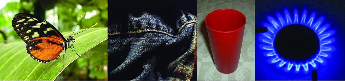
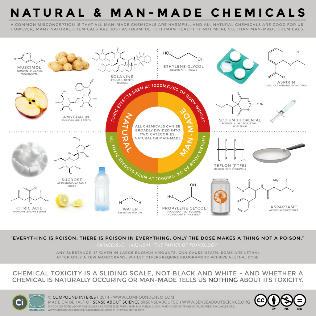
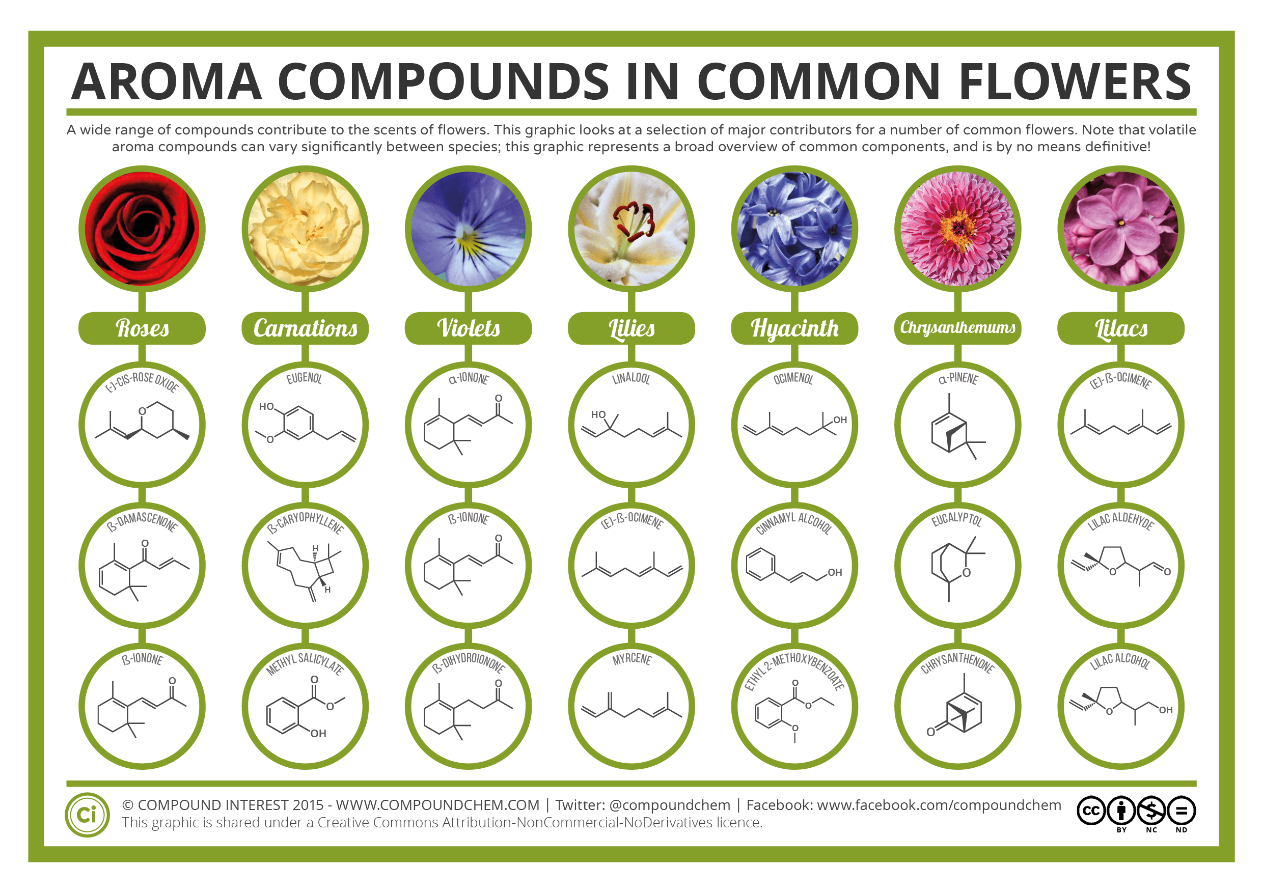
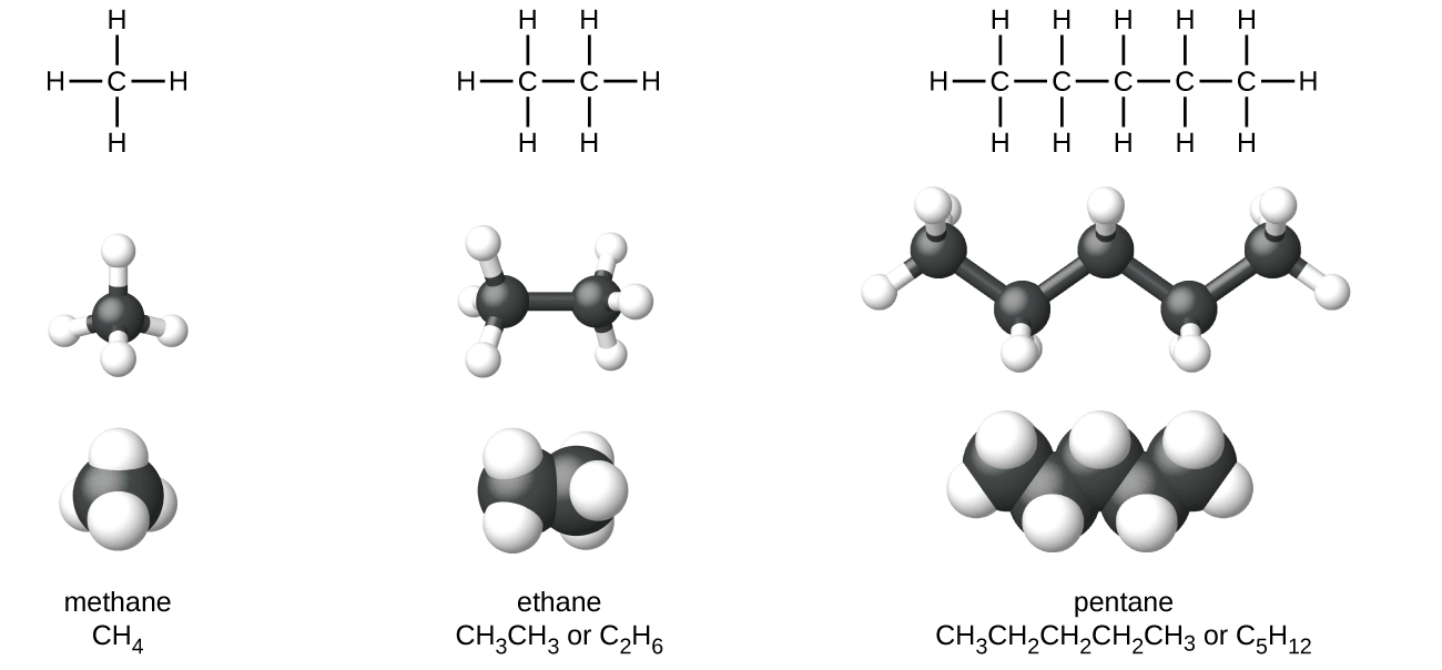
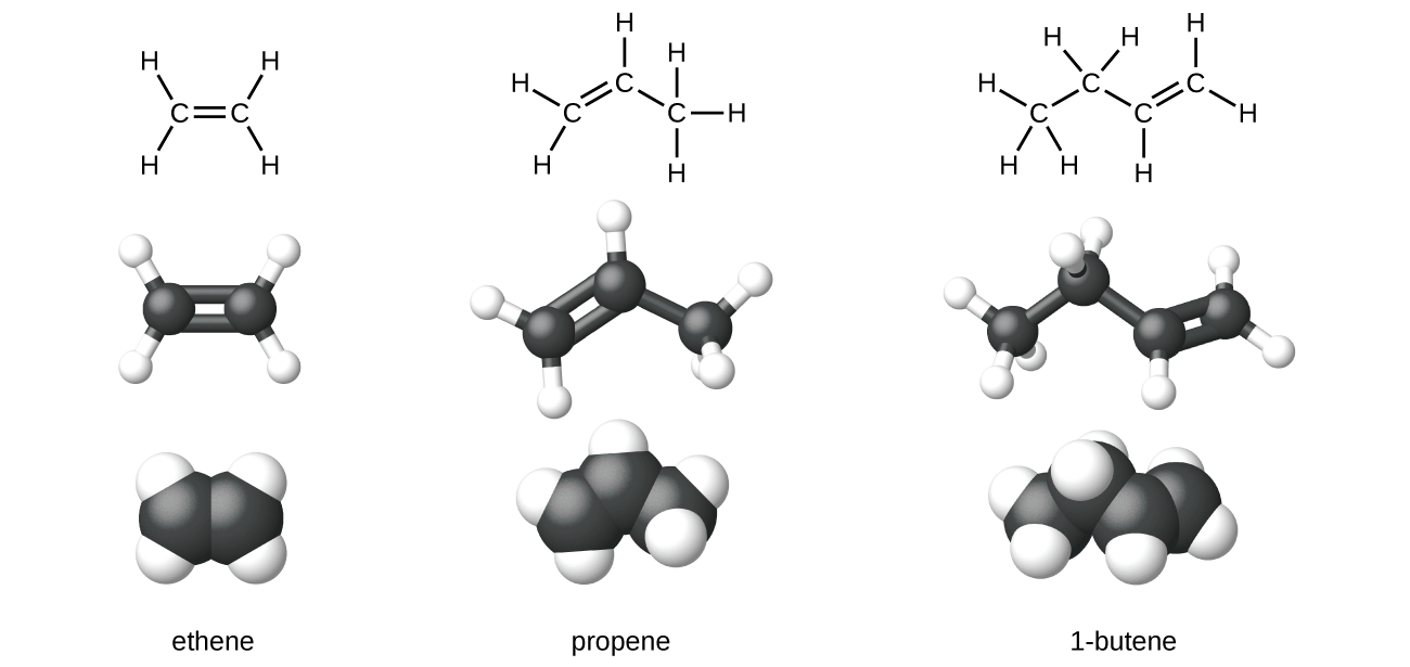
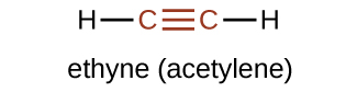
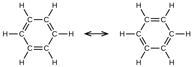
 double bond. To represent this unique bonding, structural formulas for benzene and its derivatives are typically drawn with single bonds between the carbon atoms and a circle within the ring as shown in Figure 19.1e.

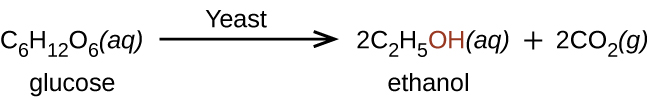
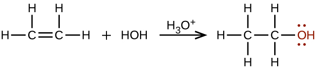
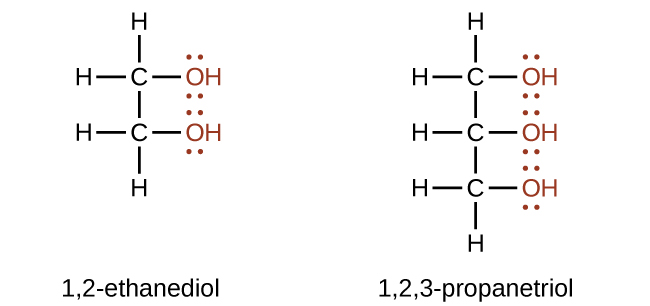

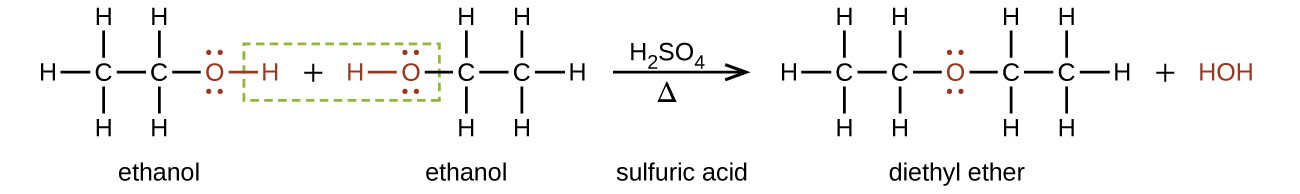

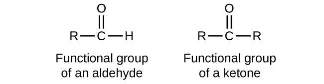
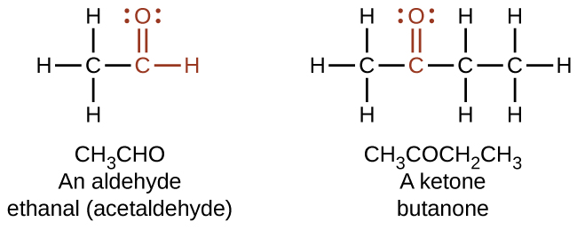
 bond in carbon dioxide, the
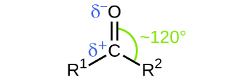
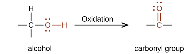
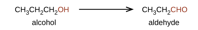
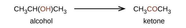
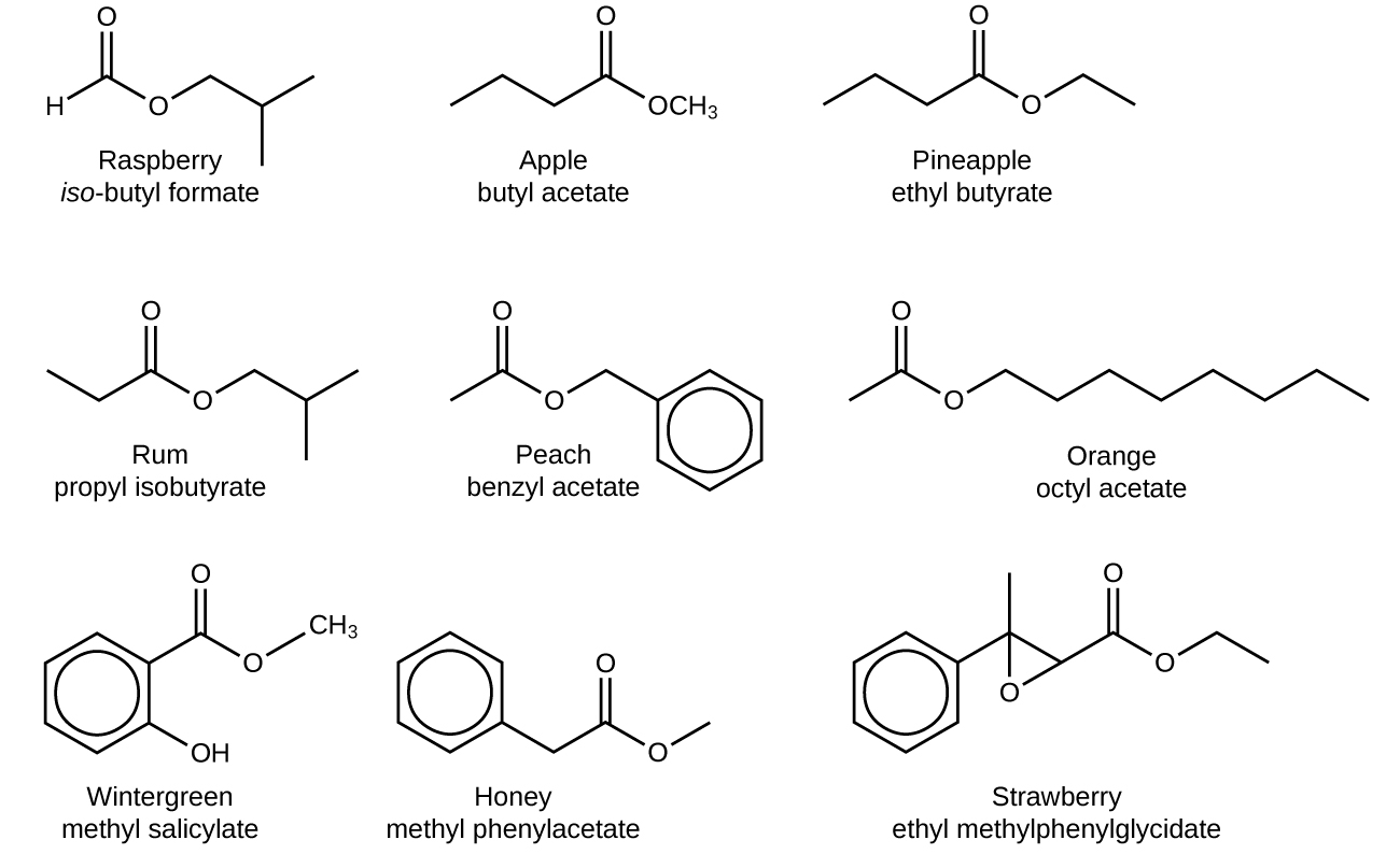
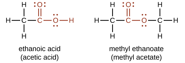
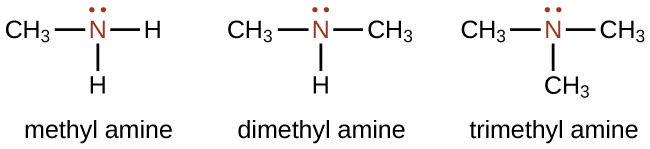
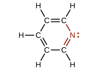
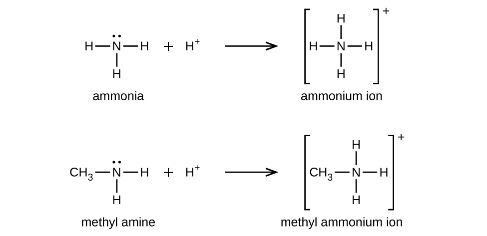
![\text{R}_3\text{N}\;+\;\text{H}_3\text{O}^{+}\;+\;\text{Cl}^{-}\;{\longrightarrow}\;[\text{R}_3\text{NH}^{+}]\;\text{Cl}^{-}\;+\;\text{H}_2\text{O}](https://atu0g9ctah.execute-api.ca-central-1.amazonaws.com/latest/latex?latex=%5Ctext%7BR%7D_3%5Ctext%7BN%7D%5C%3B%2B%5C%3B%5Ctext%7BH%7D_3%5Ctext%7BO%7D%5E%7B%2B%7D%5C%3B%2B%5C%3B%5Ctext%7BCl%7D%5E%7B-%7D%5C%3B%7B%5Clongrightarrow%7D%5C%3B%5B%5Ctext%7BR%7D_3%5Ctext%7BNH%7D%5E%7B%2B%7D%5D%5C%3B%5Ctext%7BCl%7D%5E%7B-%7D%5C%3B%2B%5C%3B%5Ctext%7BH%7D_2%5Ctext%7BO%7D&fg=000000&font=TeX&svg=1 "\text{R}_3\text{N}\;+\;\text{H}_3\text{O}^{+}\;+\;\text{Cl}^{-}\;{\longrightarrow}\;[\text{R}_3\text{NH}^{+}]\;\text{Cl}^{-}\;+\;\text{H}_2\text{O}")
![[\text{R}_3\text{NH}^{+}]\;\text{Cl}^{-}\;+\;\text{OH}^{-}\;{\longrightarrow}\;\text{R}_3\text{N}\;+\;\text{H}_2\text{O}\;+\;\text{Cl}^{-}](https://atu0g9ctah.execute-api.ca-central-1.amazonaws.com/latest/latex?latex=%5B%5Ctext%7BR%7D_3%5Ctext%7BNH%7D%5E%7B%2B%7D%5D%5C%3B%5Ctext%7BCl%7D%5E%7B-%7D%5C%3B%2B%5C%3B%5Ctext%7BOH%7D%5E%7B-%7D%5C%3B%7B%5Clongrightarrow%7D%5C%3B%5Ctext%7BR%7D_3%5Ctext%7BN%7D%5C%3B%2B%5C%3B%5Ctext%7BH%7D_2%5Ctext%7BO%7D%5C%3B%2B%5C%3B%5Ctext%7BCl%7D%5E%7B-%7D&fg=000000&font=TeX&svg=1 "[\text{R}_3\text{NH}^{+}]\;\text{Cl}^{-}\;+\;\text{OH}^{-}\;{\longrightarrow}\;\text{R}_3\text{N}\;+\;\text{H}_2\text{O}\;+\;\text{Cl}^{-}")
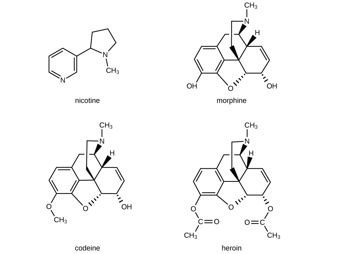

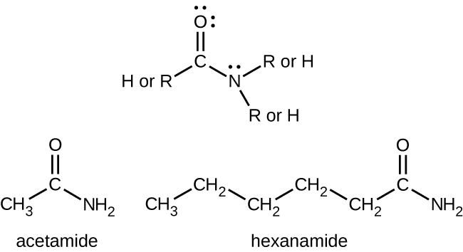

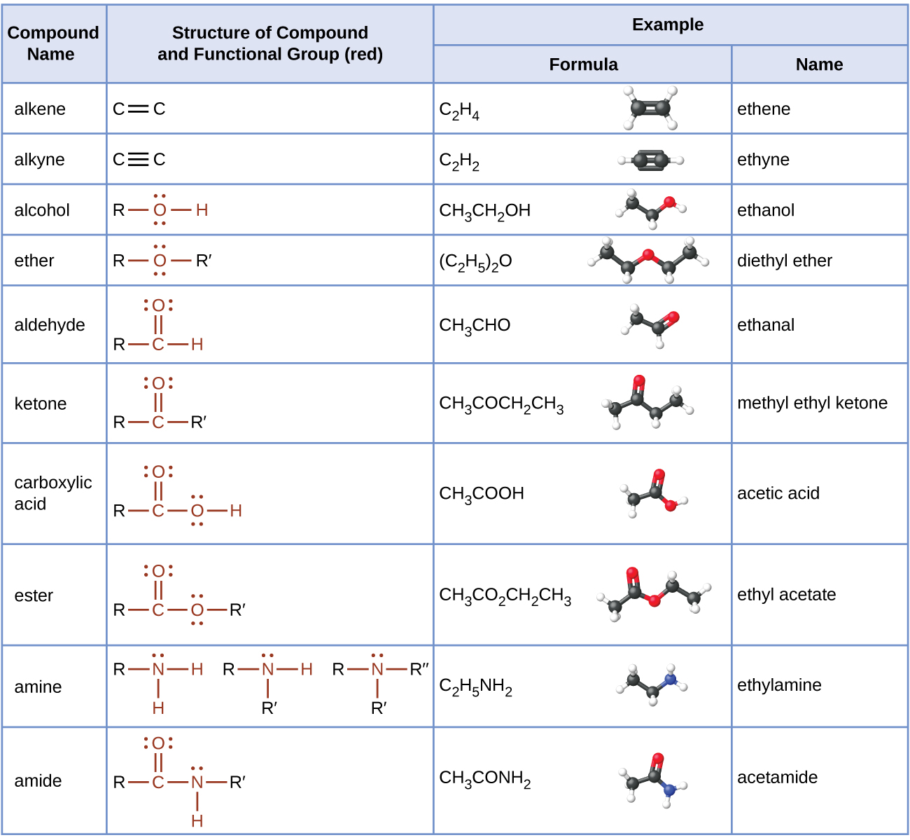
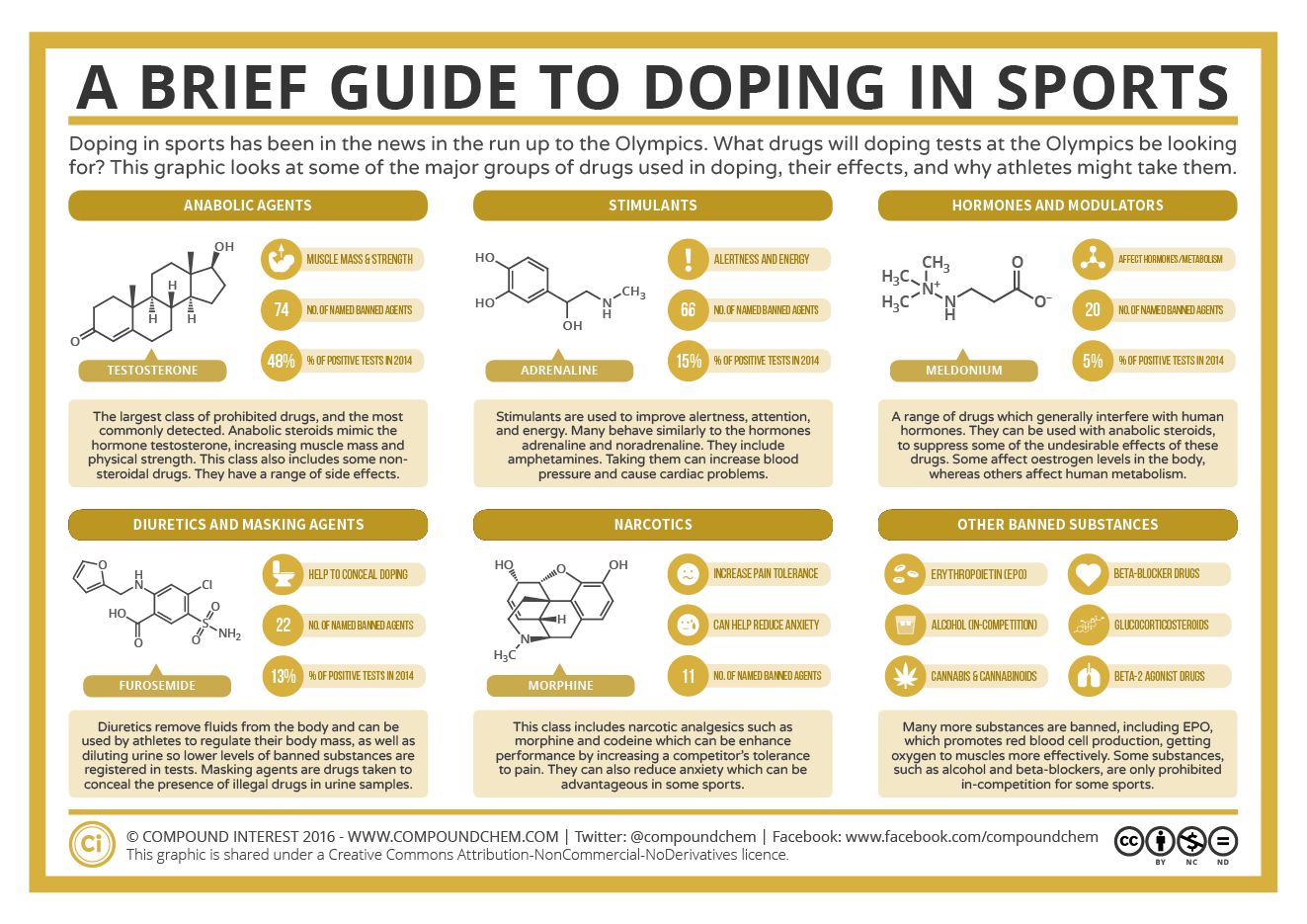
 functional group has two π bonds, alkynes typically react even more readily, and react with twice as much reagent in addition reactions. The reaction of acetylene with bromine is a typical example that is demonstrated in Figure 19.6b.
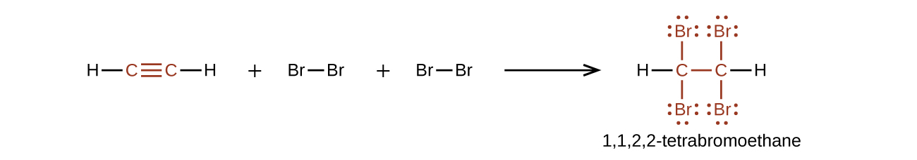
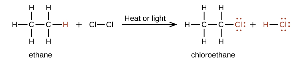
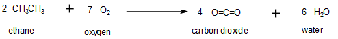
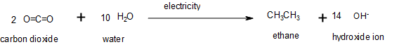
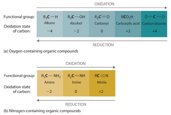
\;+\;2\text{O}_2(g)\;{\longrightarrow}\;\text{CO}_2(g)\;+\;2\text{H}_2\text{O}(g)")


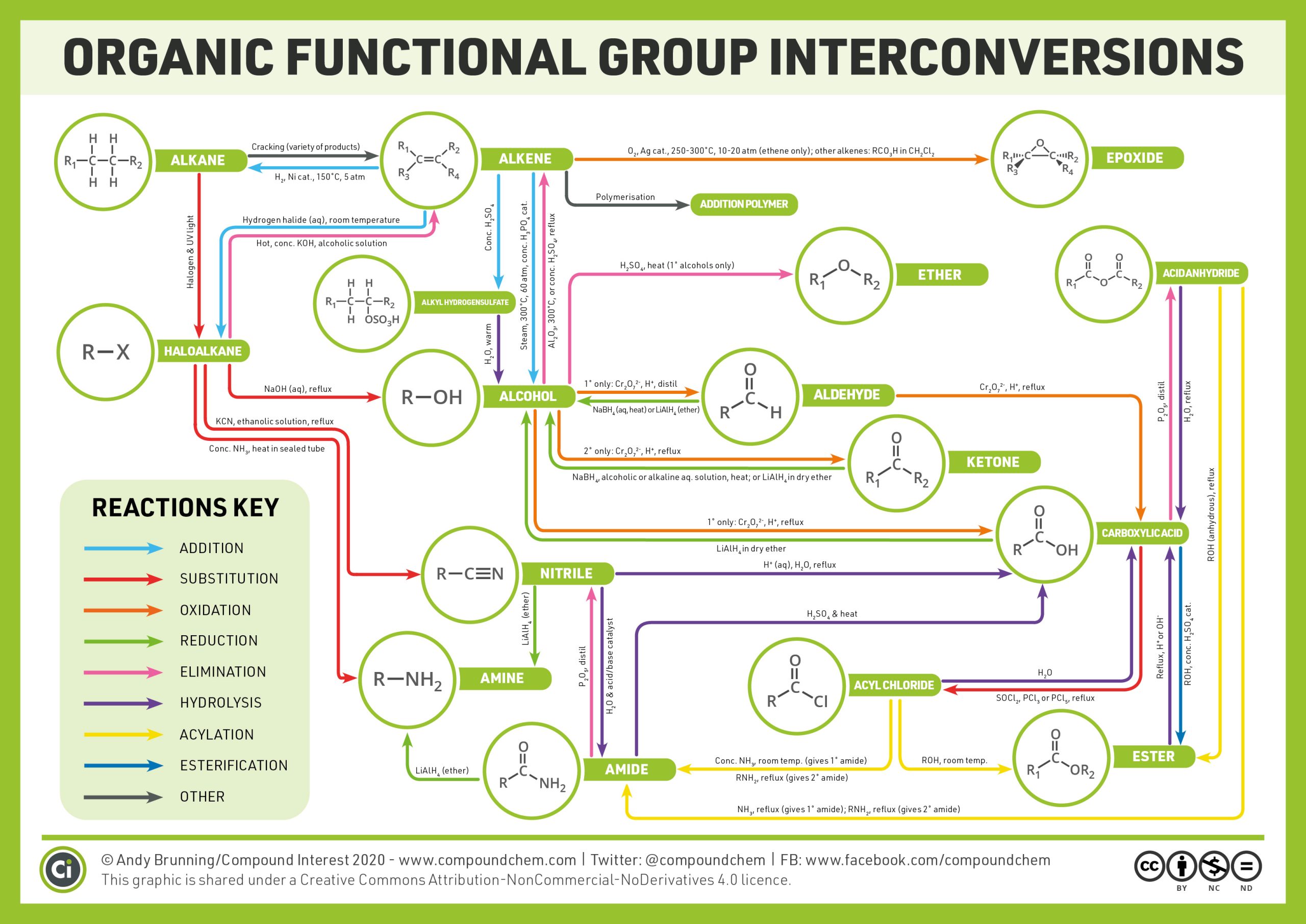
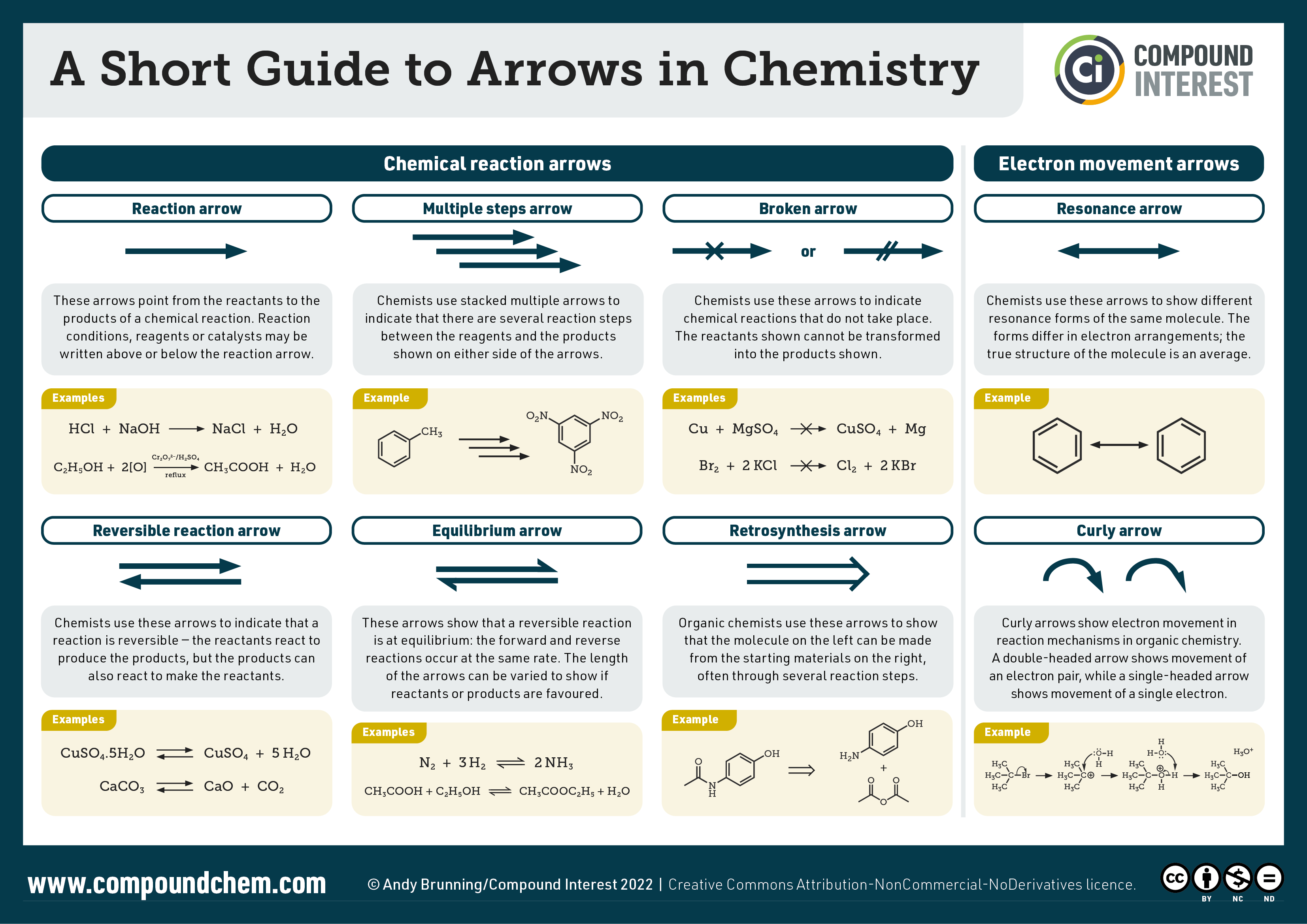


 (anhydrous), reflux
 , reflux (gives primary amide);
  , reflux (gives secondary amide)
 , reflux
 , 300 degree Celsius, or conc. H2SO4 , reflux
 , (primary alcohols only)
 ,H+ , reflux
 , distil
 (aq, heat) or
  \small}")
 ,
,  cat., 150 degree Celsius , 5 atm
 ,
,  cat., 250-300 degree Celsius, 10-20 atm (ethene only); other alkenes:
  in
in 
 cat.
cat. , distil
 in dry ether
 ,
,  or
or  , reflux
 , alcoholic solution
 , ethanolic solution, reflux
 \small}") , reflux
, reflux , alcoholic or alkaline aq. solution, heat; or
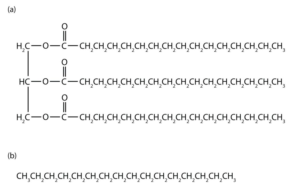

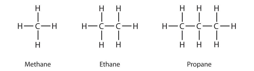
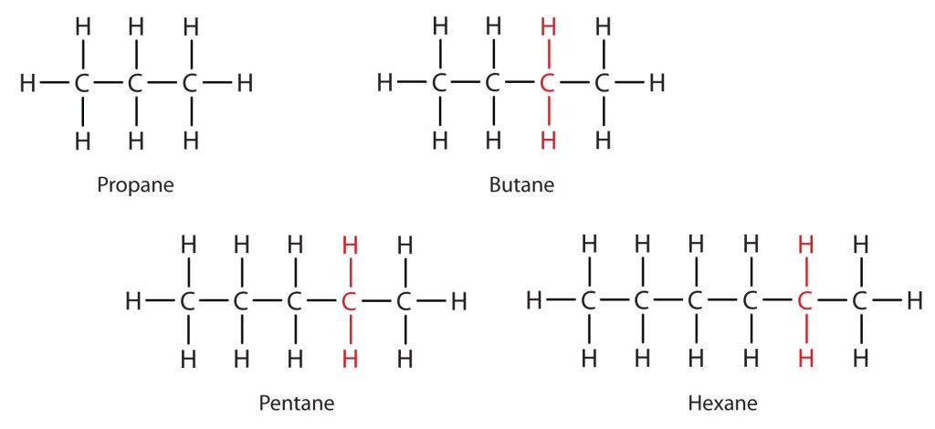

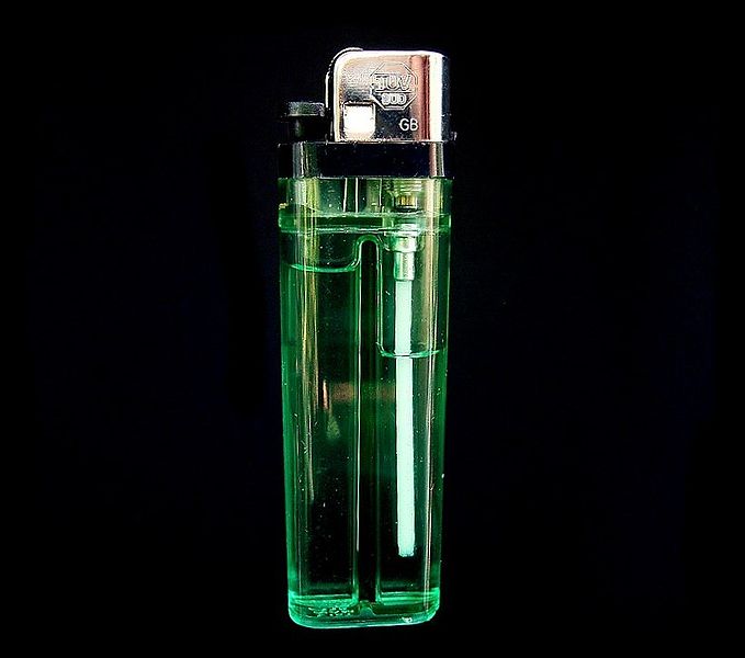
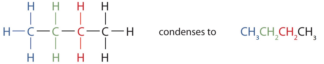
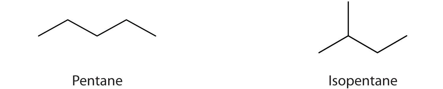
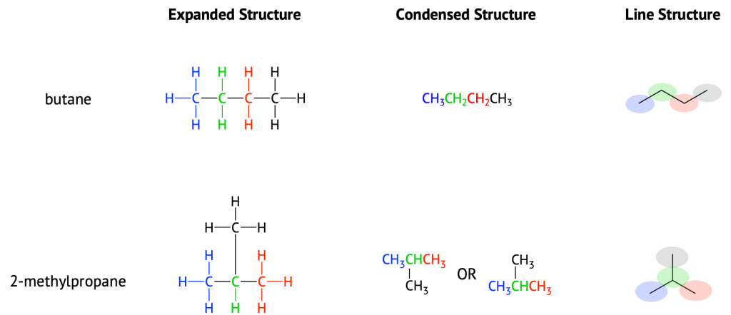
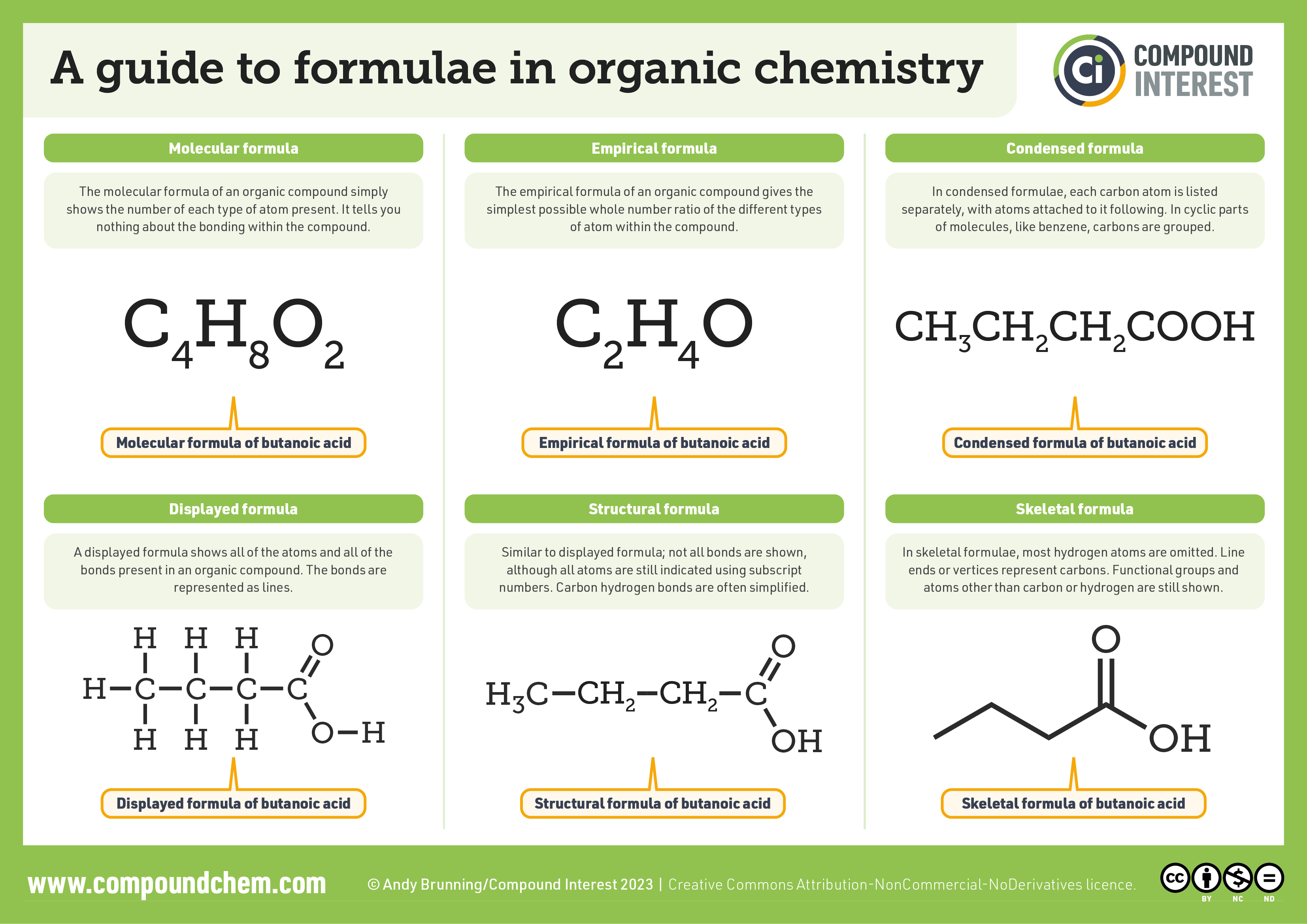
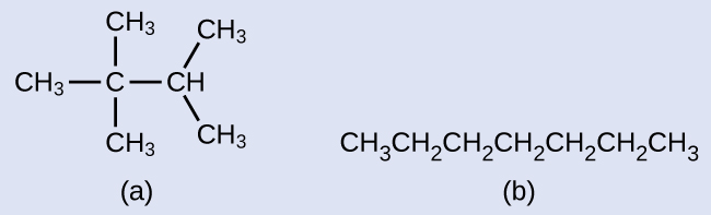
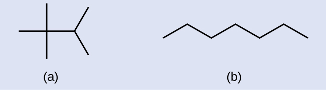
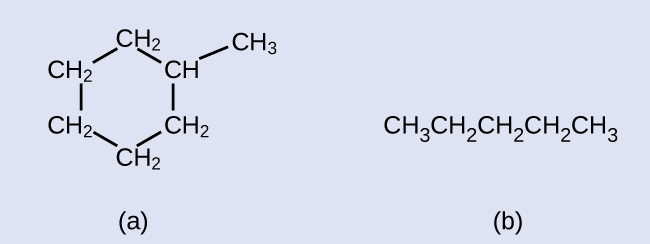
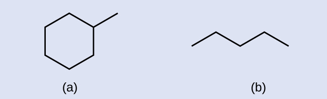
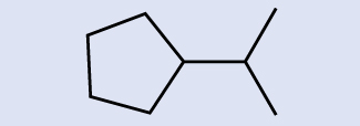

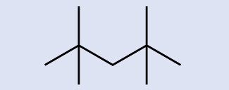
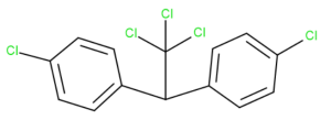
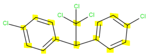 There are 14 carbon atoms. Six in each aromatic ring, one that connects the rings together and one that branches off the middle carbon.
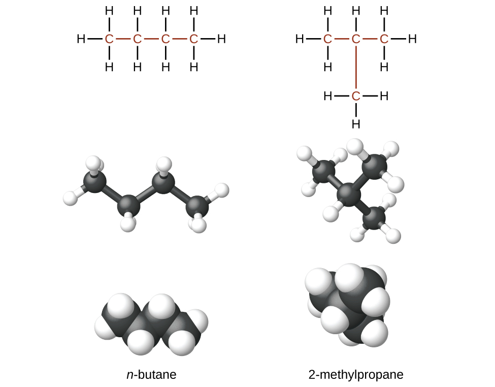
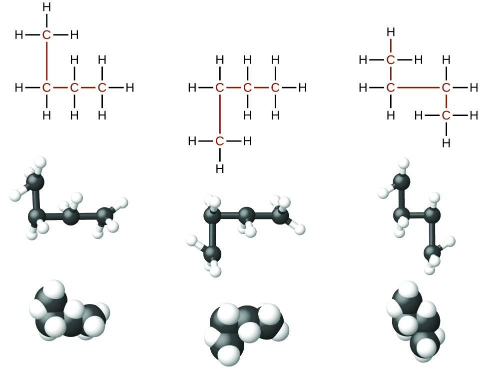
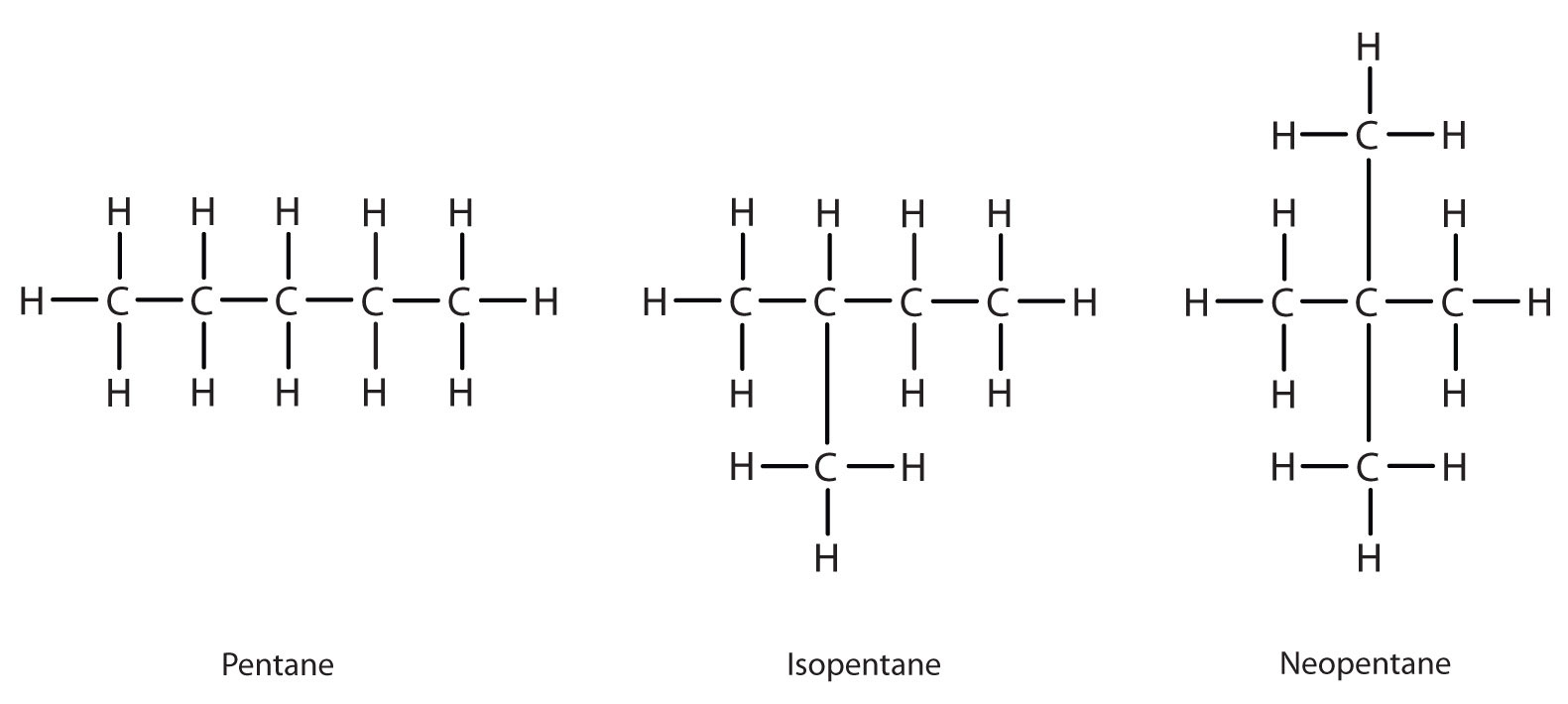
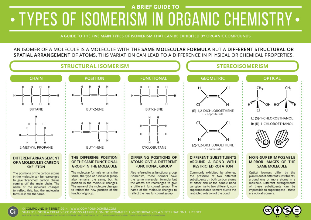
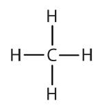
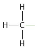
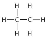
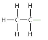
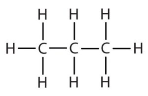
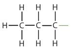
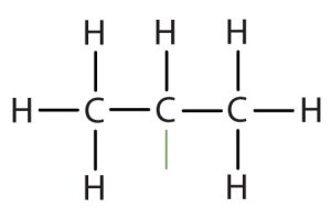
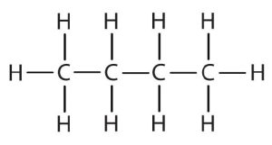
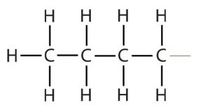

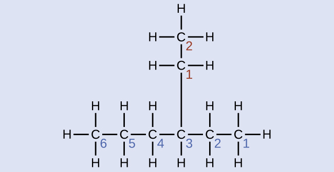
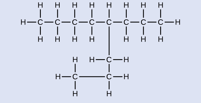
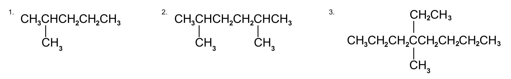
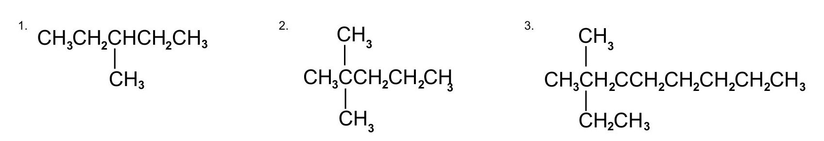


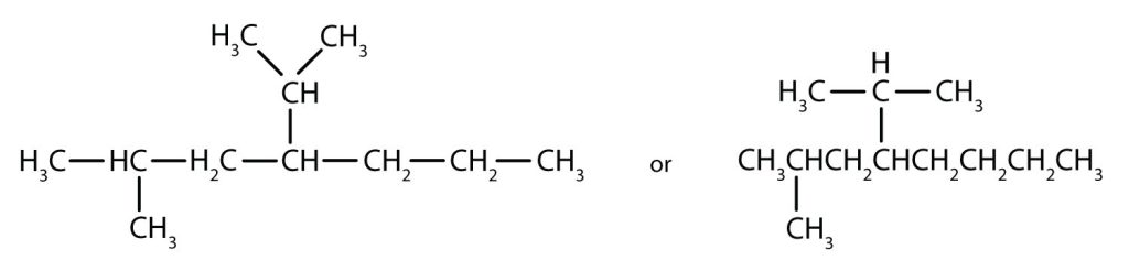
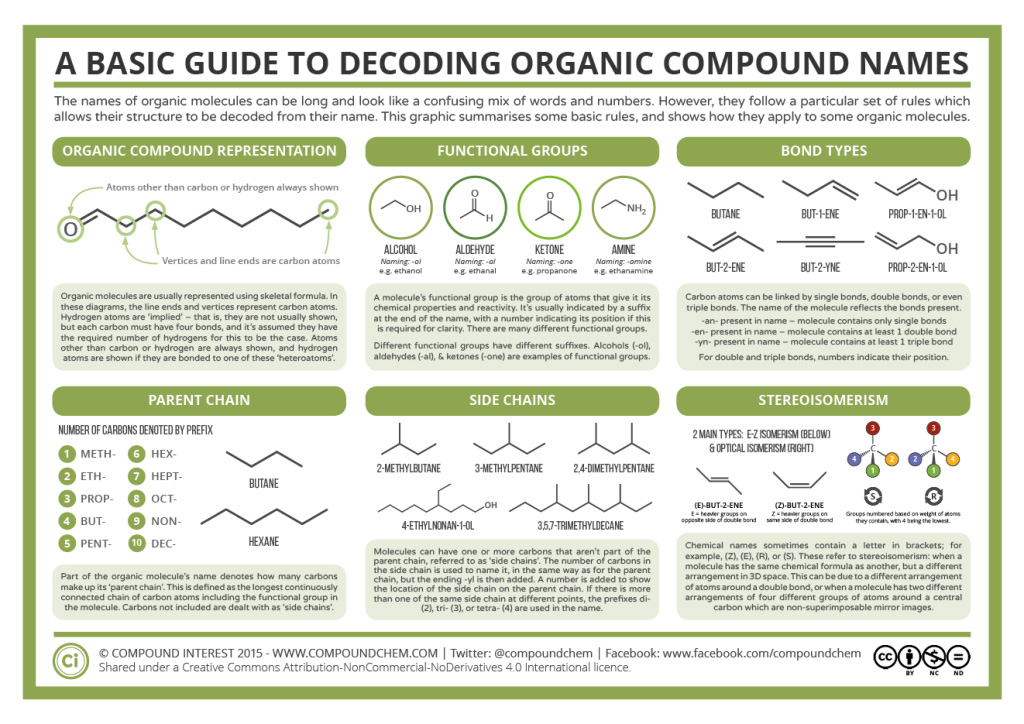
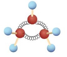
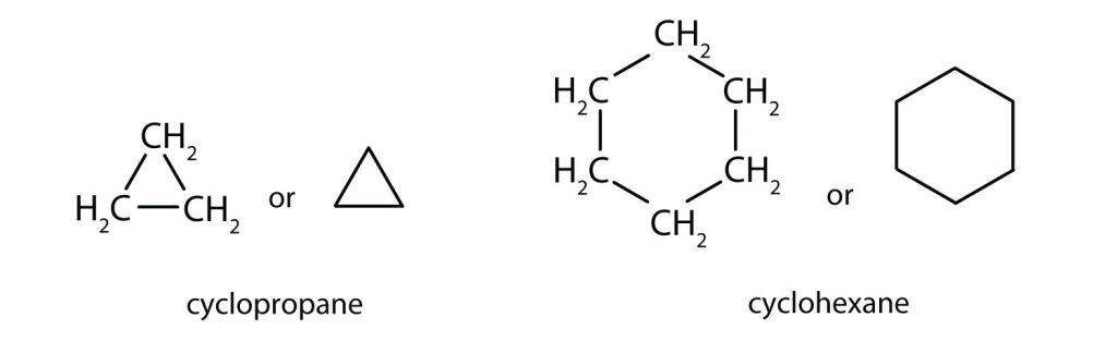
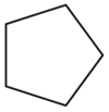


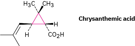
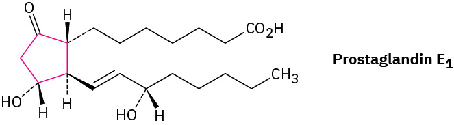
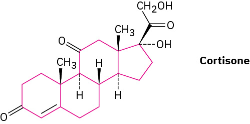
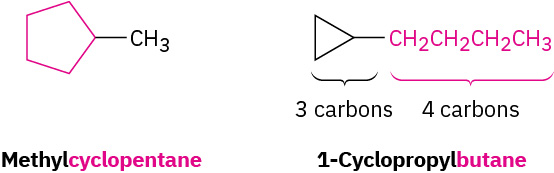
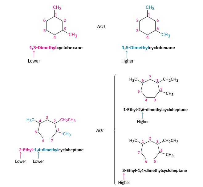
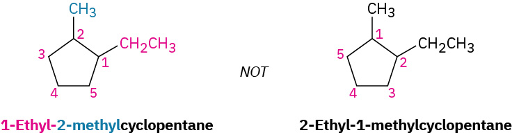
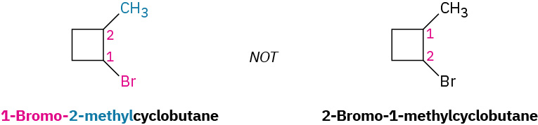
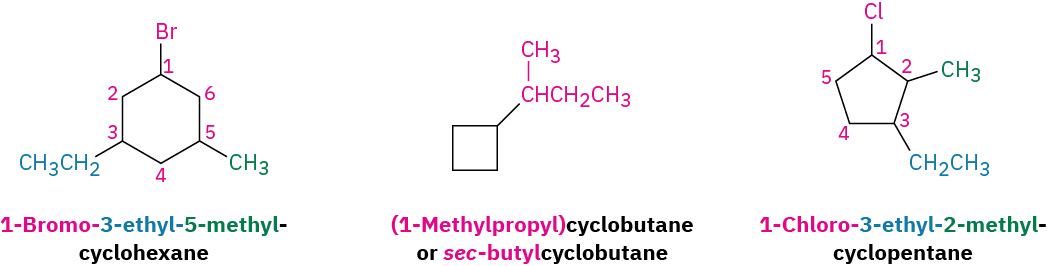
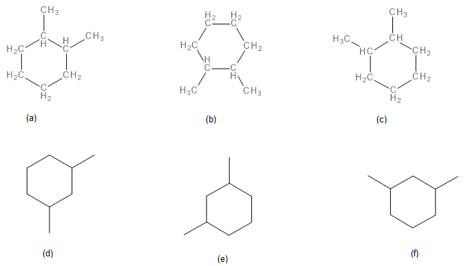
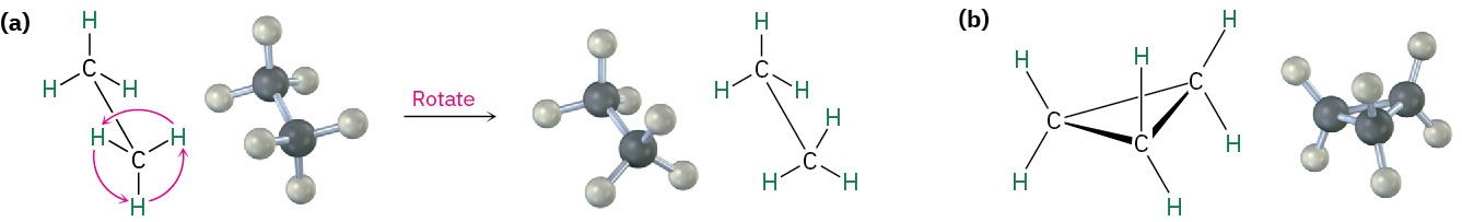
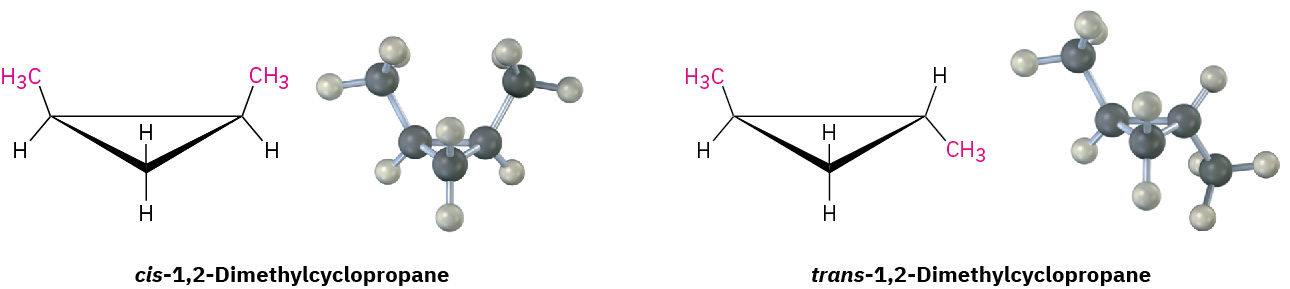
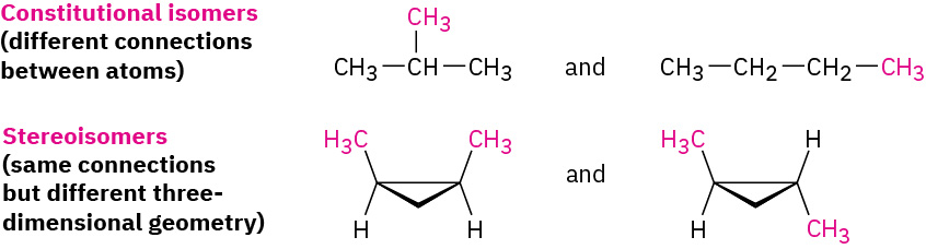
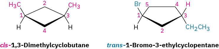
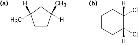


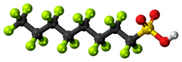
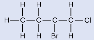
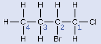

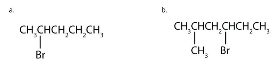
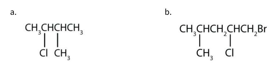
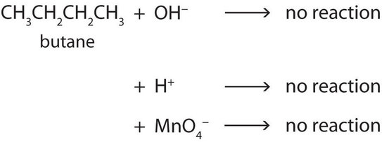
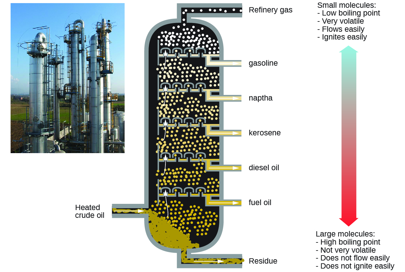
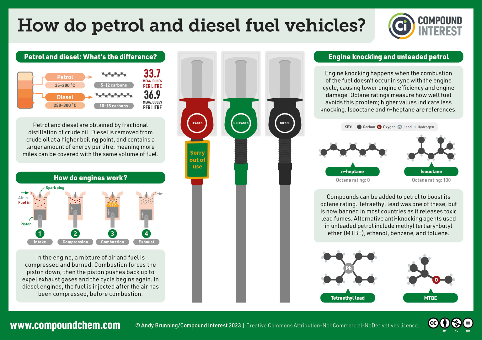
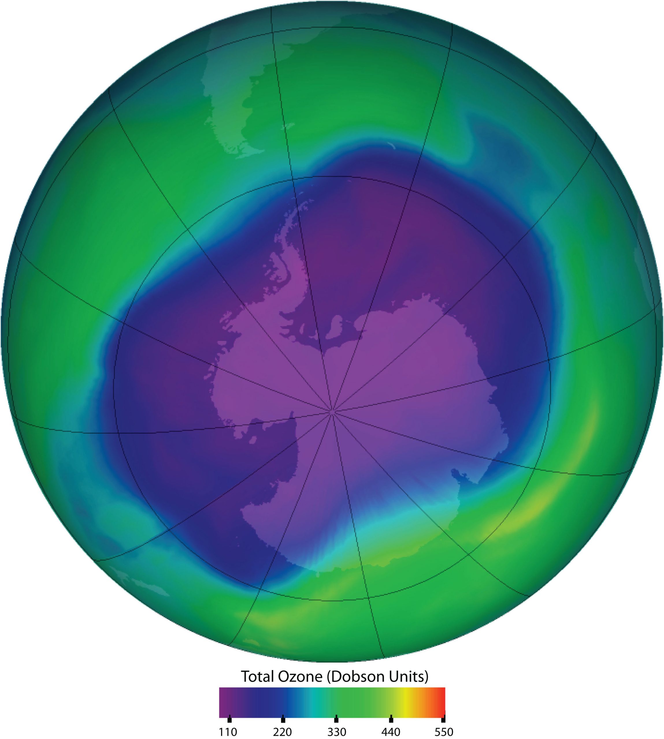
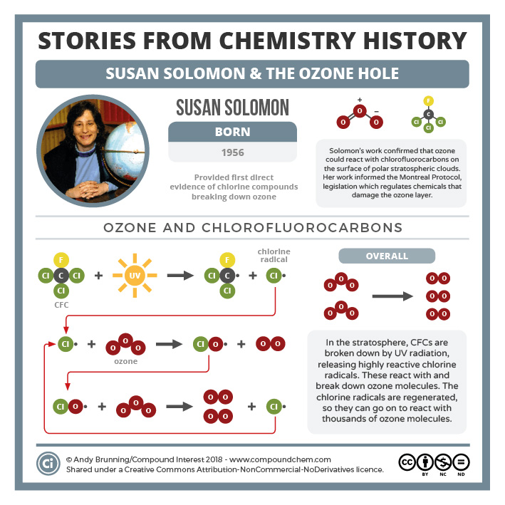


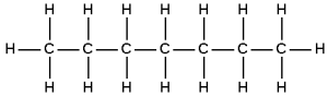
 Check answer

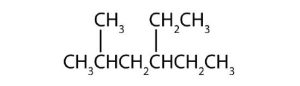
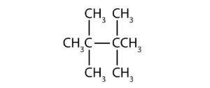

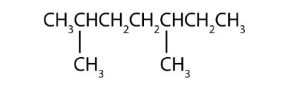
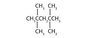


 .
. .
.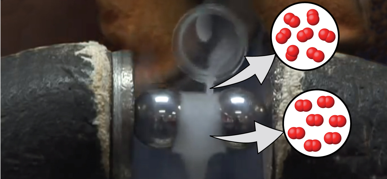

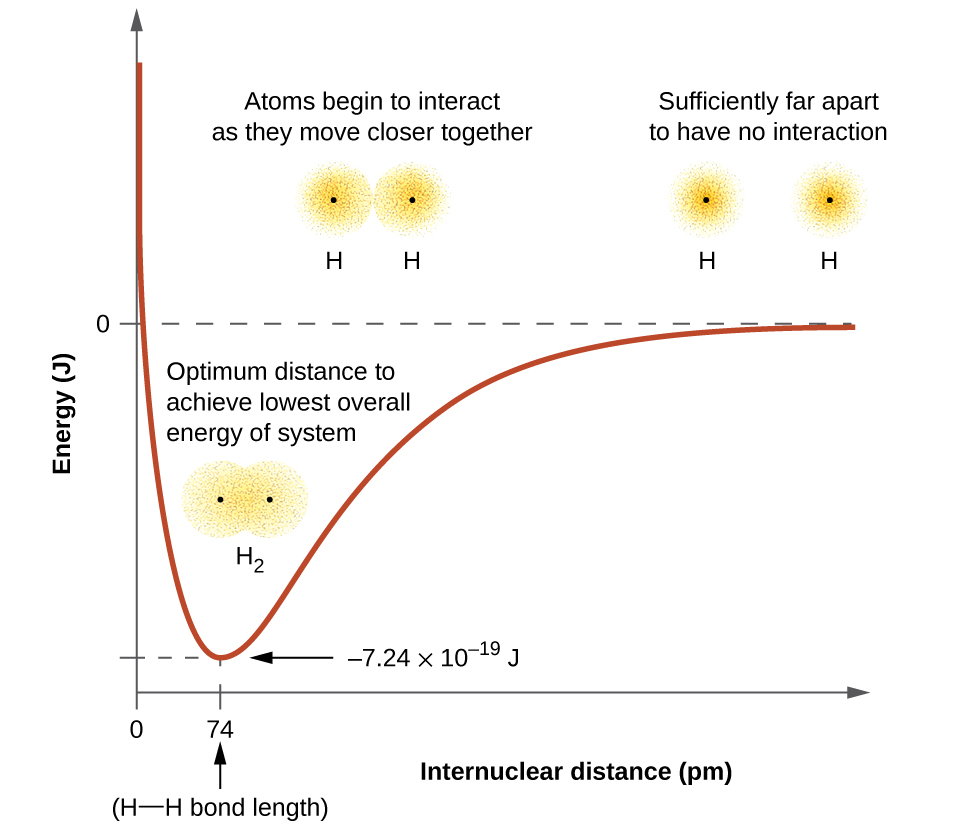
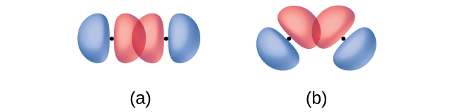
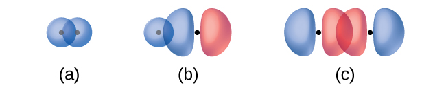

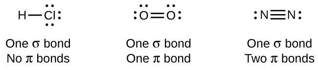
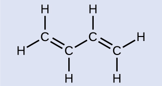

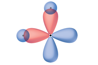
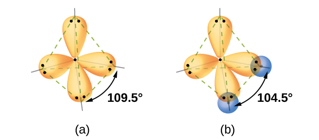
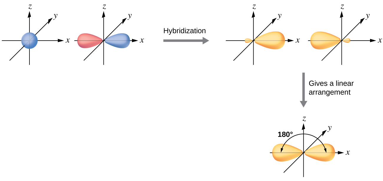
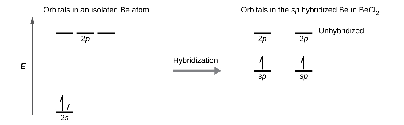
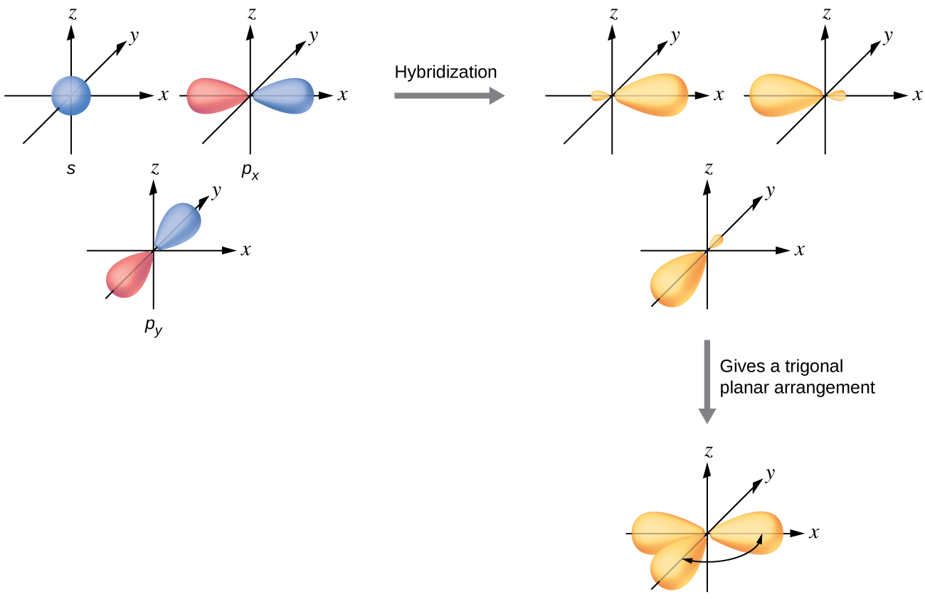
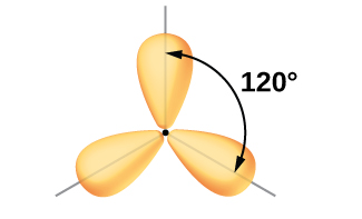
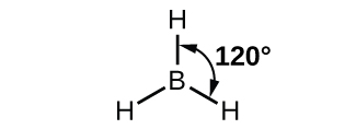
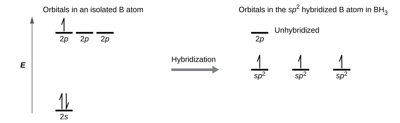
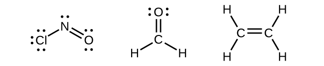
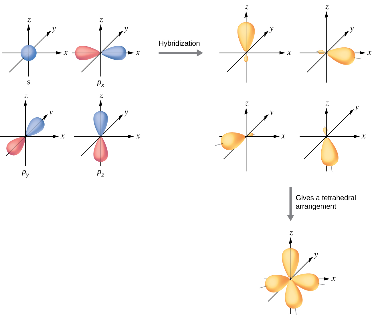
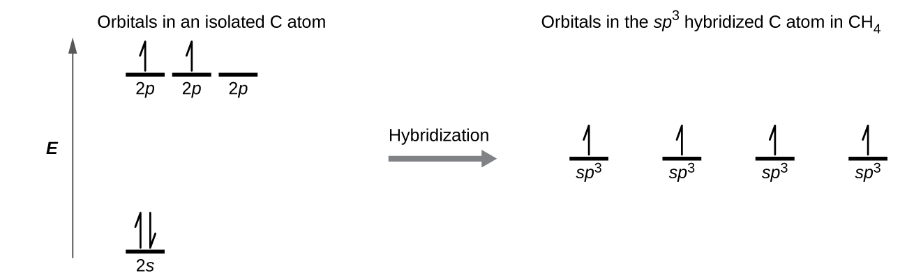
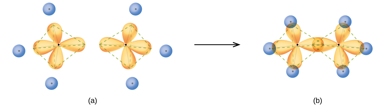
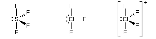
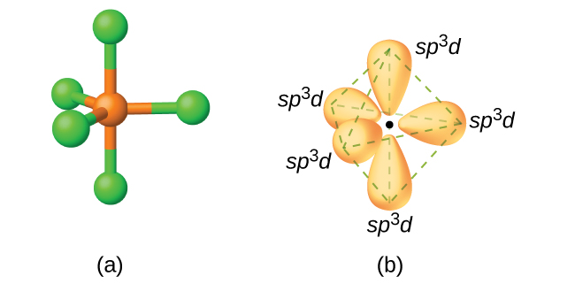
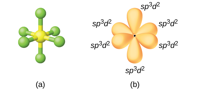
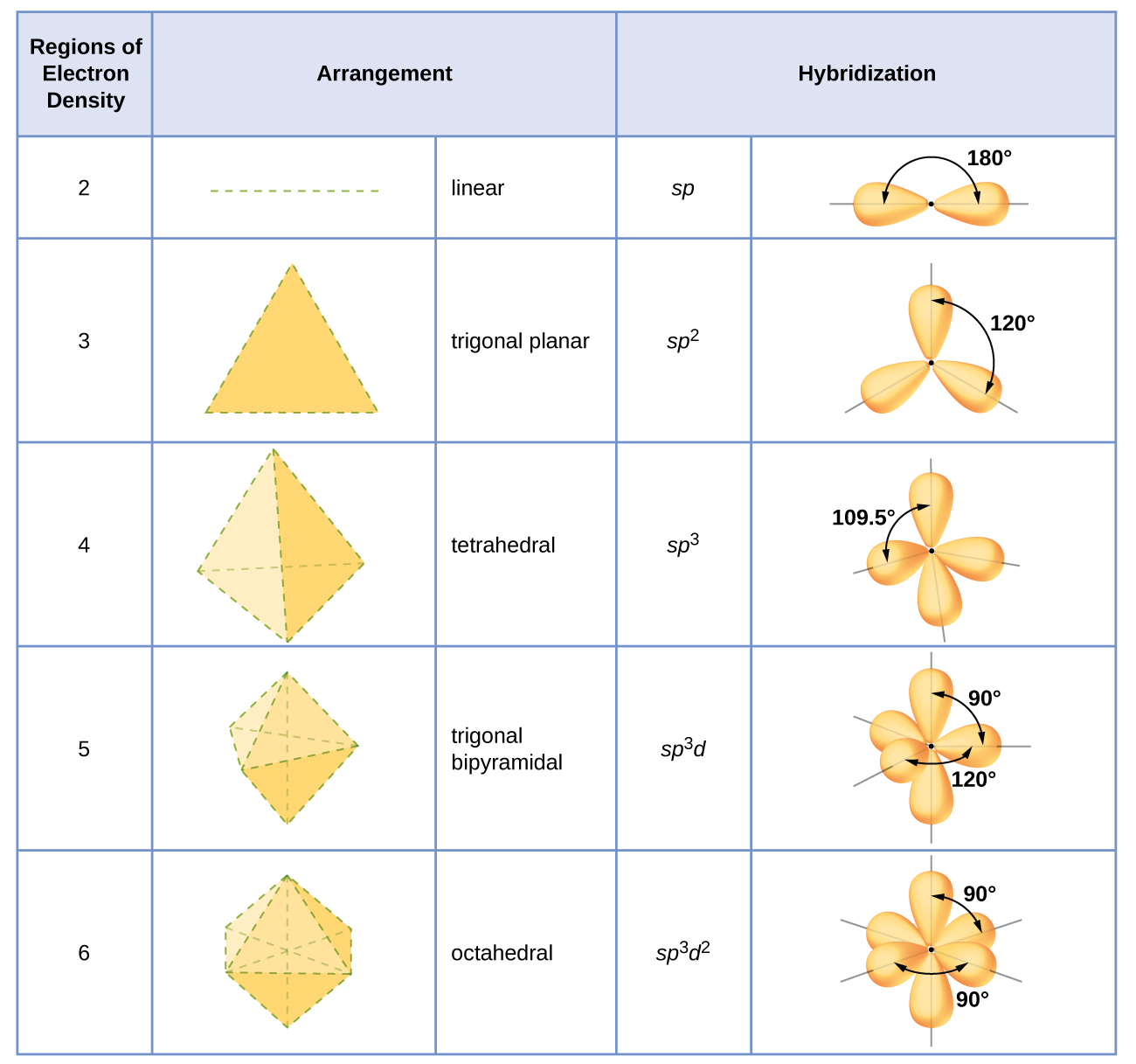
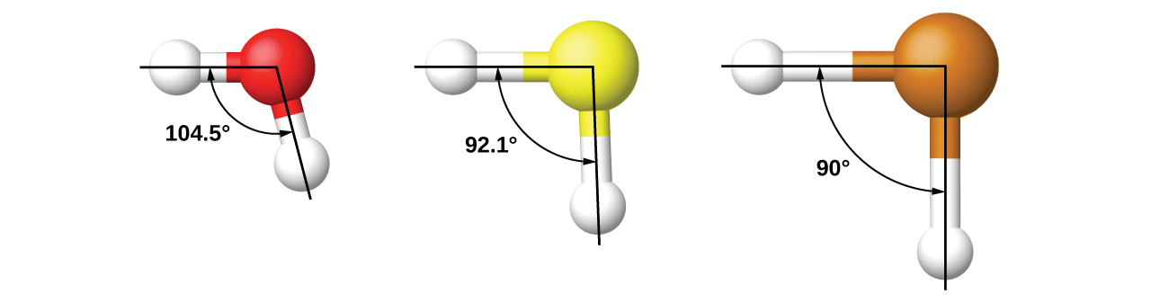
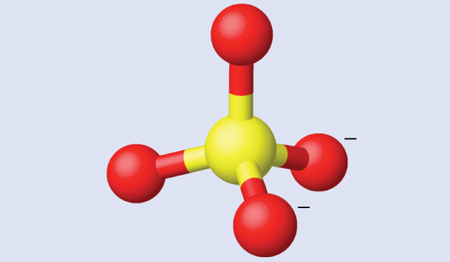
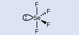
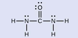
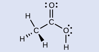
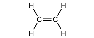
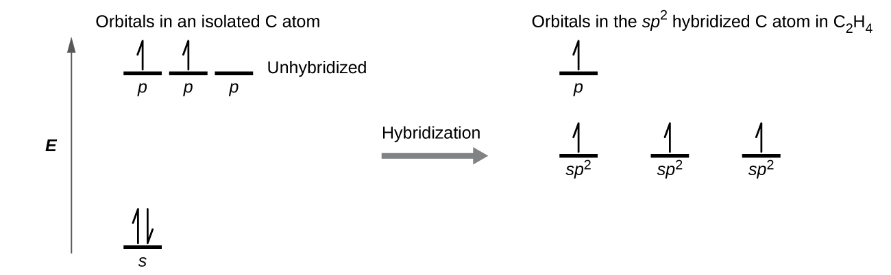
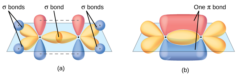
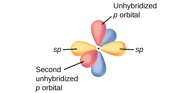

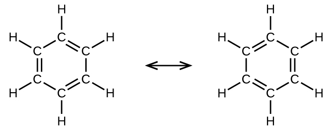


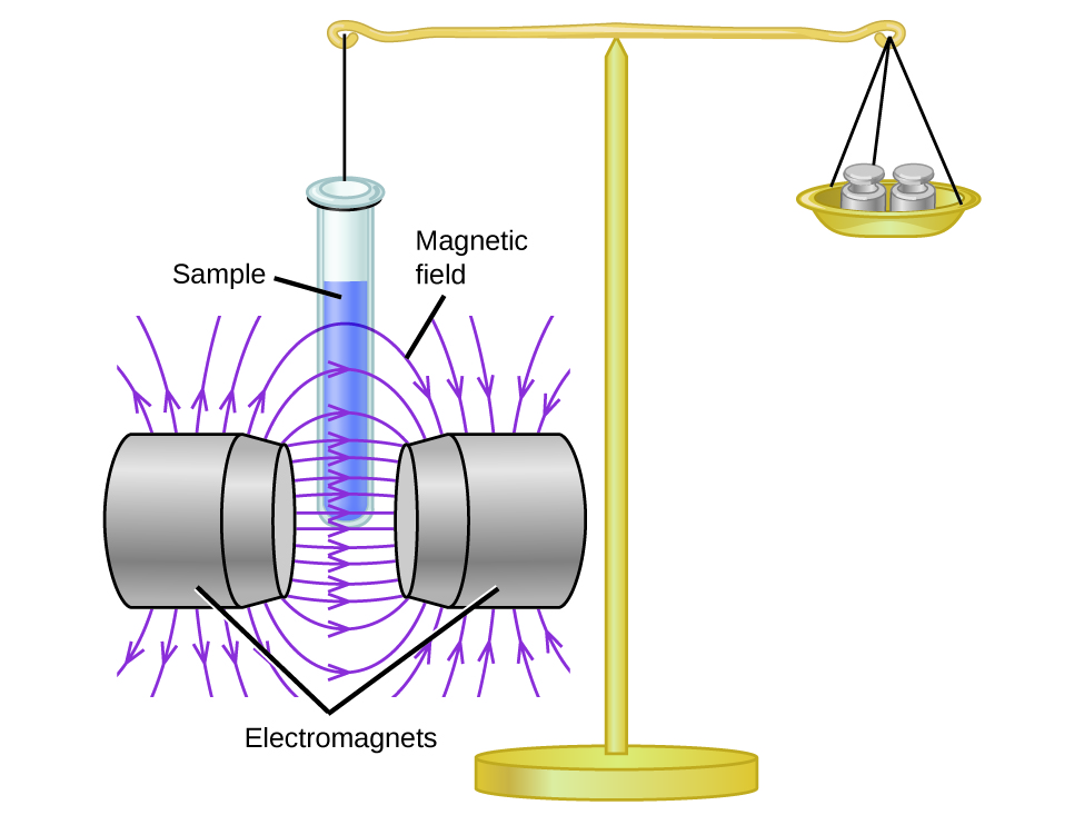
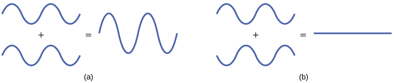
 molecular orbital (read as “sigma-s-star”) molecular orbital in which there is a node between the nuclei. The asterisk signifies that the orbital is an antibonding orbital. Electrons in a σs orbital are attracted by both nuclei at the same time and are more stable (of lower energy) than they would be in the isolated atoms. Adding electrons to these orbitals creates a force that holds the two nuclei together, so we call these orbitals
  orbitals are located well away from the region between the two nuclei. The attractive force between the nuclei and these electrons pulls the two nuclei apart. Hence, these orbitals are called
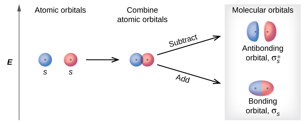
 (antibonding) (read as “sigma-p-x” and “sigma-p-x star,” respectively). Just as with s-orbital overlap, the asterisk indicates the orbital with a node between the nuclei, which is a higher-energy, antibonding orbital.
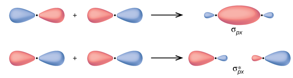
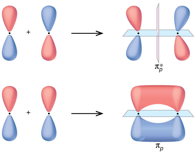
 orbitals are oriented at right angles to the πpz and
  orbitals. Except for their orientation, the πpy and πpz orbitals are identical and have the same energy; they are
  antibonding orbitals are also degenerate and identical except for their orientation. A total of six molecular orbitals results from the combination of the six atomic p orbitals in two atoms: σpx and
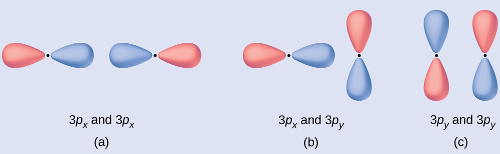
 orbital.


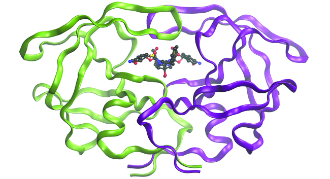
^2")
^2")
^2")
^1") . It is common to omit the core electrons from molecular orbital diagrams and configurations and include only the valence electrons.
. It is common to omit the core electrons from molecular orbital diagrams and configurations and include only the valence electrons.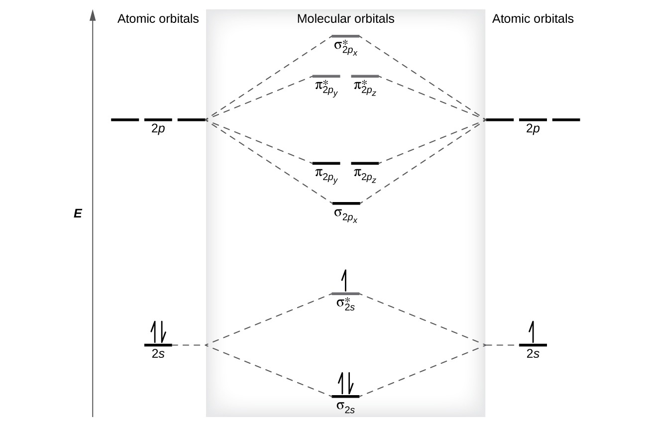
 - (\text{number of antibonding electrons})}{2}")
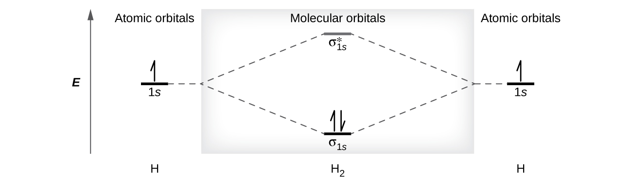
}{2} = 1")
}{2} = 0")
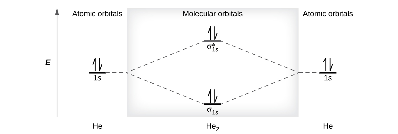
^2 (\sigma^*_{2s})^2")
^2 (\sigma^*_{2s})^2 (\pi_{2py}, \pi_{2pz})^2")
^2 (\sigma^*_{2s})^2 (\pi_{2py}, \pi_{2pz})^4")
^2 (\sigma^*_{2s})^2 (\pi_{2py}, \pi_{2pz})^4 (\sigma_{2px})^2")
^2 (\sigma^*_{2s})^2 (\sigma_{2px})^2 (\pi_{2py}, \pi_{2pz})^4 (\pi^*_{2py}, \pi^*_{2pz})^2")
^2 (\sigma^*_{2s})^2 (\sigma_{2px})^2 (\pi_{2py}, \pi_{2pz})^4 (\pi^*_{2py}, \pi^*_{2pz})^4")
^2 (\sigma^*_{2s})^2 (\sigma_{2px})^2 (\pi_{2py}, \pi_{2pz})^4 (\pi^*_{2py}, \pi^*_{2pz})^4 (\sigma^*_{2px})^2")
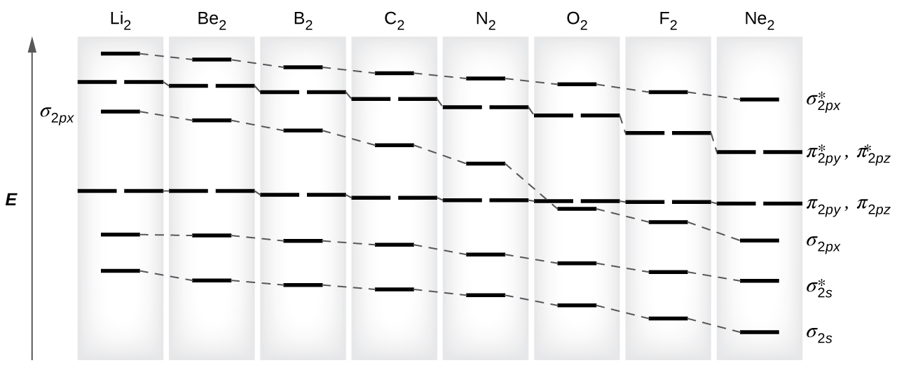
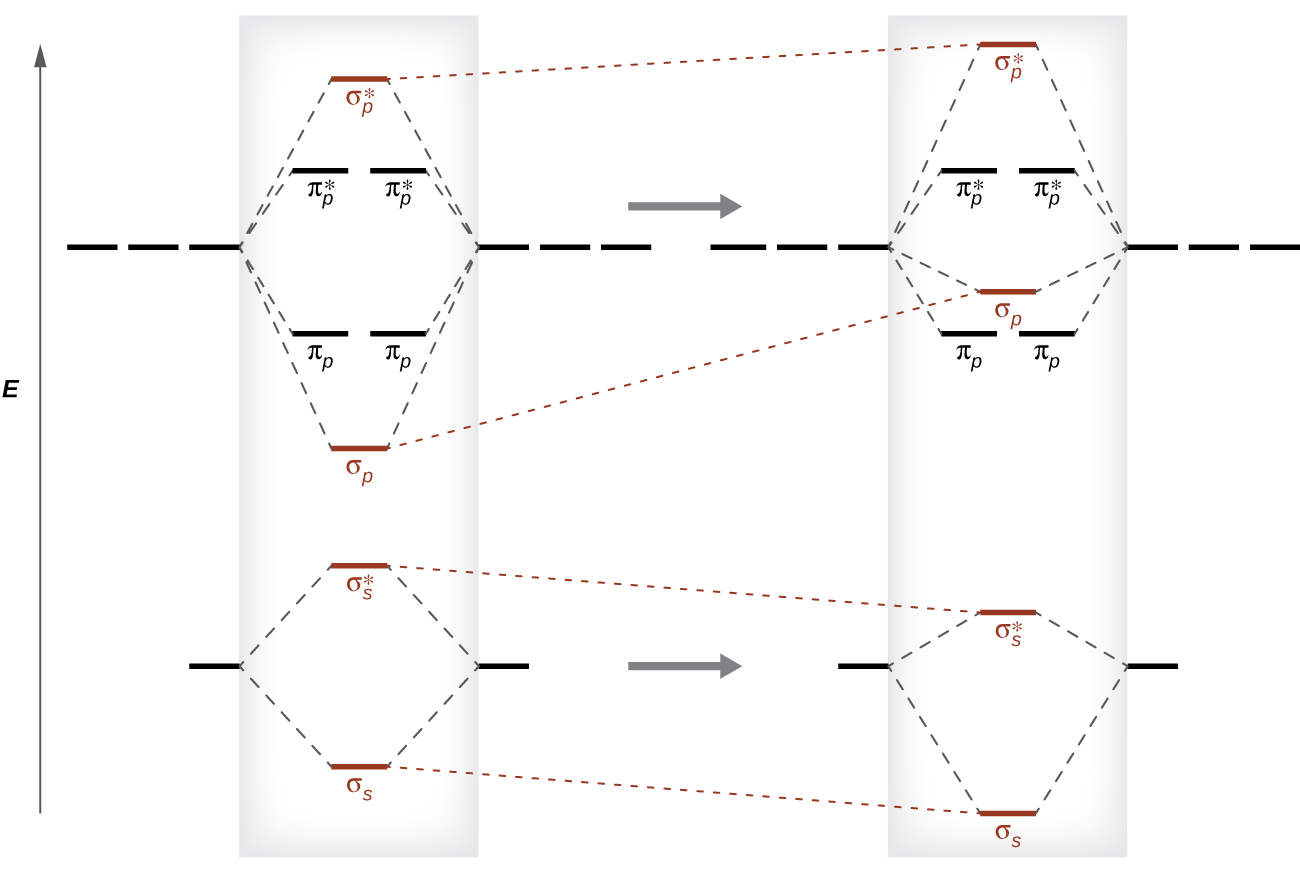
 ,
,  ) level. We expect the two electrons that occupy these two degenerate orbitals to be unpaired, and this molecular electronic configuration for O2 is in accord with the fact that the oxygen molecule has two unpaired electrons (Figure 21.4n.). The presence of two unpaired electrons has proved to be difficult to explain using Lewis structures, but the molecular orbital theory explains it quite well. In fact, the unpaired electrons of the oxygen molecule provide a strong piece of support for the molecular orbital theory.
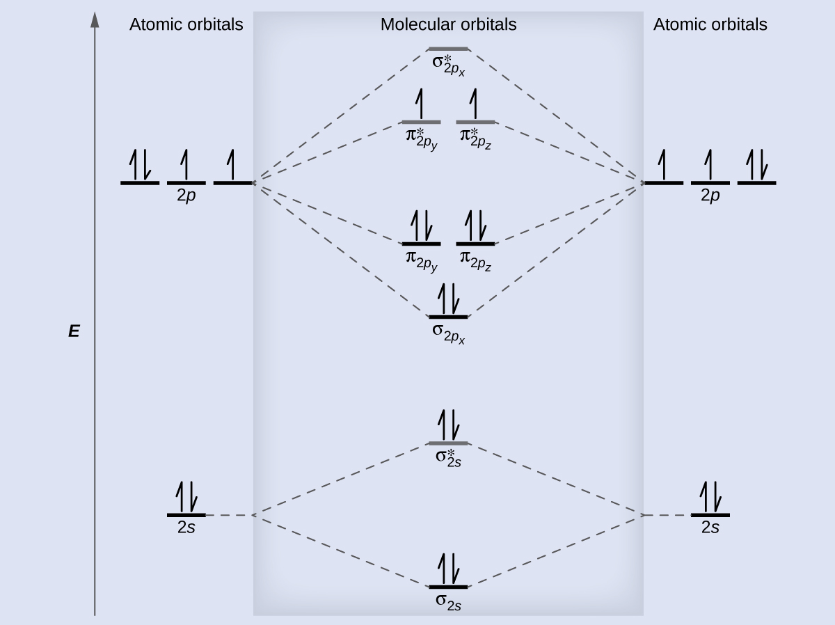
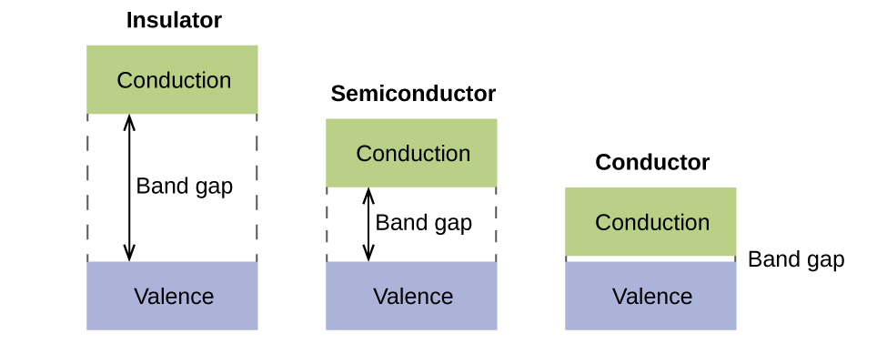

 - (\text{number of antibonding electrons})}{2}")
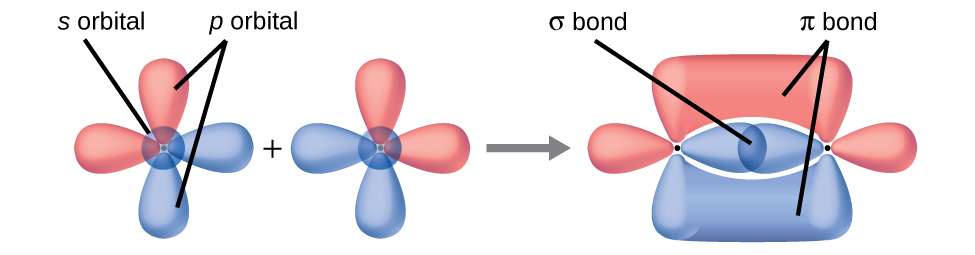
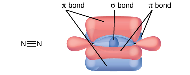
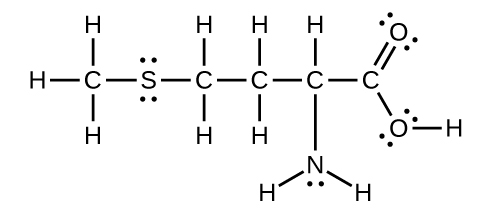
 + 8 \text{O}_2(g) \longrightarrow 8\text{SO}_2(g)")
 + \text{O}_2(g) \longrightarrow 2\text{SO}_3(g)")
 + \text{H}_2 \text{O}(l) \longrightarrow \text{H}_2 \text{SO}_4(l)")
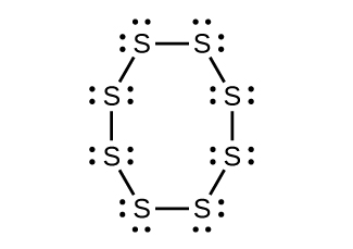
 Bent (120°), sp2 (c)
 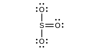 Trigonal planar, sp2
 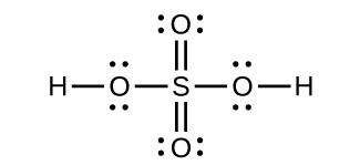 Tetrahedral, sp3
 \longrightarrow \text{C}_2\text{H}_4(g) + \text{C}_3\text{H}_6(g) + \text{CH}_4(g) + \text{H}_2(g)")
 (c) linear (d) sp3d
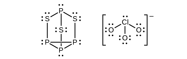 (b) P atoms, trigonal pyramidal; S atoms, bent, with two lone pairs; Cl atoms, trigonal pyramidal; (c) Hybridization about P, S, and Cl is, in all cases, sp3; (d) Oxidation states P +1,
  , Cl +5, O –2. Formal charges: P 0; S 0; Cl +2: O –1
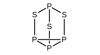

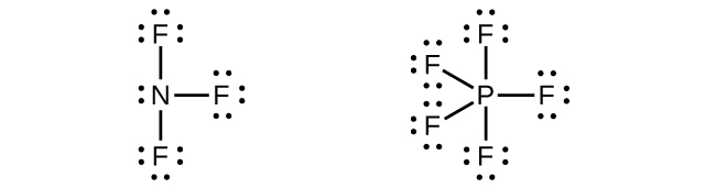 Phosphorus and nitrogen can form sp3 hybrids to form three bonds and hold one lone pair in PF3 and NF3, respectively. However, nitrogen has no valence d orbitals, so it cannot form a set of sp3d hybrid orbitals to bind five fluorine atoms in NF5. Phosphorus has d orbitals and can bind five fluorine atoms with sp3d hybrid orbitals in PF5.
  (b) The terminal carbon atom uses sp3 hybrid orbitals, while the central carbon atom is sp hybridized. (c) Each of the two π bonds is formed by overlap of a 2p orbital on carbon and a nitrogen 2p orbital.
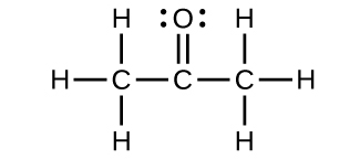

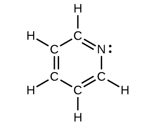
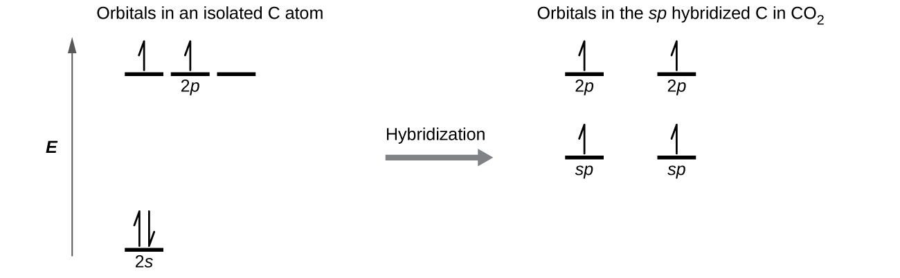 Each of the four electrons is in a separate orbital and overlaps with an electron on an oxygen atom.
 ^2 (\sigma^*_{2s})^2 (\sigma_{2px})^2 (\pi_{2py} , \pi_{2pz})^4 (\pi^*_{2py} , \pi^*_{2pz})^3")
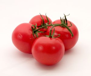

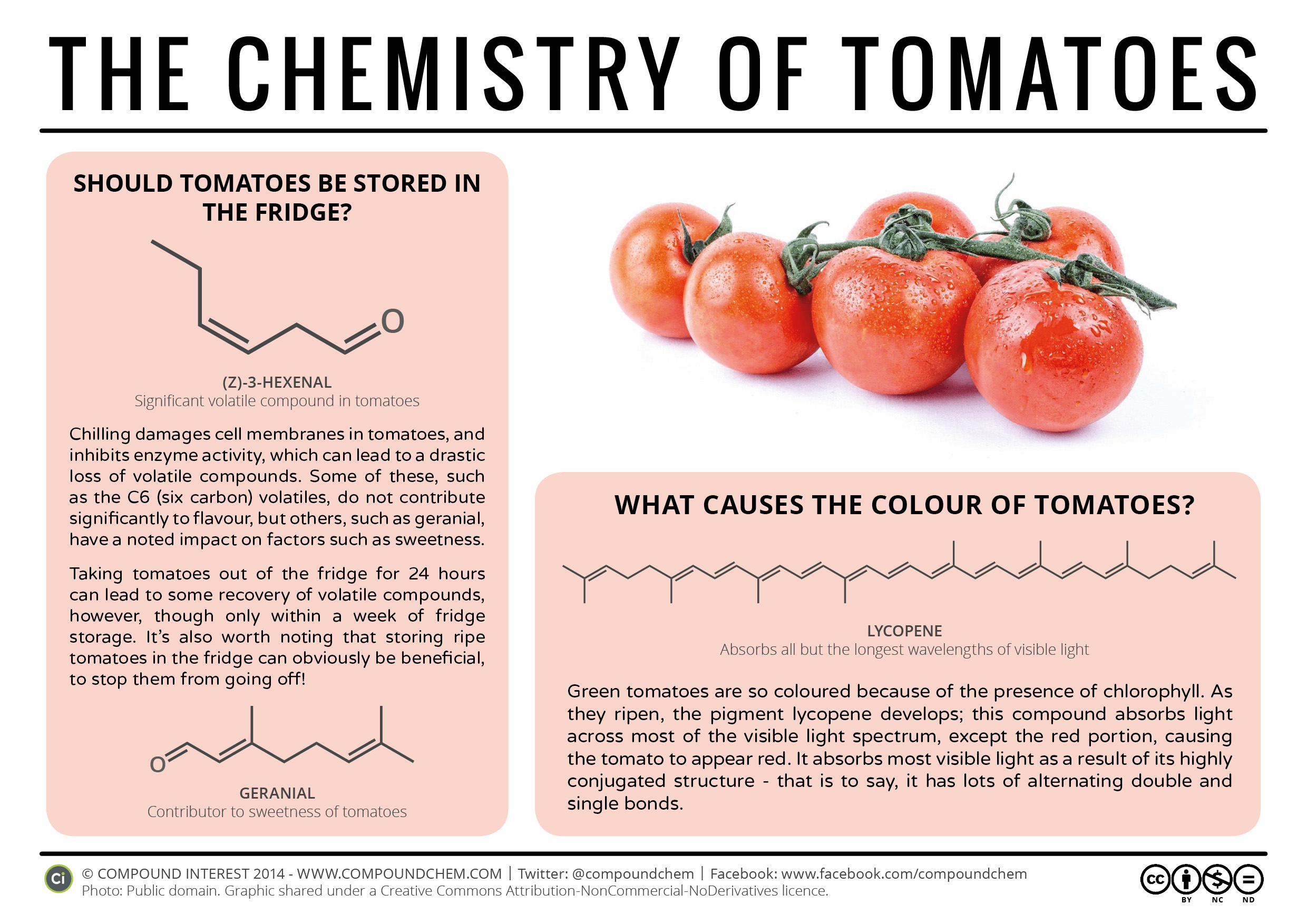
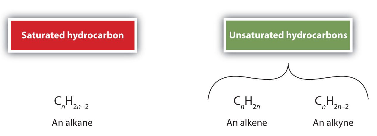
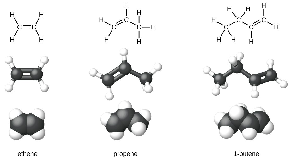
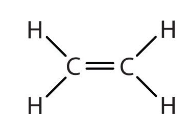
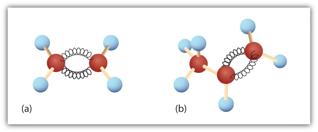
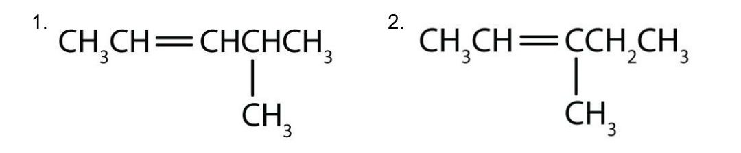
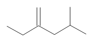
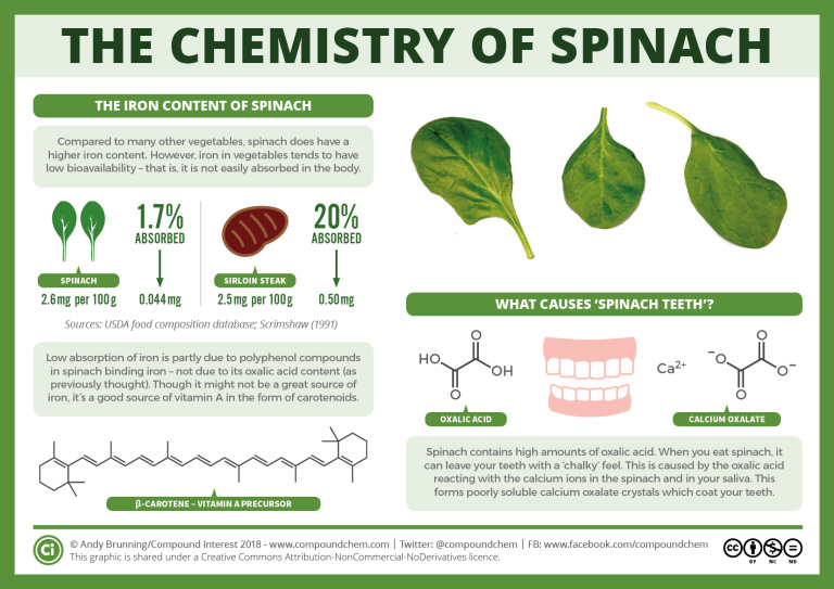
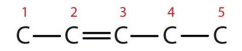
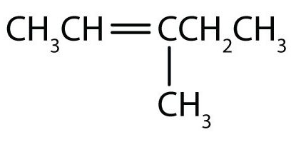

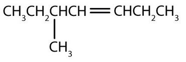


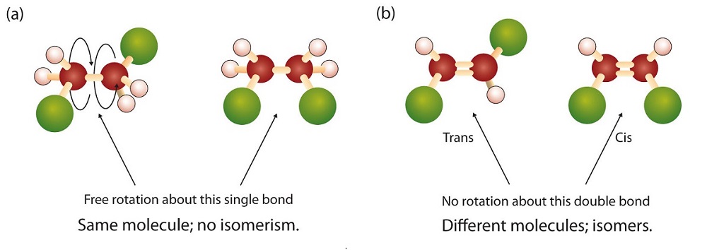

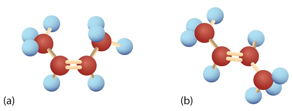
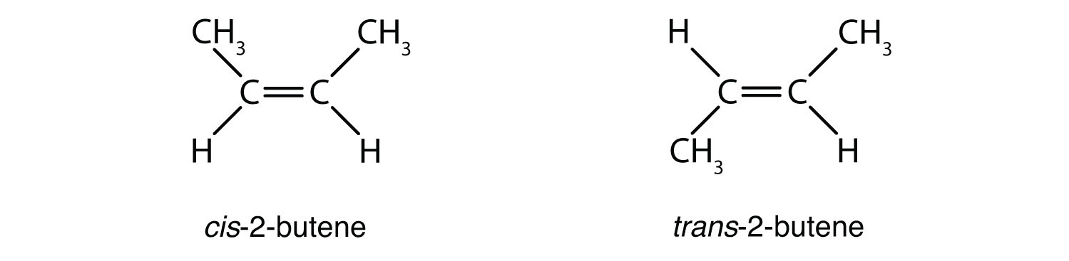

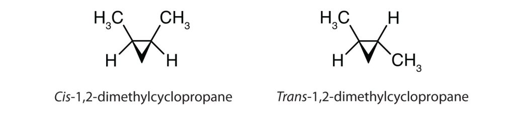

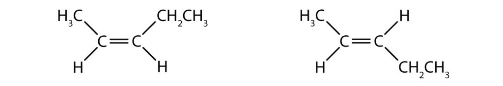


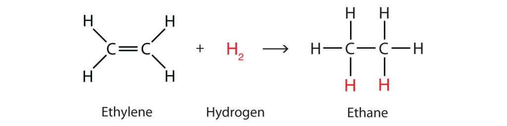
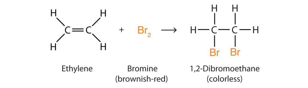
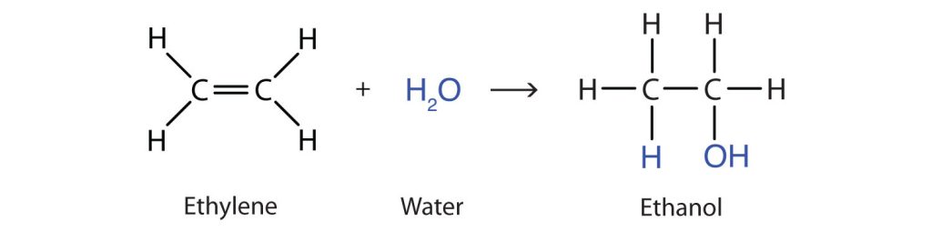
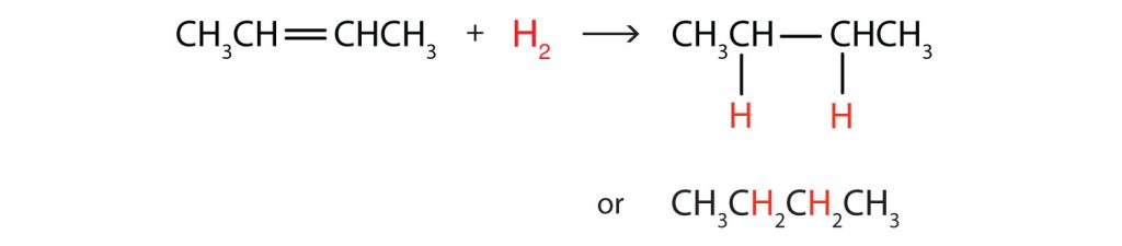
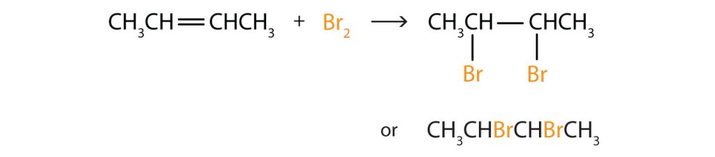
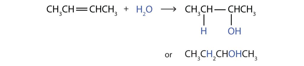
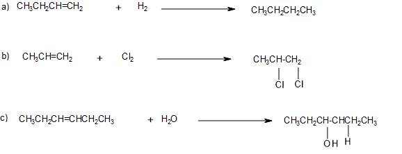
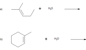
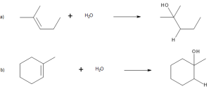
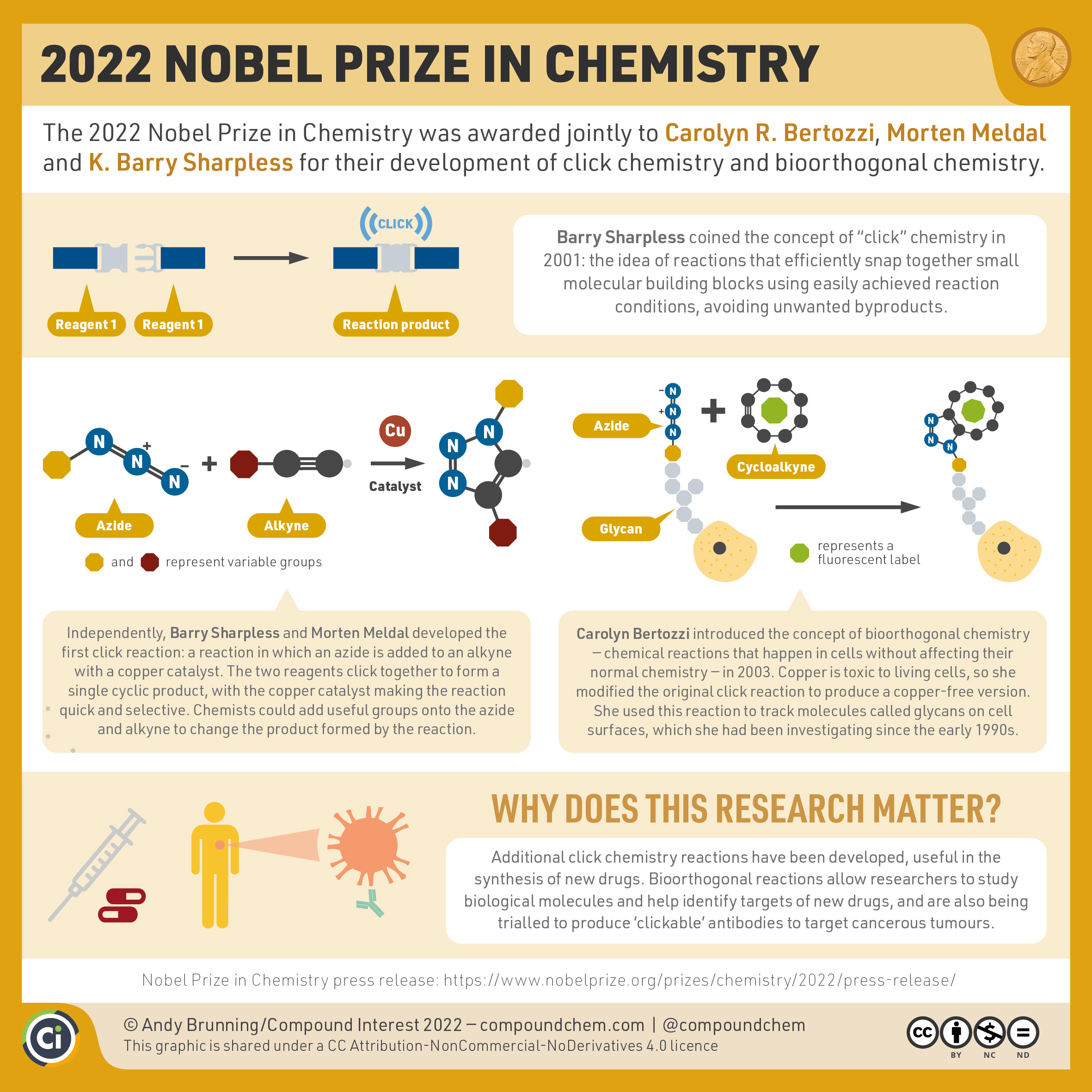
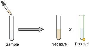
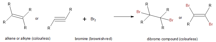
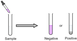
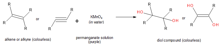
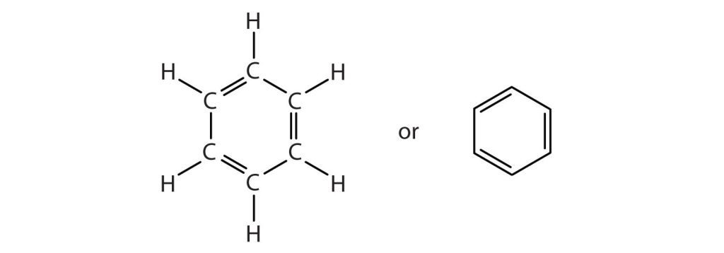


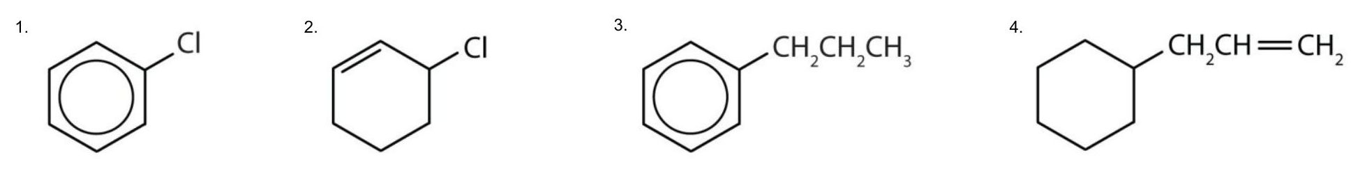
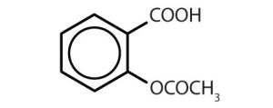
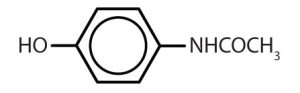
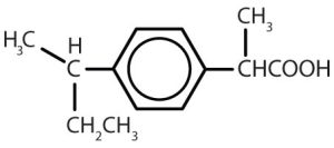
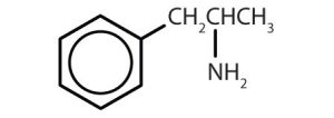
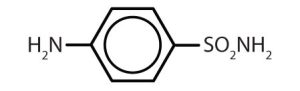
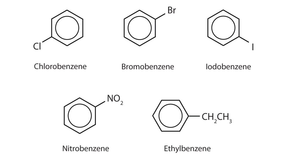
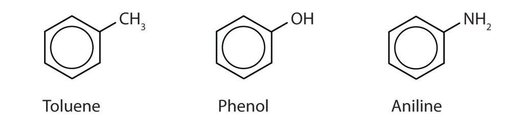
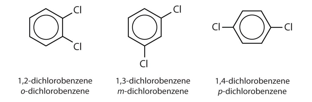
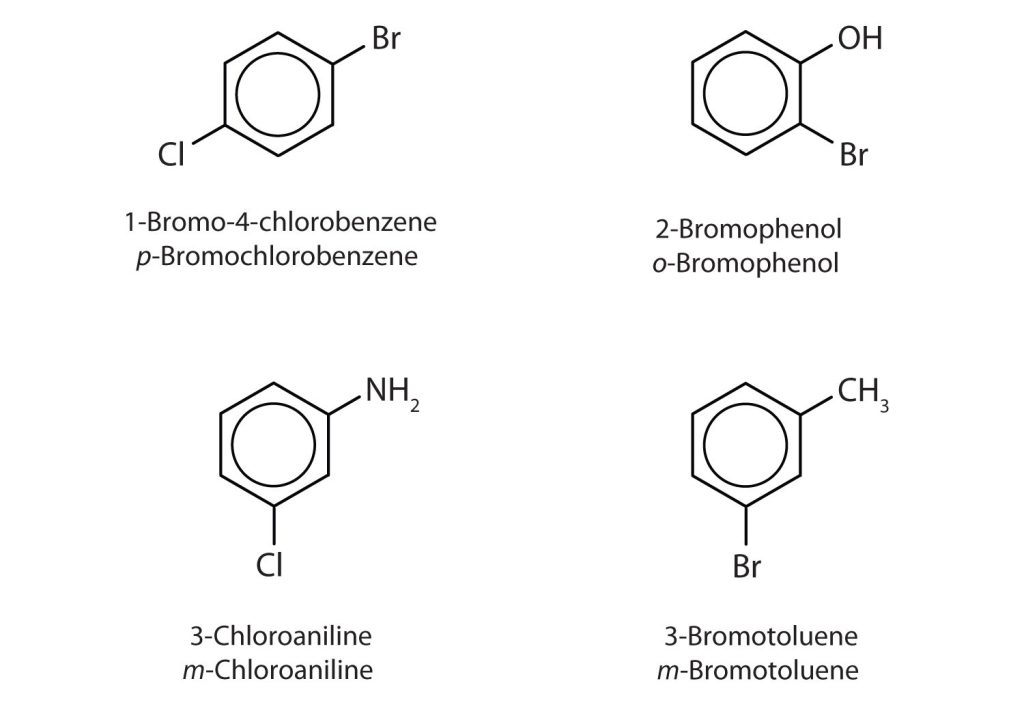
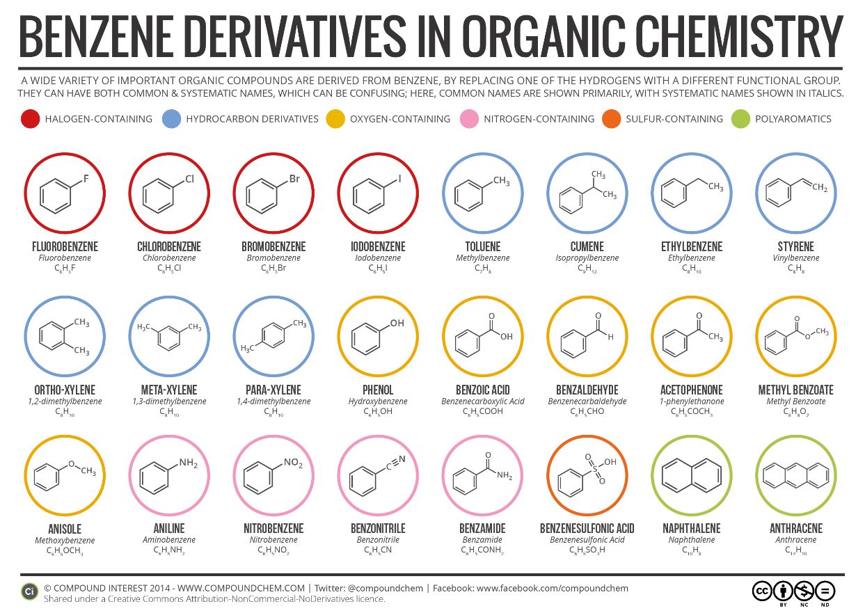
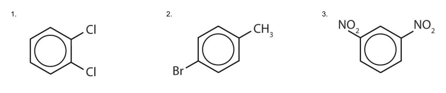


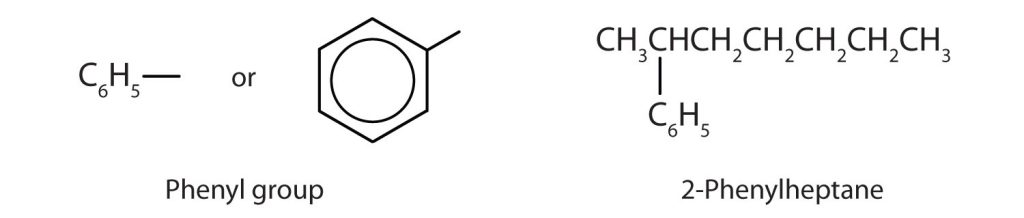
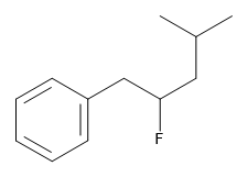
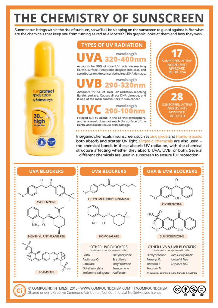
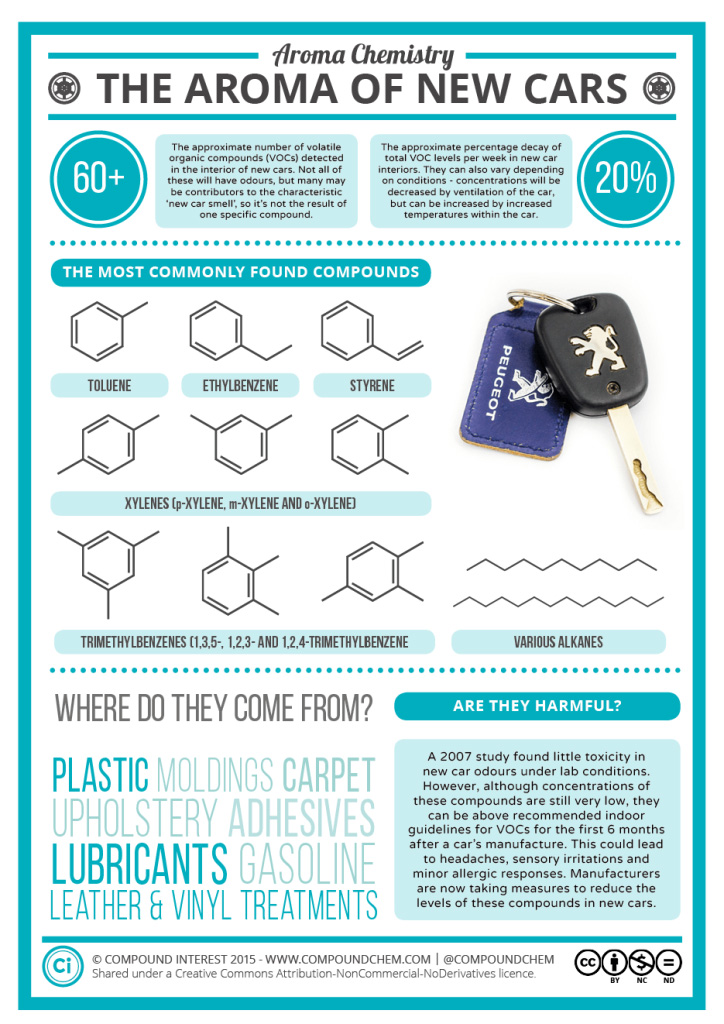
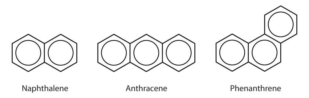
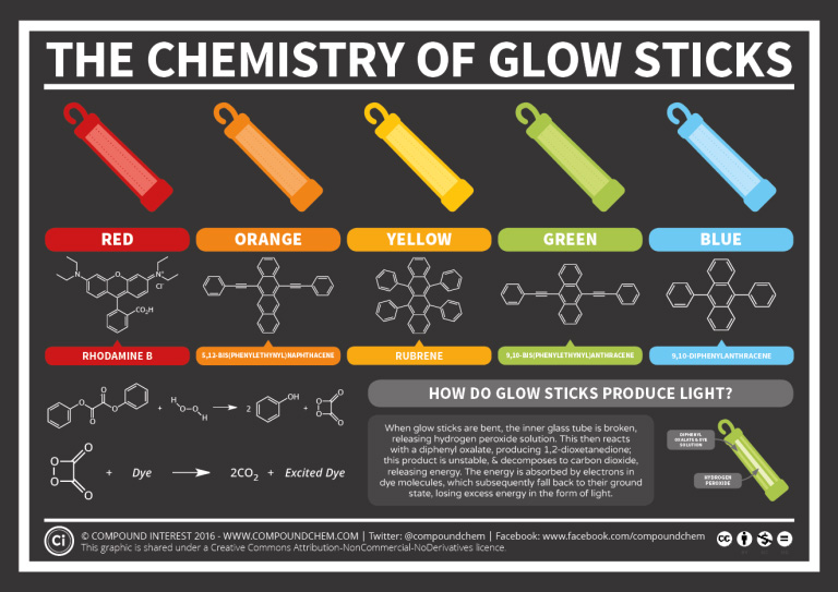
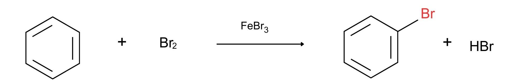
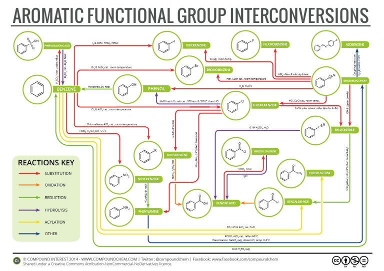
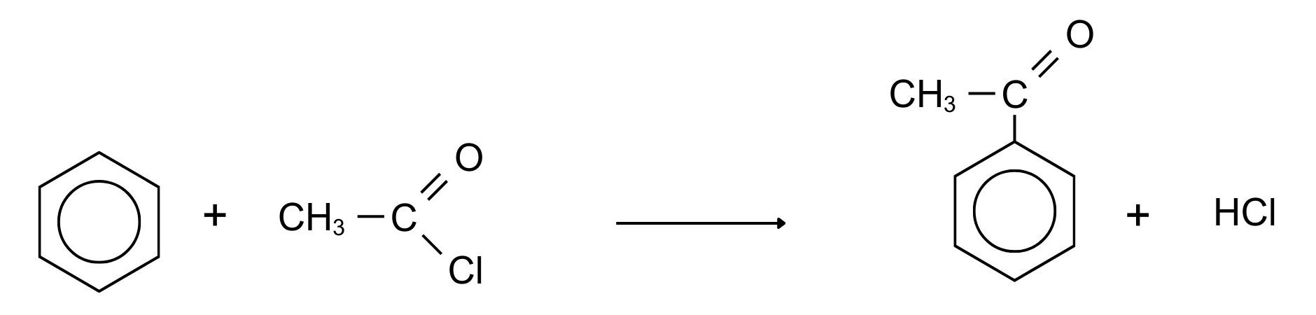
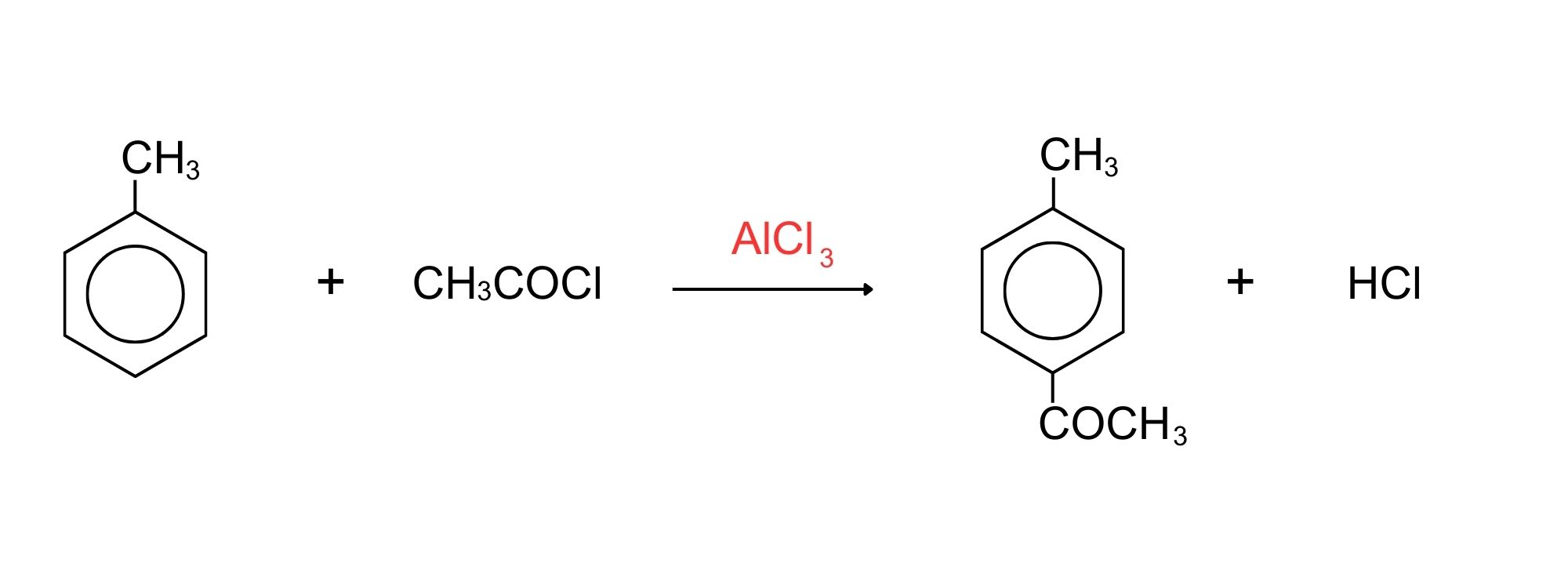
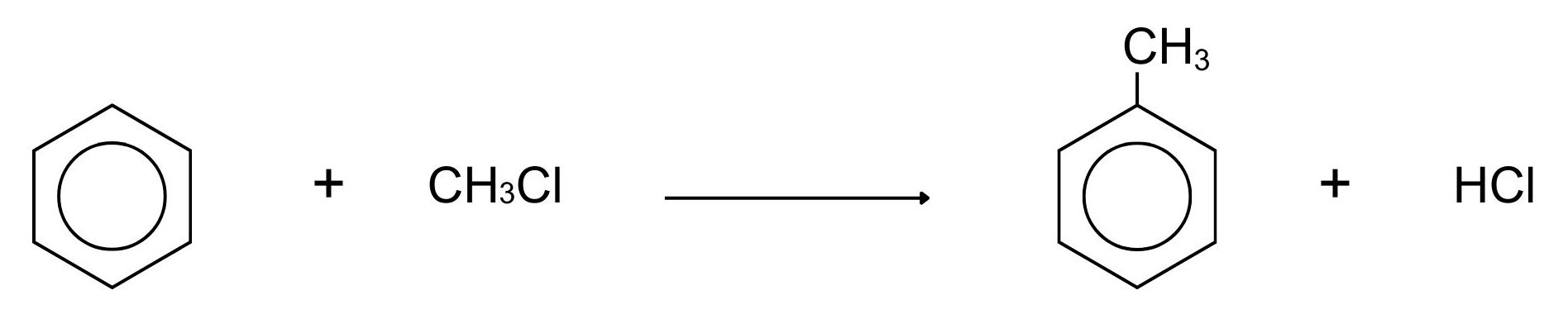
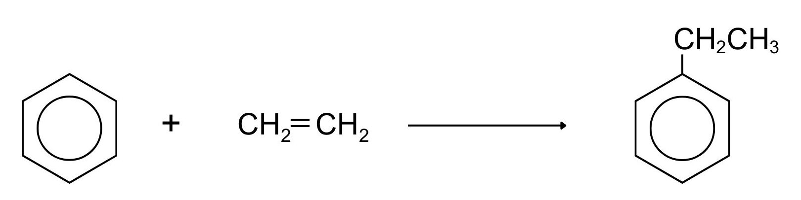
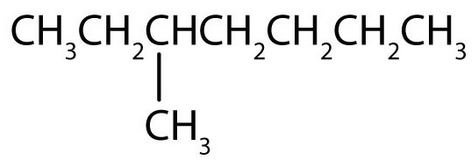
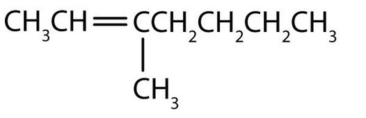
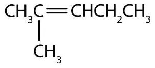
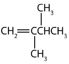
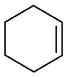
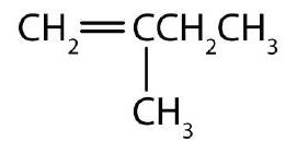
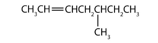
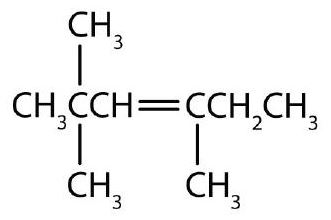
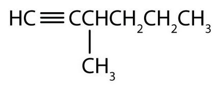
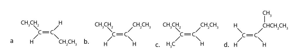
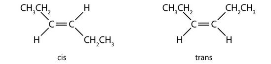
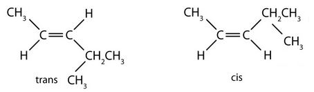
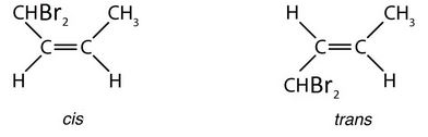
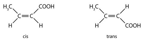
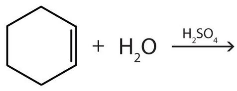
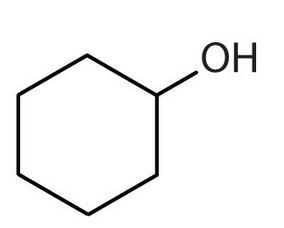
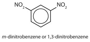 ortho = 1,2 disubstitution
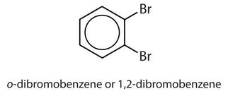 para = 1,4 disubstitution or 1-bromo-4-chlorobenzene
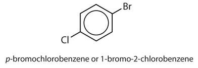
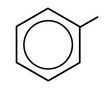 3-phenyloctane:
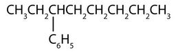
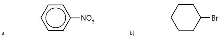
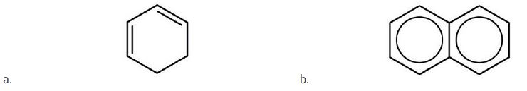
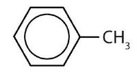
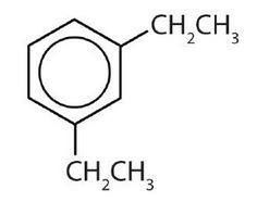
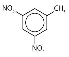
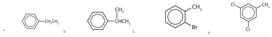
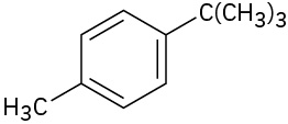
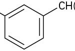


 ,
,  filter off solid, dry and heat
 ,
,  cat., room temperature
 ,
,  cat., room temperature
 \small}") , room temperature
, room temperature \small}")
 & conc.
  . reflux
 &
&  cat., room temperature
 &
&  cat., room temperature
 ,
,  ,
,  \small}") ,
,  -33 degrees Celsius, then dilute acid
 &
&  , dry ether
 with
with  salt cat., 200 atm and 350 degree Celsius then
  , polar solvent, reflux (also for
  )
)
 , heat
 \small}") , dilute
, dilute 
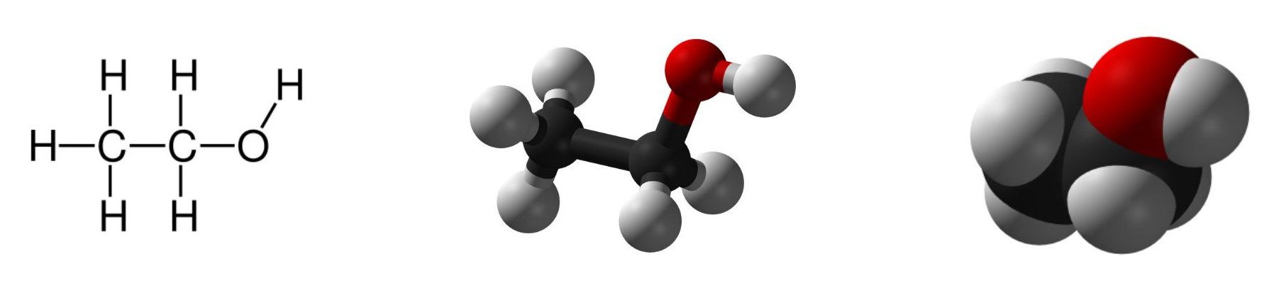
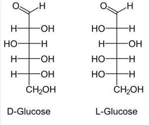


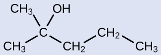
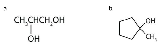


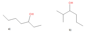
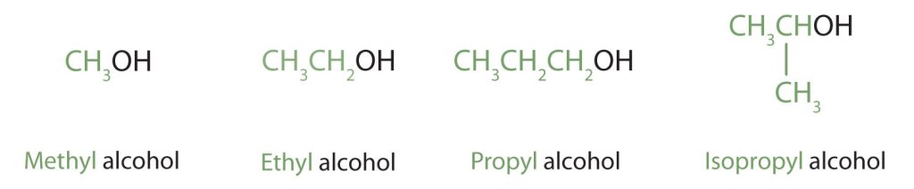


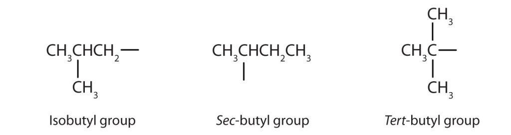
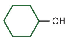
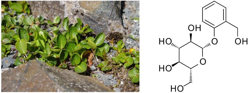
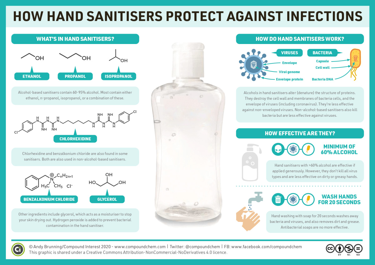


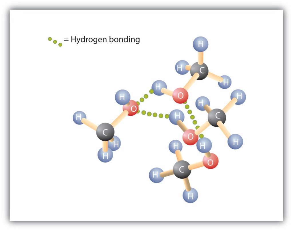
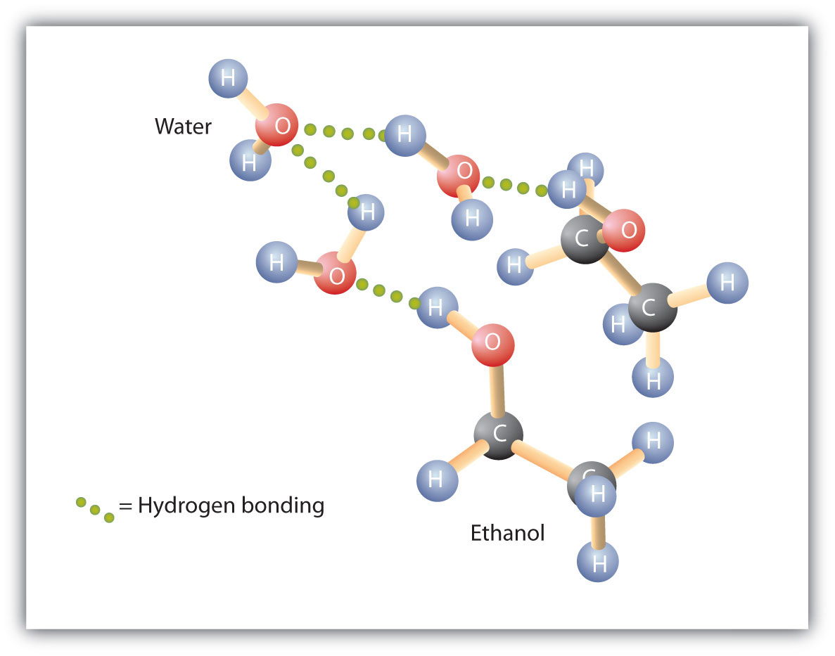
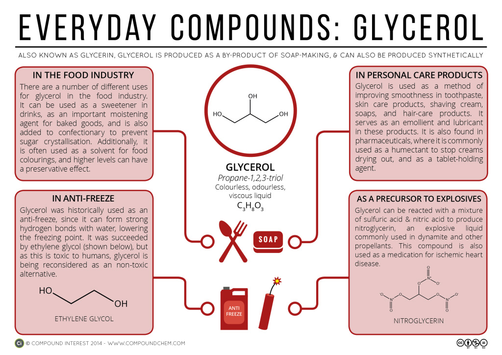

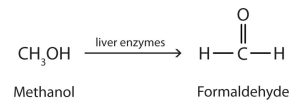
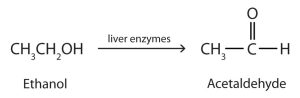
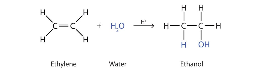
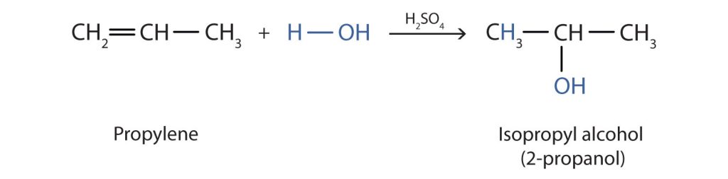

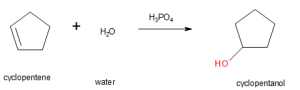

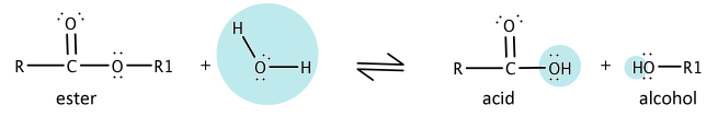
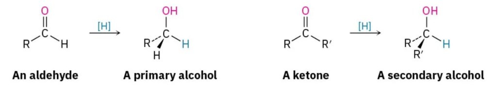
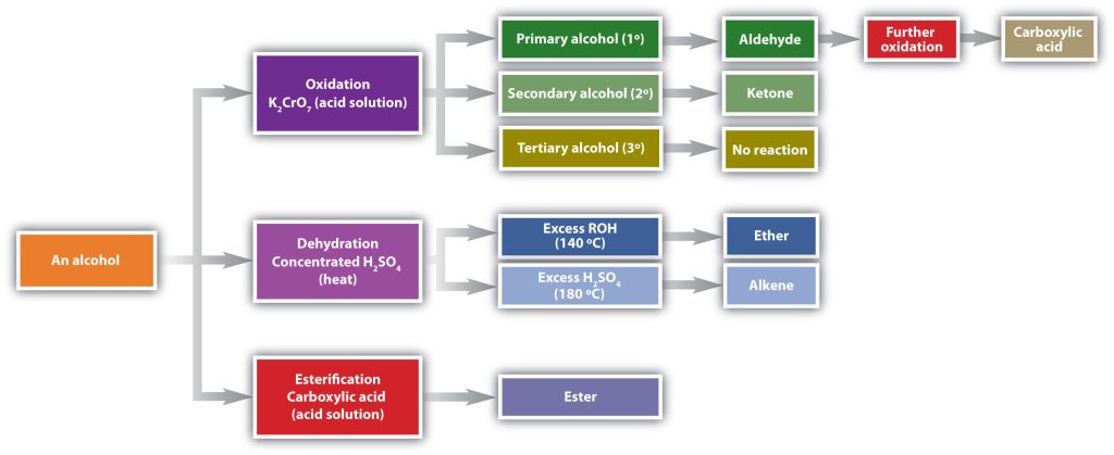


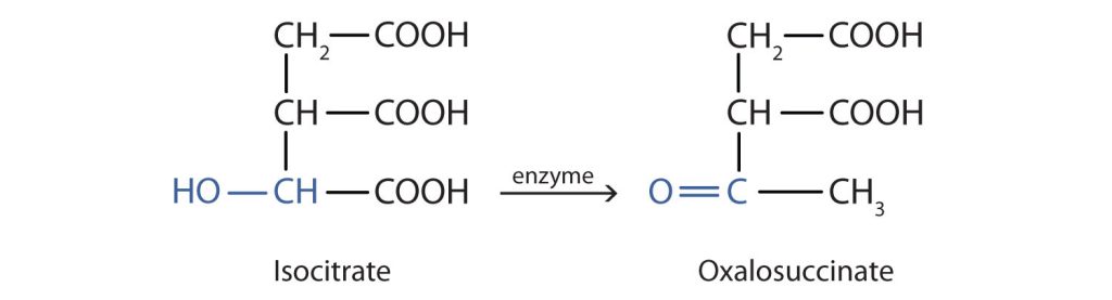
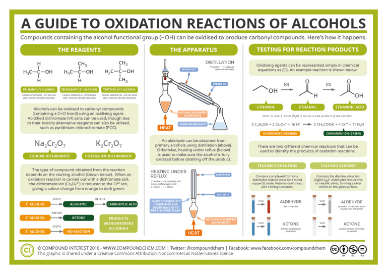
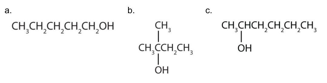
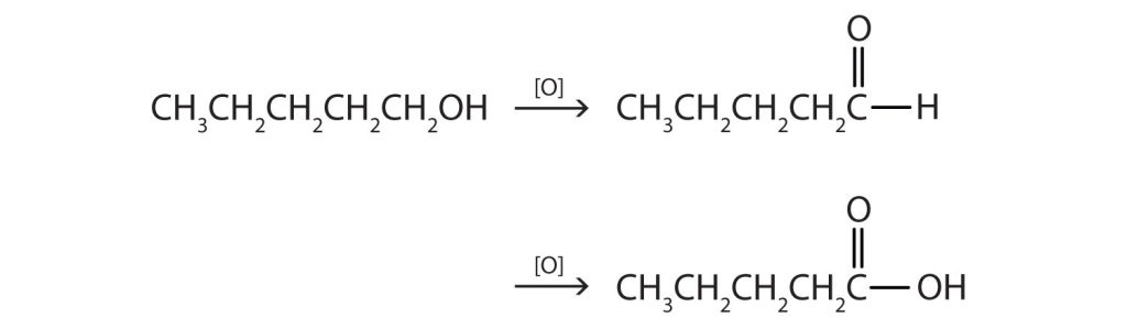


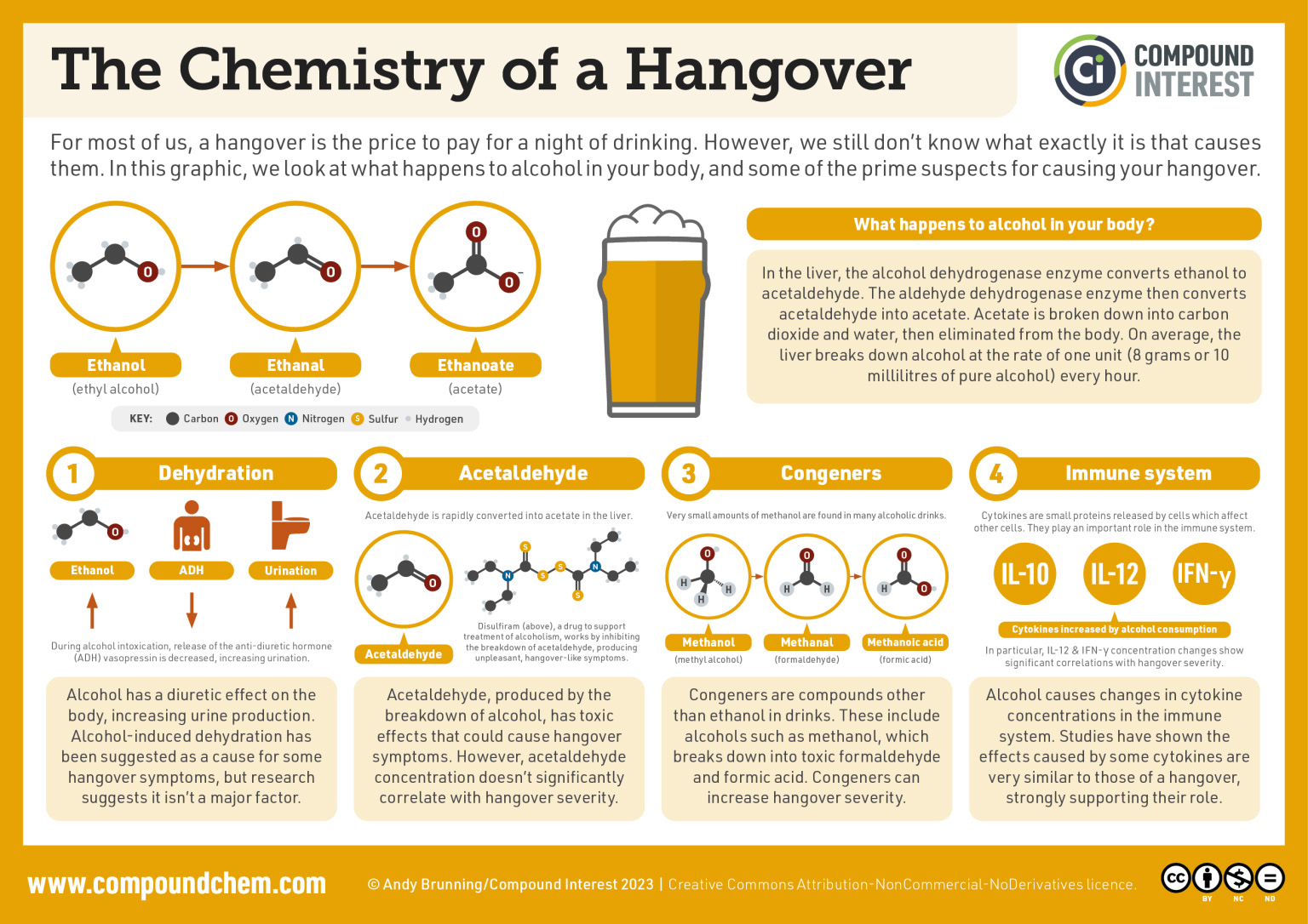
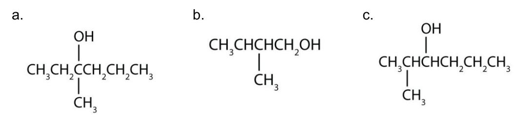
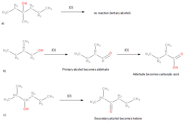
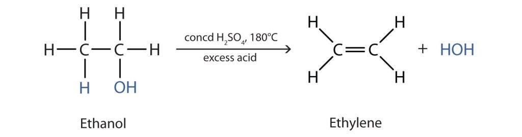
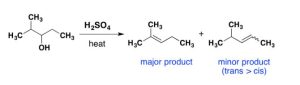
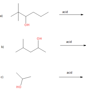
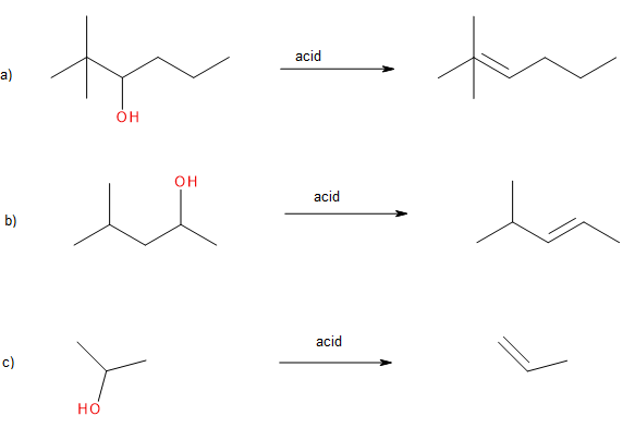
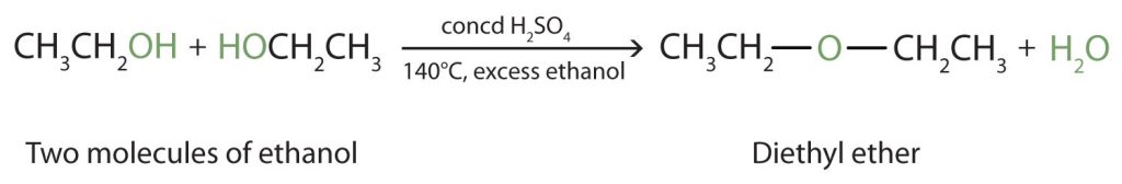
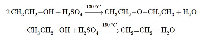
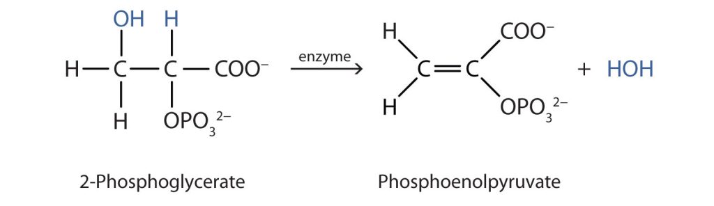
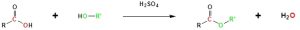
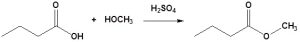


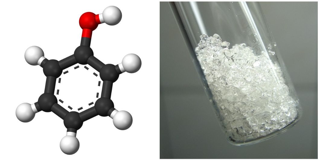

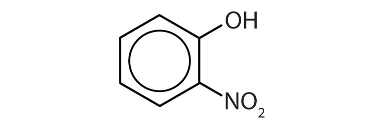

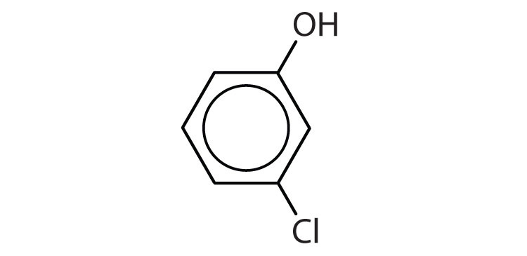
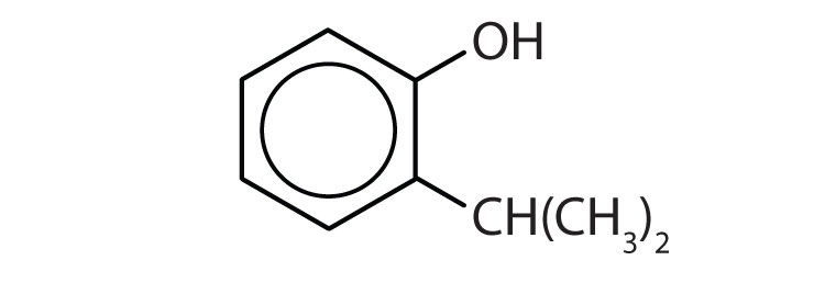

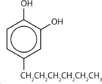
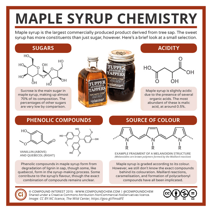
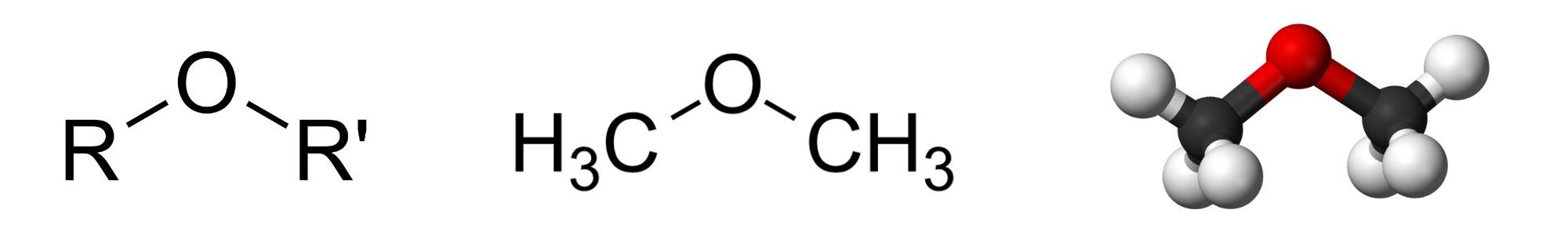


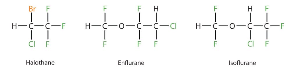

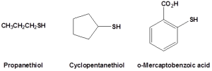
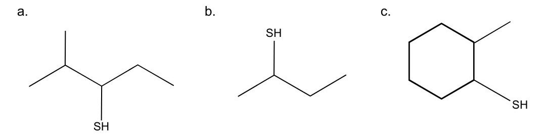
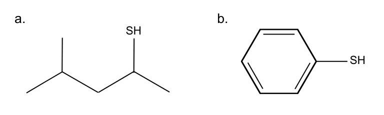
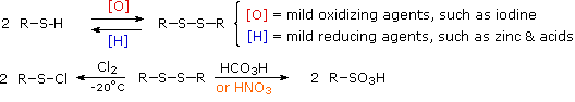
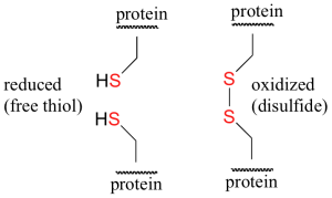
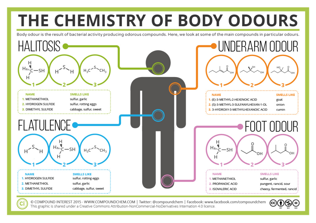


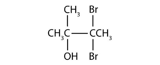
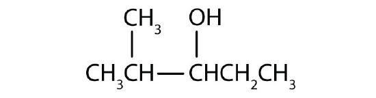


 b)
b)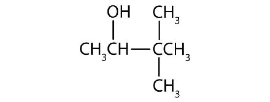 c)
c)


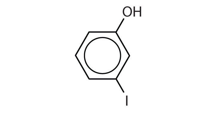 b)
b)


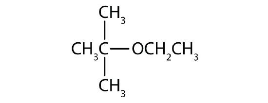
 .
.


 ions. Aldehydes reduce these ions to red copper (I) oxide. Ketones don’t react with Fehling’s solution.
_{2}\sqsupset ^{+}") . Aldehydes reduce this to metallic silver, forming a silver mirror on the glass surface.
. Aldehydes reduce this to metallic silver, forming a silver mirror on the glass surface. Sodium ion concentration and
  Calcium ion concentration.
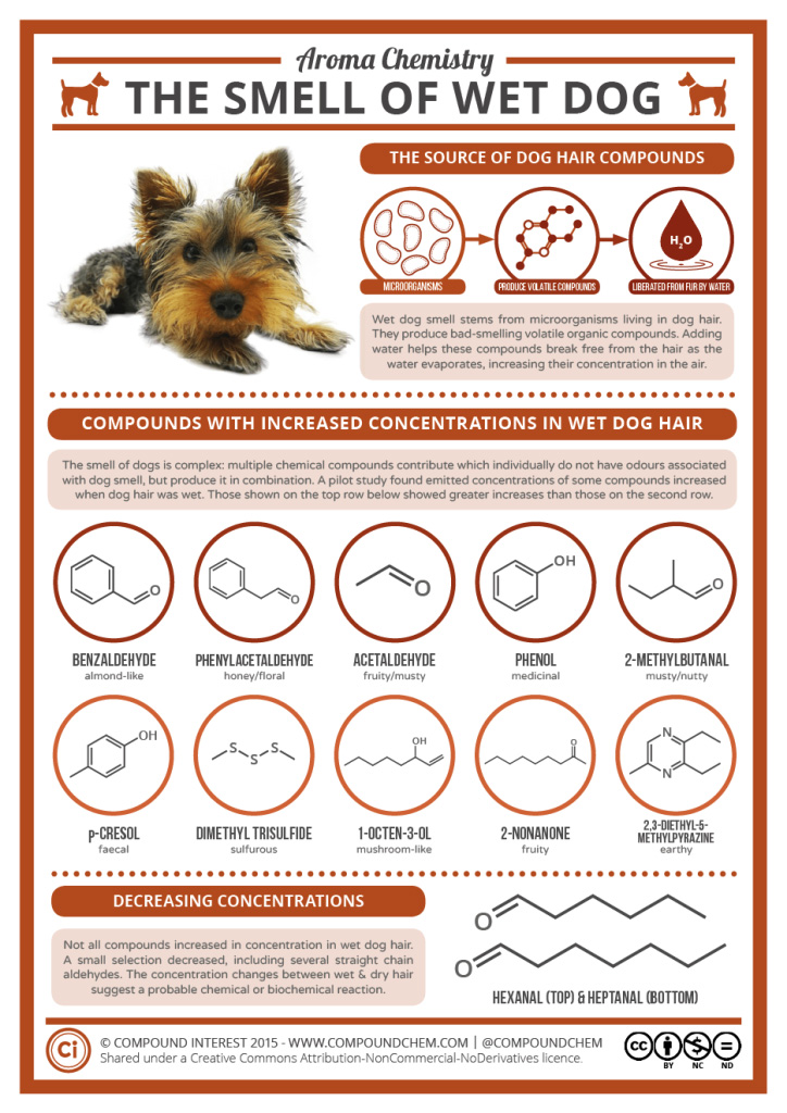


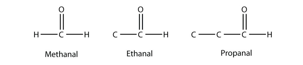

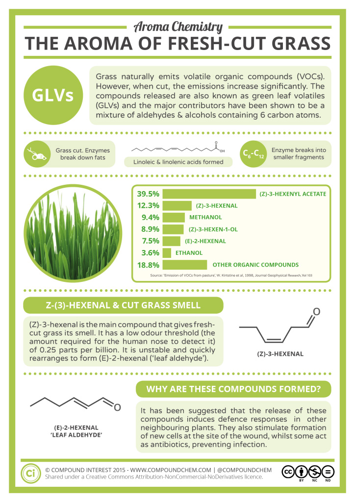
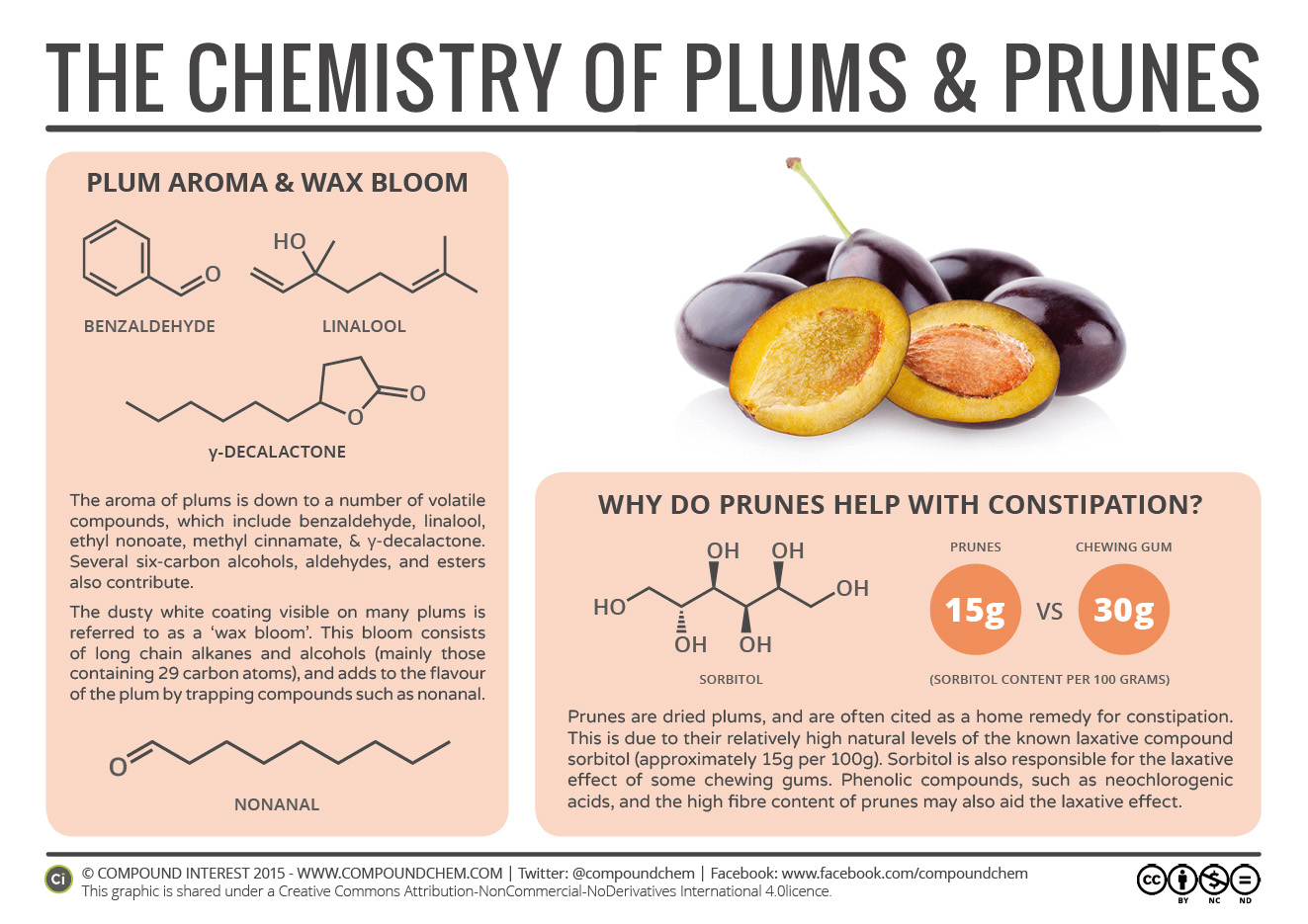
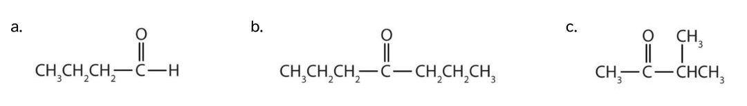
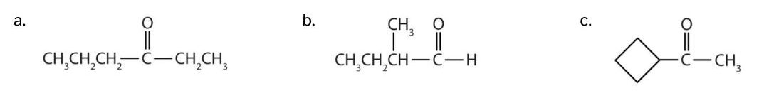
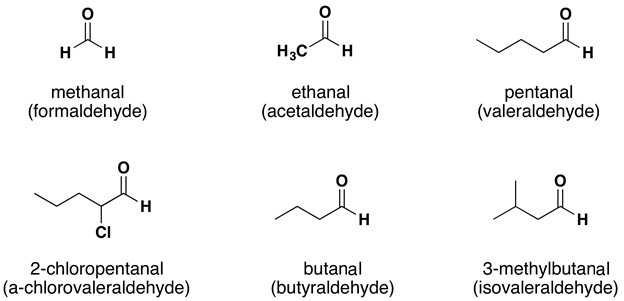

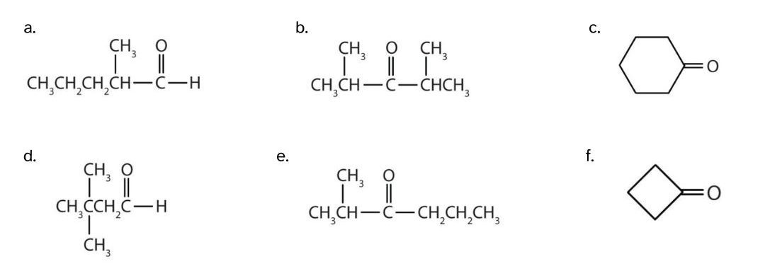
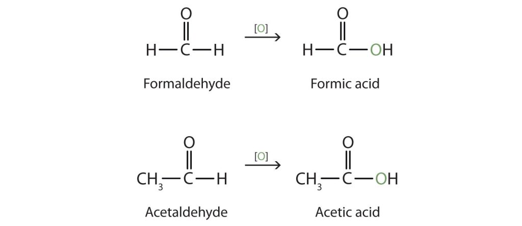


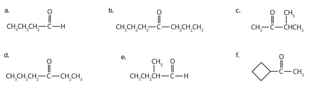


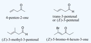


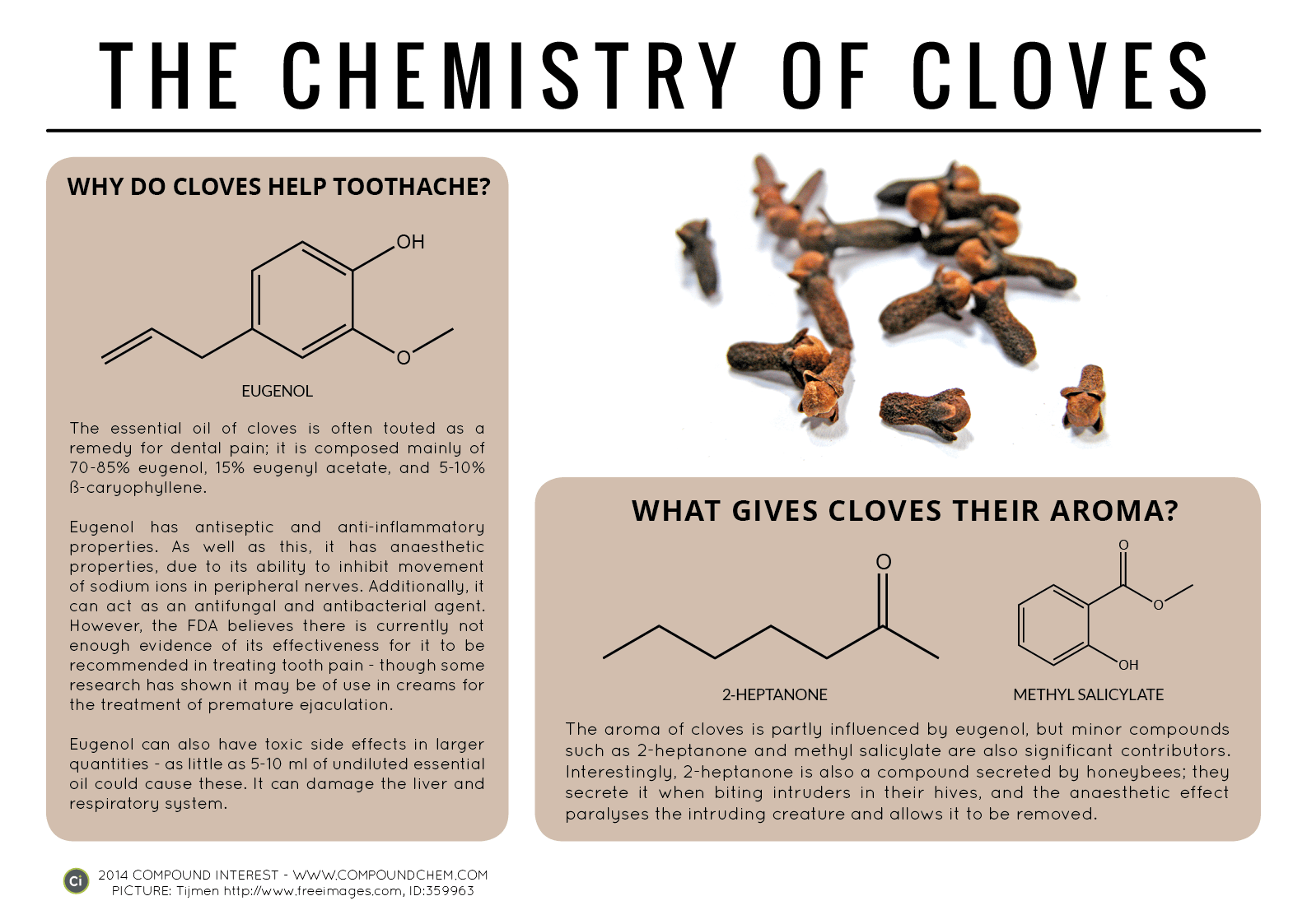
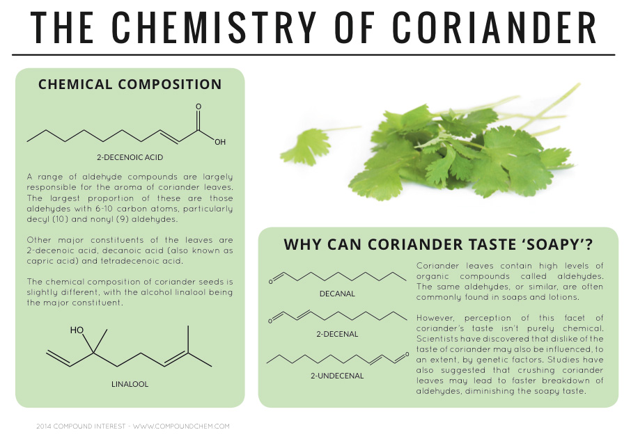
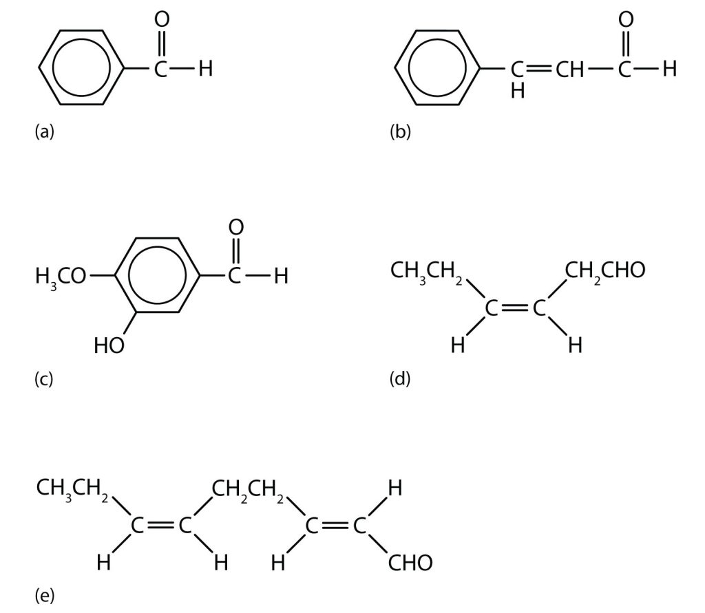
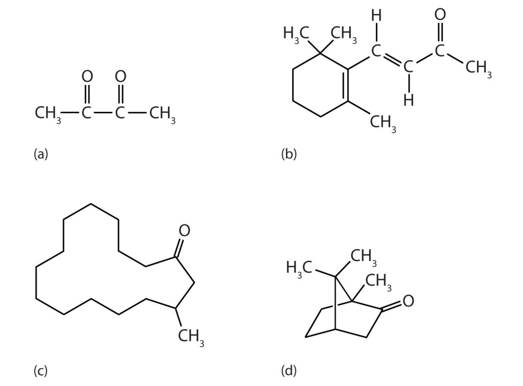


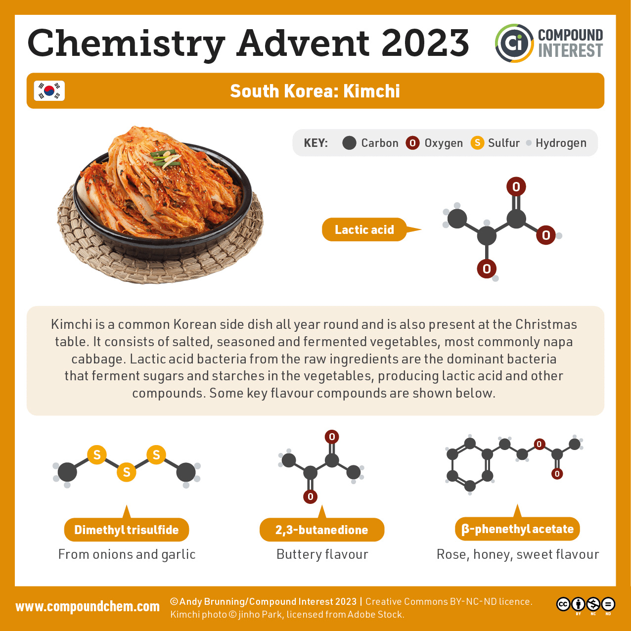
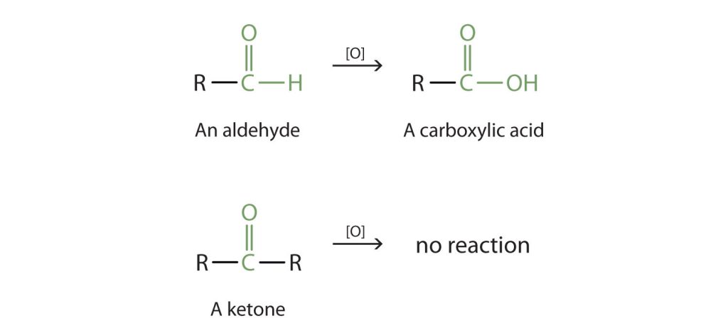

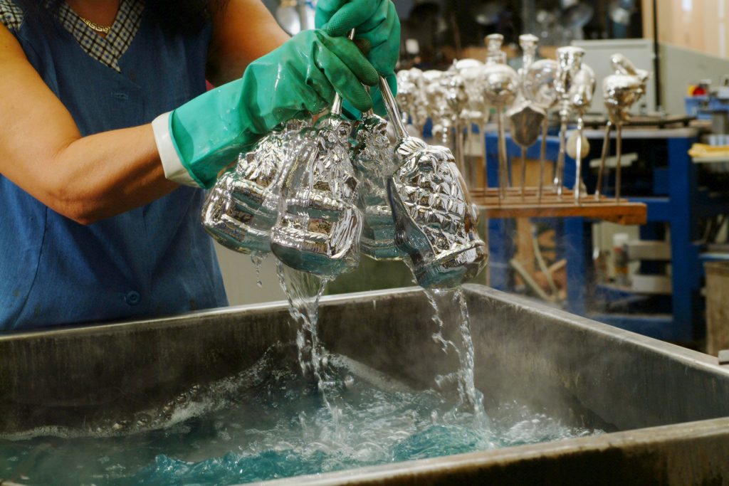
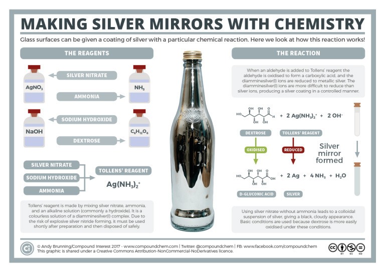
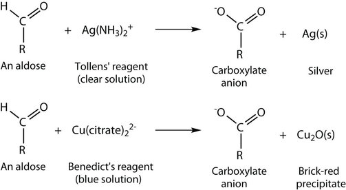
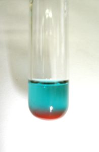
![Reduction of a carbonyl group (where the oxygen is depicted in a pink colour) to an alcohol (the OH group is depicted in a pink colour). The reducing agent used is [H]](https://ecampusontario.pressbooks.pub/app/uploads/sites/3009/2023/12/reduction.jpg)
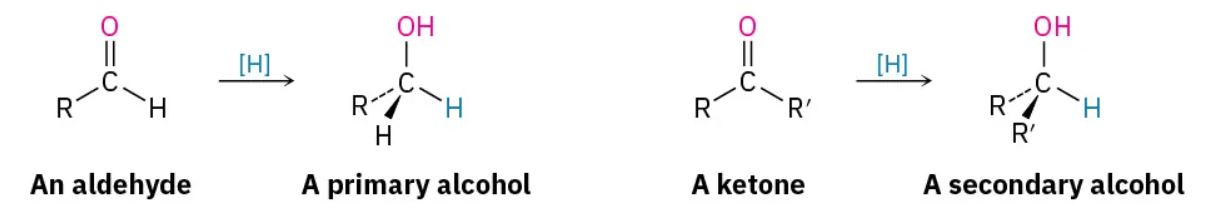
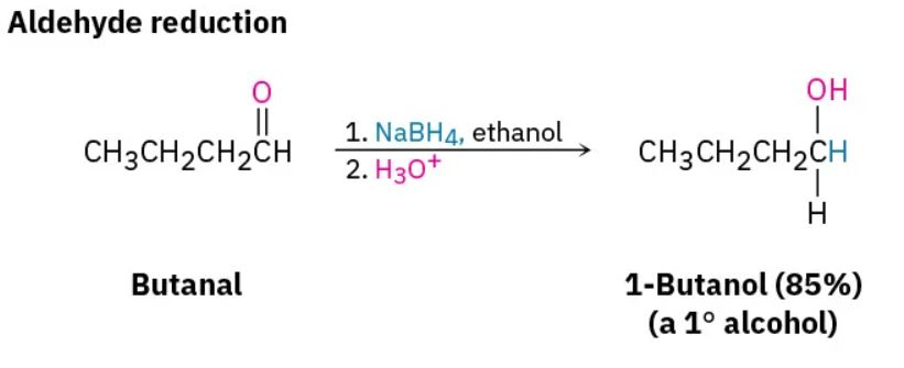
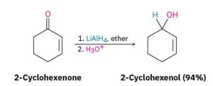
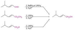
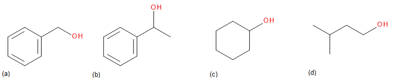
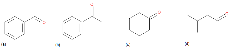
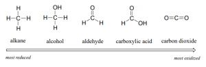
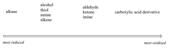


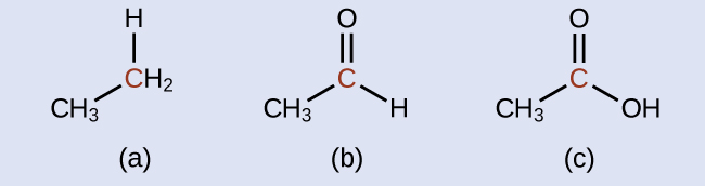
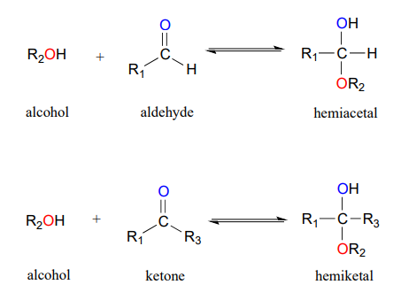
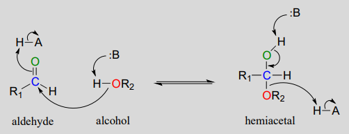
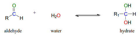

 RCH(OH)CN (a cyanohydrin)
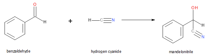

 3-bromobenzaldehyde
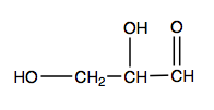
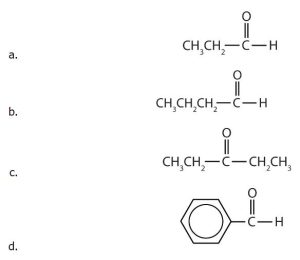
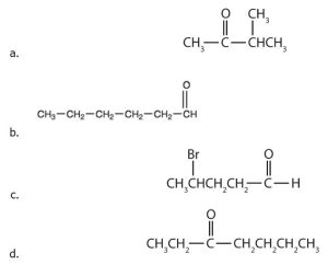
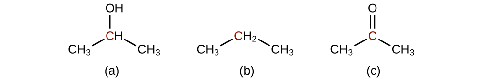
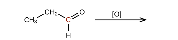 Check answer


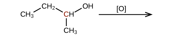


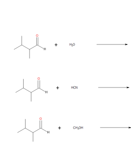
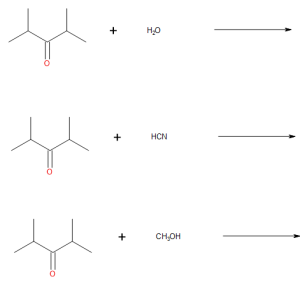
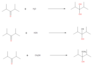


_{2^{+}}") contains silver nitrate, sodium hydroxide and ammonia.
contains silver nitrate, sodium hydroxide and ammonia._{2^{2}} + 2OH^{-} \small}") where Dextrose is oxidized and Tollen’s reagent is reduced to silver results in the silver mirror forming
where Dextrose is oxidized and Tollen’s reagent is reduced to silver results in the silver mirror forming  .
.
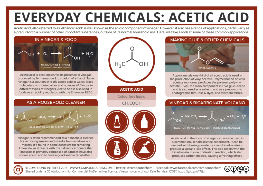
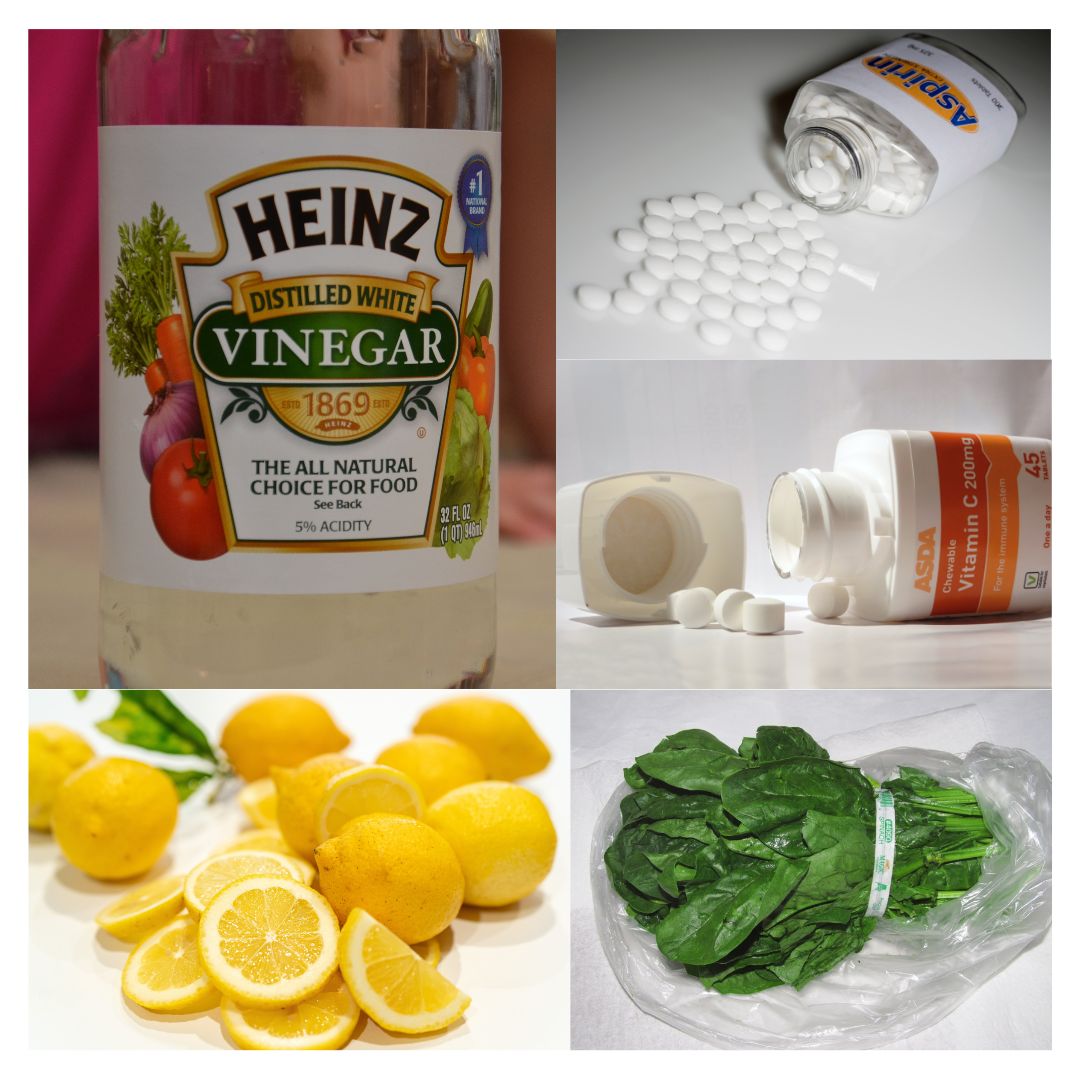
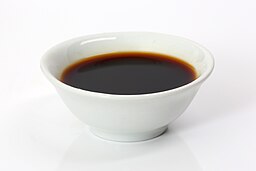

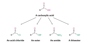
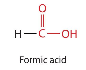
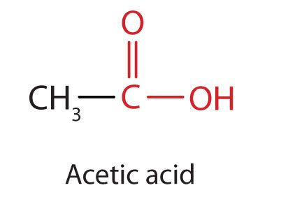
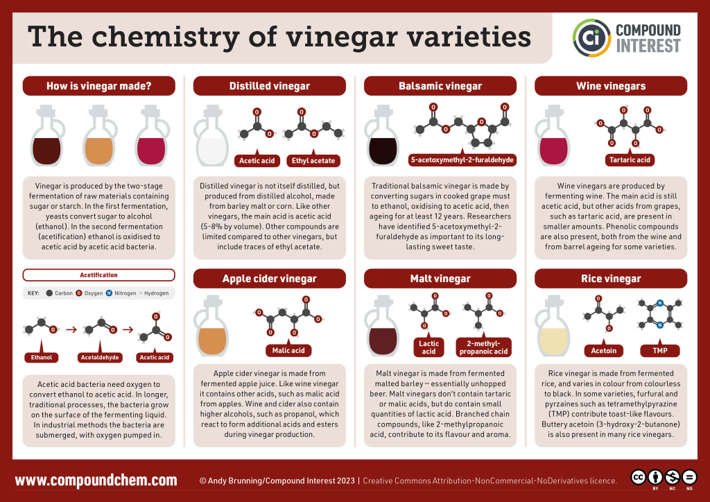
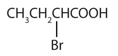
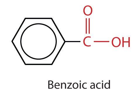
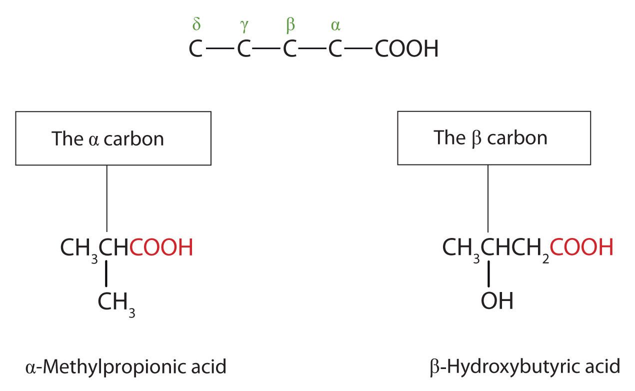
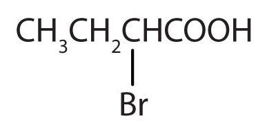
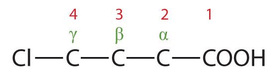
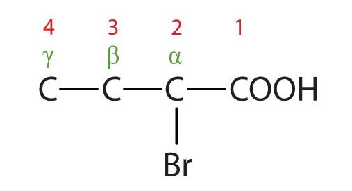
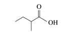
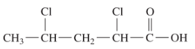
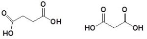
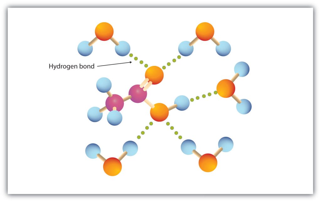

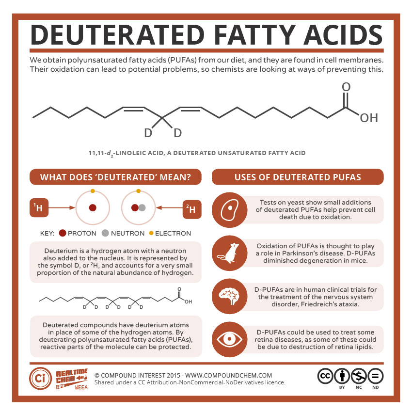
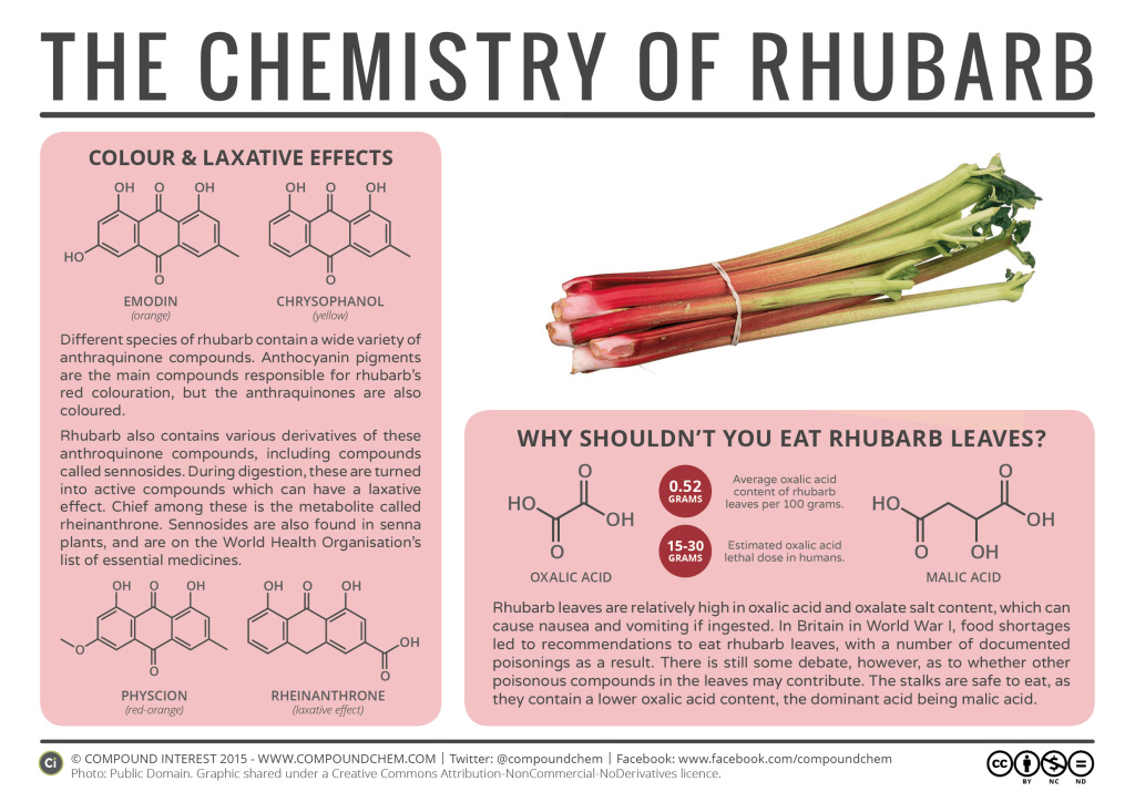
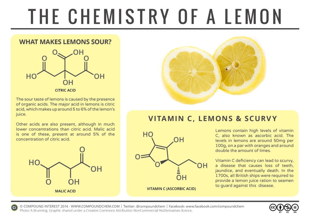
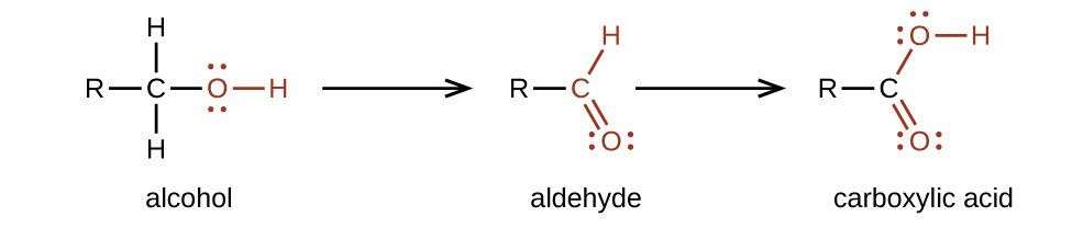


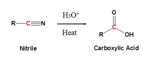
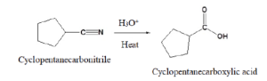
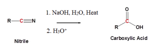
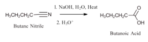
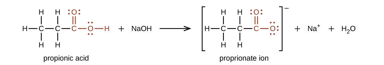

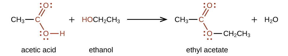
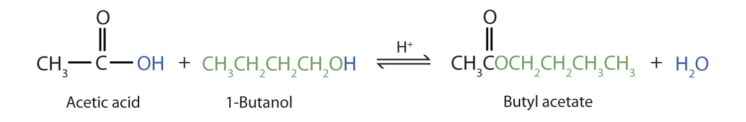

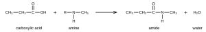
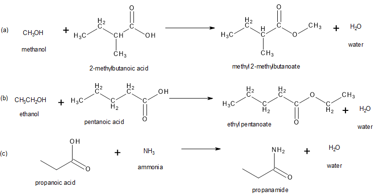
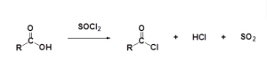
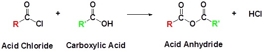
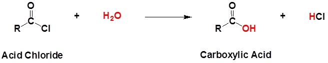
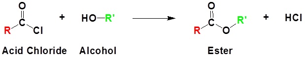
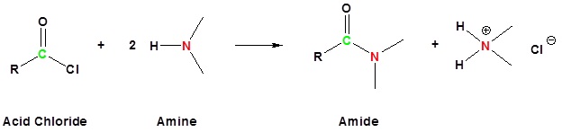
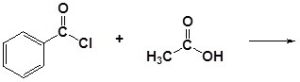
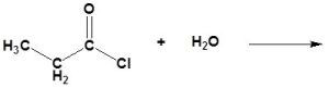
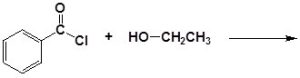
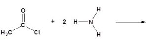
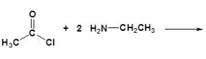
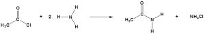


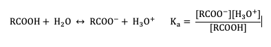
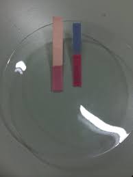
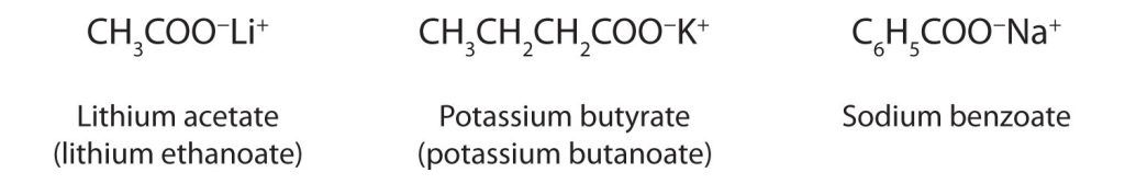
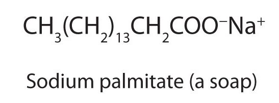
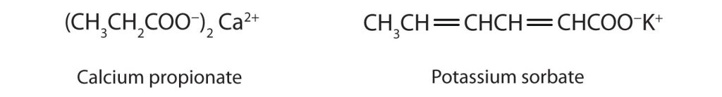
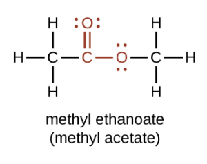
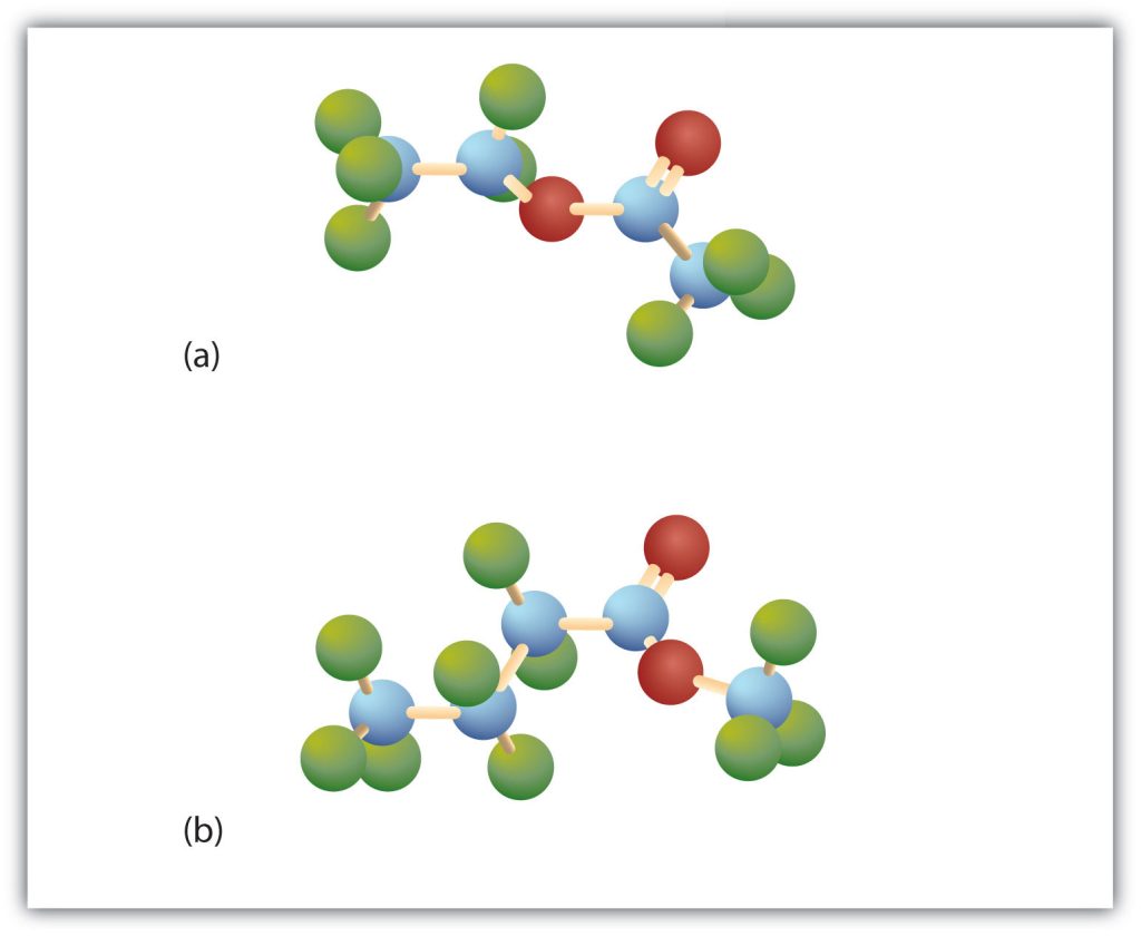


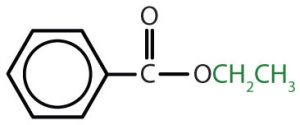
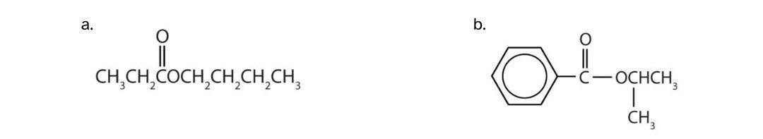
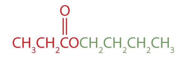
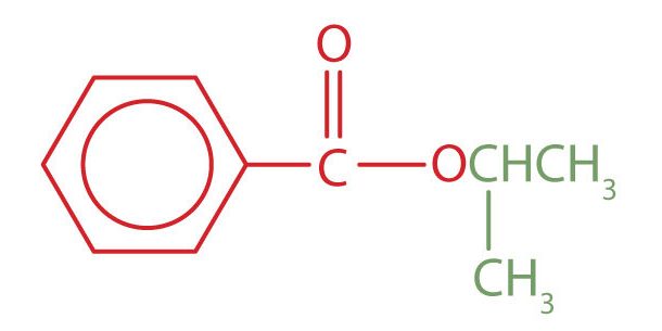
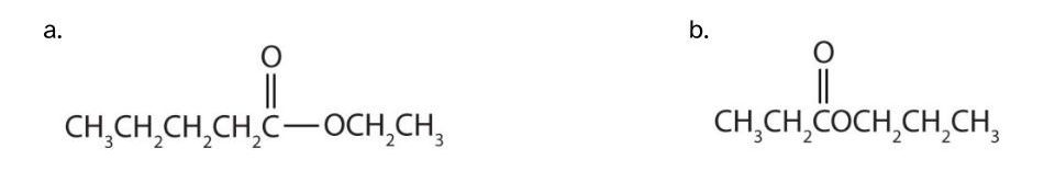
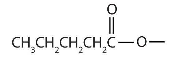
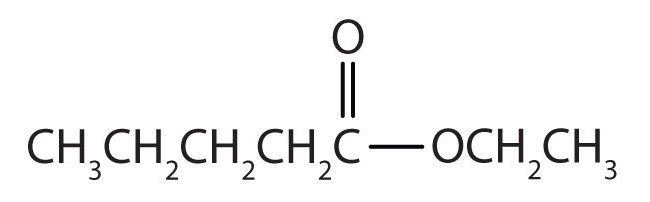
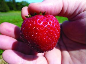


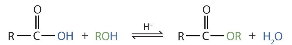
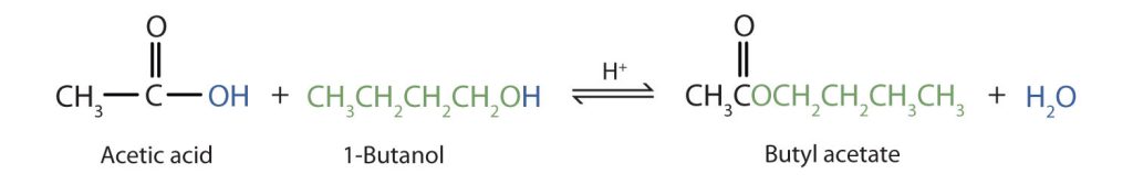
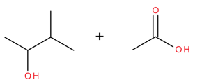
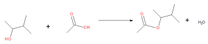
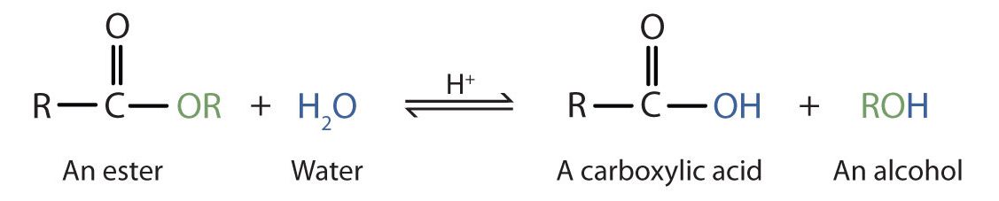
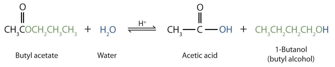
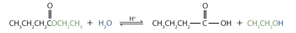
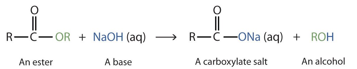
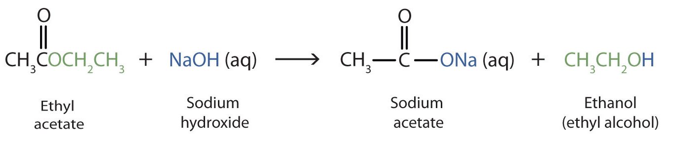
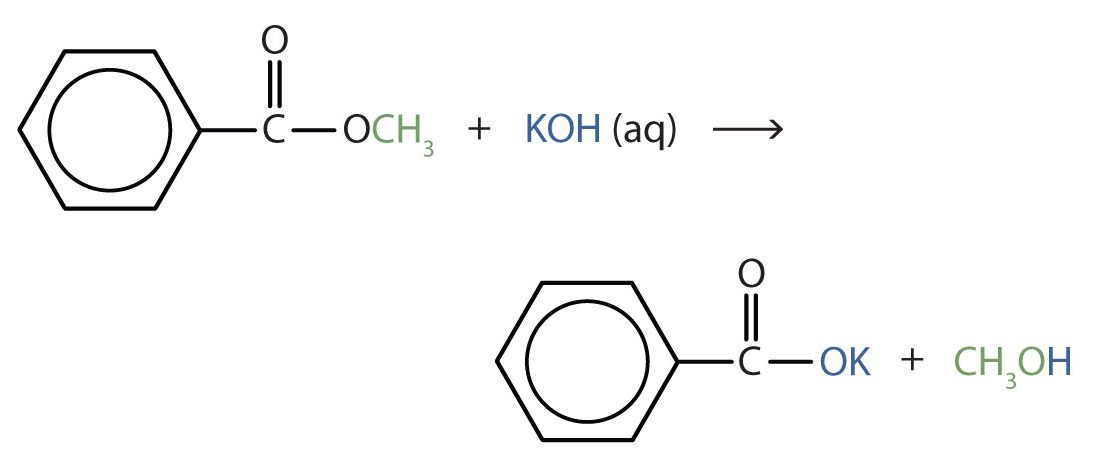
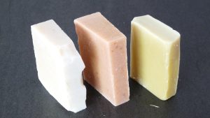
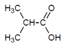 Check answer
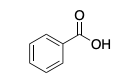 Check answer
 Check answer
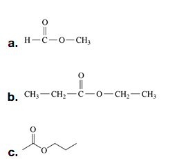
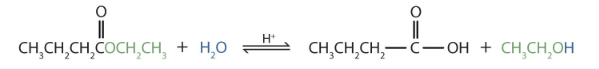


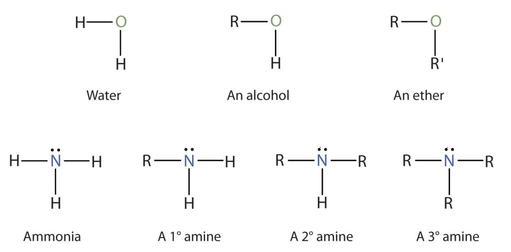
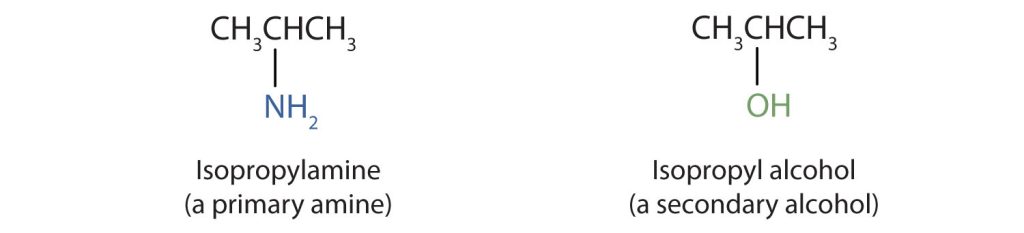


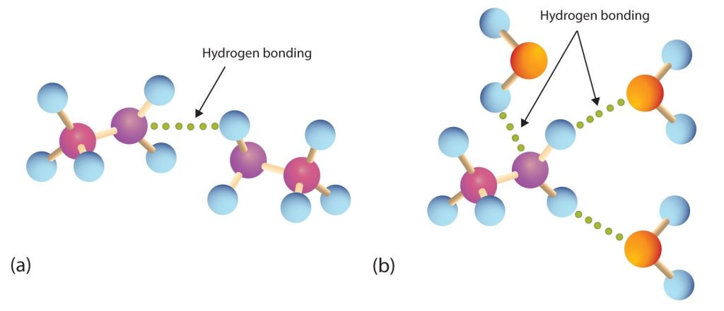

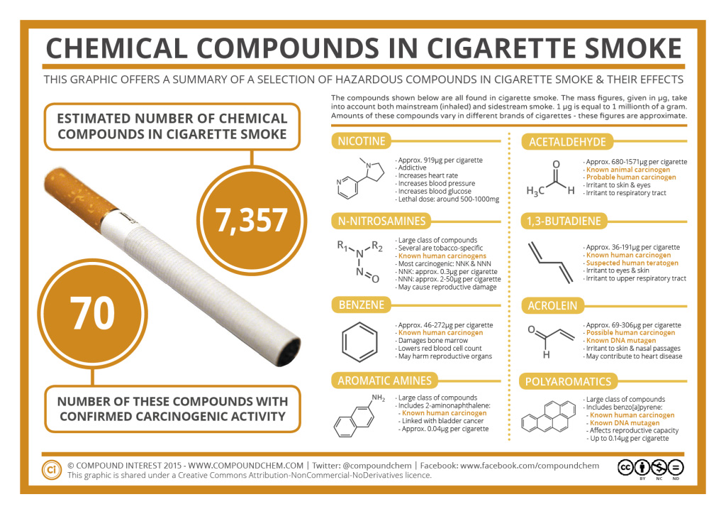

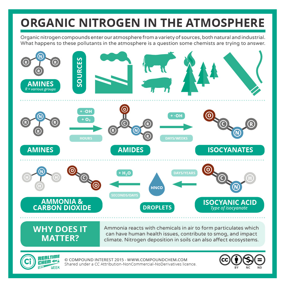
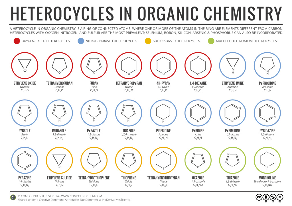
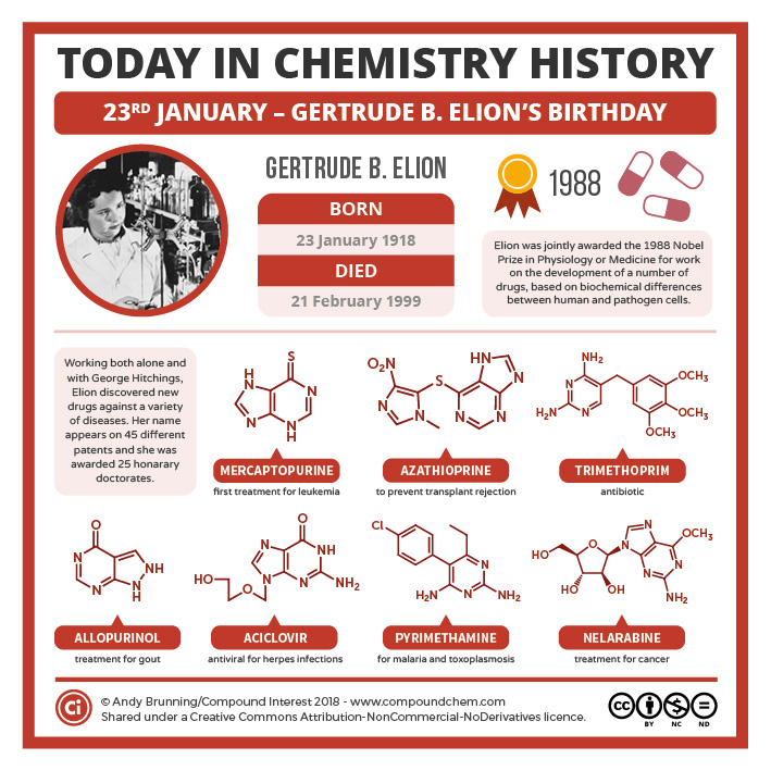
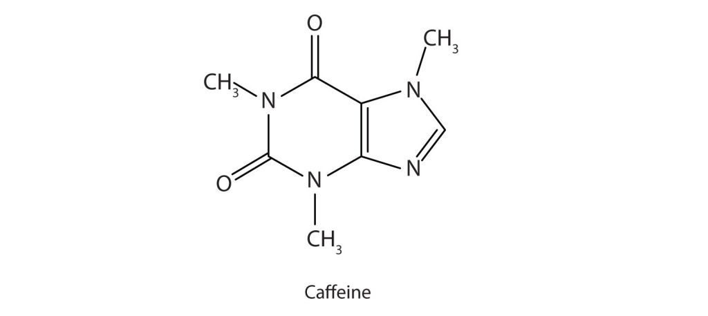
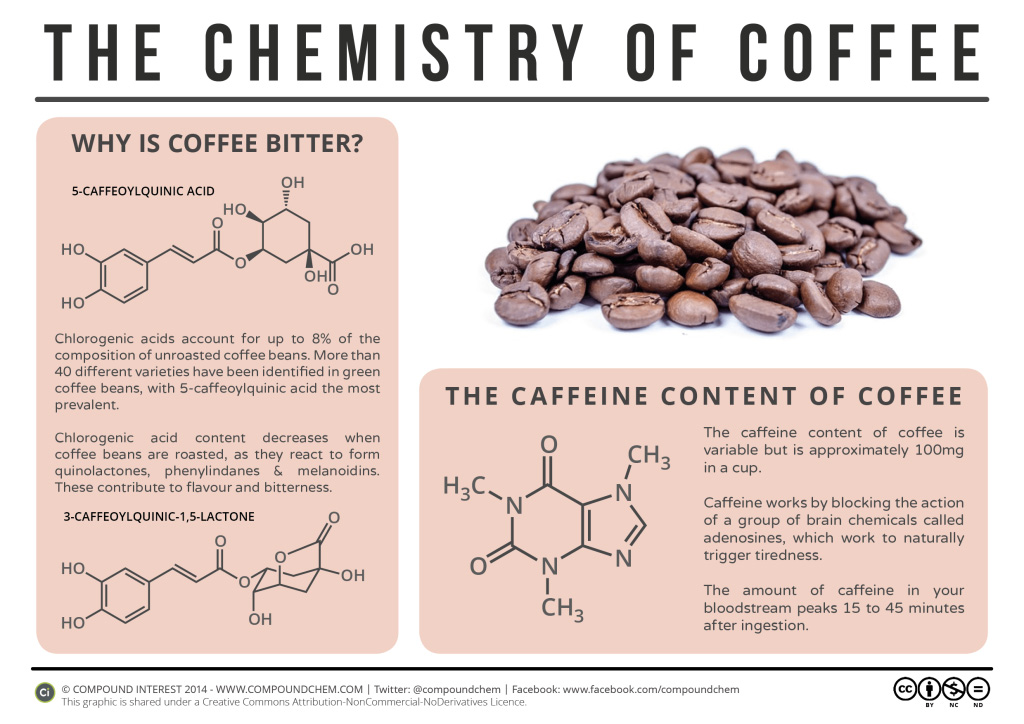
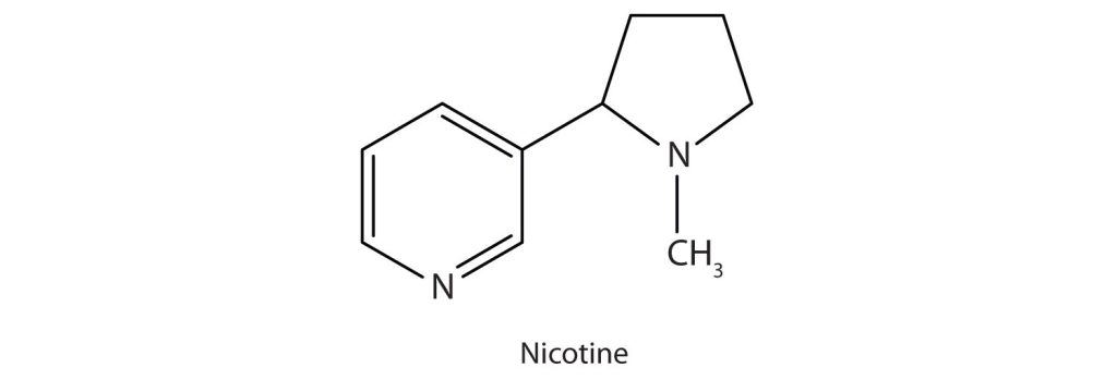
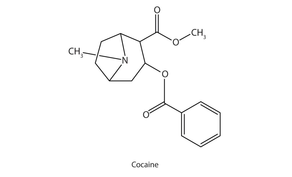

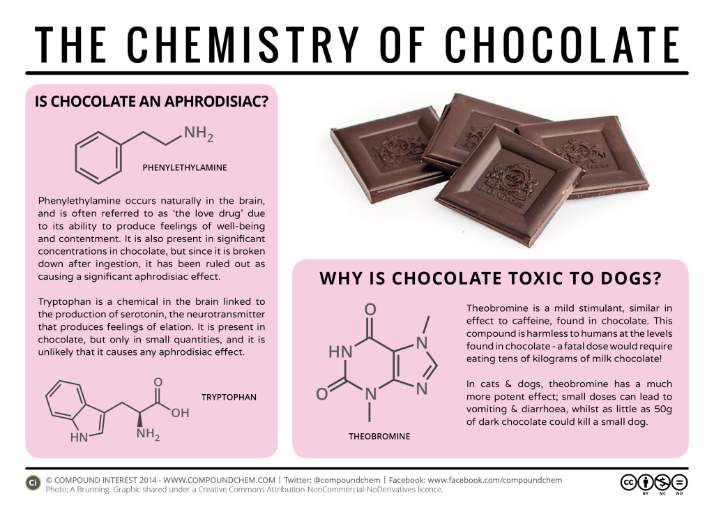
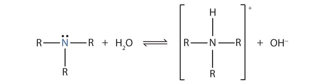

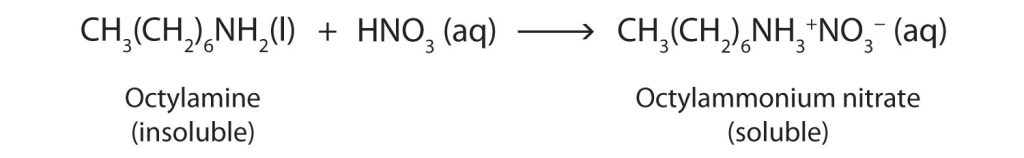
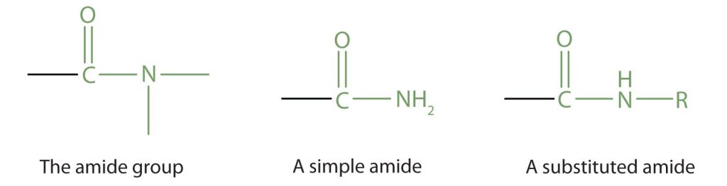
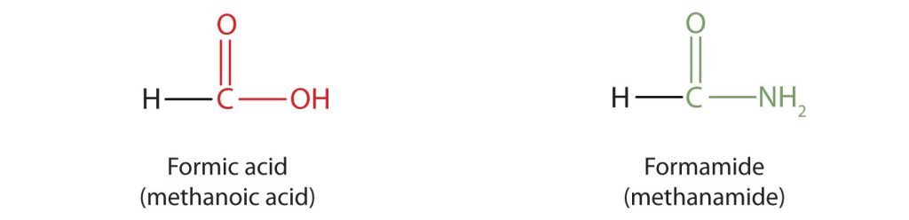
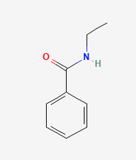
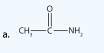
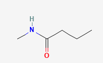
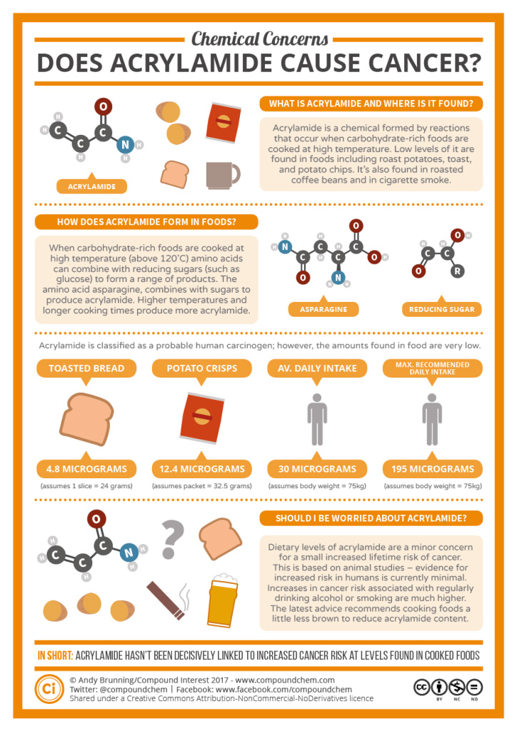

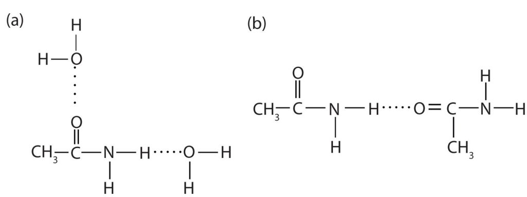
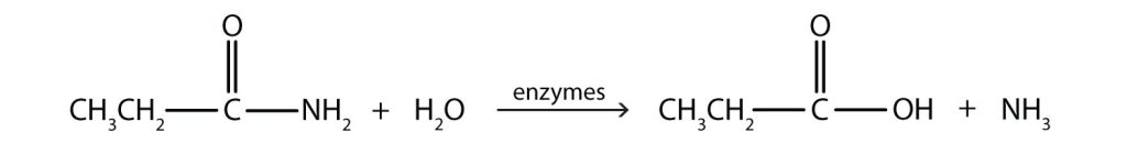
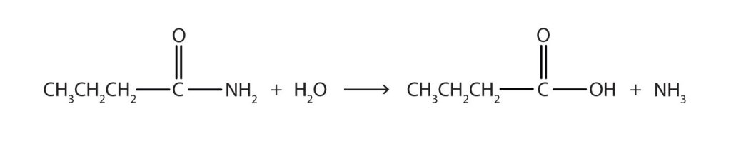
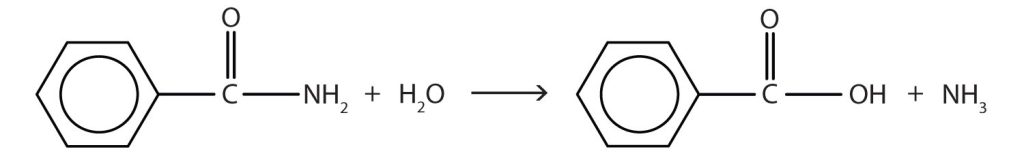


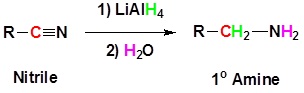
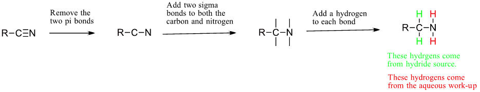
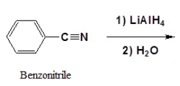
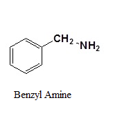
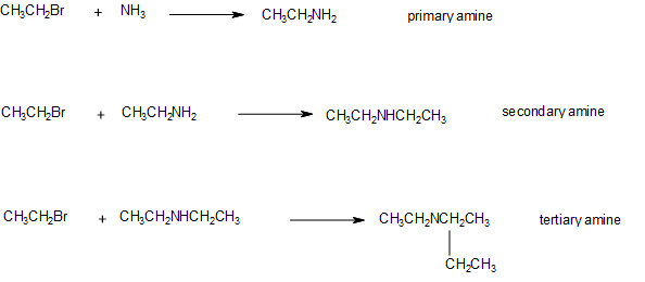
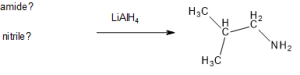
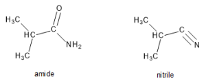
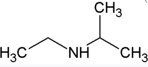
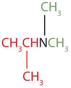 The name indicates that there are an isopropyl group (in red) and two methyl groups (in green) attached to the nitrogen atom; the amine is tertiary.
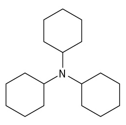
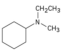
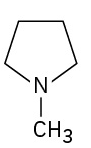

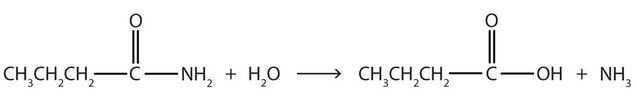

 g, take into account both mainstream (inhaled) and sidestream smoke. 1
![{\normalsize \text{Amines}\xrightarrow[Hours]{+\cdot OH{+O_{3}}}\text{Amides}\xrightarrow[Days/Weeks]{+\cdot OH}\text{Isocyantes} \normalsize}](https://atu0g9ctah.execute-api.ca-central-1.amazonaws.com/latest/latex?latex=%7B%5Cnormalsize%20%5Ctext%7BAmines%7D%5Cxrightarrow%5BHours%5D%7B%2B%5Ccdot%20OH%7B%2BO_%7B3%7D%7D%7D%5Ctext%7BAmides%7D%5Cxrightarrow%5BDays%2FWeeks%5D%7B%2B%5Ccdot%20OH%7D%5Ctext%7BIsocyantes%7D%20%5Cnormalsize%7D&fg=000000&font=TeX&svg=1 "{\normalsize \text{Amines}\xrightarrow[Hours]{+\cdot OH{+O_{3}}}\text{Amides}\xrightarrow[Days/Weeks]{+\cdot OH}\text{Isocyantes} \normalsize}")
![{\scriptsize \text{Ammonia \& Carbon Dioxide}\xleftarrow[Seconds/Days]{+H_{2}O}\text{HNCO (dropl)}\xleftarrow[Days/Years]{}\text{Isocynaic Acid (type of isocynanate)} \scriptsize}](https://atu0g9ctah.execute-api.ca-central-1.amazonaws.com/latest/latex?latex=%7B%5Cscriptsize%20%5Ctext%7BAmmonia%20%5C%26%20Carbon%20Dioxide%7D%5Cxleftarrow%5BSeconds%2FDays%5D%7B%2BH_%7B2%7DO%7D%5Ctext%7BHNCO%20%28dropl%29%7D%5Cxleftarrow%5BDays%2FYears%5D%7B%7D%5Ctext%7BIsocynaic%20Acid%20%28type%20of%20isocynanate%29%7D%20%5Cscriptsize%7D&fg=000000&font=TeX&svg=1 "{\scriptsize \text{Ammonia \& Carbon Dioxide}\xleftarrow[Seconds/Days]{+H_{2}O}\text{HNCO (dropl)}\xleftarrow[Days/Years]{}\text{Isocynaic Acid (type of isocynanate)} \scriptsize}")


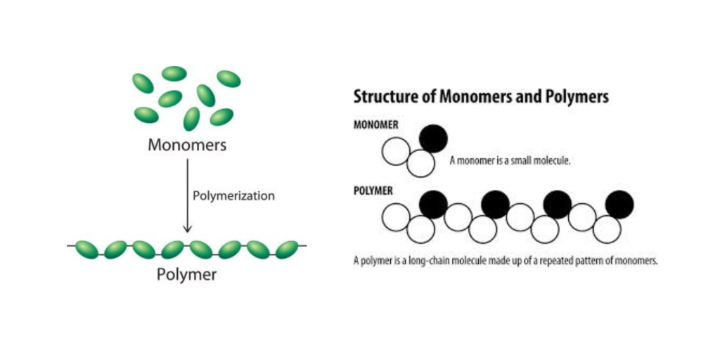

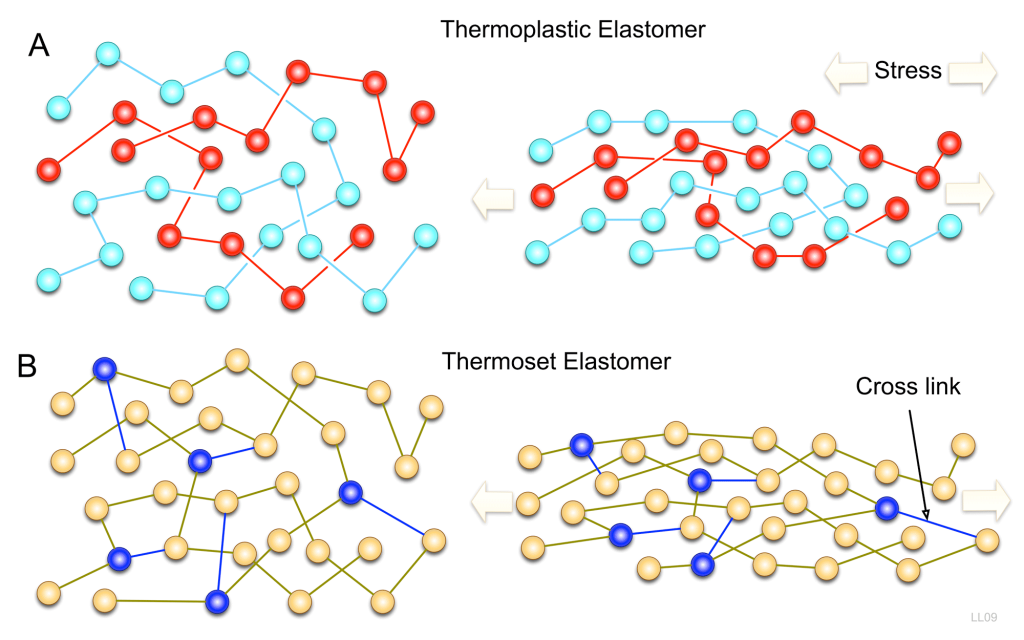
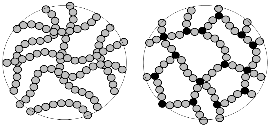

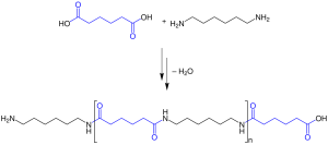
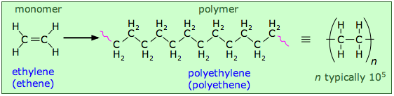
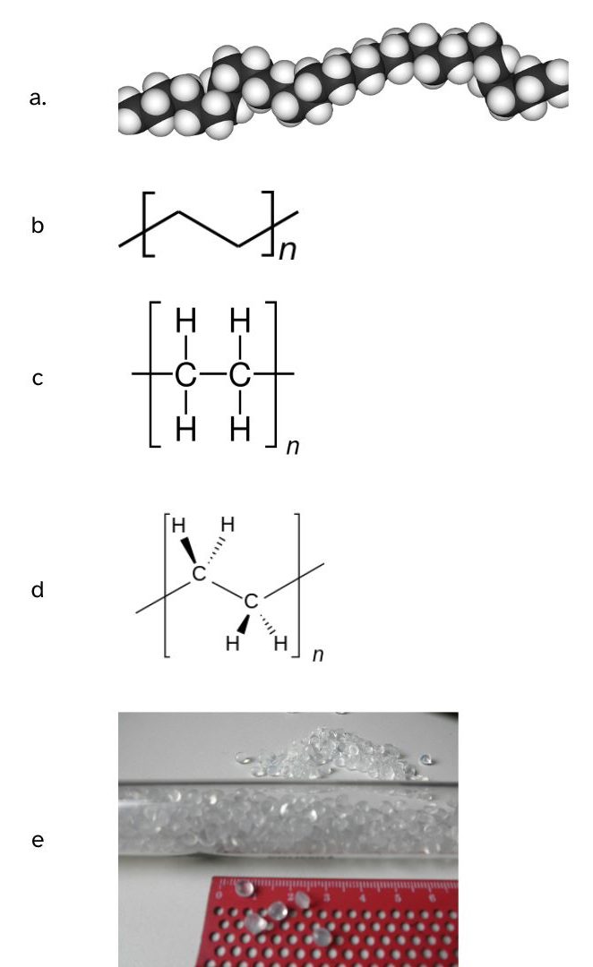
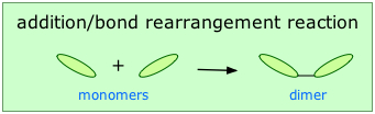
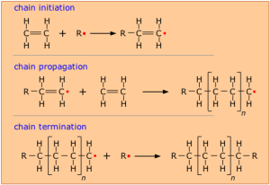
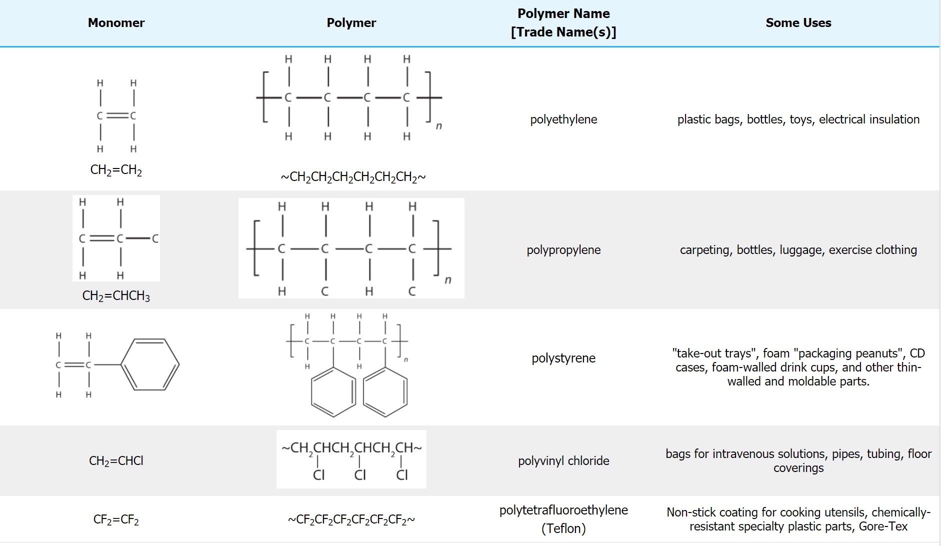
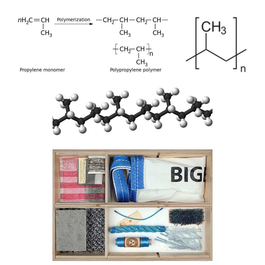
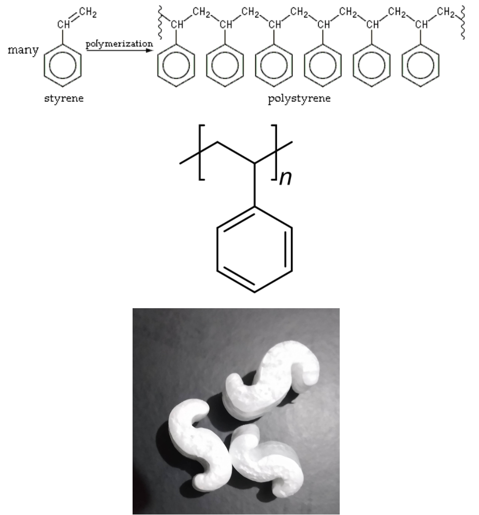


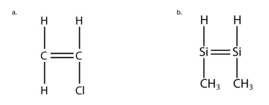


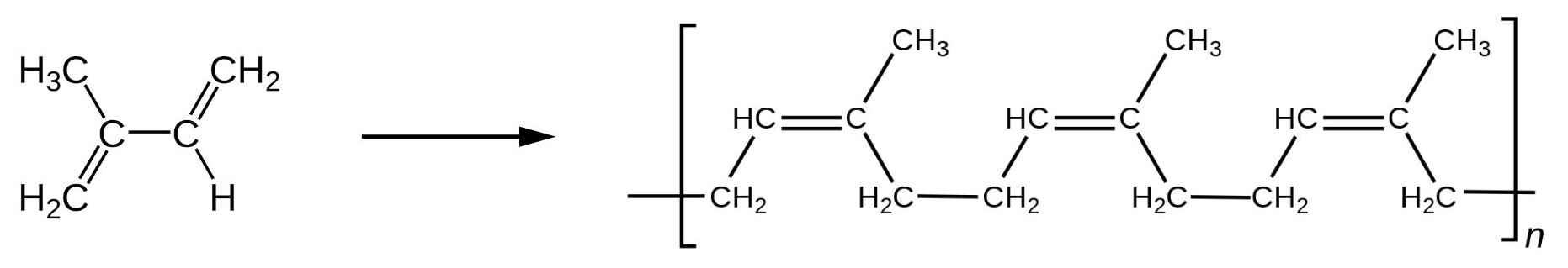
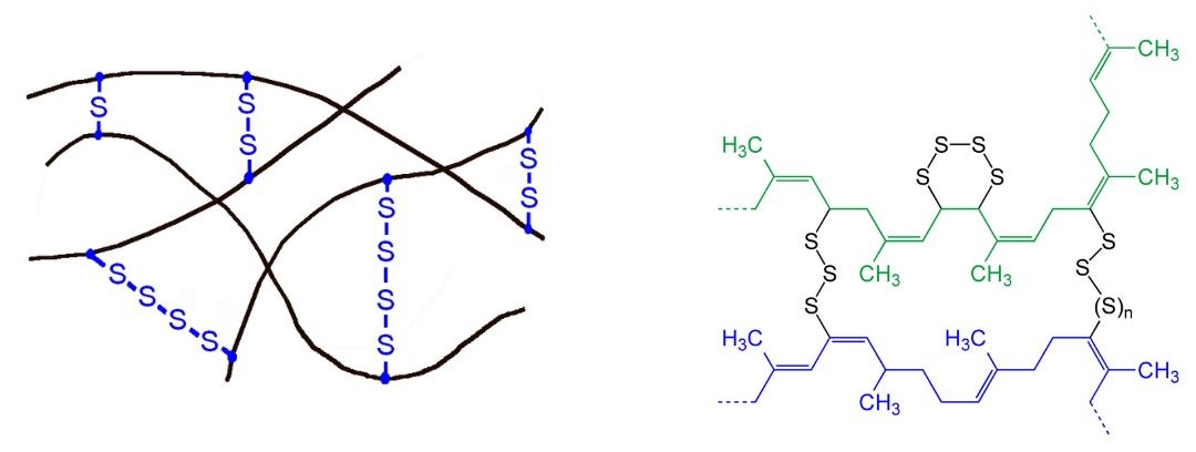
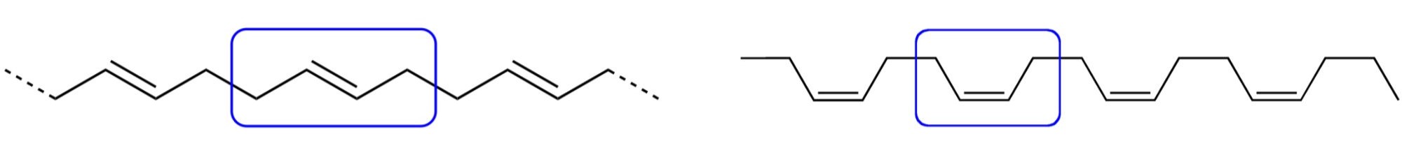
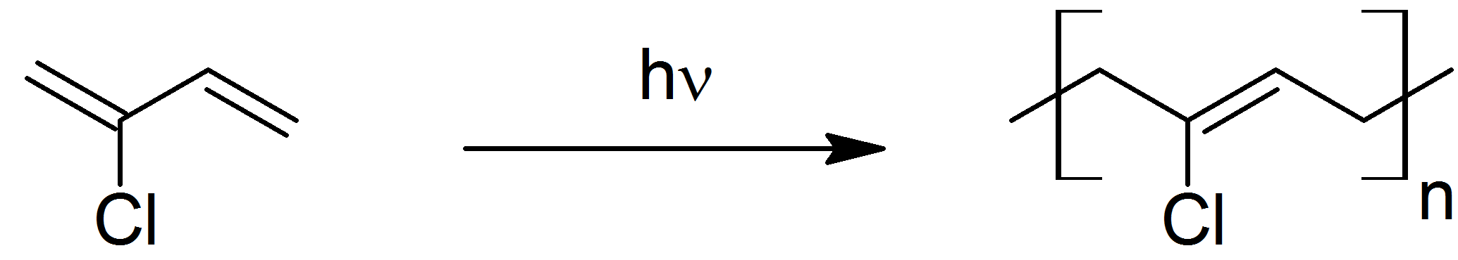


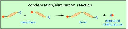


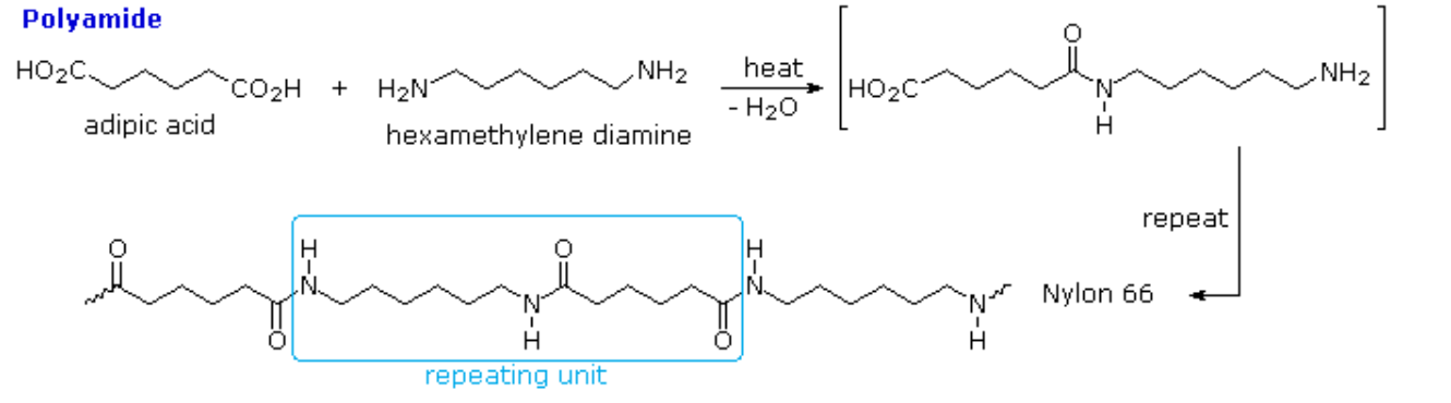
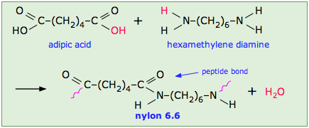

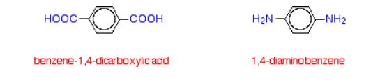
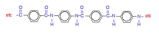
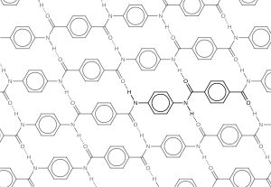

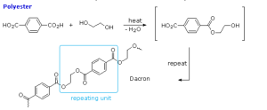
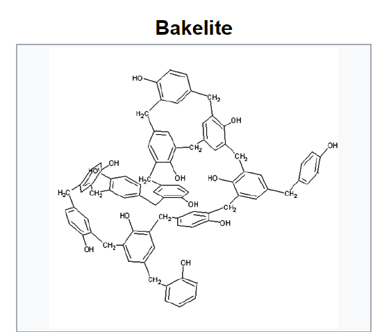
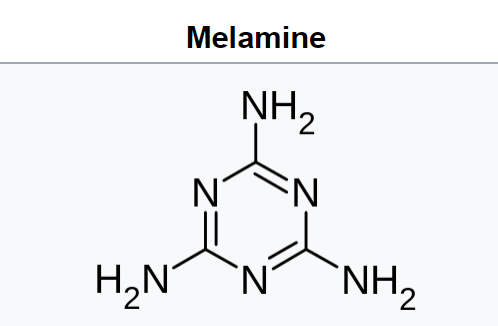

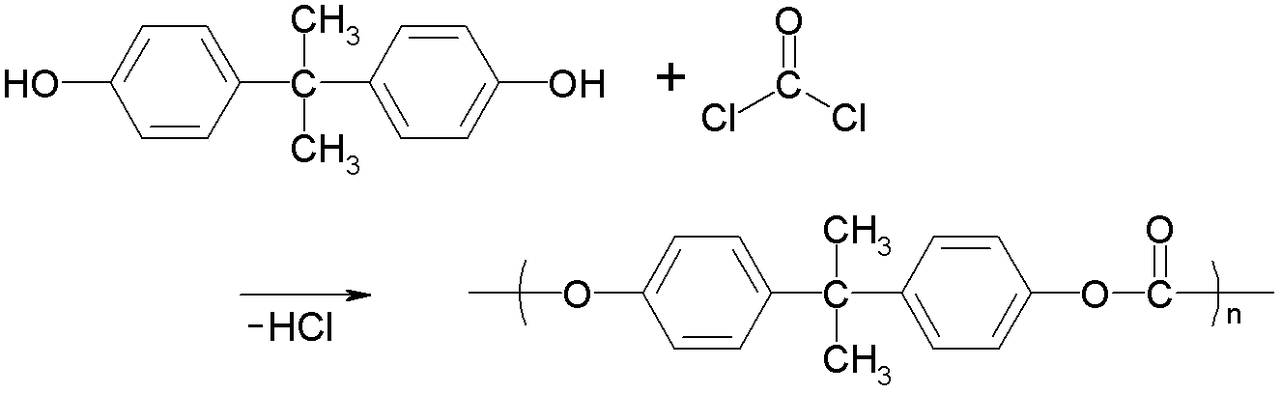

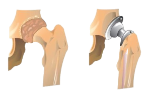


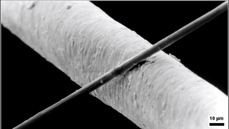
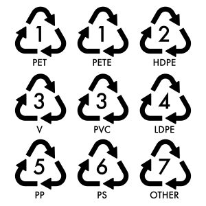


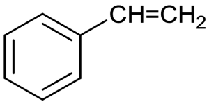


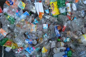


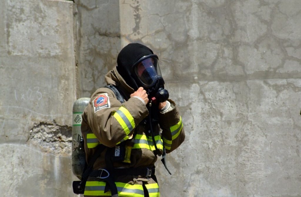

 The well-known “new car smell” is largely due to plasticizer release from upholstery and internal trim. According to 2014 data, the total global market for plasticizers was 8.4 million metric tonnes including 1.3 million metric tonnes in Europe.
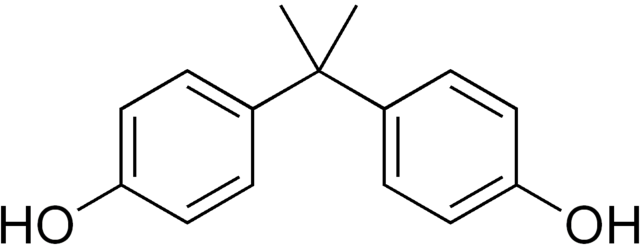


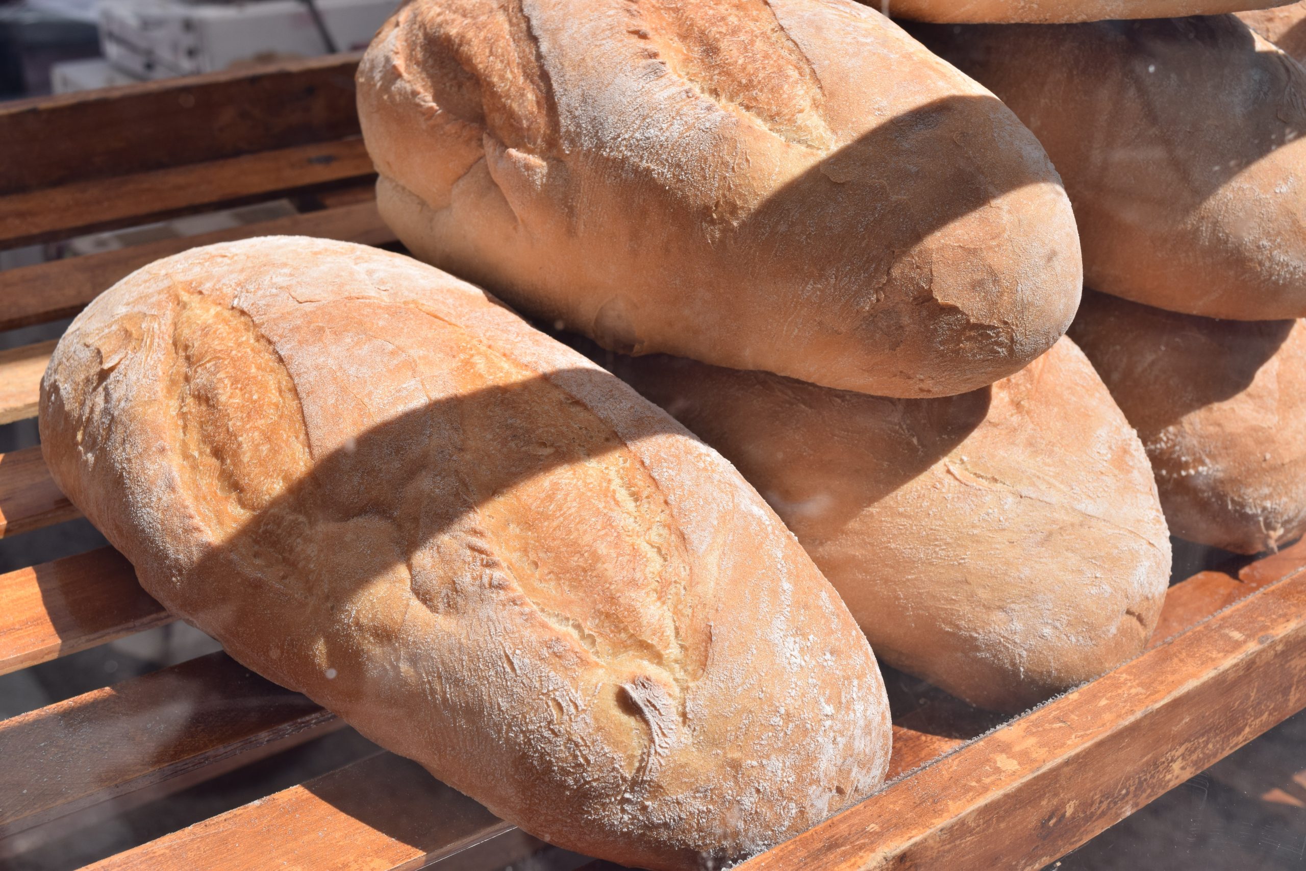
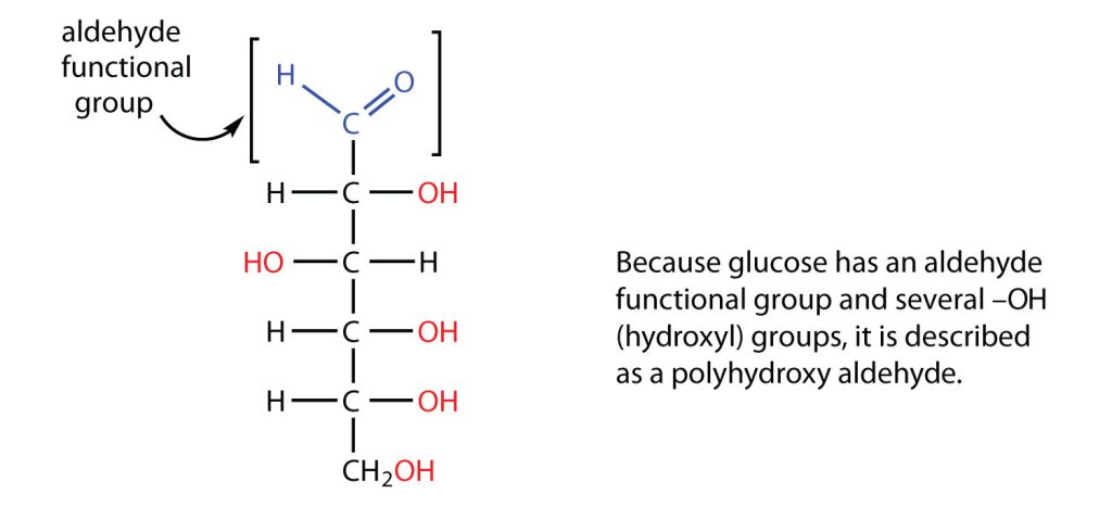
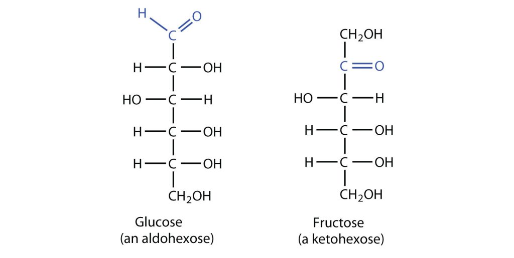

 Example source:
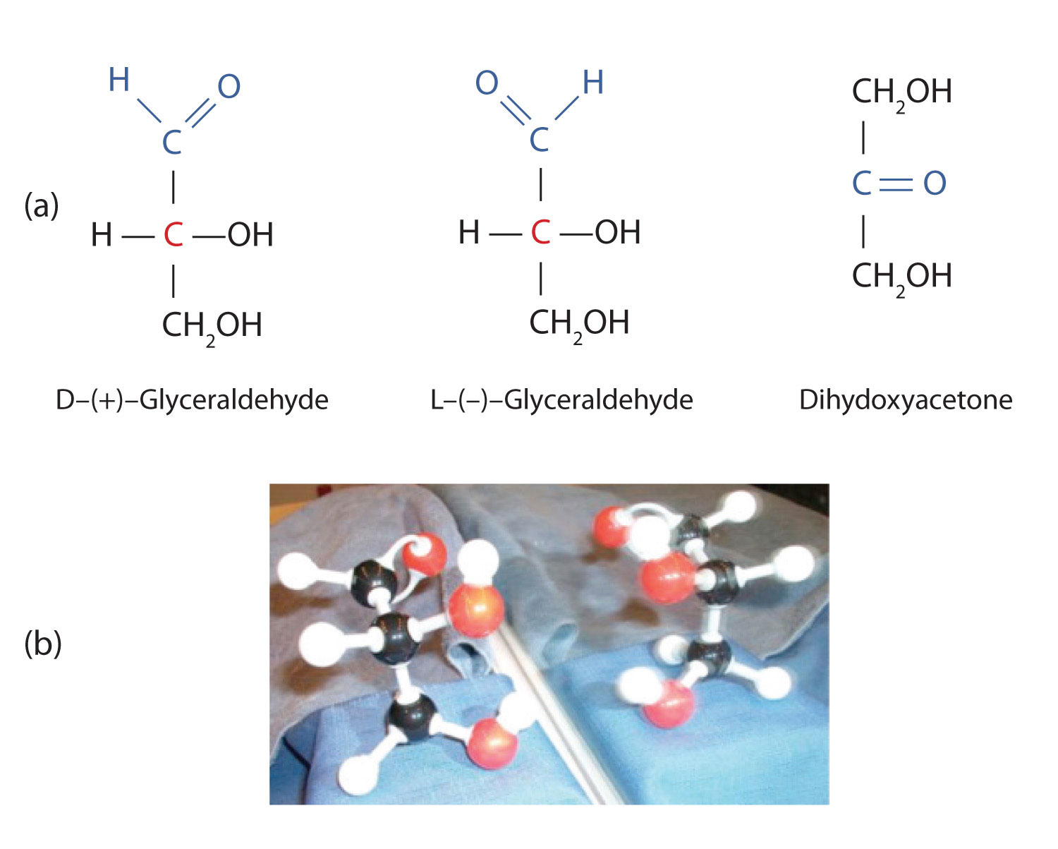
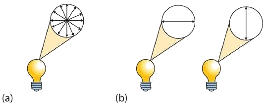

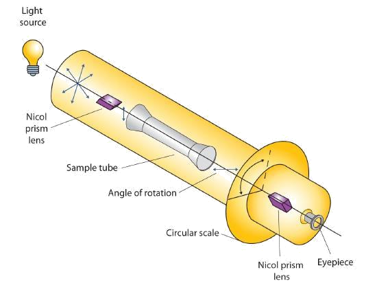
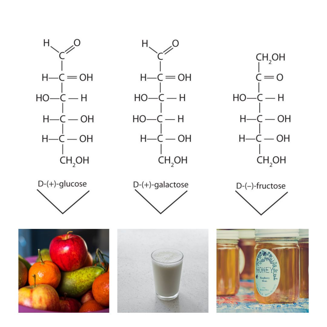
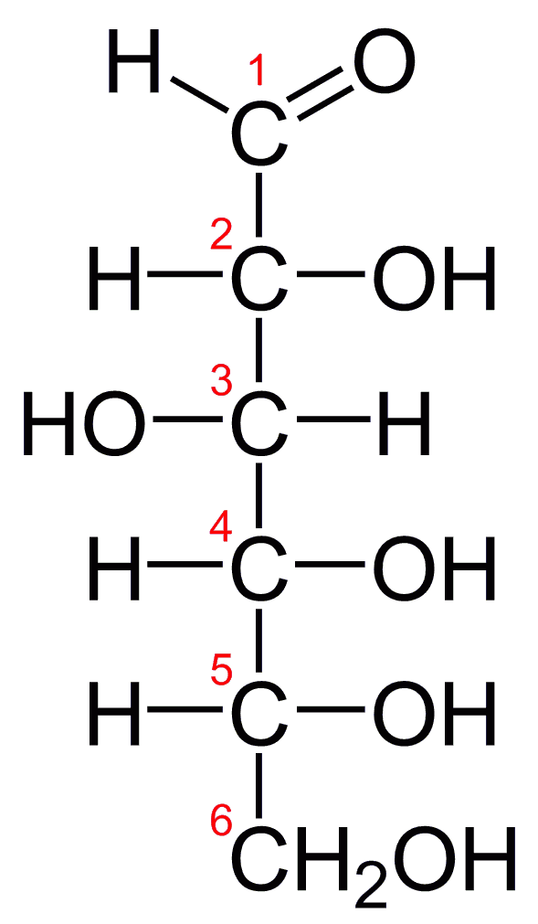
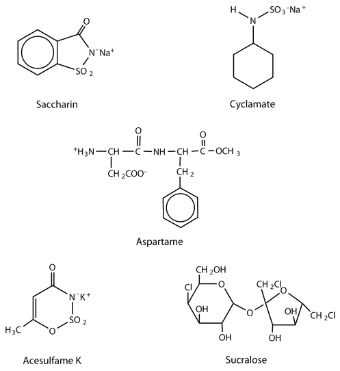
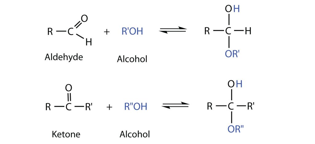
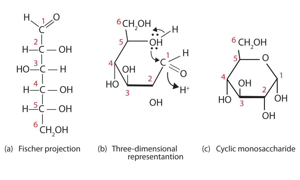
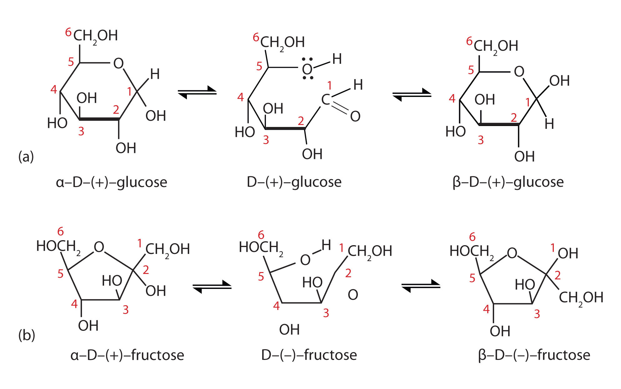
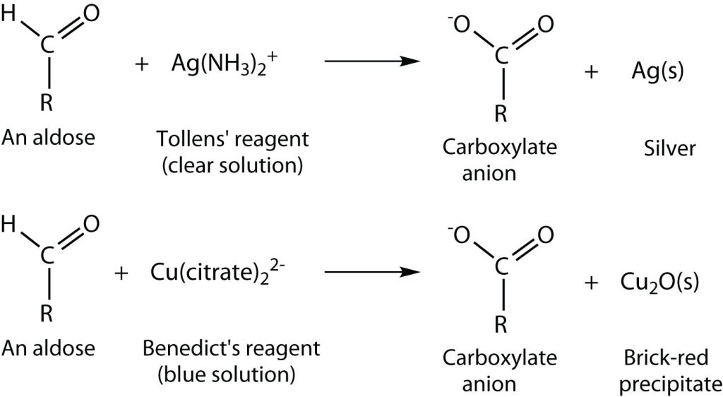
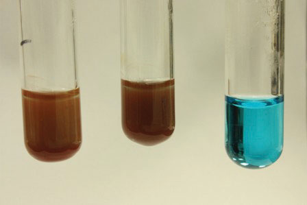
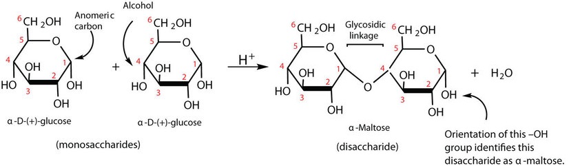

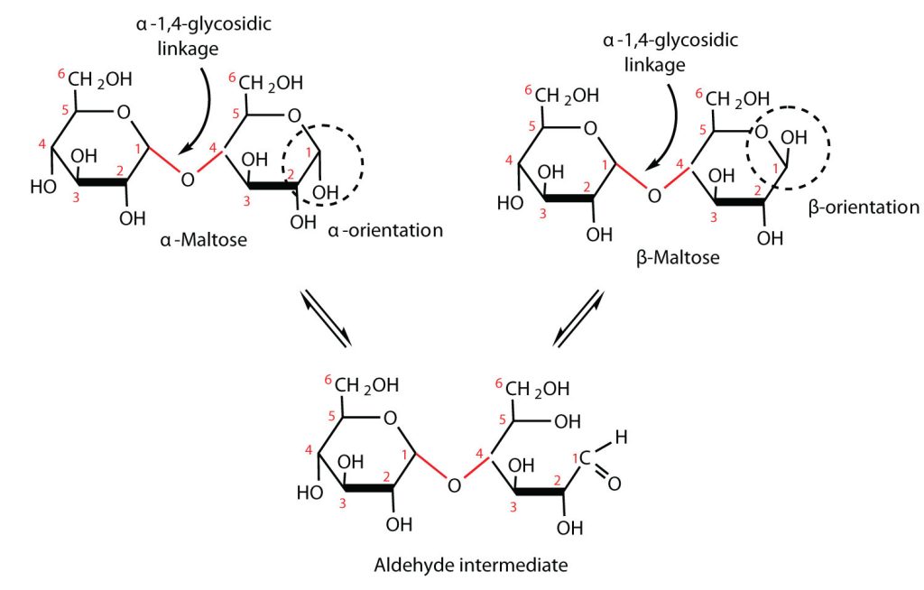
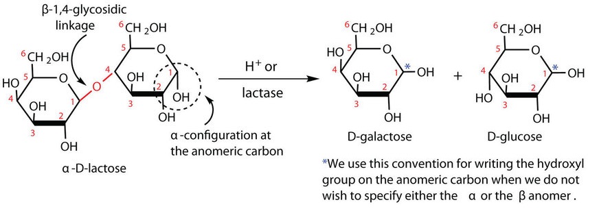
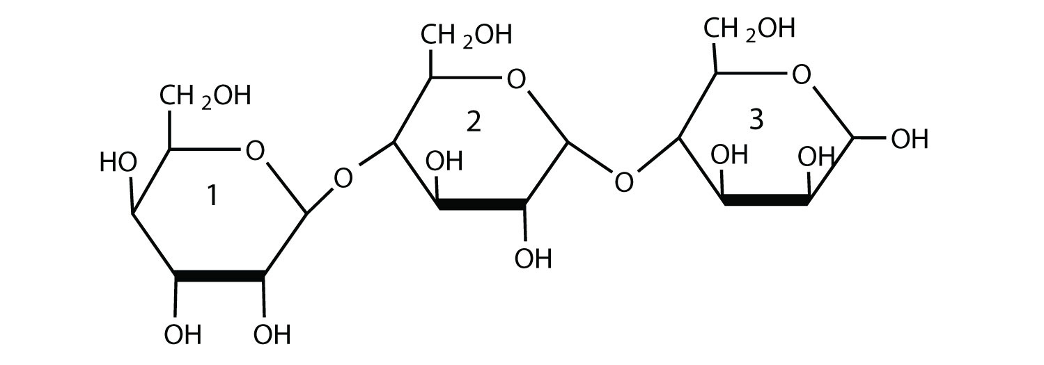
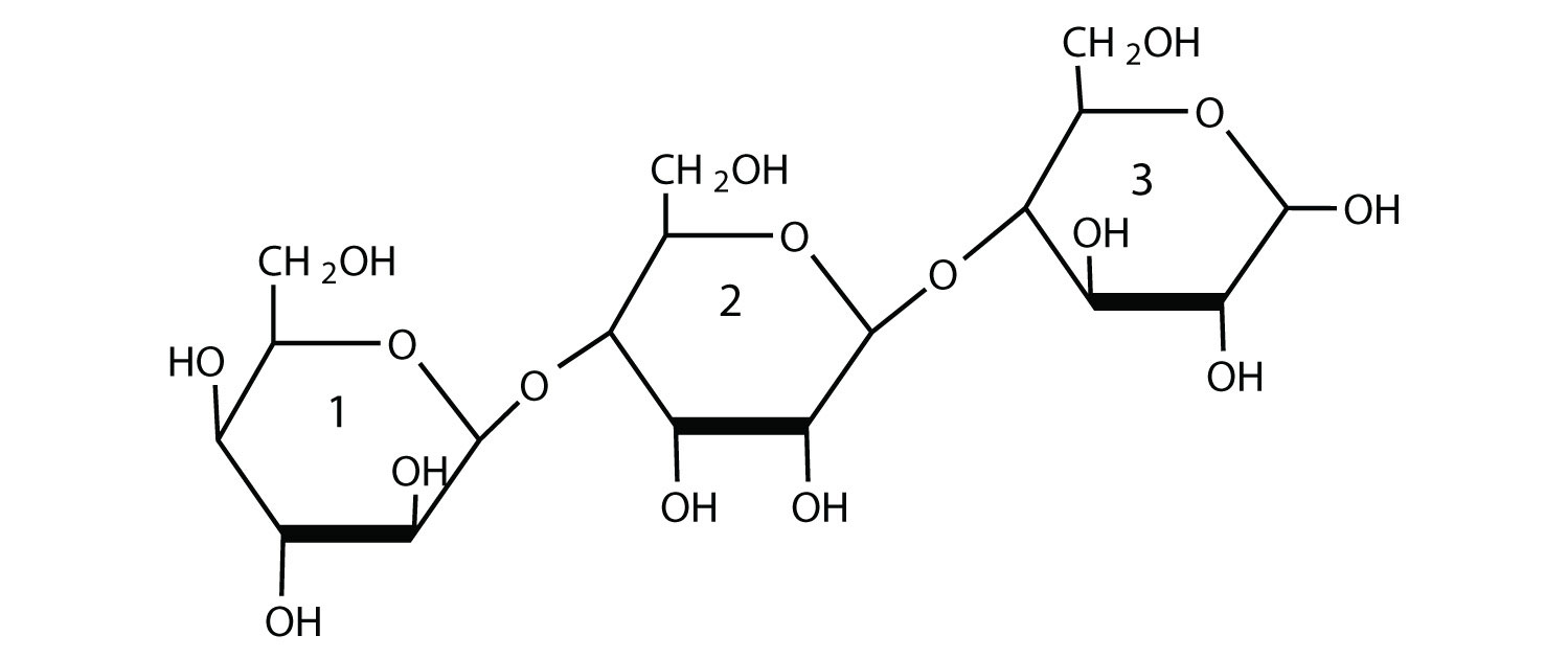
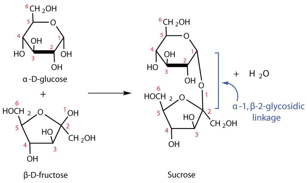
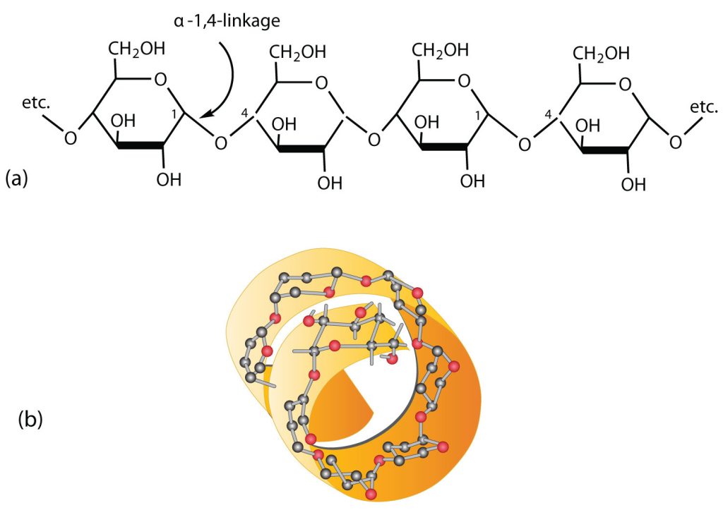
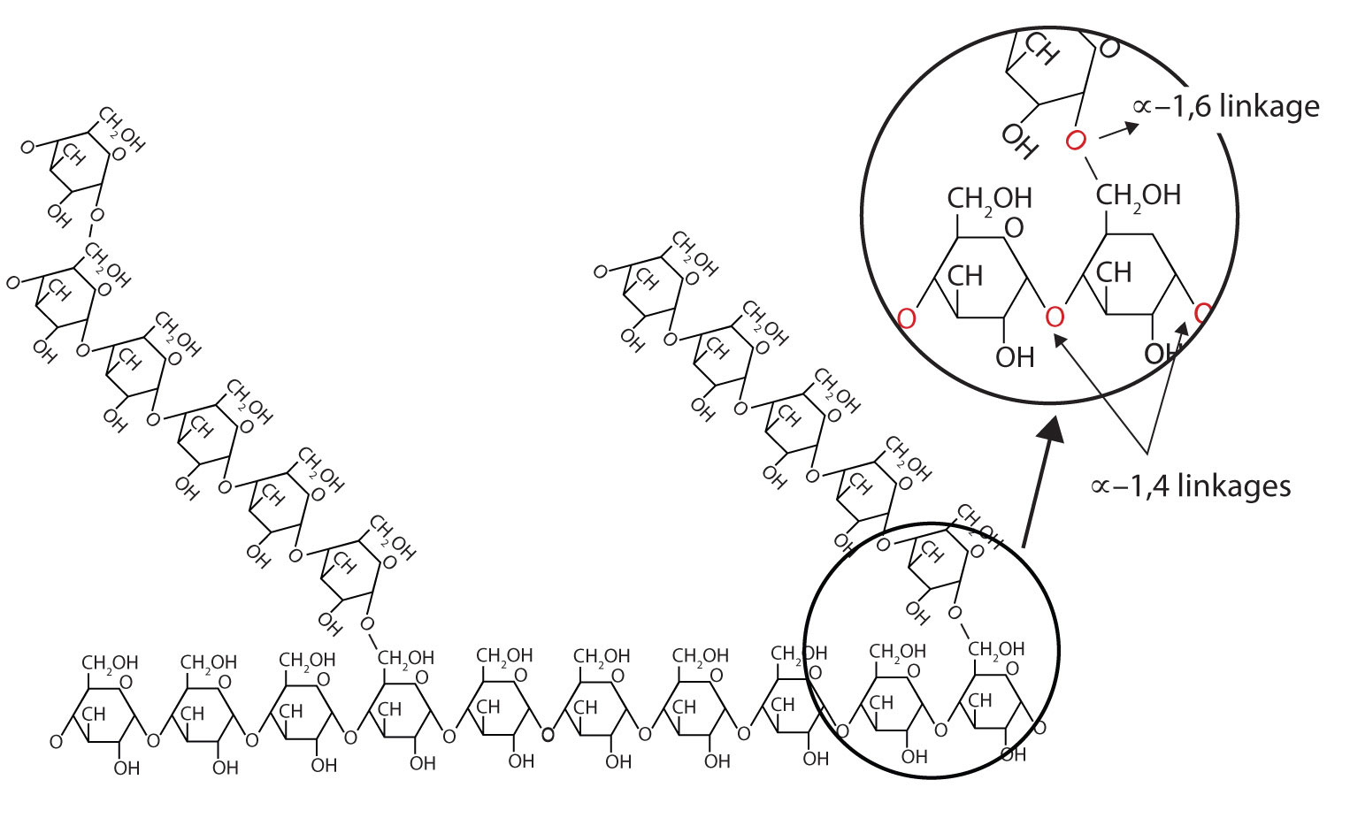
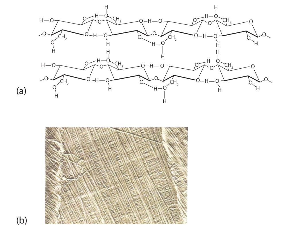
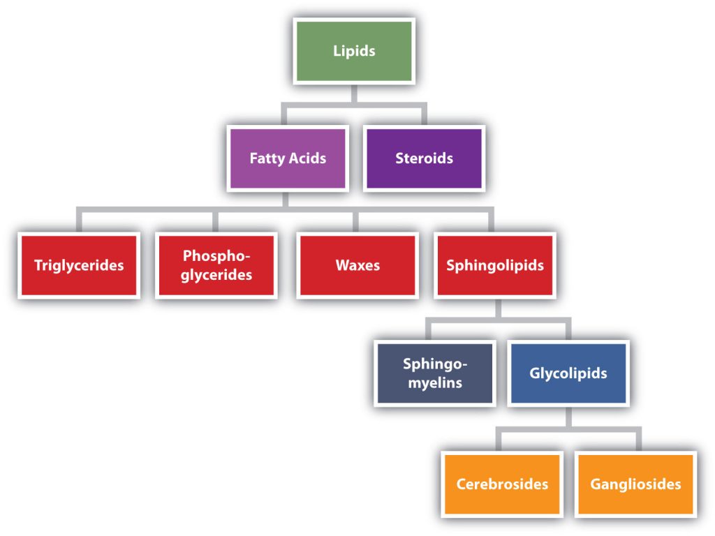
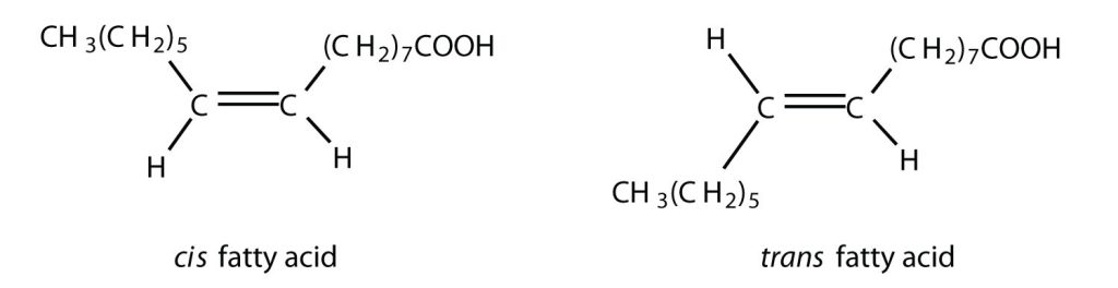
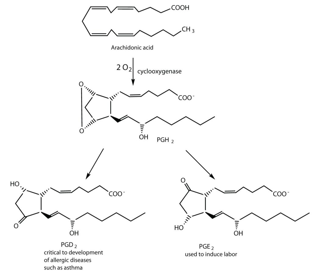
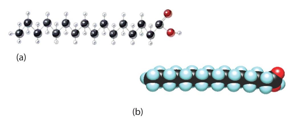
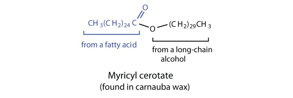
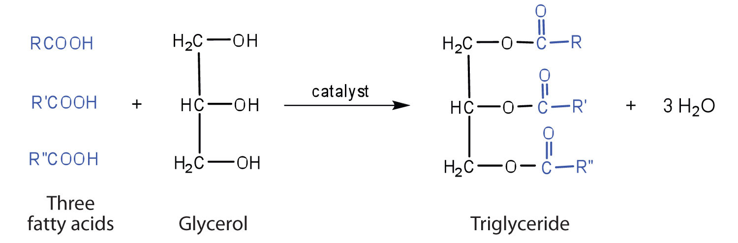
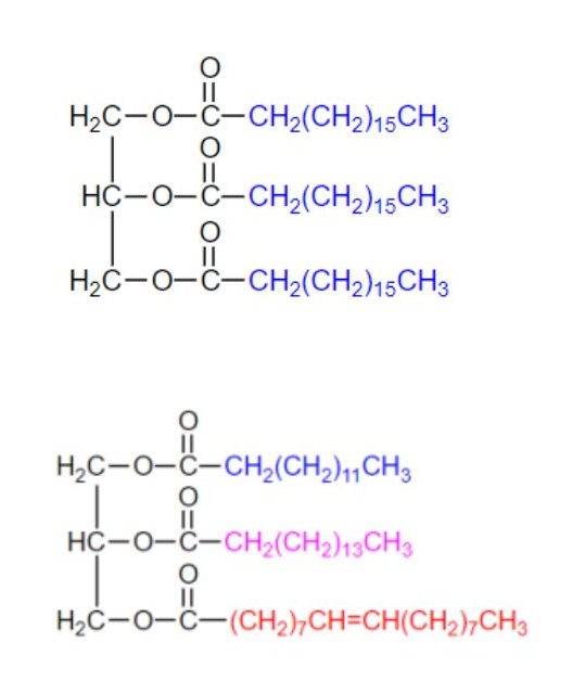

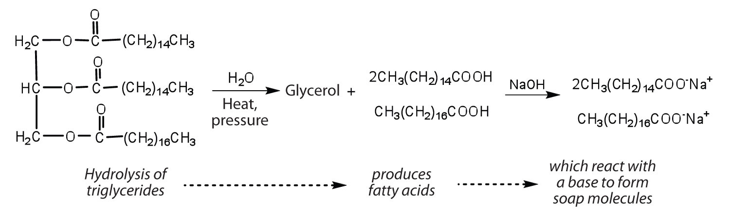
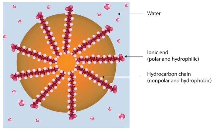

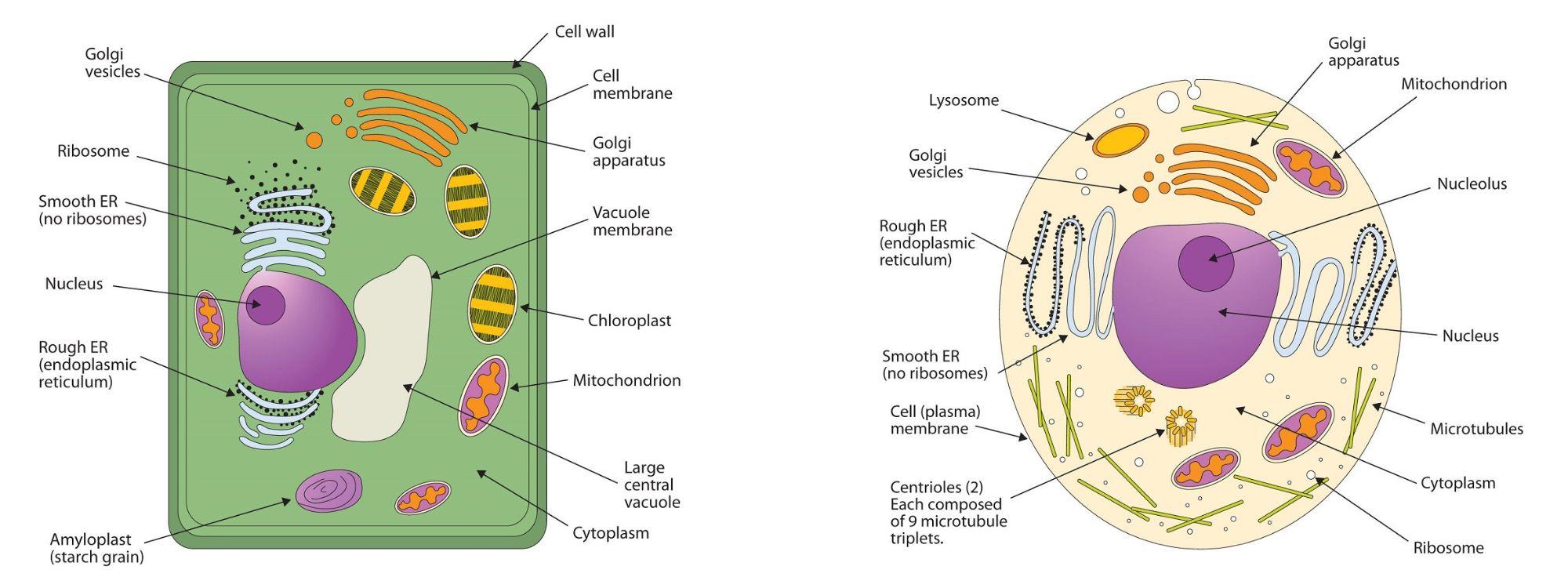
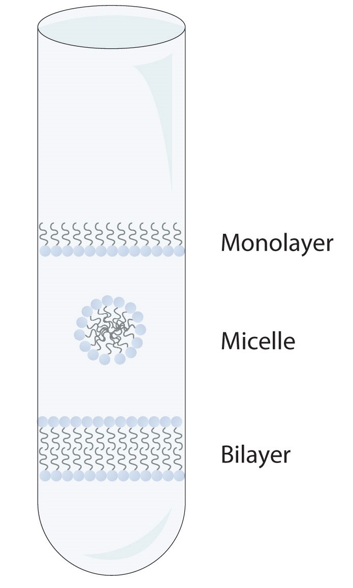
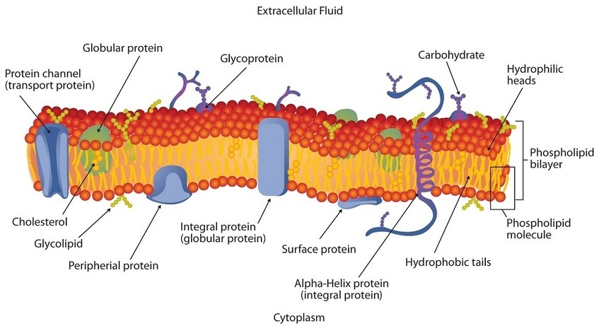
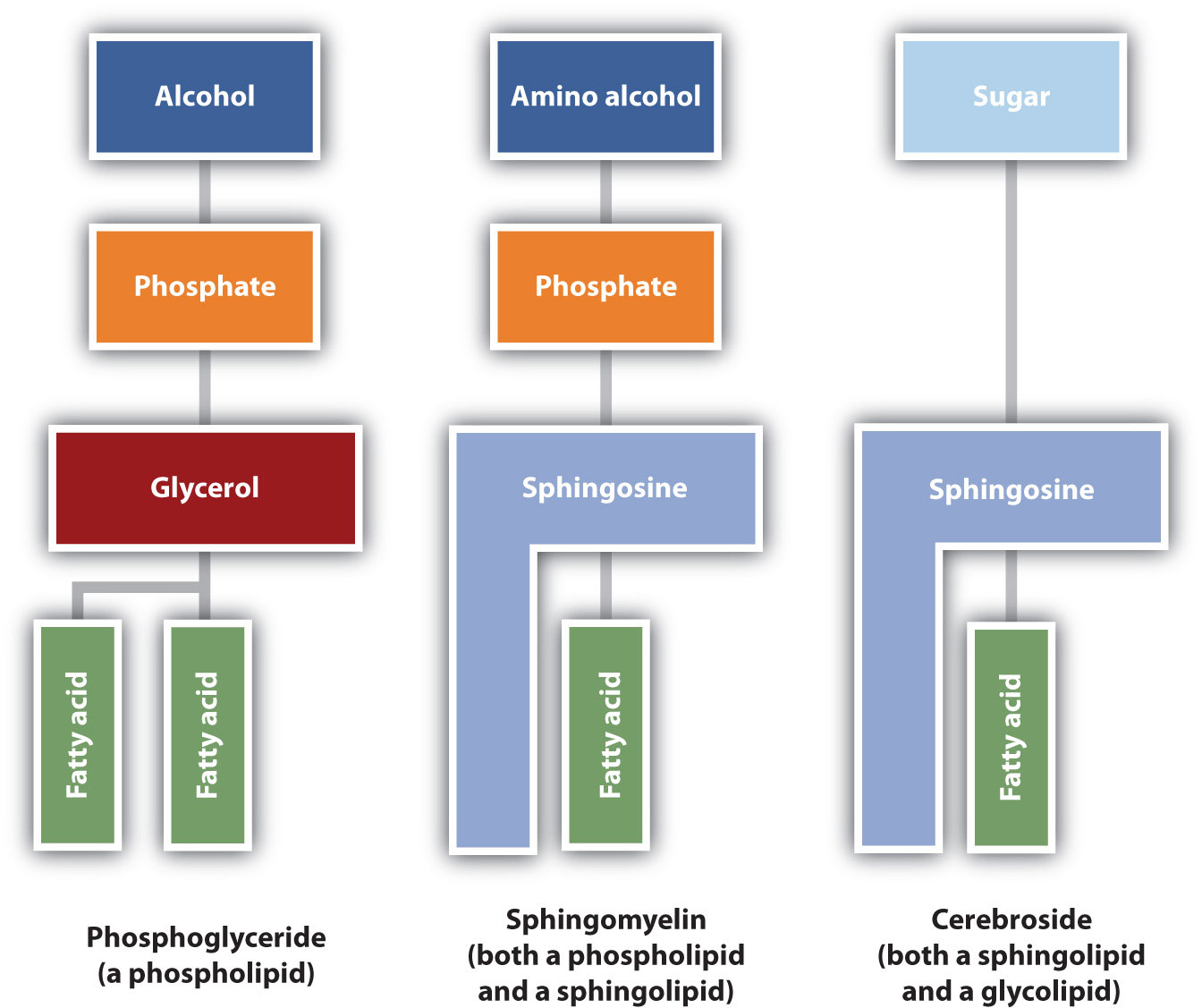
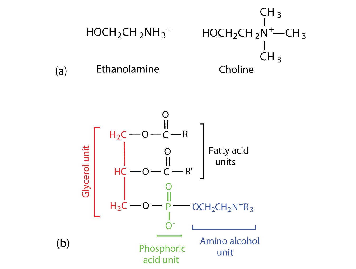
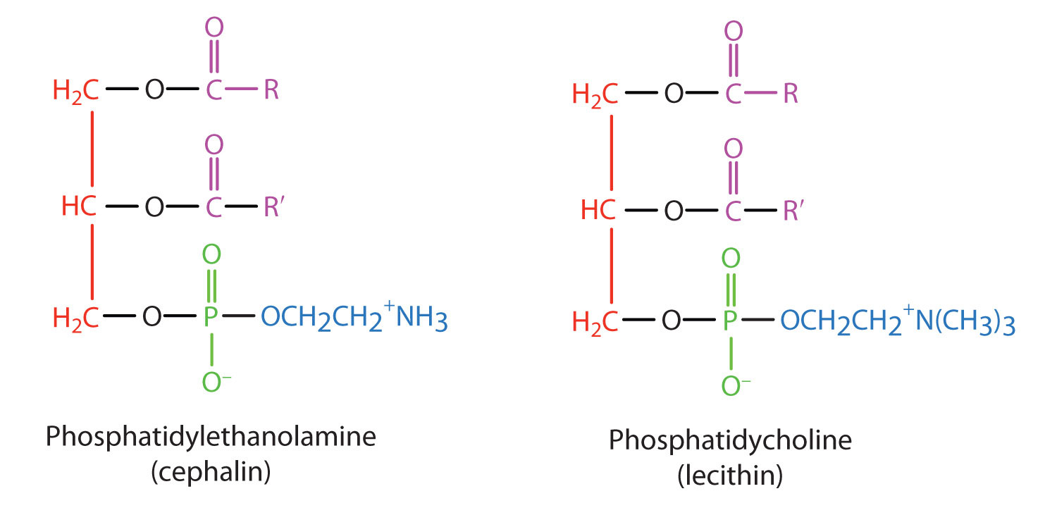
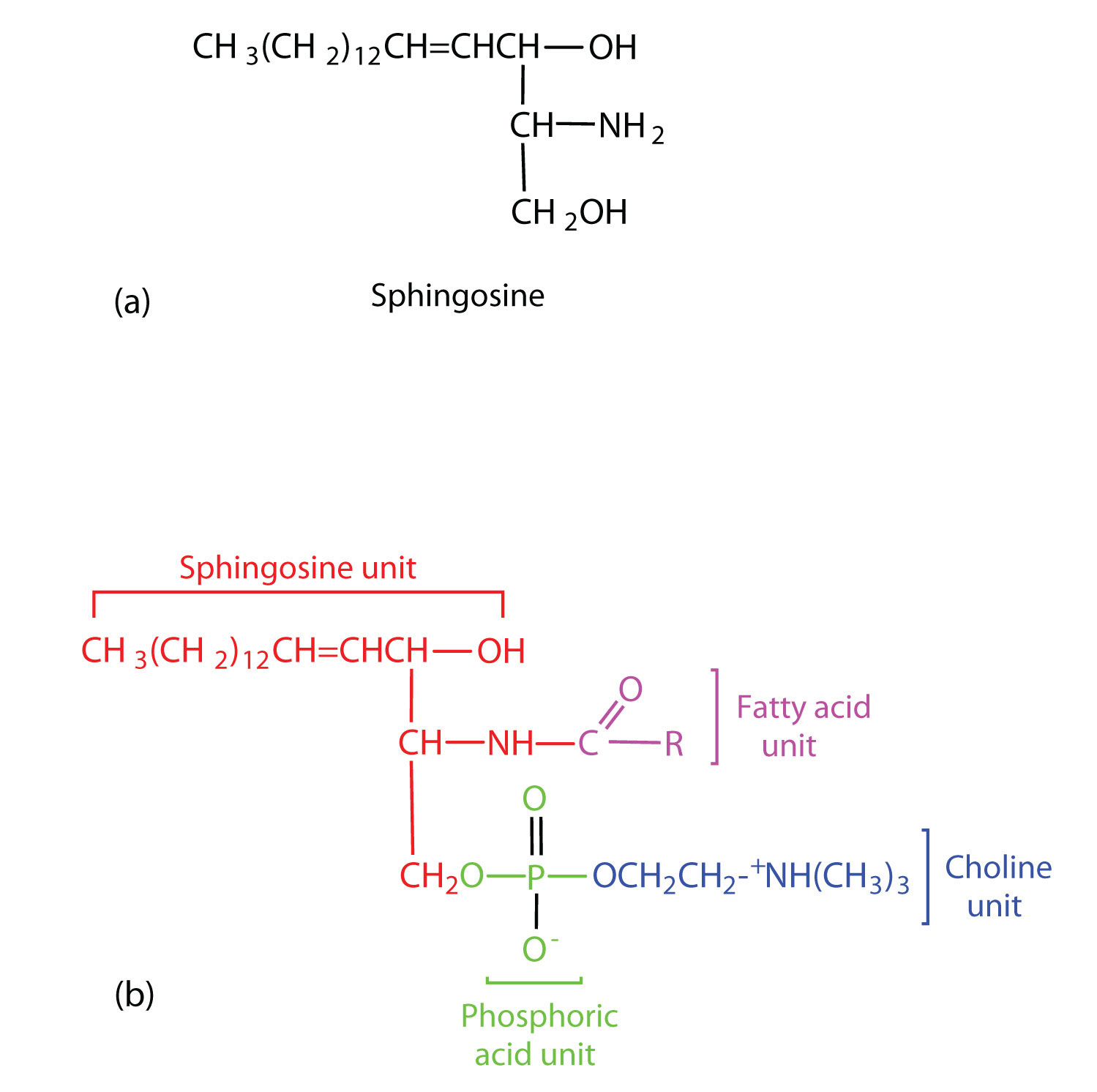
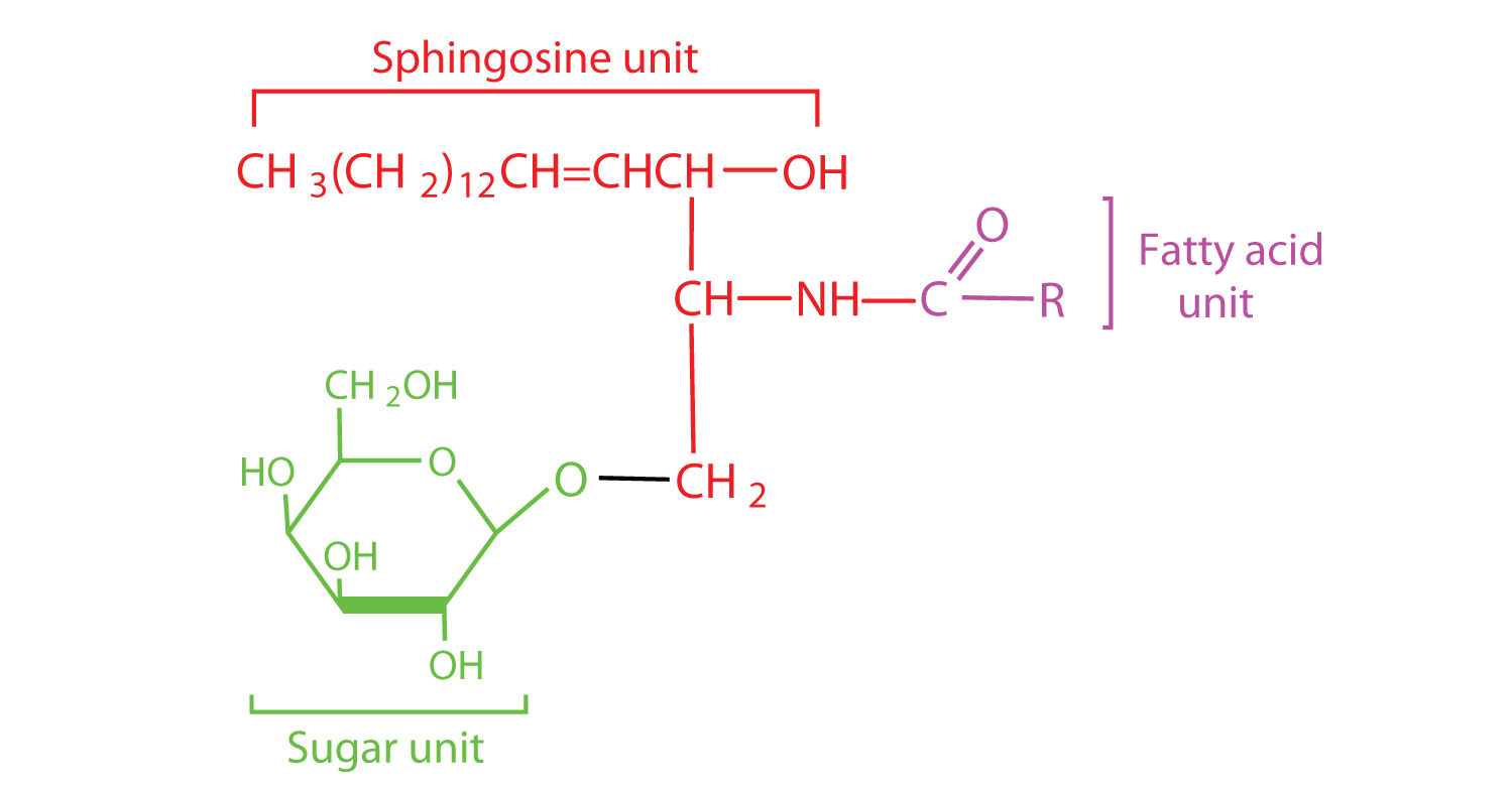
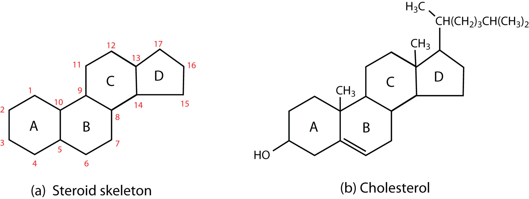
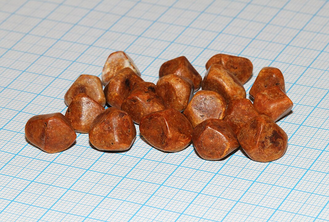
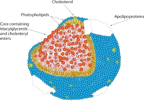
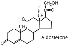
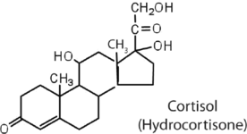
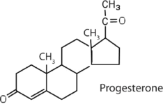
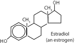
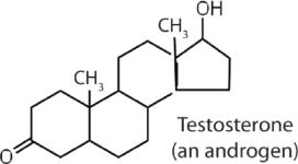
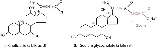

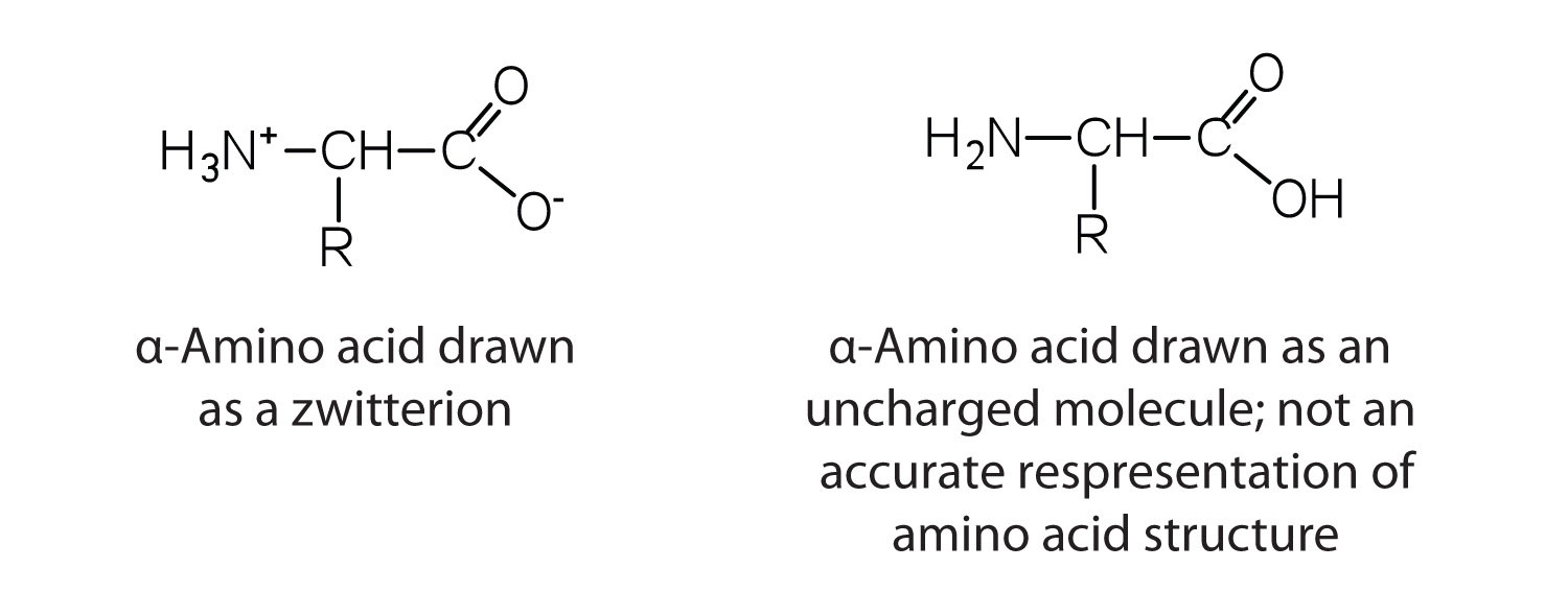
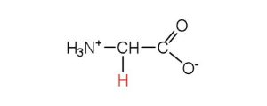
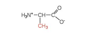

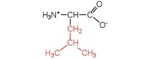
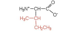
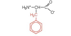
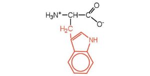
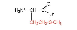
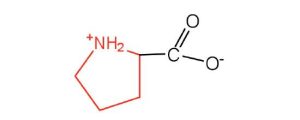

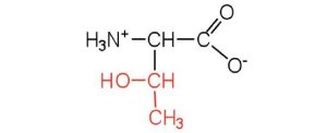
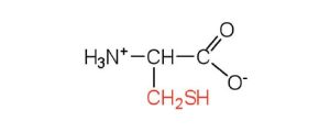
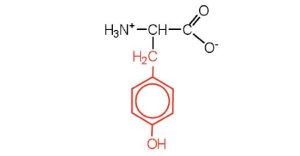
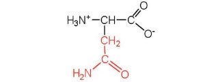
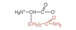
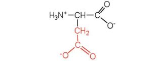
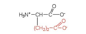
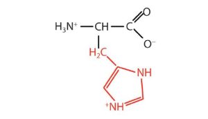
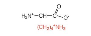
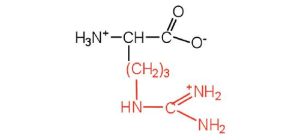
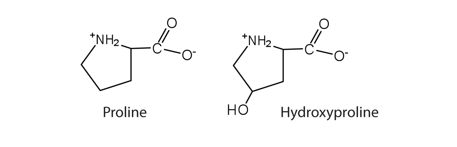
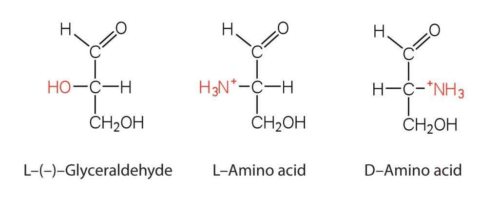
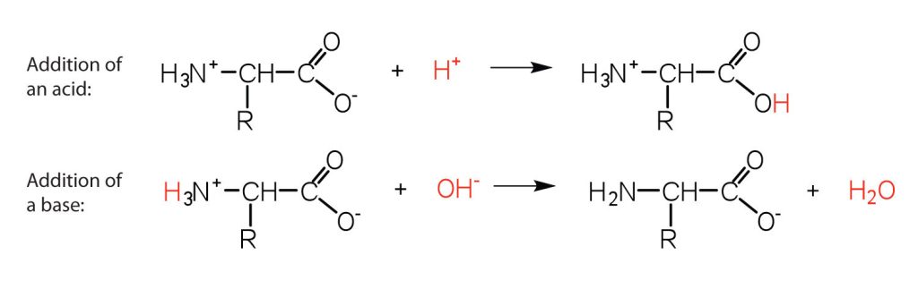


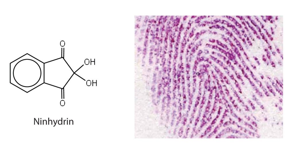
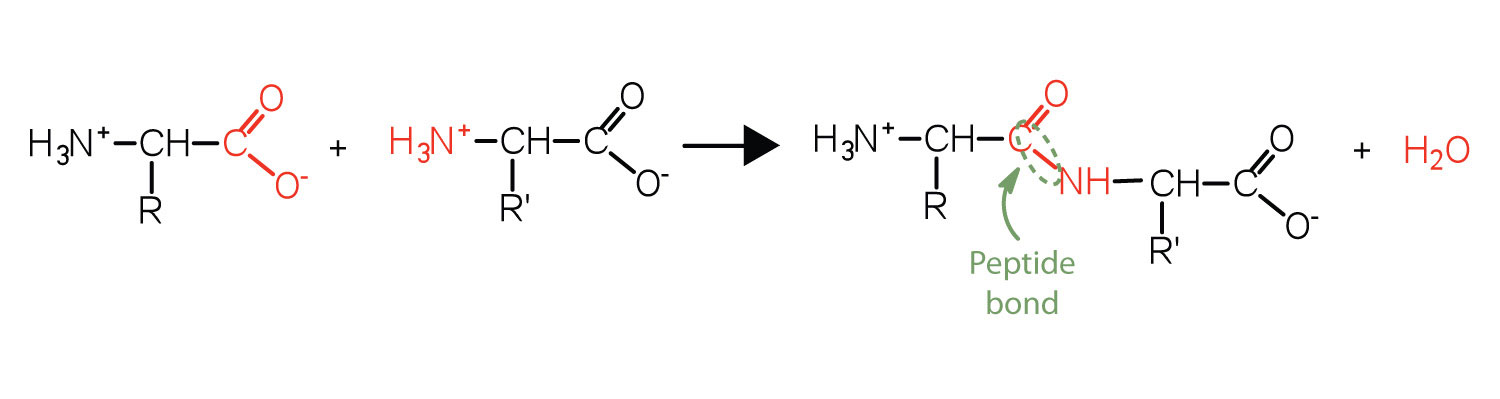
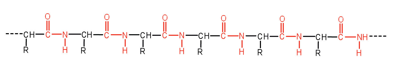
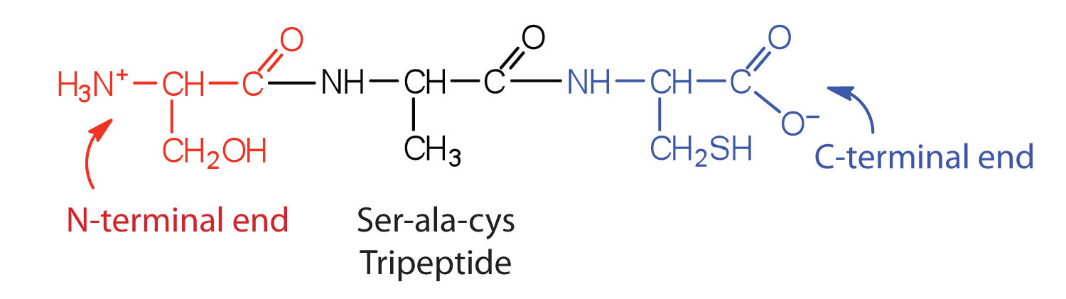
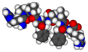
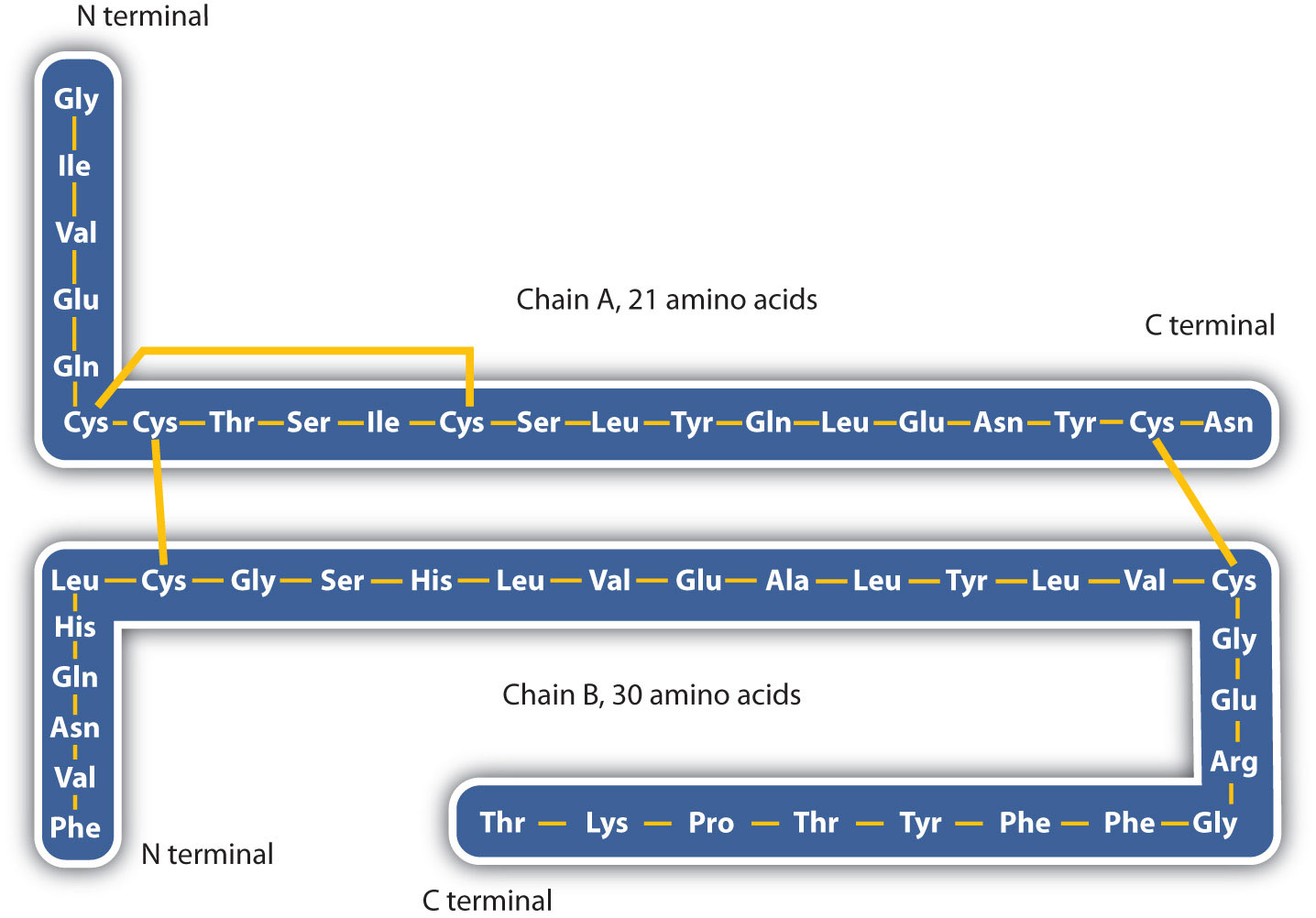
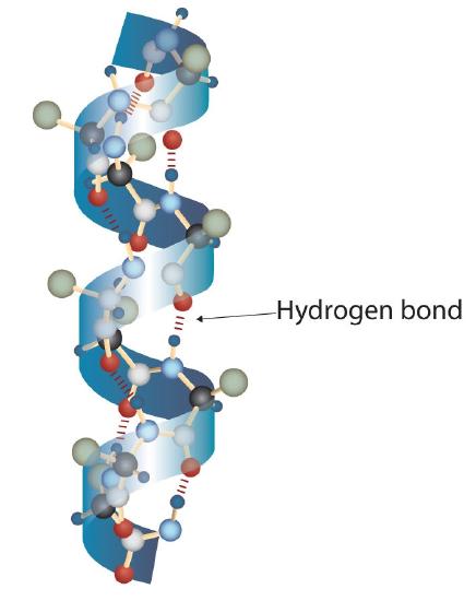
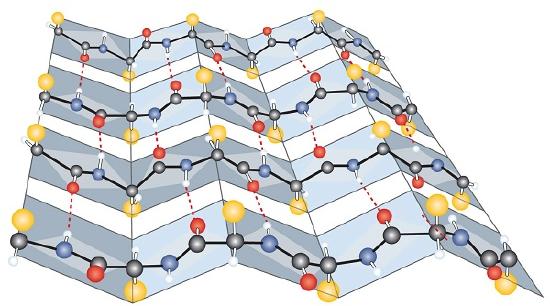
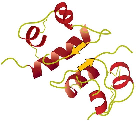
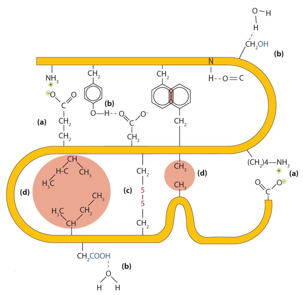
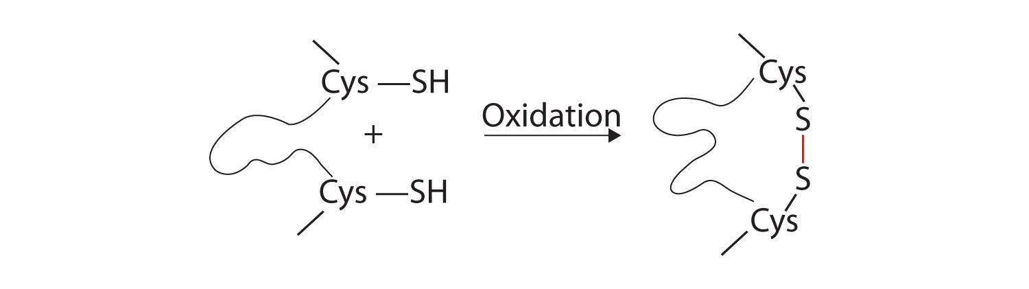
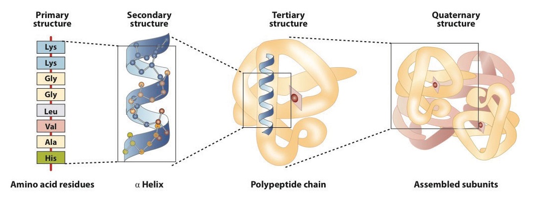
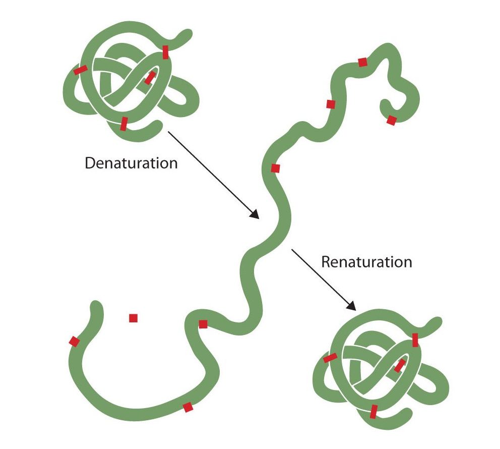
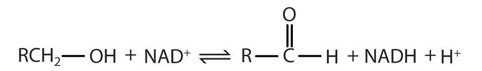


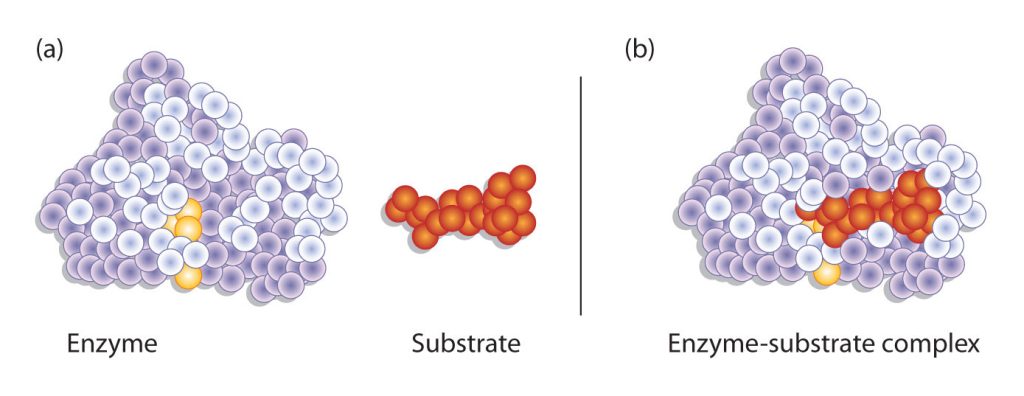
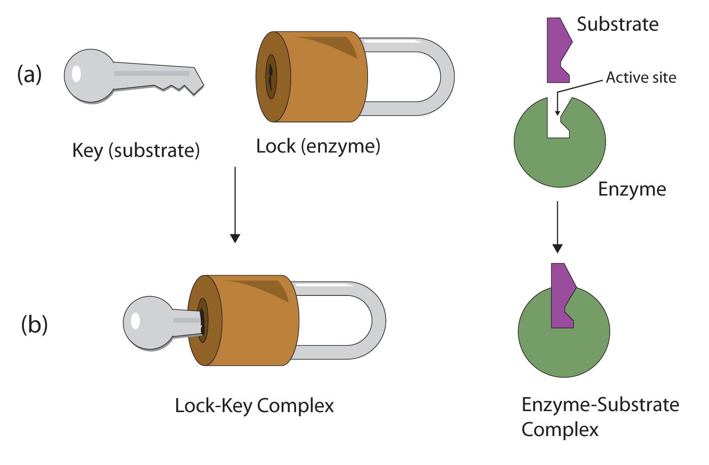
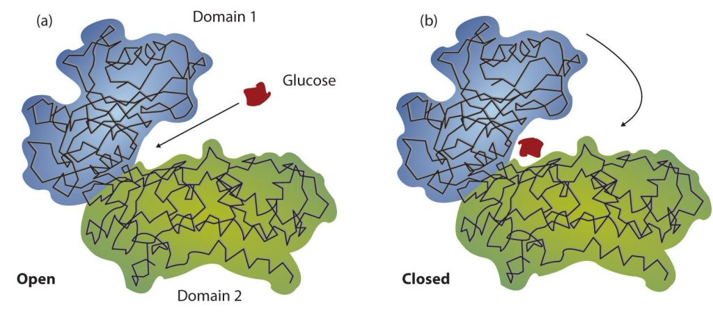
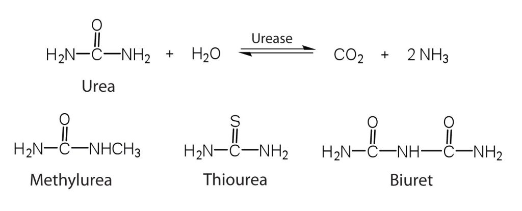
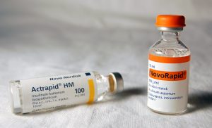

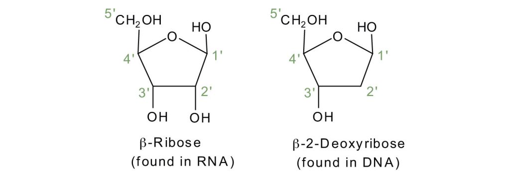
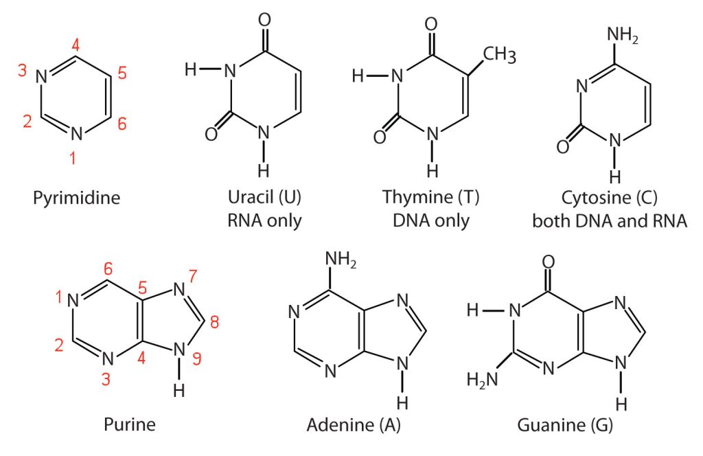
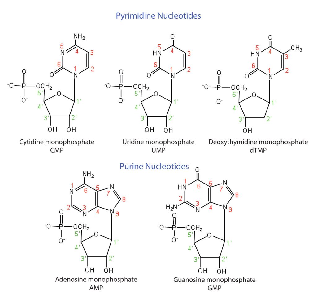
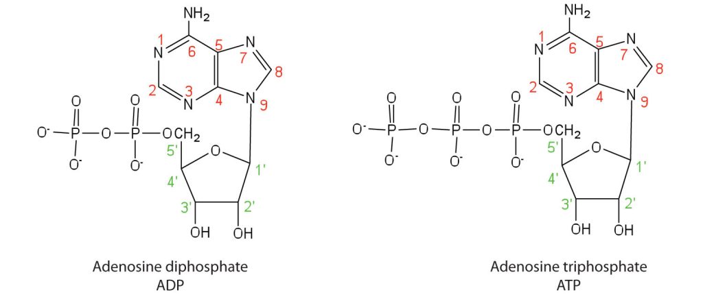
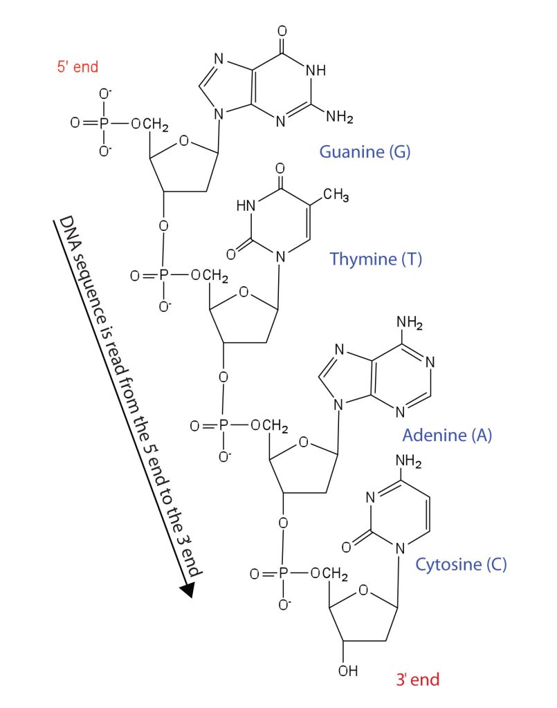
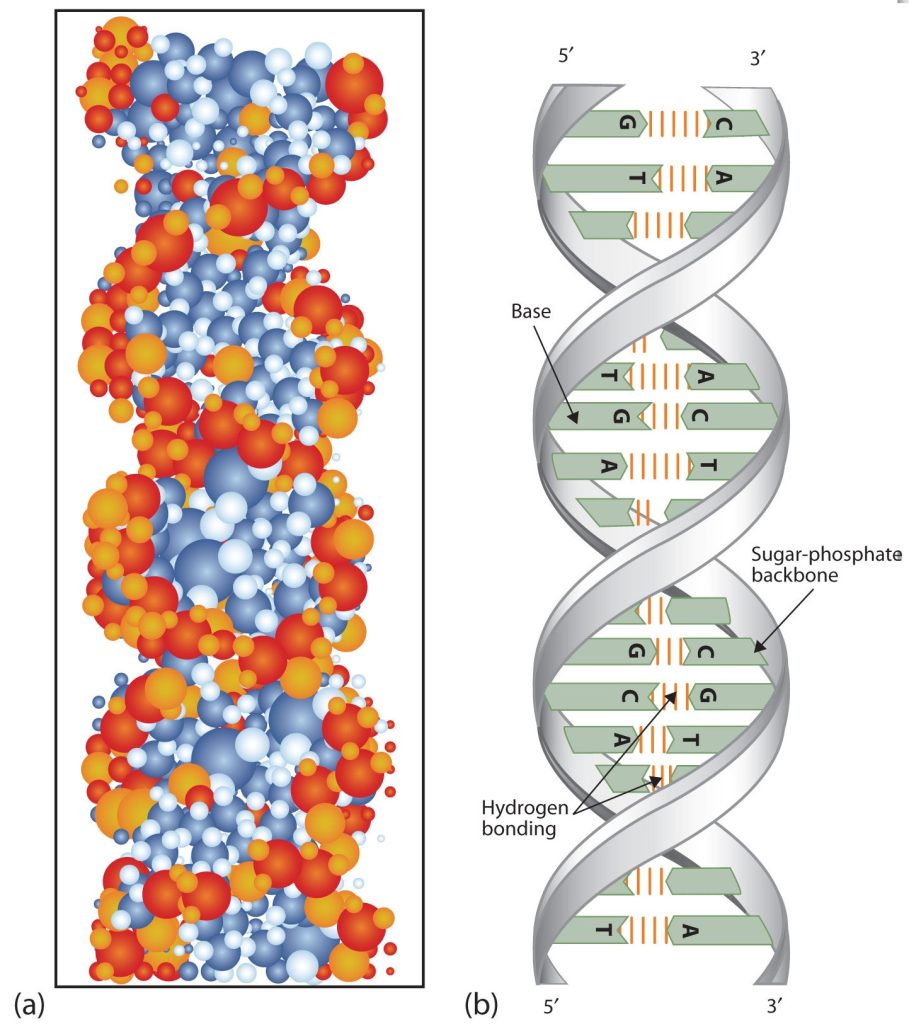
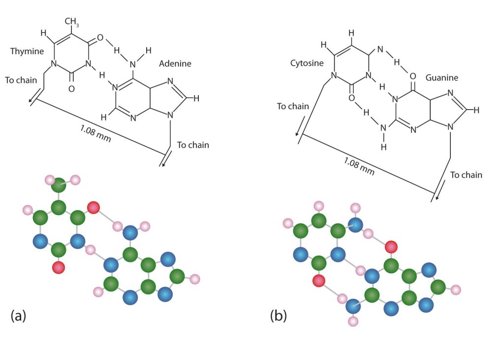
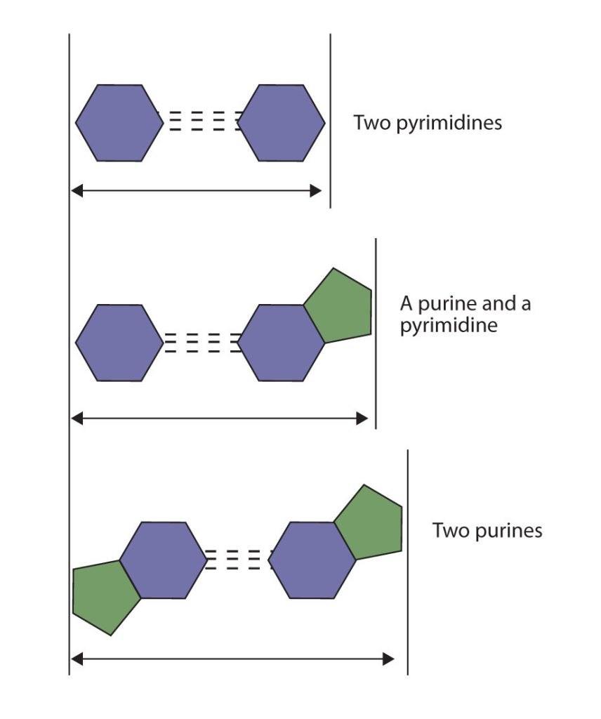
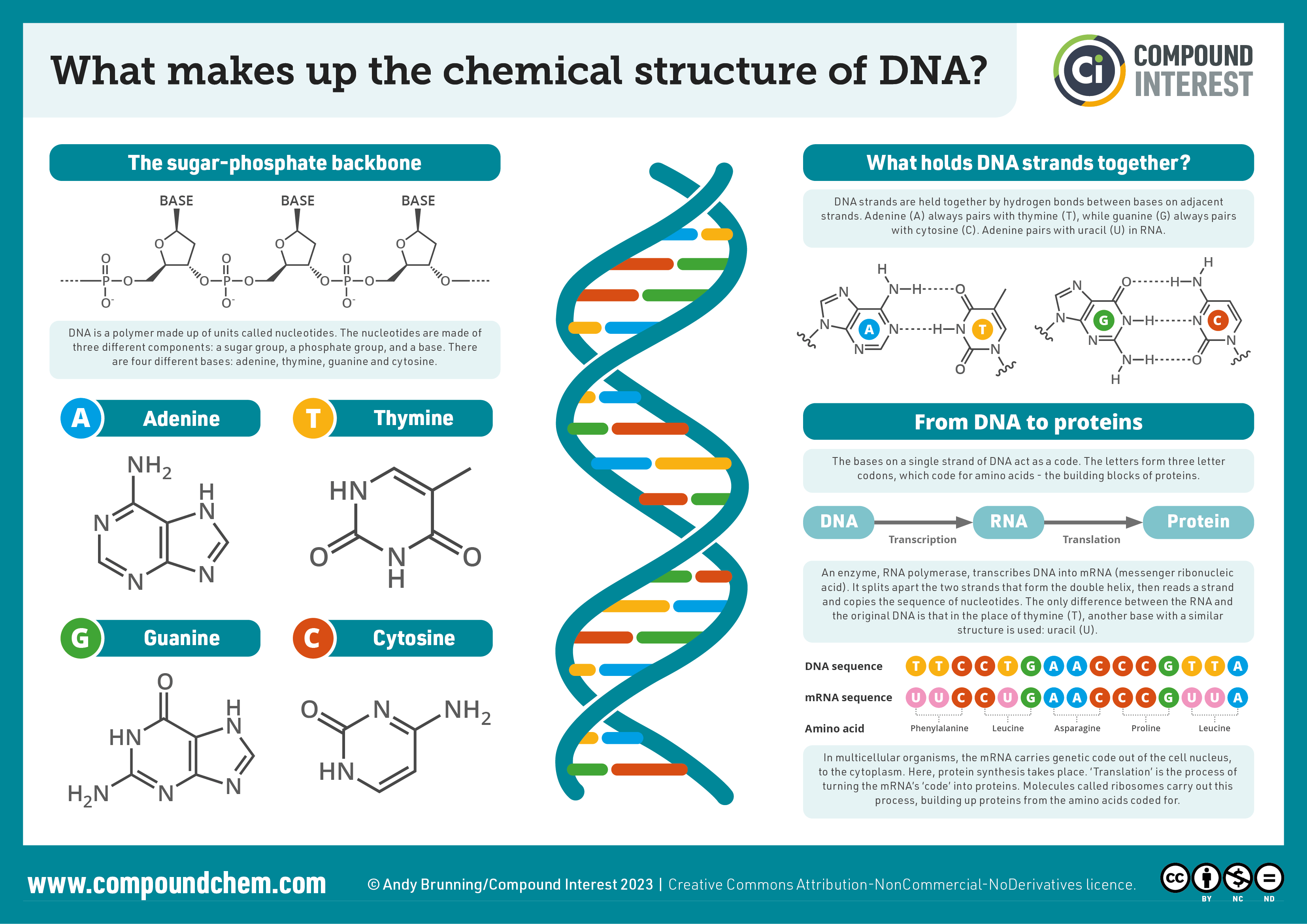
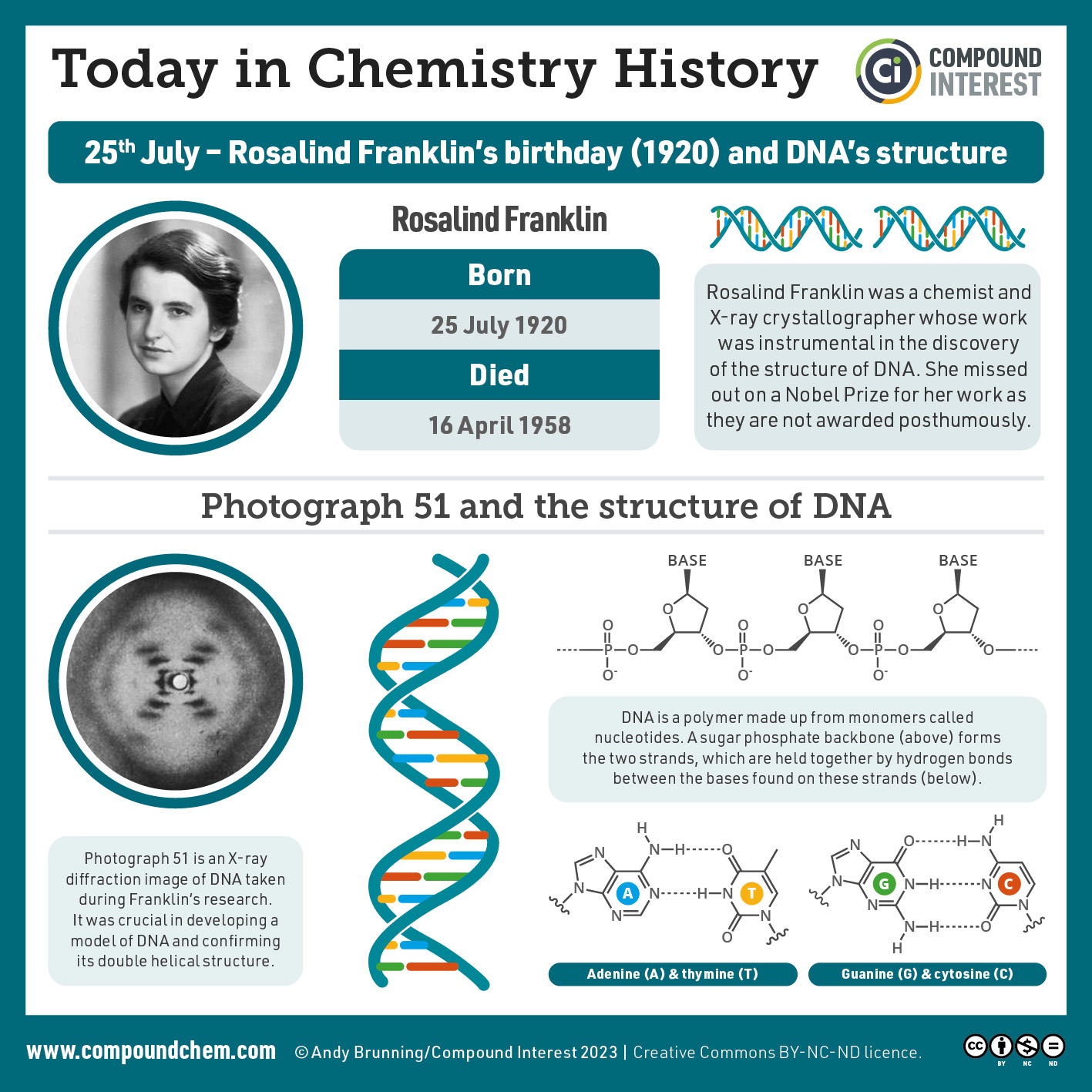
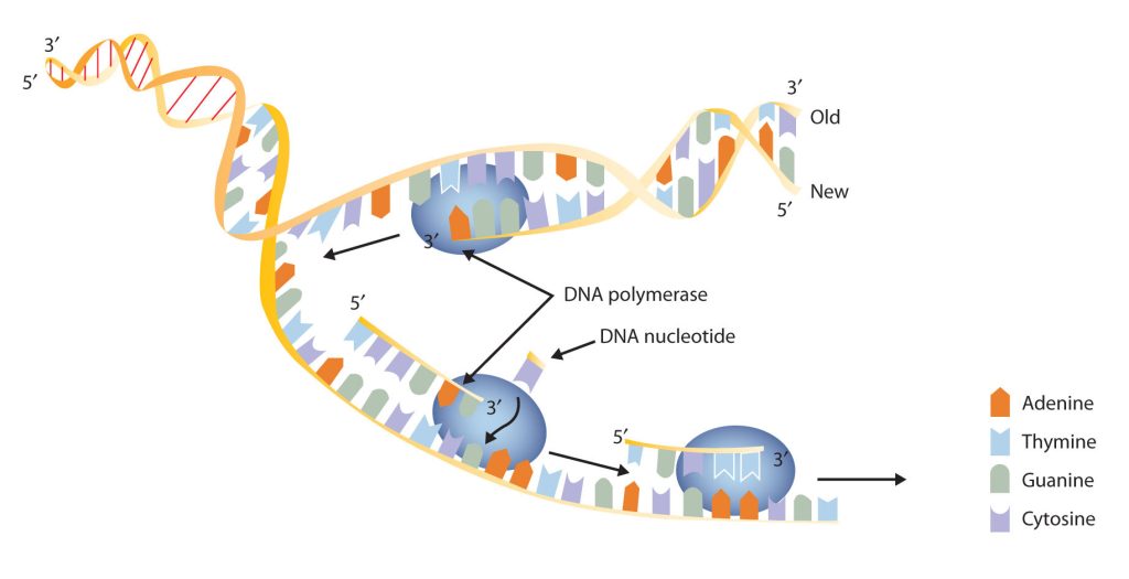
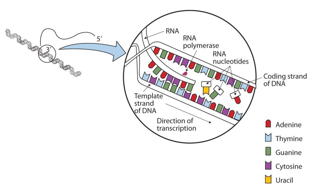
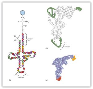
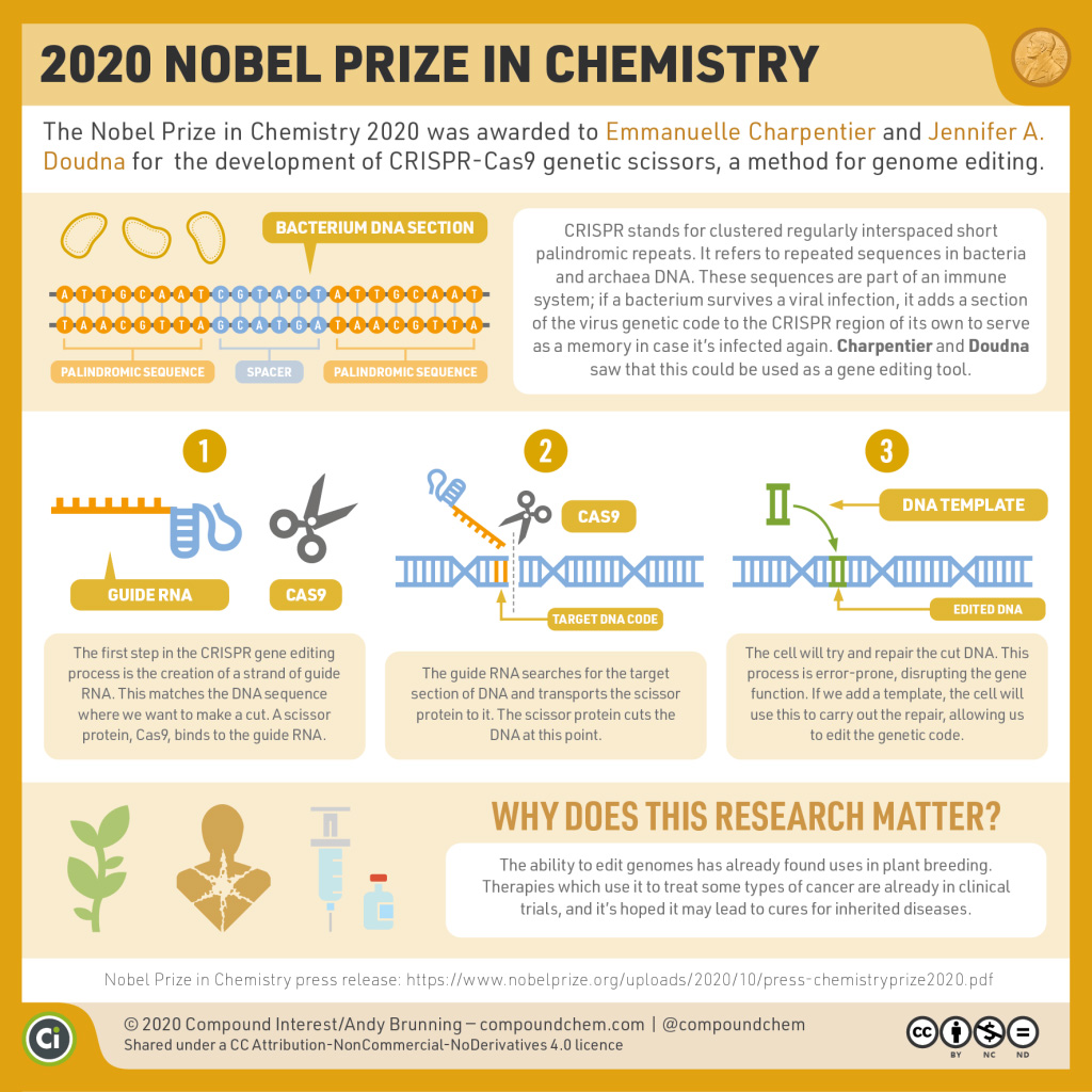
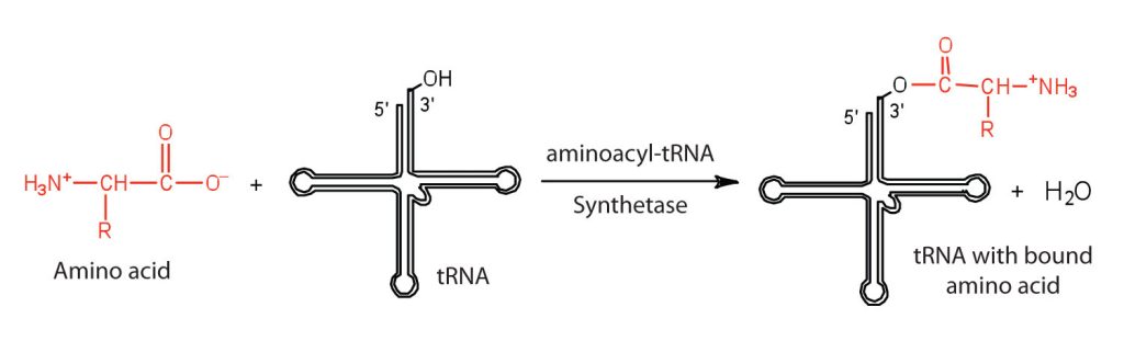
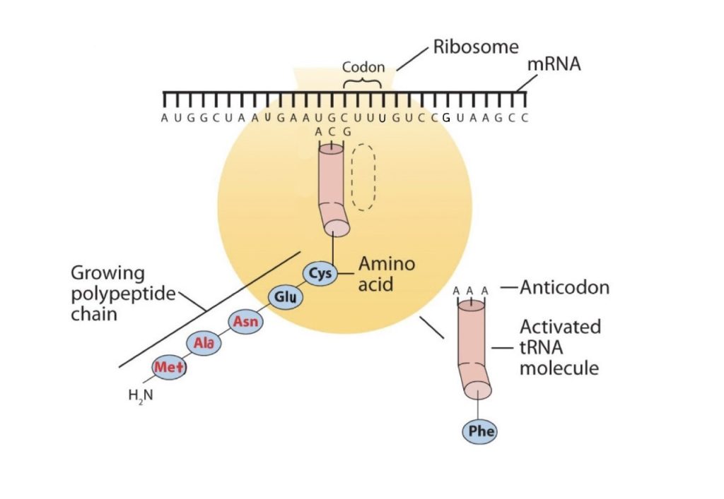

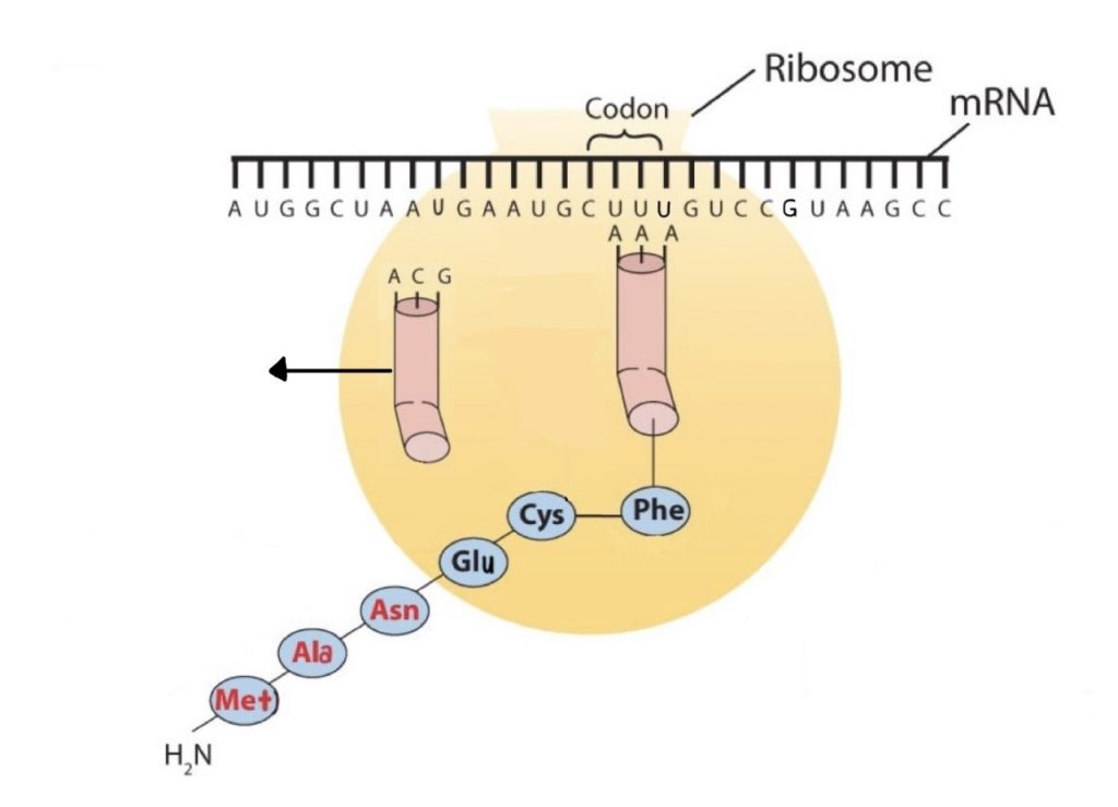
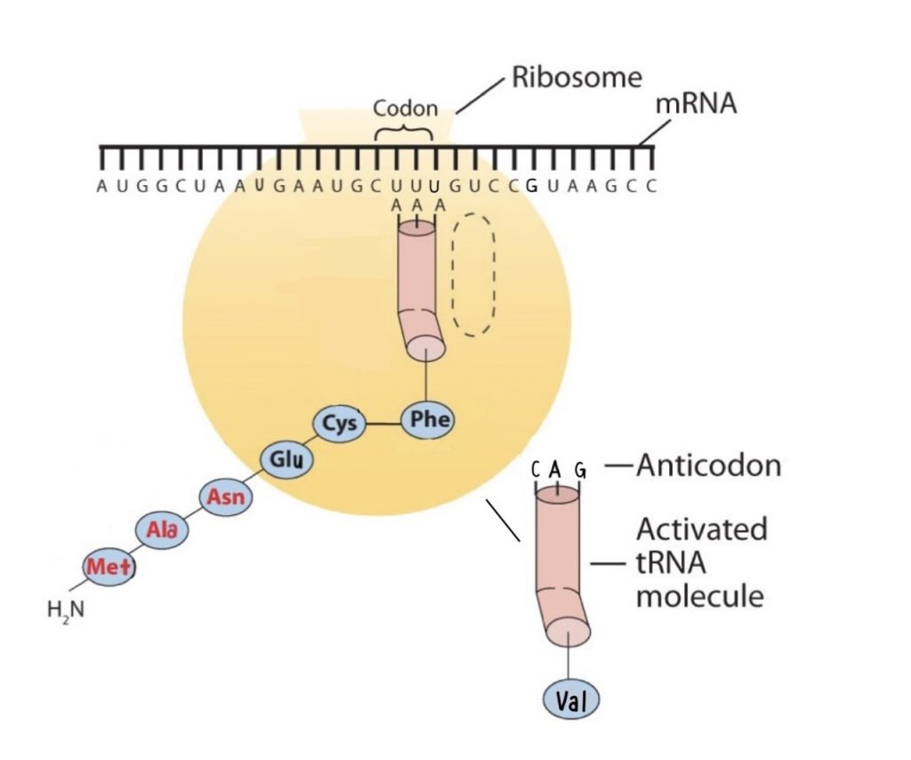
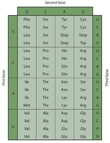
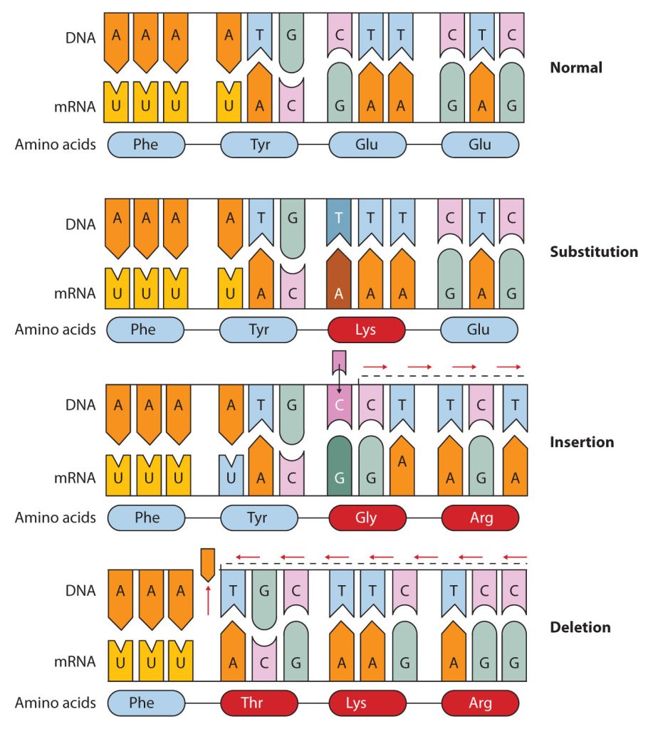
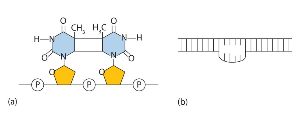
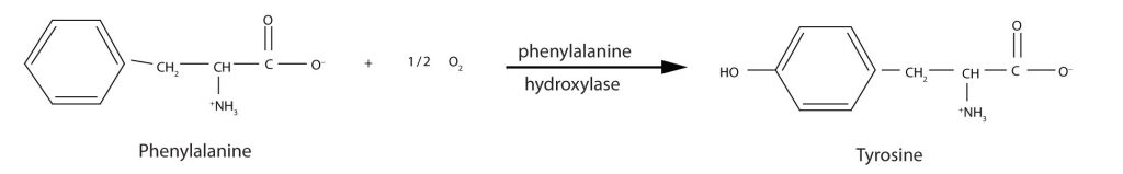
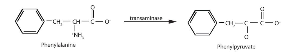
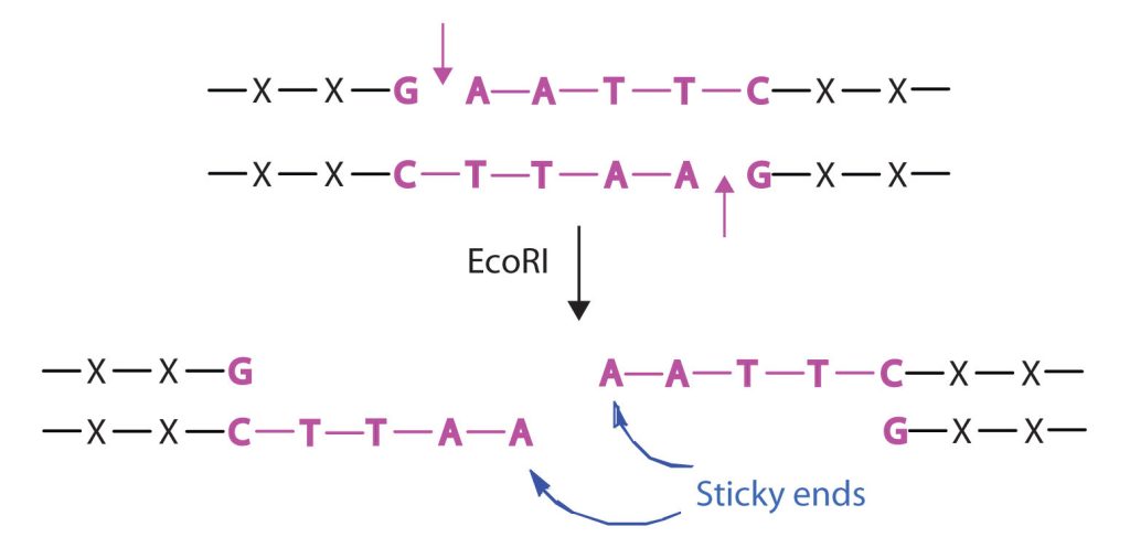
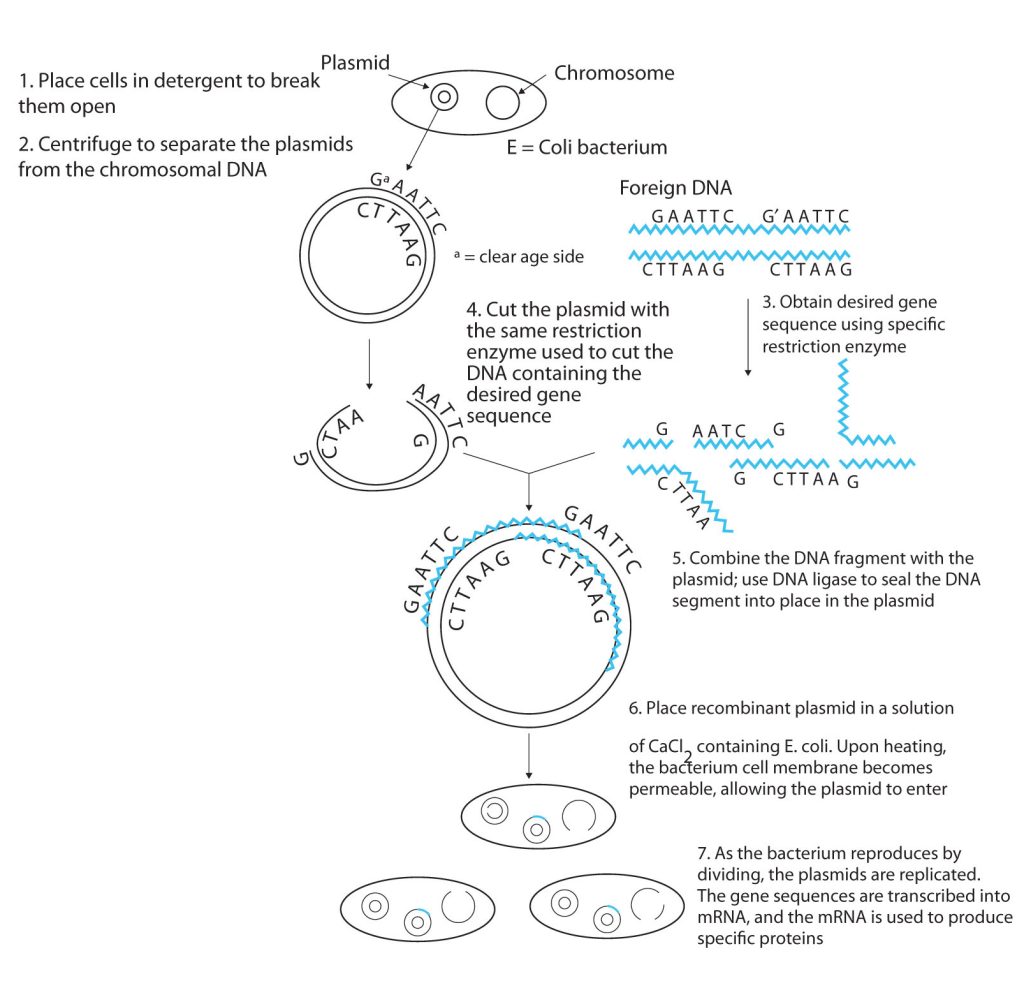
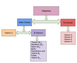
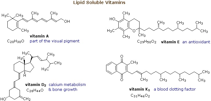
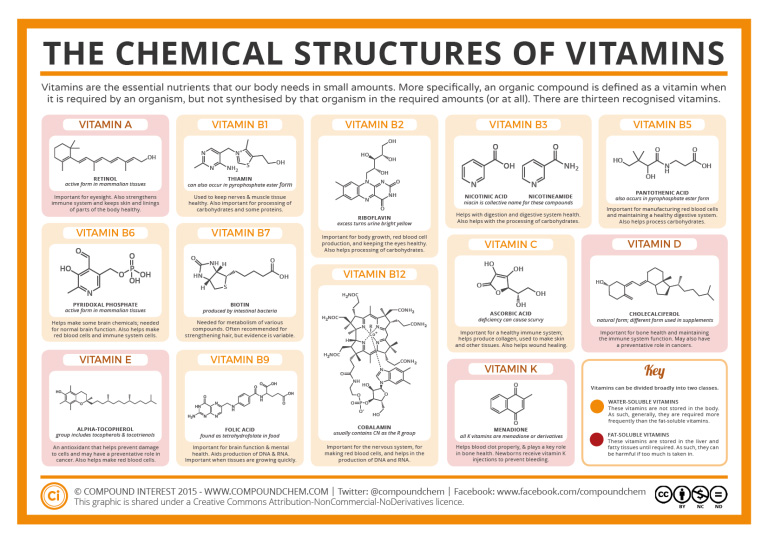

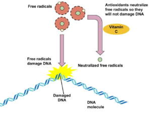

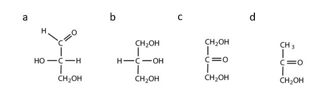
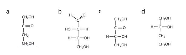
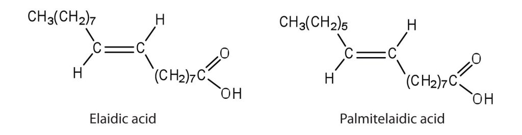

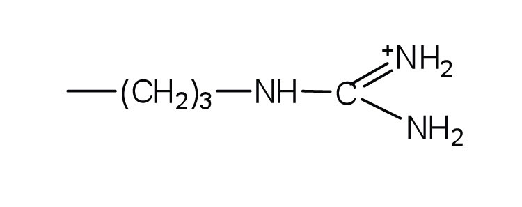

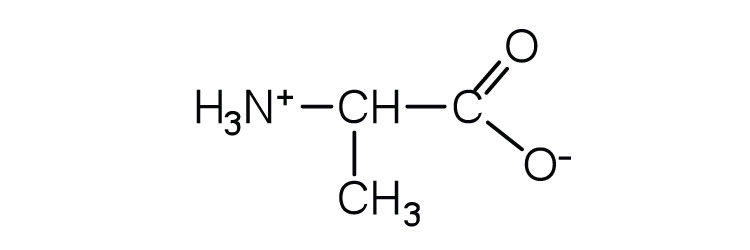
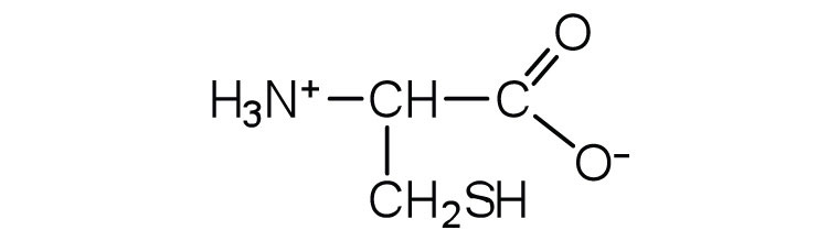
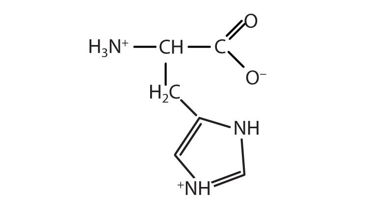


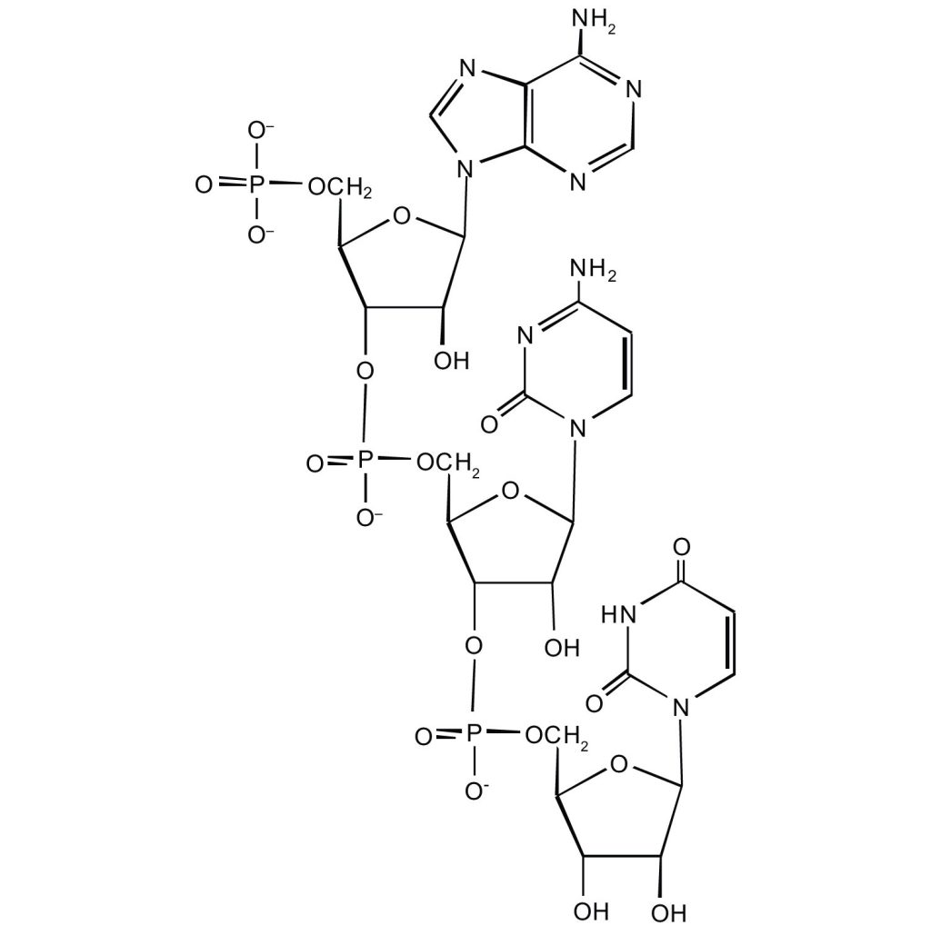
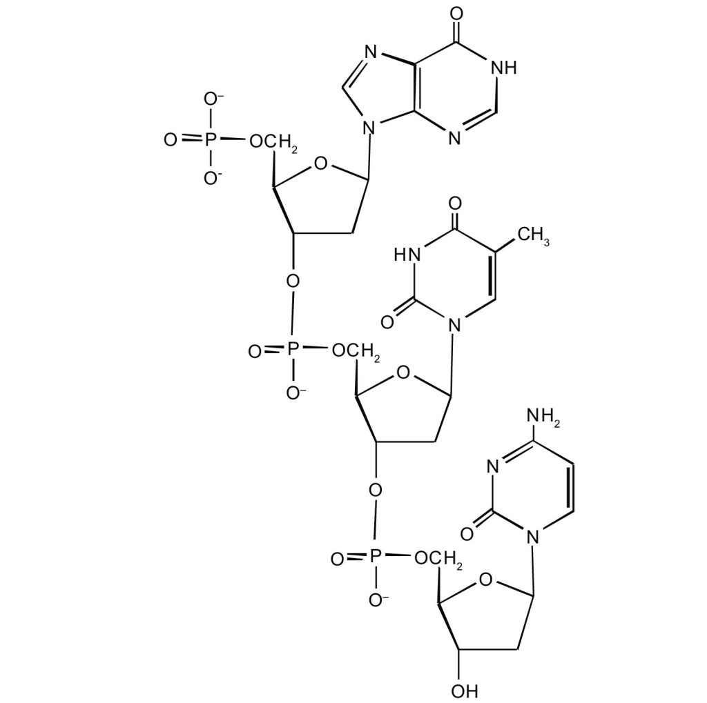
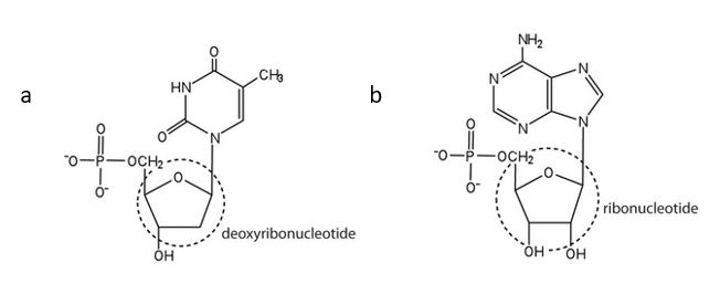
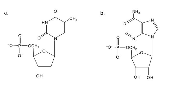
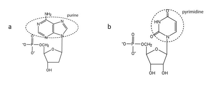
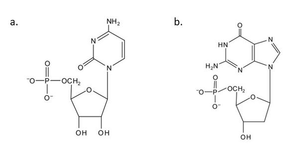
![\text {DNA}\xrightarrow[Transcription]{}\text{RNA}\xrightarrow[Transcription]{}\text{Protein}](https://atu0g9ctah.execute-api.ca-central-1.amazonaws.com/latest/latex?latex=%5Ctext%20%7BDNA%7D%5Cxrightarrow%5BTranscription%5D%7B%7D%5Ctext%7BRNA%7D%5Cxrightarrow%5BTranscription%5D%7B%7D%5Ctext%7BProtein%7D&fg=000000&font=TeX&svg=1 "\text {DNA}\xrightarrow[Transcription]{}\text{RNA}\xrightarrow[Transcription]{}\text{Protein}")


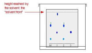

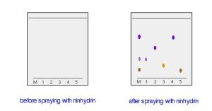
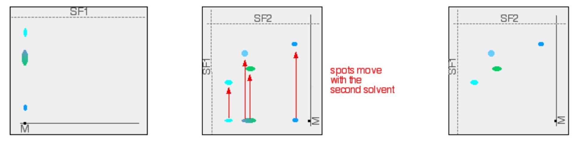
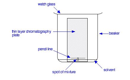
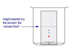

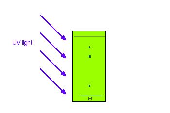

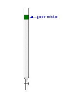
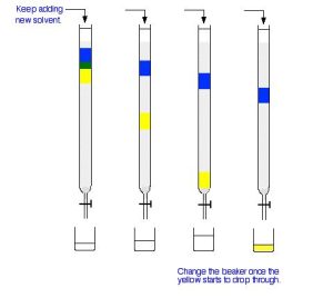
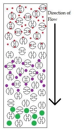


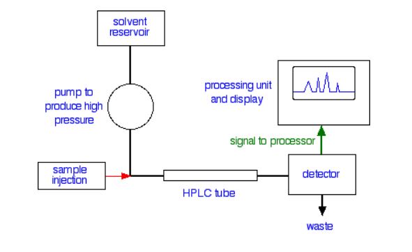
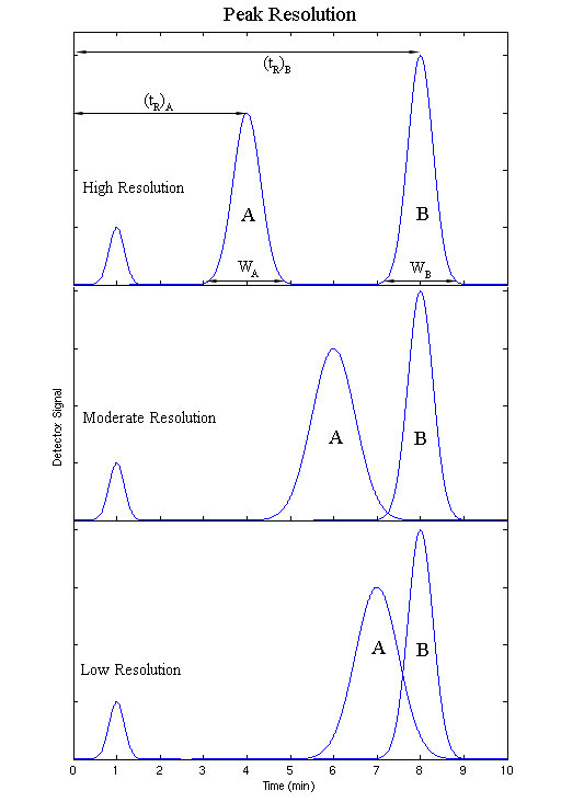
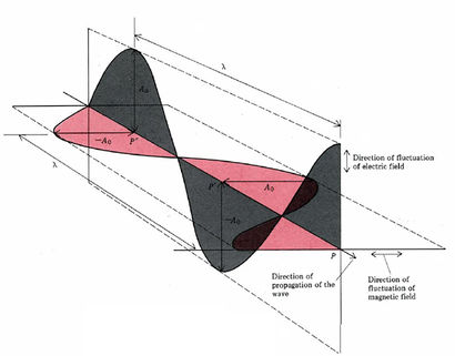
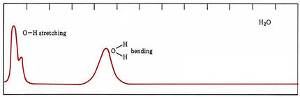


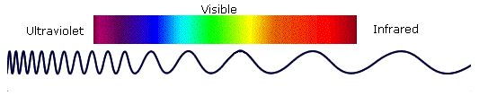
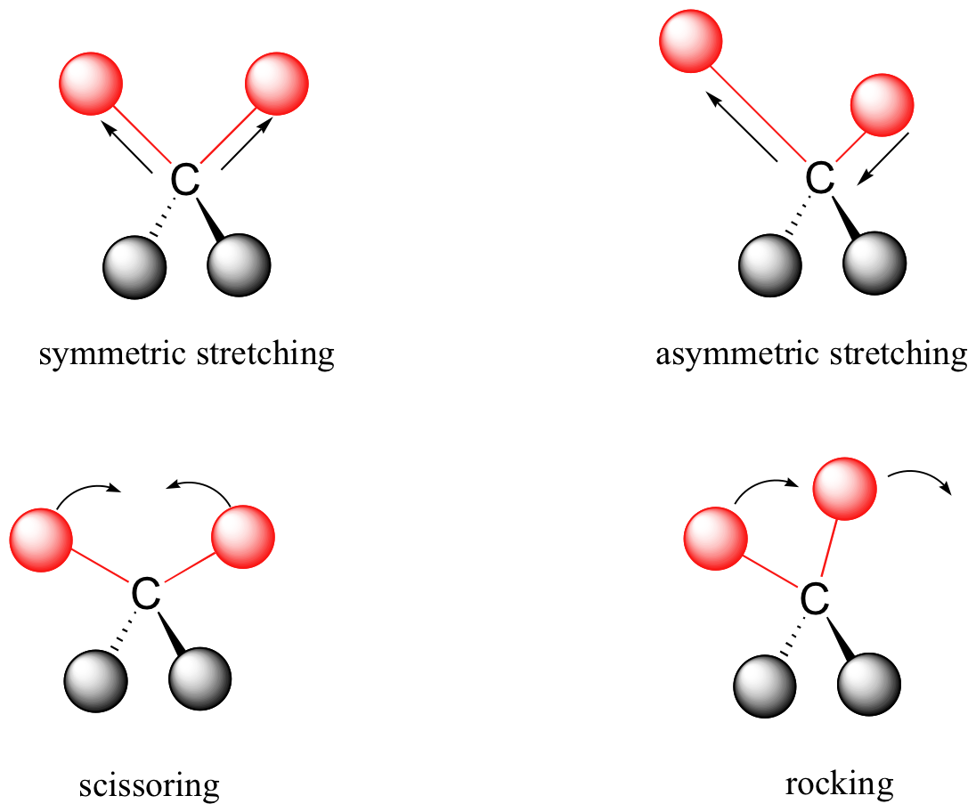
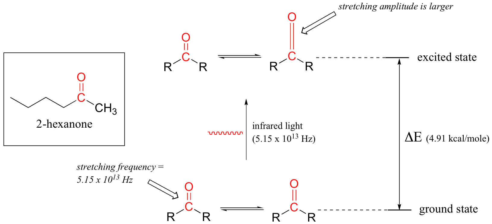

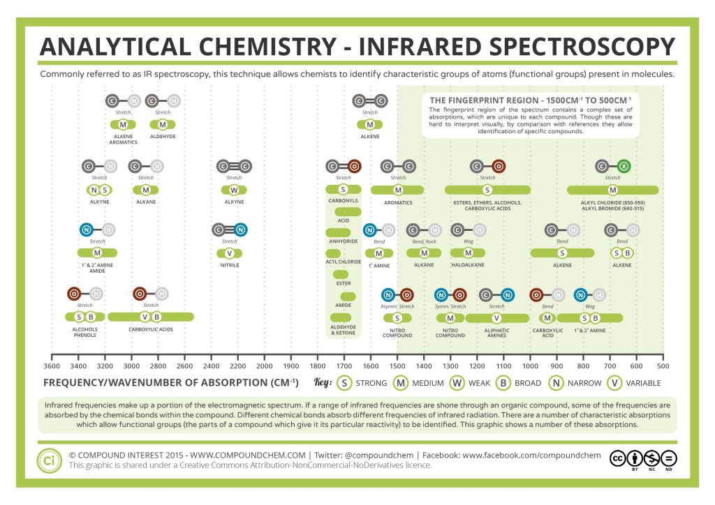


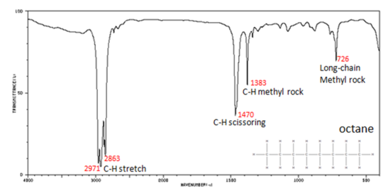
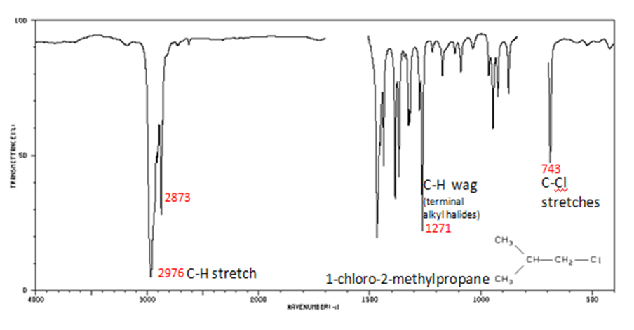
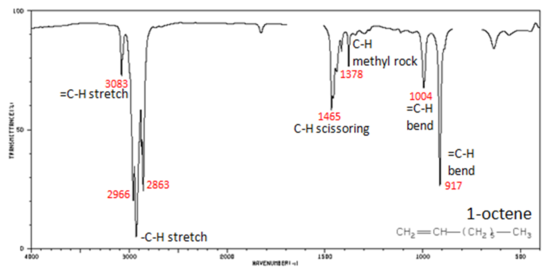
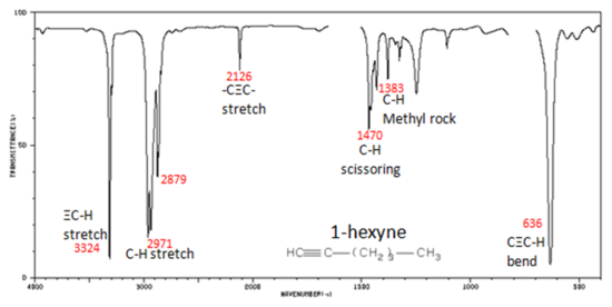
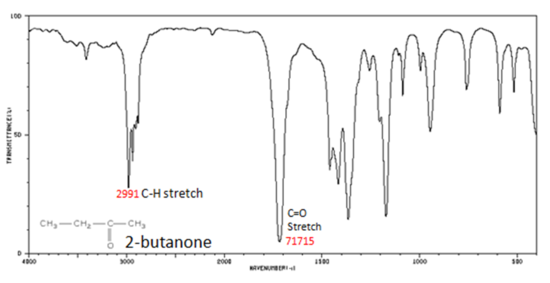
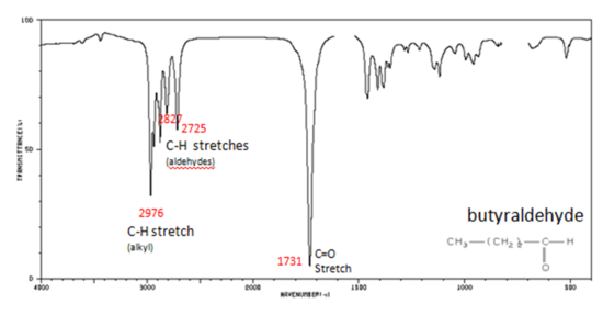
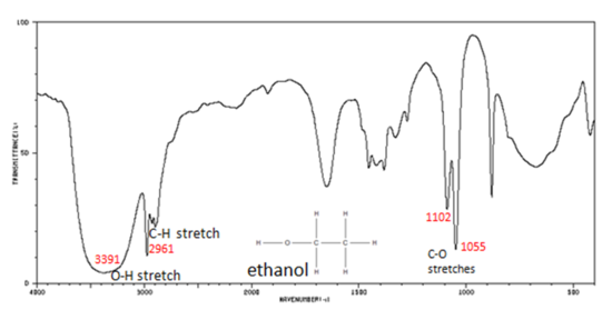
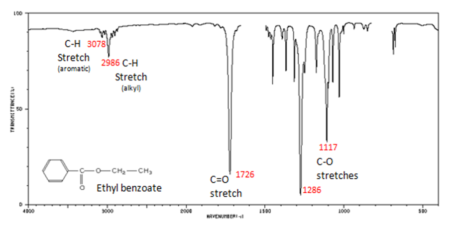
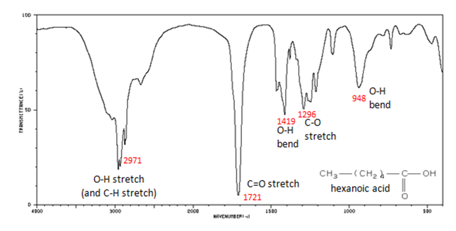
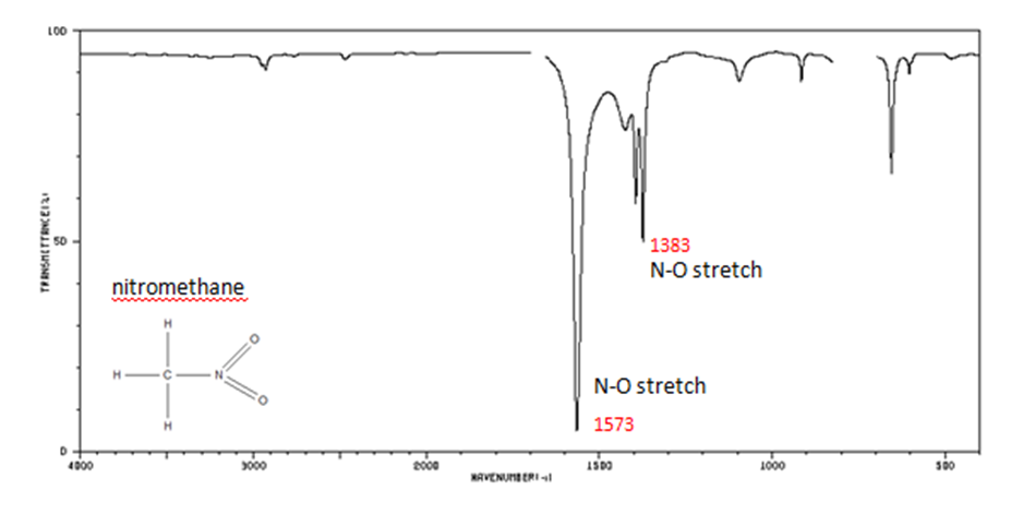
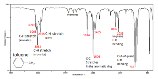
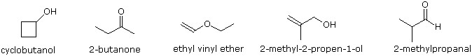


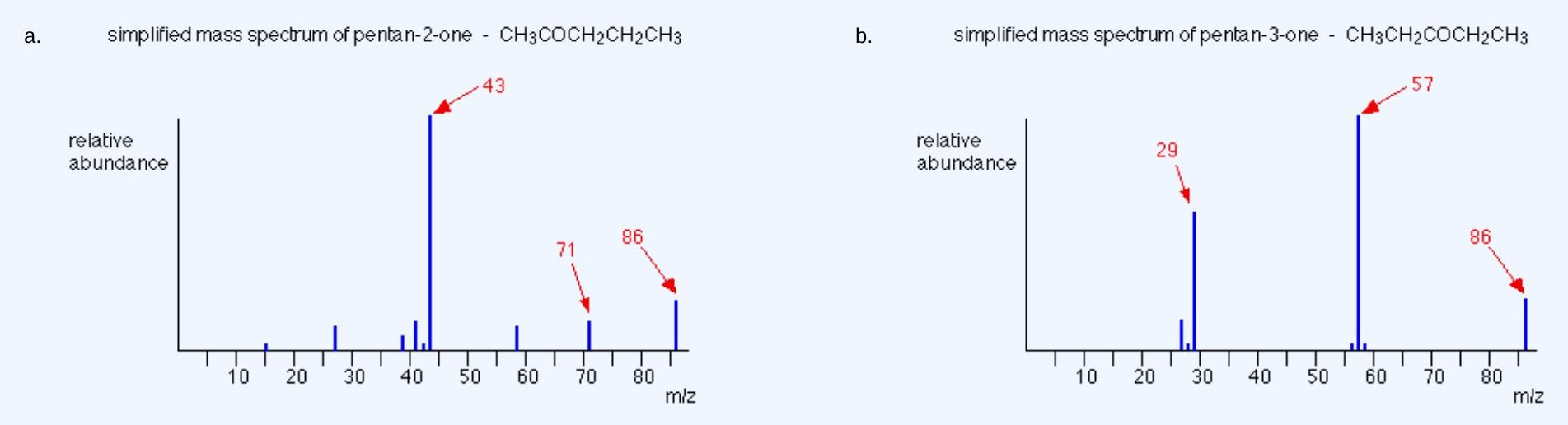
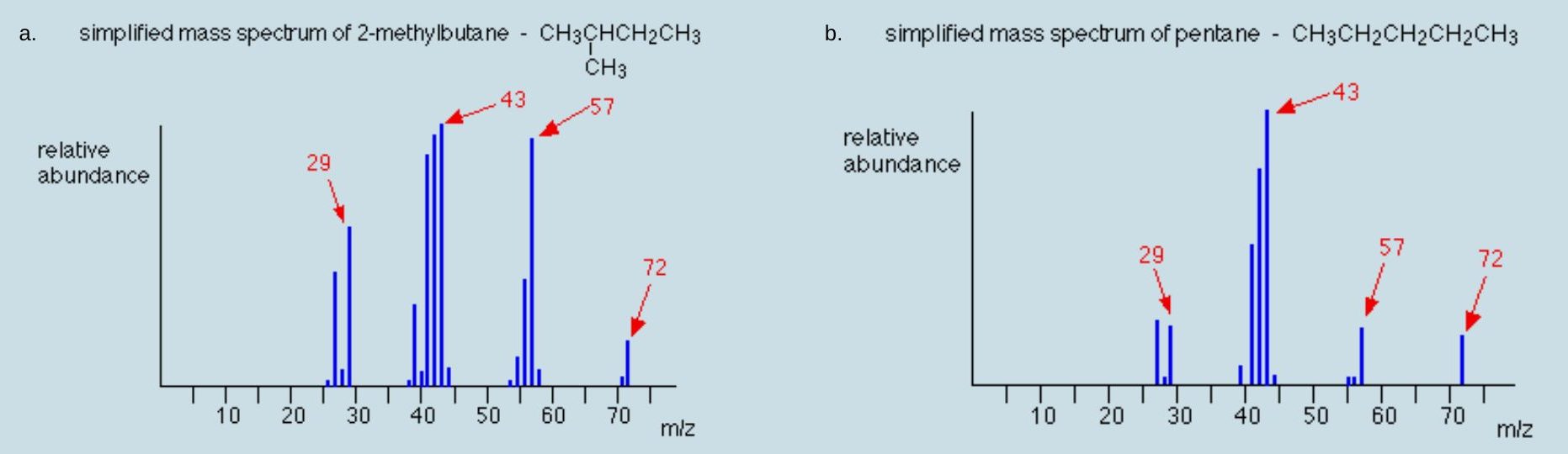
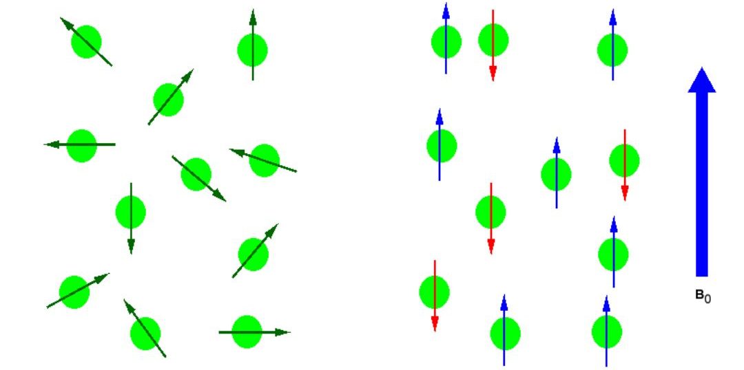
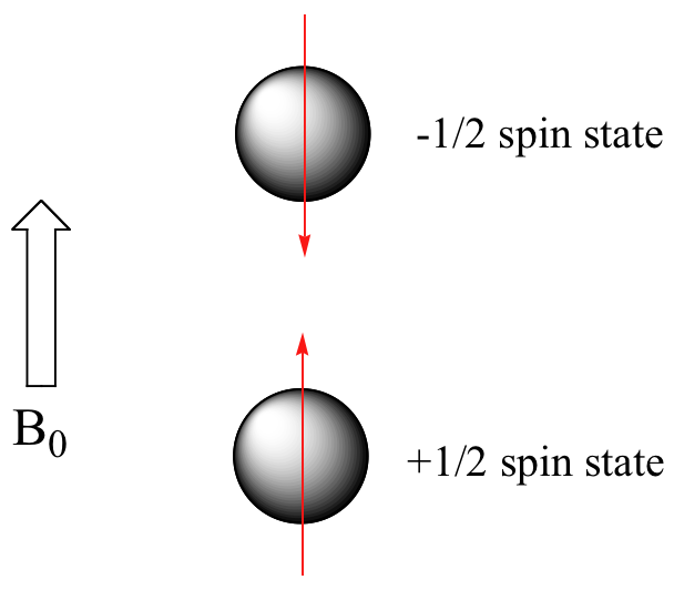
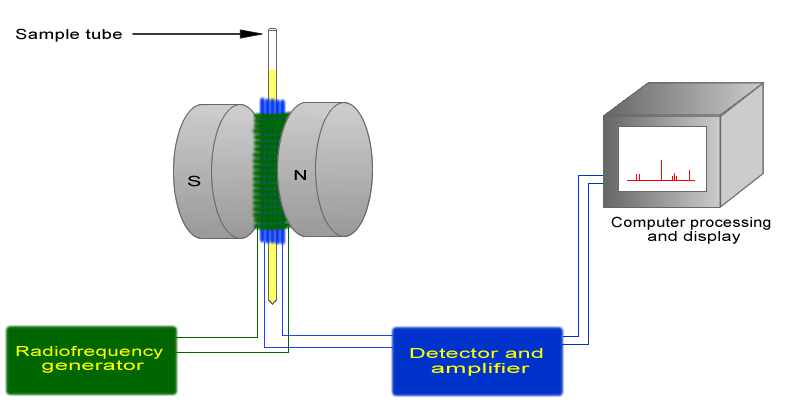
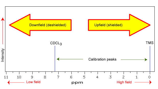
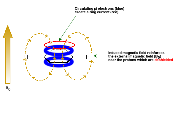
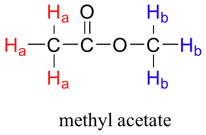
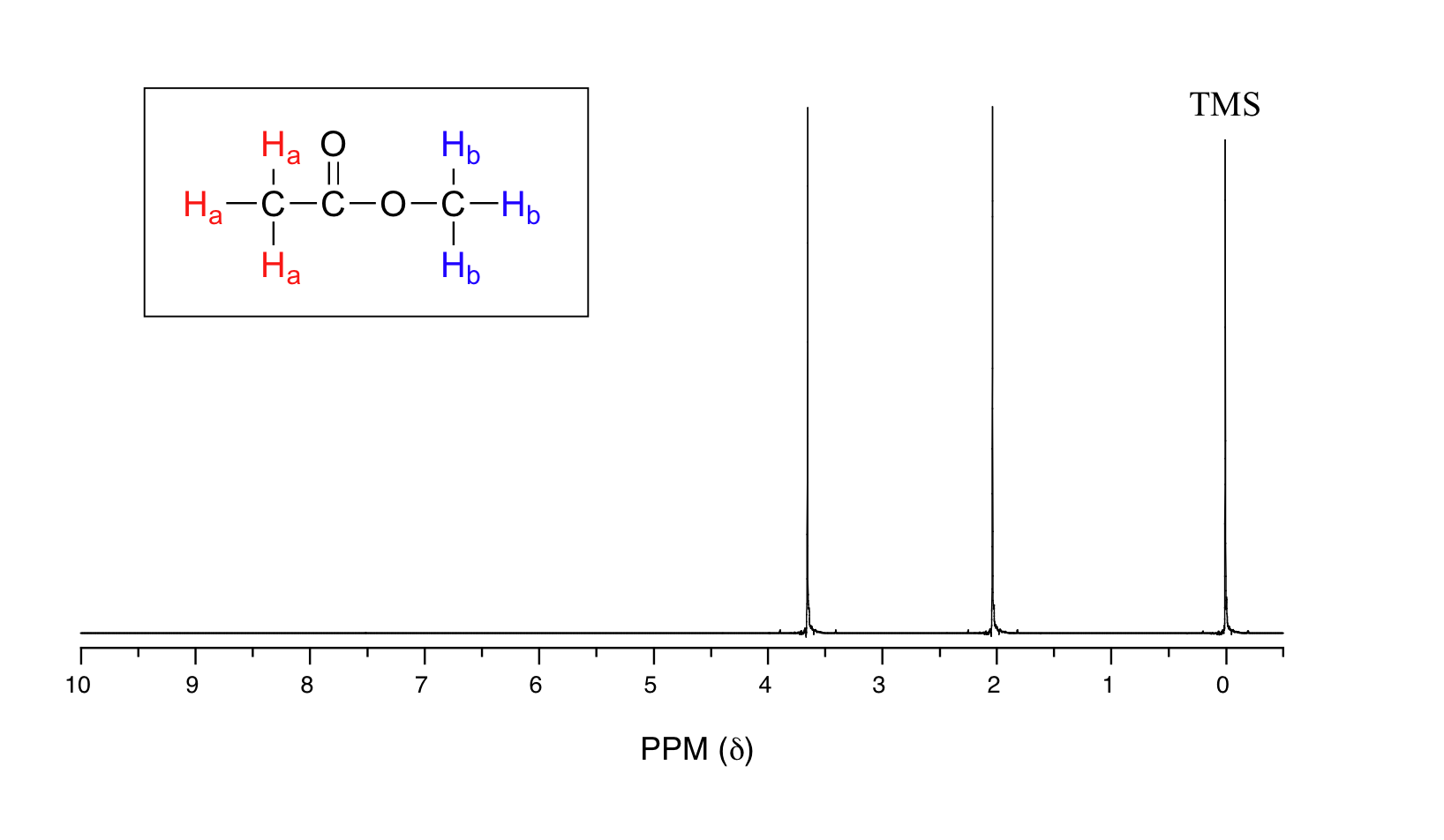
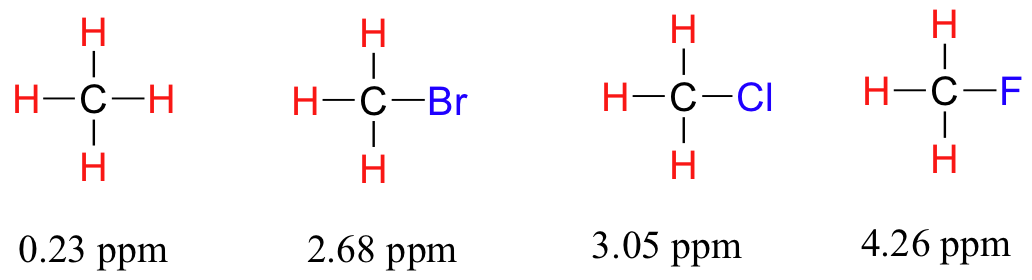
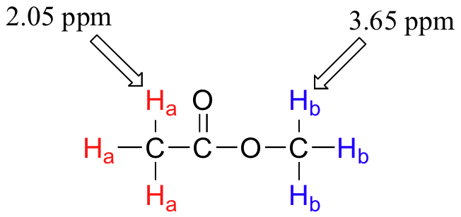
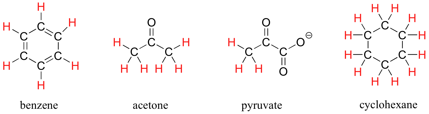
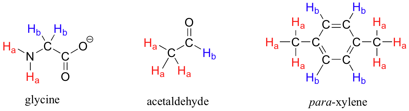
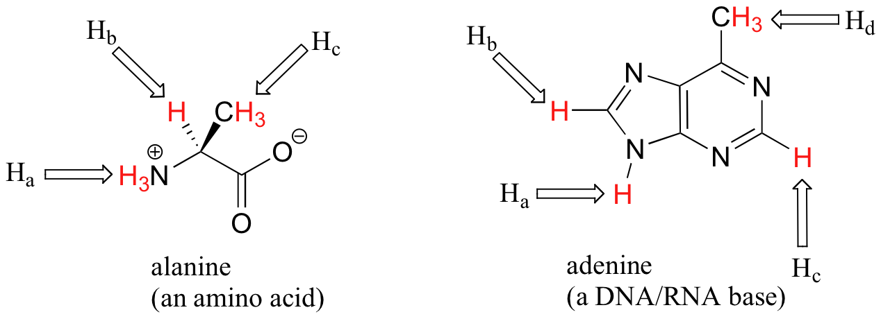
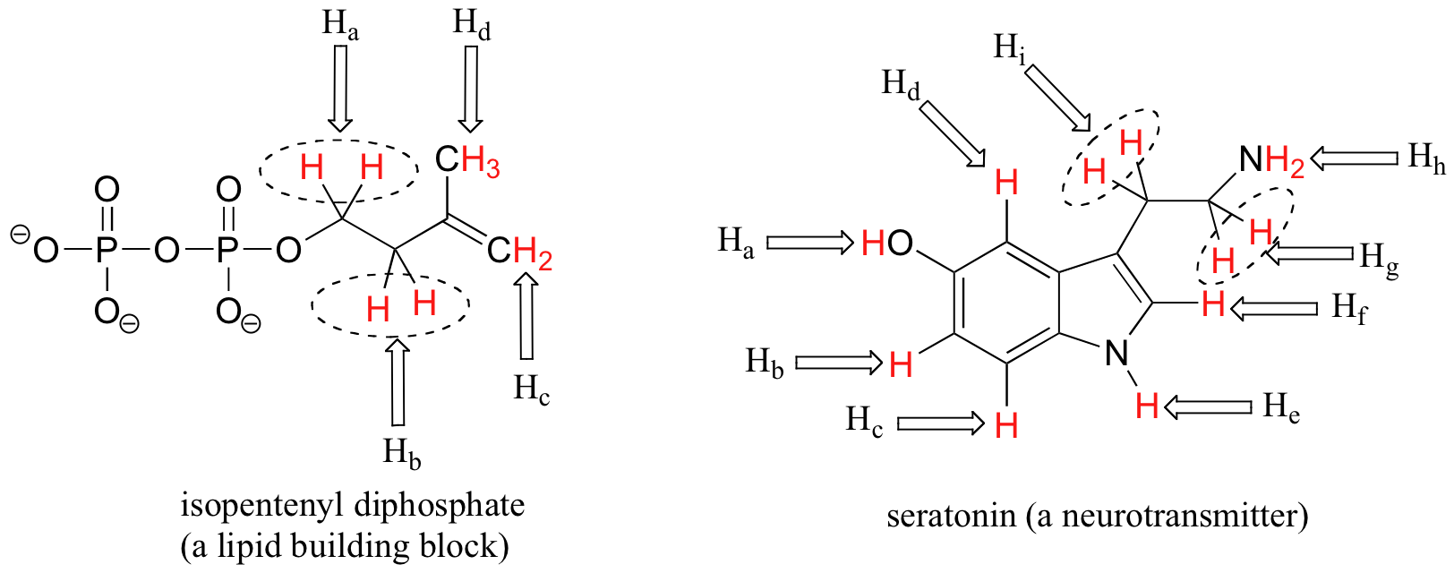
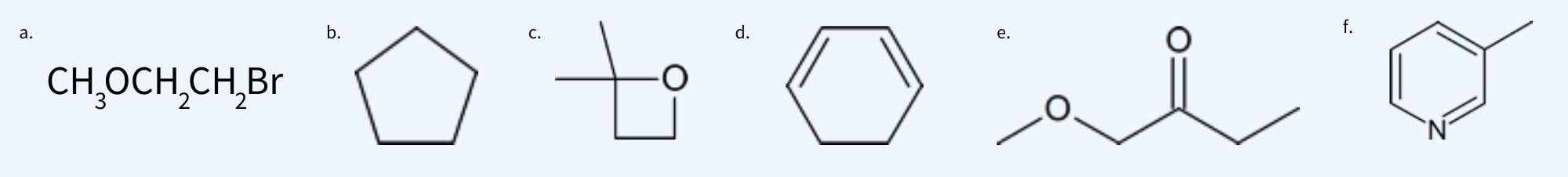


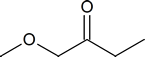


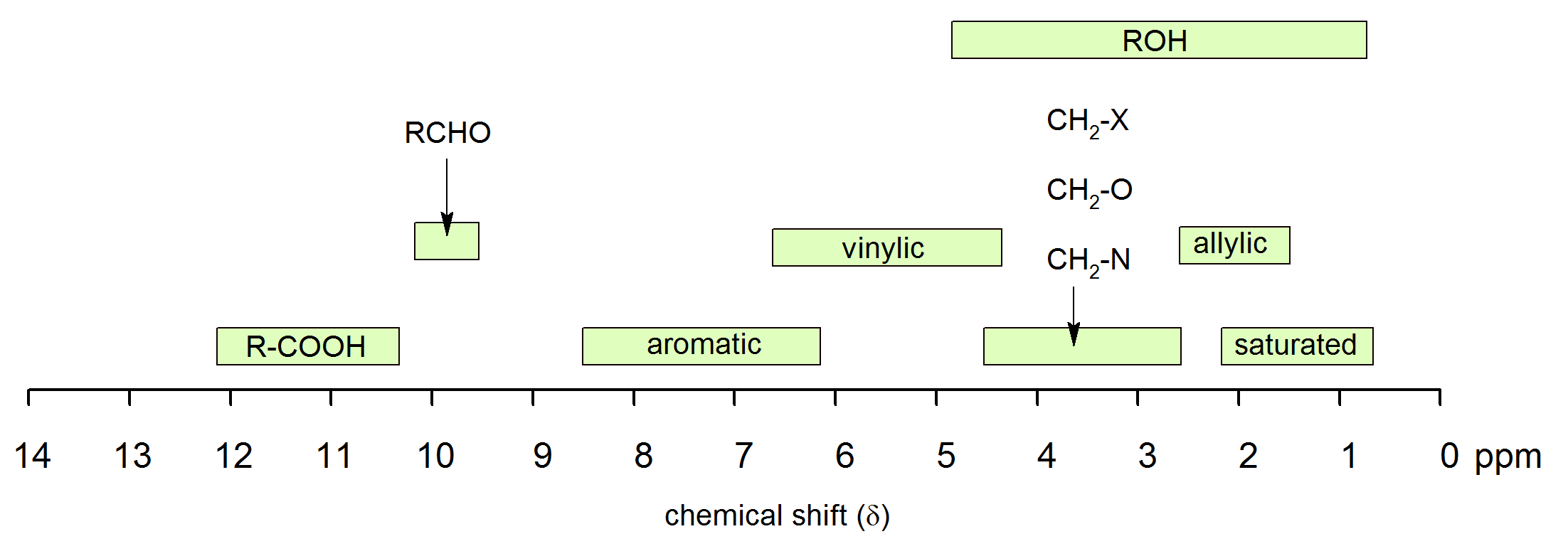


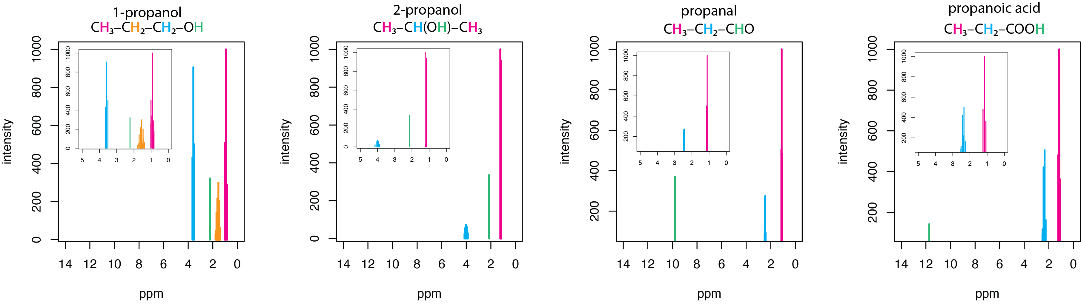
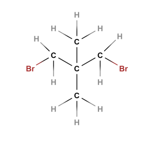
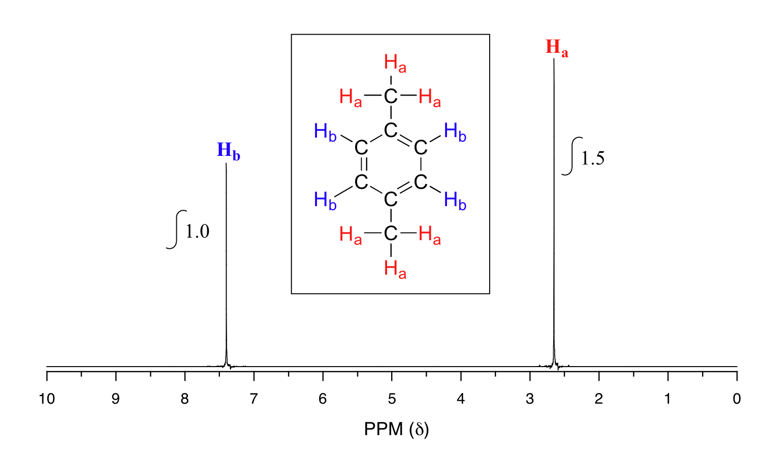
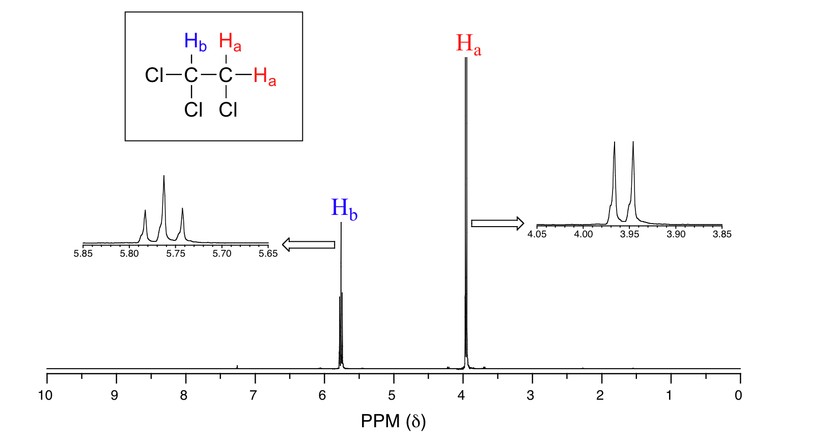
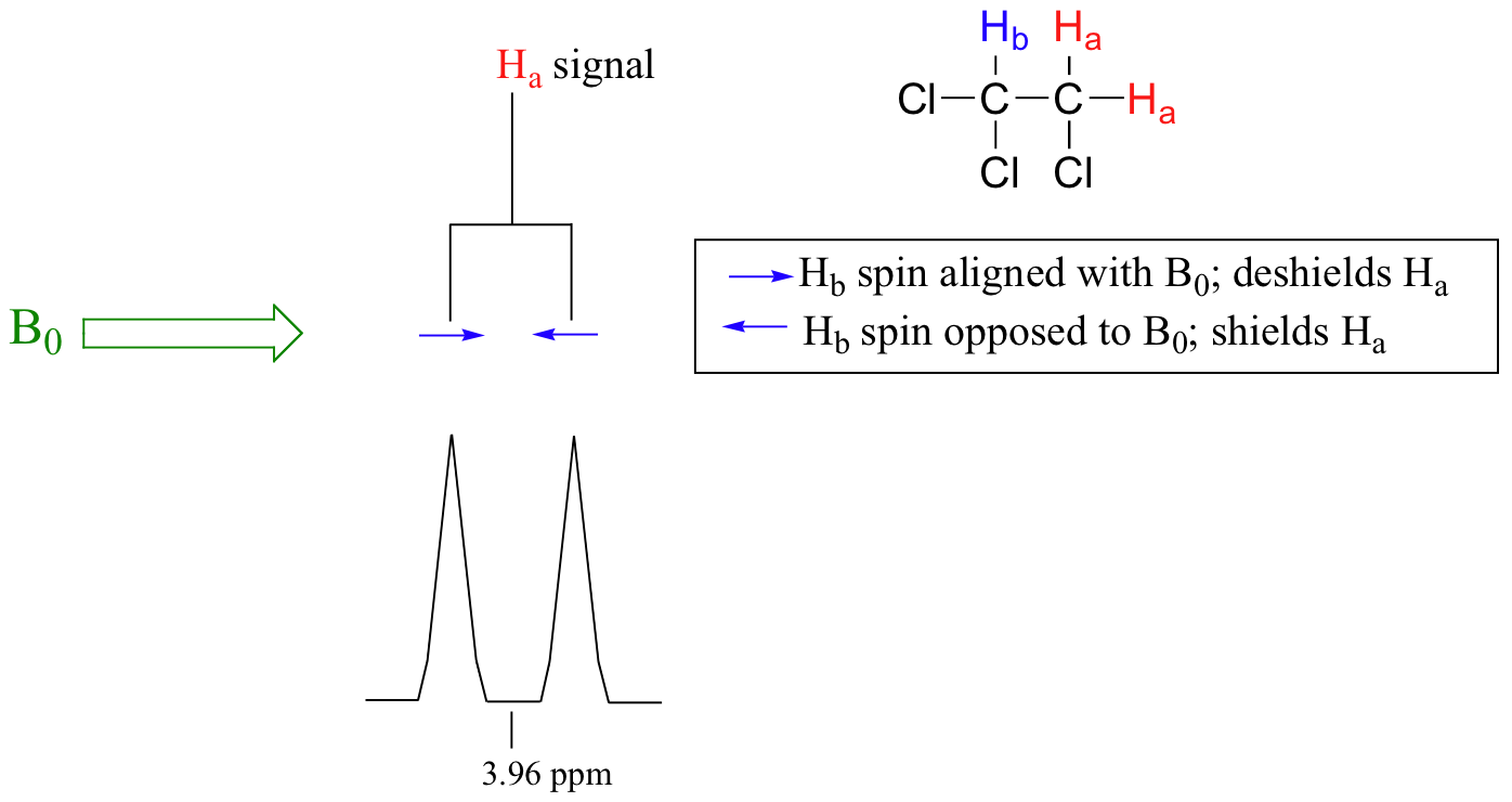
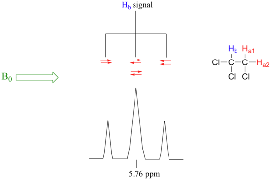


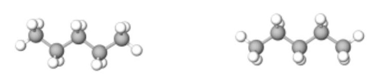
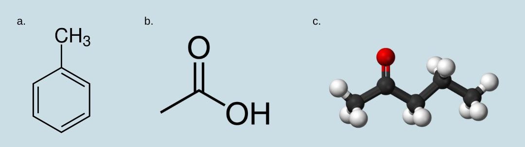
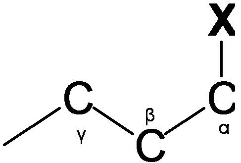

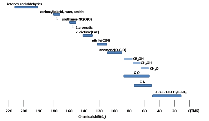


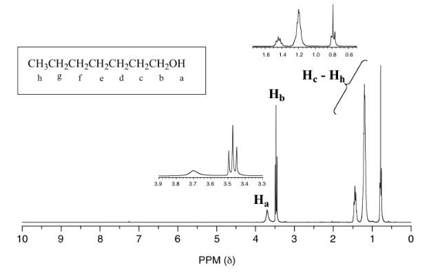

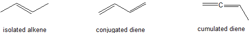
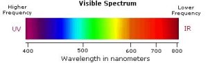
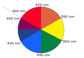
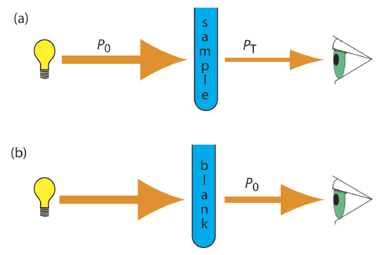
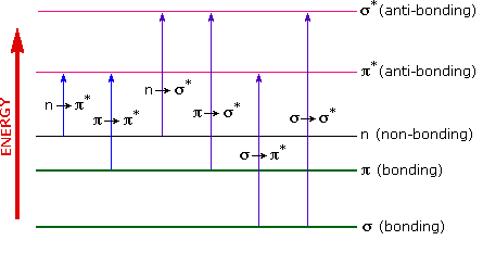
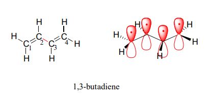
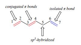

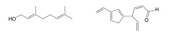
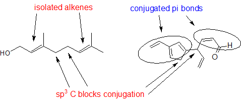
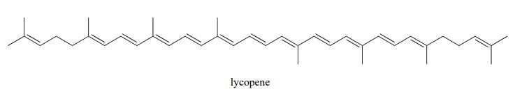
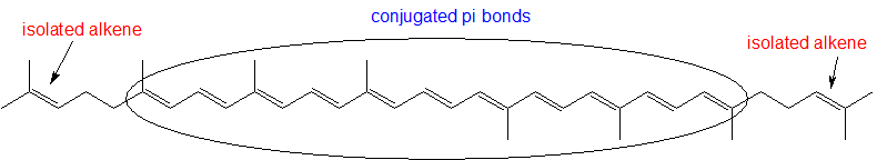
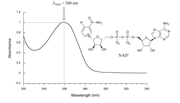
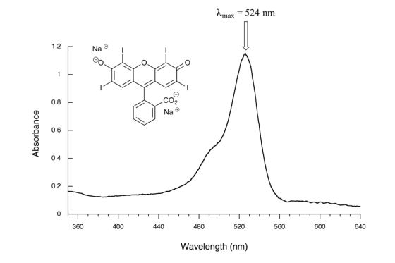
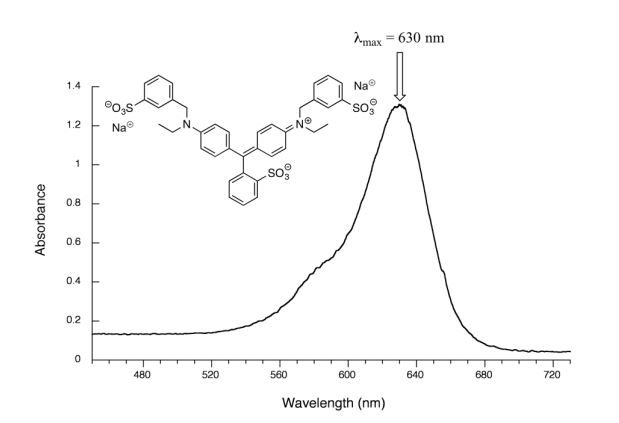
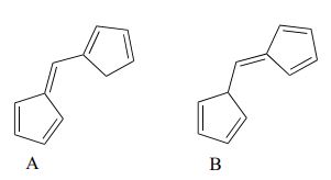

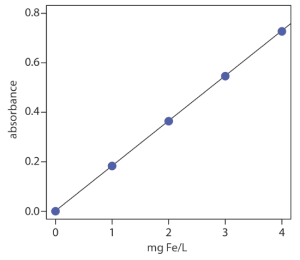


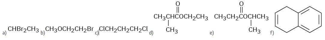
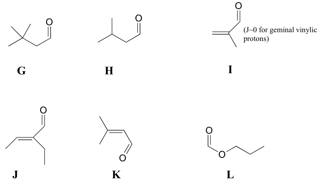
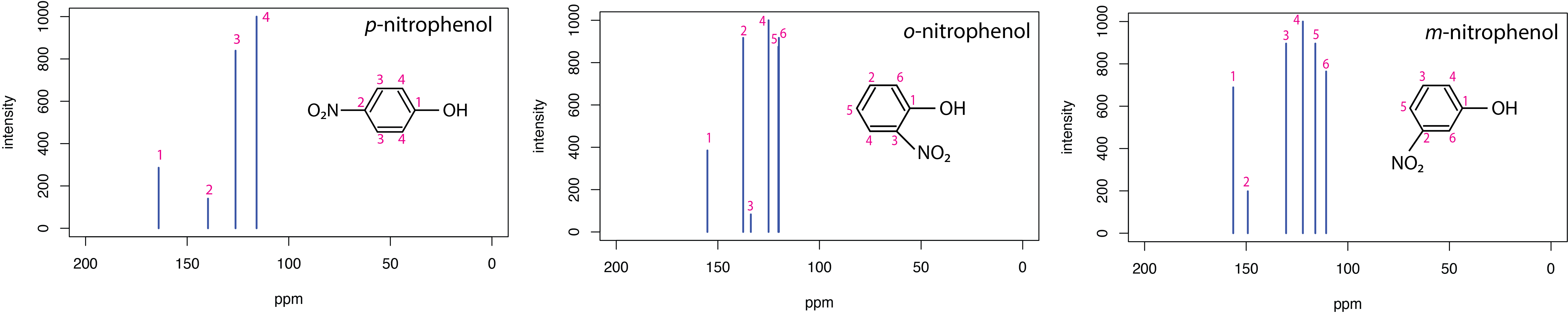


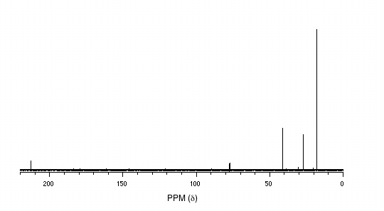

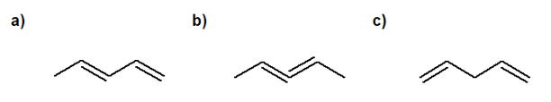


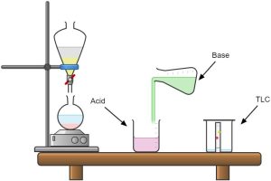
.svg)
.jpg)
.jpg)
.jpg)
,_Fuglesangen,_Svalbard.jpg)
.jpg)