
Human Anatomy Lab Manual by Malgosia Wilk-Blaszczak is licensed under a Creative Commons Attribution 4.0 International License, except where otherwise noted.
Images are individually licensed as noted in the back matter.
Human Anatomy Lab Manual by Malgosia Wilk-Blaszczak is licensed under a Creative Commons Attribution 4.0 International License, except where otherwise noted.
Images are individually licensed as noted in the back matter.
Creation of this resource was supported by Mavs Open Press, operated by the University of Texas at Arlington Libraries (UTA Libraries). Mavs Open Press offers no-cost services for UTA faculty, staff, and students who wish to openly publish their scholarship. The Libraries’ program provides human and technological resources that empower our communities to publish new open access journals, to convert traditional print journals to open access publications, and to create or adapt open educational resources (OER). Resources published by Mavs Open Press are openly licensed using Creative Commons licenses and are offered in various e-book formats free of charge. Optional print copies may be available through the UTA Bookstore or can be purchased through print-on-demand services, such as Lulu.com.
OER are free teaching and learning materials that are licensed to allow for revision and reuse. They can be fully self-contained textbooks, videos, quizzes, learning modules, and more. OER are distinct from public resources in that they permit others to use, copy, distribute, modify, or reuse the content. The legal permission to modify and customize OER to meet the specific learning objectives of a particular course make them a useful pedagogical tool.
Pressbooks is a web-based authoring tool based on WordPress, and it is the primary tool that Mavs Open Press (in addition to many other open textbook publishers) uses to create and adapt open textbooks. In May 2018, Pressbooks announced their Accessibility Policy, which outlines their efforts and commitment to making their software accessible. Please note that Pressbooks no longer supports use on Internet Explorer as there are important features in Pressbooks that Internet Explorer doesn’t support.
The following browsers are best to use for Pressbooks:
This publication was designed to work best online and features hyperlinks in the text. We have retained the blue font for hyperlinks in the print version to make it easier to find the URL in the “Links by Chapter” section at the back of the book.
Information about open education at UTA is available online. If you are an instructor who is using this OER for a course, please let us know by filling out our OER Adoption Form. Contact us at pressbooks@uta.edu for other inquires related to UTA Libraries publishing services.
This is a lab manual for a college-level human anatomy course (BIOL 3446 at UTA). Despite the abundance of information readily available via Google, the mastery of anatomy requires a fair amount of memorization for quick recall. The activities in this manual encourage students to engage with new vocabulary in many ways, including grouping key terms, matching terms to structures, recalling definitions, and written exercises.
As the majority of college campuses do not have easy access to a cadaver, most of the activities in this manual utilize anatomical models. Also included are several dissections of animal tissues, and a significant amount of histological examinations.
Each unit includes both pre- and post-lab questions and six lab exercises designed for a classroom where students move from station to station during a three-hour period. Effort was put into equalizing the time required to perform each lab exercise, to facilitate class flow. The vocabulary terms used in each unit are listed at the end of the manual and serve as a checklist for practicals.
When Malgosia Wilk-Blaszczak began teaching human anatomy at UTA she realized that while there are many commercially available manuals which incorporate a lot of human physiology, none of them focus solely on anatomy. She decided to create a manual for anatomy labs that could fill that void. The first version of this work was created and used in anatomy labs at UTA.
The idea of publishing the lab manual as an OER came to her courtesy of Michelle Reed, Open Education Librarian at UTA. To make this leap to an open platform, she enlisted the help of some of her best students. In Fall 2017, one year prior to the publication of this work, Wilk recruited a group of three excellent undergraduate teaching assistants. These students worked with UTA Libraries to identify openly licensed images and incorporate them into the text. Libraries’ staff assisted in migrating the resource to Pressbooks, where it could be easily exported into a variety of formats. Furthermore, we conducted student surveys to gather feedback. Wilk’s teaching assistants have always been an important part of her pedagogy. With their assistance, she was able to complete and openly publish this anatomy lab manual. The students put in the hard work to change all illustrations to Creative Commons licensed images and ensure proper attribution of all the images used. The student contributors, Kevin Alford, Andrea Compo-Valez, and Victoria Dorch, now alumni, reviewed and edited the resource, and are listed as co-authors of this manual.
Ultimately, open manuals reduce the cost to students while customizing the information and visuals required for class. In addition, the digital copy of the manual allows students to access homework and exercises wherever they are and is easily obtainable on the first day of class. Open manuals are also dynamic works that can be adapted to suit the needs of other institutions or groups that wish to explore the topic but do not have a solid framework to do so. The resulting OER is being piloted in human anatomy labs in Fall 2018 and will be revised following the pilot period with input from current students and lab instructors. It is our hope that this extension of Wilk’s class will open the door to connecting our courses to broader collaborations and student input.
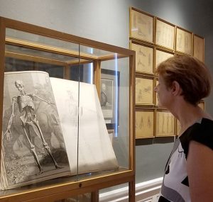
In the International Museum of Surgical Science, Chicago, IL
Dr. Malgosia Wilk-Blaszczak has taught human anatomy and human physiology courses for 30 years to medical and nursing students, and currently to undergraduate students at University of Texas at Arlington. She holds an M.D. and a Ph.D. in Neuroscience from the Warsaw Medical University. Ever since she discovered her father’s anatomical fold-out “manikin” as a child, Dr. Wilk has has been enamored by all aspects of the human body. In addition to teaching, she loves old medical illustration and never misses the chance to see them in museums when she travels.
I would like to dedicate this section to all my undergraduate teaching assistants, past and present. Every semester, I pick the most gifted students from previous semesters to serve as teaching assistants. I appreciate your commitment, passion, and hard work, but most of all, the amazing times we have had together. Special thanks to Clint Hassell and Natalie Winter who have served as my teaching assistants for many semesters, and have been good friends ever since. You have always done more than what was expected, and have given so much of your time and effort to support students to really grow and surprise us.
Malgosia Wilk-Blaszczak, M.D., Ph.D. – Professor of Instruction, University of Texas at Arlington
Kevin A. Alford, B.S. – University of Texas at Arlington alumnus
Andrea Campo-Velez, B.S. – University of Texas at Arlington alumna
Victoria Dorch, B.S. – University of Texas at Arlington alumna
Michelle Reed and Thomas Perappadan of UTA Libraries for assisting in the publication of this resource.
Jodi Wiley, B.S, UTA alumna, for creating and formatting class handouts that became the foundation for this OER.
Bradford Dimos, UTA graduate student, and Collin Funkhouser, UTA alumnus, for class-testing the previous version of this resource.
Kyle Pinkos, UTA Libraries’ Marketing Coordinator, designed the cover for this OER. The images used are in the public domain. Featured images, from Ontleding Des Menschelyken Lichaams by Govard Bidloo, are available from the U.S. National Library of Medicine.
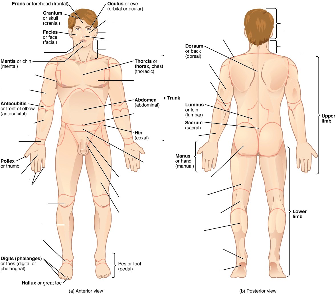
A solid foundation is essential when learning any new skill. Understanding anatomical directions, articulations, planes, and regions are the foundation for learning anatomy.
The standard anatomical position of the human body is facing towards the observer, legs hip-width apart, feet facing forward, arms out slightly at either side with palms facing forward. When determining a structure’s relative position, be sure to use this frame of reference. For example, it can be easy to confuse which side is the anterior aspect of the hands, therefore, one might incorrectly assume that the thumb is medial to the little finger. Remember, the anterior aspect of the hand is the palm, therefore the thumb is furthest from the center of the body and is lateral.
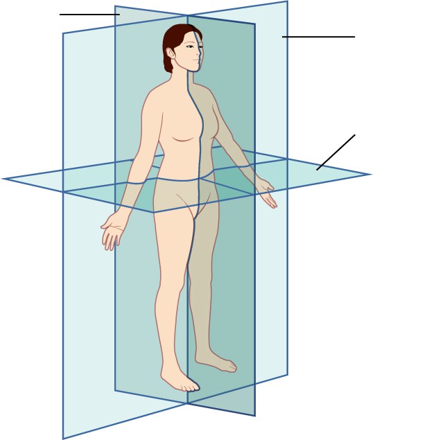
The archetypal body planes are frontal, sagittal and transverse planes. The frontal plane splits the body into anterior and posterior halves. The sagittal plane splits the body into left and right halves. The transverse plane splits the body into superior and inferior (top and bottom) halves. It is important to be able to identify a given plane so that you can orient yourself when a specimen, model or diagram is depicted a certain way. This same reasoning applies to the necessity of understanding directional terms such as anterior, inferior, distal and medial. It is recommended that you read the content prior to attending lab to make the most of your time.
Vocabulary for Anatomical Language on page(s) 160-161.
(5 points)
Last Name: _______________________ First Name: _______________________
Fill in the table below with the appropriate terms. Note: For this lab only, you may use any anatomical structure of the human body to fill in the table.
For the remaining pages of the prelab, label the designated planes, regions, and directions.
(1 point)
| Name of a structure | is | directional term | to | Name of the second structure |
|---|---|---|---|---|
| forearm | is | proximal | to | hand |
| head | is | superior | to | |
| is | inferior | to | tibia | |
| breast | is | anterior | to | |
| is | distal | to | upper arm | |
| brain | is | medial | to | |
| is | lateral | to | trunk |
Label the planes of the body. (1 point)
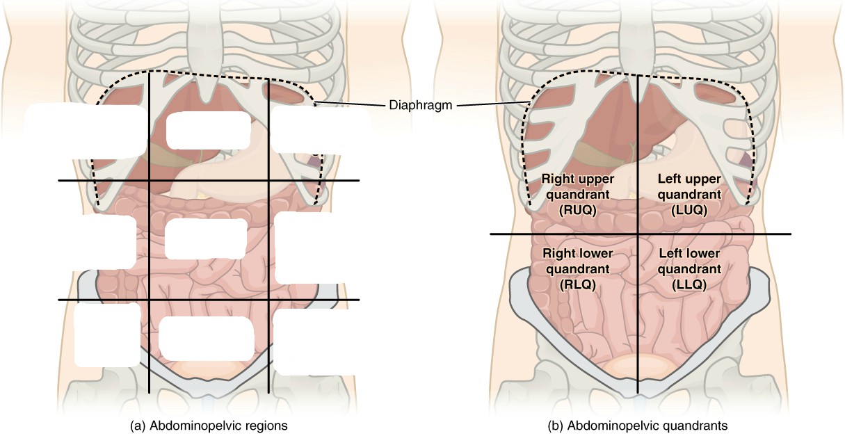
Label all nine regions of the abdomen. (1 point)
Label the anatomical directions designated by the lines and arrows. (1 point)
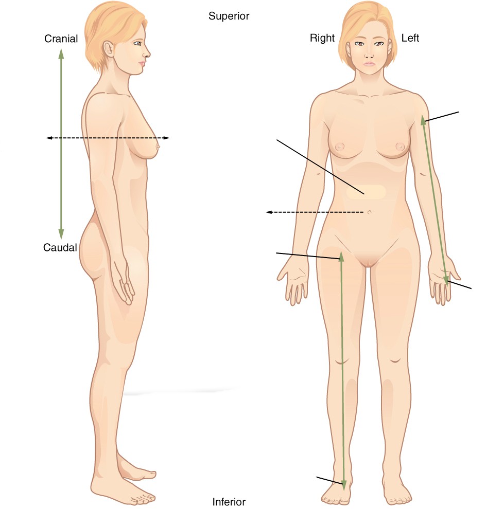
Label the regions of the body. (1 point)
For this lab only, there will be three stations for each group to cycle through, stations one, two and three; stations four, five and six will mirror these stations for this lab only. A list of words is provided below that you are expected to identify, learn, and label on the models provided. You must use all the words provided. Using the colored tape provided, write the number that corresponds to the organ/structure and place it on your model. When complete, notify your TA so they may check your work.
Note: Do not simply label the models, it is crucial that you understand how to apply all of these terms in each system, for the rest of the semester!
For each additional station, directions will be provided for the particular activity.
This is an advanced biology class, therefore you all likely have experience with microscopes. However, use these stations to refresh your memory of proper microscope etiquette, how to focus on a slide, and identify key features. For the remainder of this class, you will be expected to identify various tissues under the microscope. Be sure to ask your TA for assistance, and remember taking a picture of the slide to study later is not helpful if you don’t take the time to study it in lab and understand which aspects are most important.
Basic instructions for use:
Sketch the slides available for today’s lab and indicate the magnitude at which you are observing/sketching. Be sure to identify, include, and label your sketch with the corresponding structures listed beneath each slide. Use the images provided to guide you through this process.
 | |
| Monocyte | Compact Bone |
 |  |
| |
Spinal Cord | Kidney |
 | 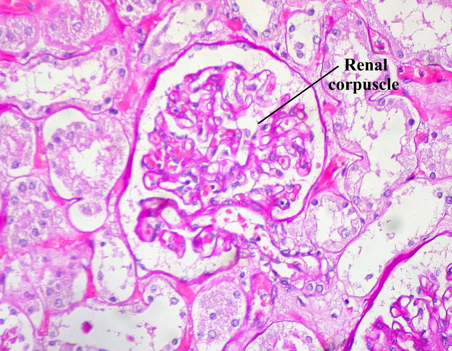 |
The terms in the following tables are important in understanding the relationship between different organs and structures of the body. Using the models and diagrams in your atlas, learn how to identify the different body planes and the appropriate use of directional terms. When trying to understand body movements, it is helpful to act them out yourself.
Label the torso models of this station with the number that corresponds to the appropriate regions of the abdominal cavity using the colored tape. When you are finished, ask your TA to check your labeling. Before leaving the station, remove all of the labels you have placed on the model. Note the locus of each organ within each region.
#1 frontal | #2 transverse | #3 sagittal |
#4 anterior | #7 inferior | #10 proximal | #13 superficial |
#5 posterior | #8 lateral | #11 distal | #14 parietal |
#6 superior | #9 medial | #12 deep | #15 visceral |
#16 right hypochondriac region | #19 right lumbar region | #22 right iliac region |
#17 epigastric region | #20 umbilical region | #23 hypogastric region |
#18 left hypochondriac region | #21 left lumbar region | #24 left iliac region |
Label the models of this station with the number that corresponds to the appropriate structure of the peripheral nervous system using the colored tape. When you are finished, ask your TA to check your labeling. Before leaving the station, remove all the labels you have placed on the model.
#1 cephalic | #11 brachial | #21 abdominal | #31 femoral |
#2 cranial | #12 cubital | #22 hepatic | #32 patellar |
#3 ocular (orbital) | #13 antecubital | #23 renal | #33 popliteal |
#4 auricular (otic) | #14 olecranal | #24 umbilical | #34 crural |
#5 buccal | #15 antebrachial | #25 lumbar | #35 sural |
#6 nasal | #16 carpal (carpus) | #26 pelvic | #36 tarsal (tarsus) |
#7 oral | #17 palmar | #27 inguinal | #37 calcaneal |
#8 cervical | #18 digital (phalangeal) | #28 pubic | #38 pedal |
#9 acromial | #19 thoracic | #29 sacral | #39 plantar |
#10 scapular | #20 mammary | #30 gluteal |
The following terms are useful to know and understand as they will reappear throughout this course.
#40 process | #45 sulcus | #50 facet | #54 septum |
#41 tuberosity | #46 gyrus | #51 fossa | #55 raphe |
#42 condyle | #47 foramen | #51 fundus | #56 ampulla |
#43 epicondyle | #48 foramina | #52 hilum | |
#44 fissure | #49 meatus | #53 isthmus |
(2 points)
Last Name: _______________________ First Name: _______________________
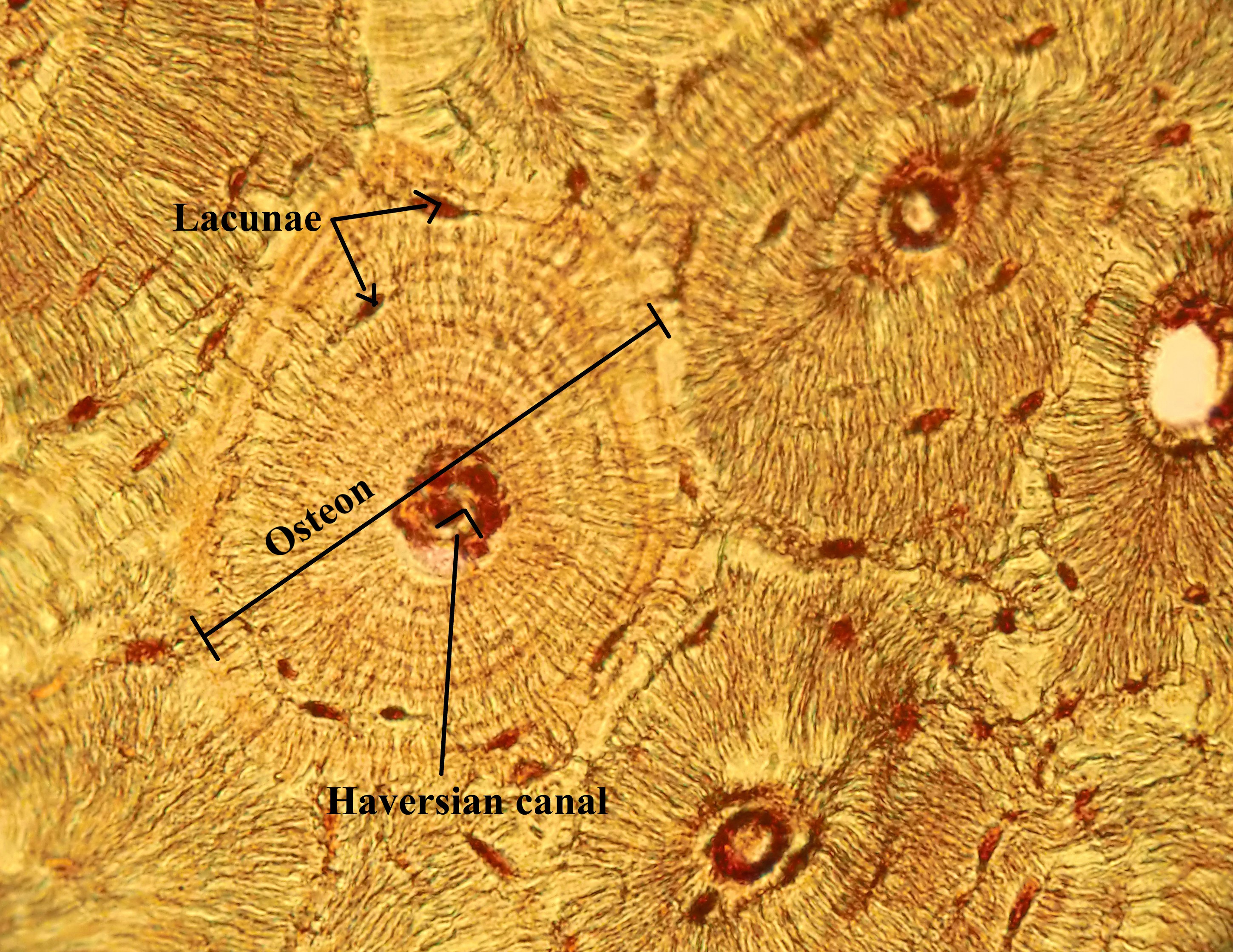
The skeletal system is the primary structural organ system of the body. Many people think of the skeletal system as being static in that it is unchanging, however, this is not the case. Bones, like other organ systems, have specialized cells which allow them to perform a variety of essential tasks. Osteoblast are responsible for secreting the bony matrix necessary for bone formation. Osteoclast, meanwhile, are large multinucleated cells responsible for the dissolution and reabsorption of bone. It is made mostly of collagen, which gives bone its soft framework, and calcium phosphate which adds strength and hardness to the structure. It is divided into the axial and the appendicular skeleton. The axial skeleton consists of the skull, hyoid bone, vertebral column, sternum, and ribs. Whereas the appendicular skeleton consists of the clavicle, scapula and the rest of the upper and lower limbs. Without the foundational structure of the skeletal system, there would be nothing to support the body and provide points of attachment for muscles. Bones function to protect internal organs, assist body movements, store and release calcium and phosphorous, participate in blood cell production and store fat in the yellow marrow. Bones also function to protect internal organs, assist body movements, and the storage and release of ions such as calcium and phosphorous. Furthermore, long bones contain both hemopoietic (red) and stromal (yellow) marrow which produce red blood cells and fat cells respectively. Each of these cells have specific functions that are key to the development and repair of a bone over time. The two types of bone tissue are compact and spongy bone. Compact bone is typically found along the perimeter of bones and makes up the majority of the diaphysis of long bones. It is stronger than spongy bone and provides more stability. Compact bone is made up of circular units called osteons. Osteons are composed of rings called lamellae that spiral down into a central canal, known as the Haversian canal. This central canal is the passage for nerves, blood vessels, and lymphatics. Spongy bone, on the other hand, is typically the deepest layer of a bone’s composition. It is made of trabeculae which give spongy bone its characteristic lighter weight. There are five classifications of bones based on their shape, long bones, short bones, flat bones, irregular bones and sesamoid bones. The shape and composition of each bone allow them to function as mentioned above.
Vocabulary for Bones and Bone Markings on page(s) 161-162.
(5 points)
Last Name: _______________________ First Name: _______________________
Fill in the table with the appropriate terms. For the remaining illustrations, label the structures indicated.
(1 point)
| Name of a structure | is | directional term | to | Name of the second structure |
|---|---|---|---|---|
| radius | is | proximal | to | ulna |
| femur | is | superior | to | |
| is | inferior | to | thoracic vertebrae | |
| patella | is | anterior | to | |
| is | distal | to | metacarpals | |
| tibia | is | medial | to | |
| is | lateral | to | sternum |
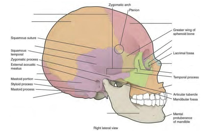
Label the cranial structures and bones. (0.5 points)
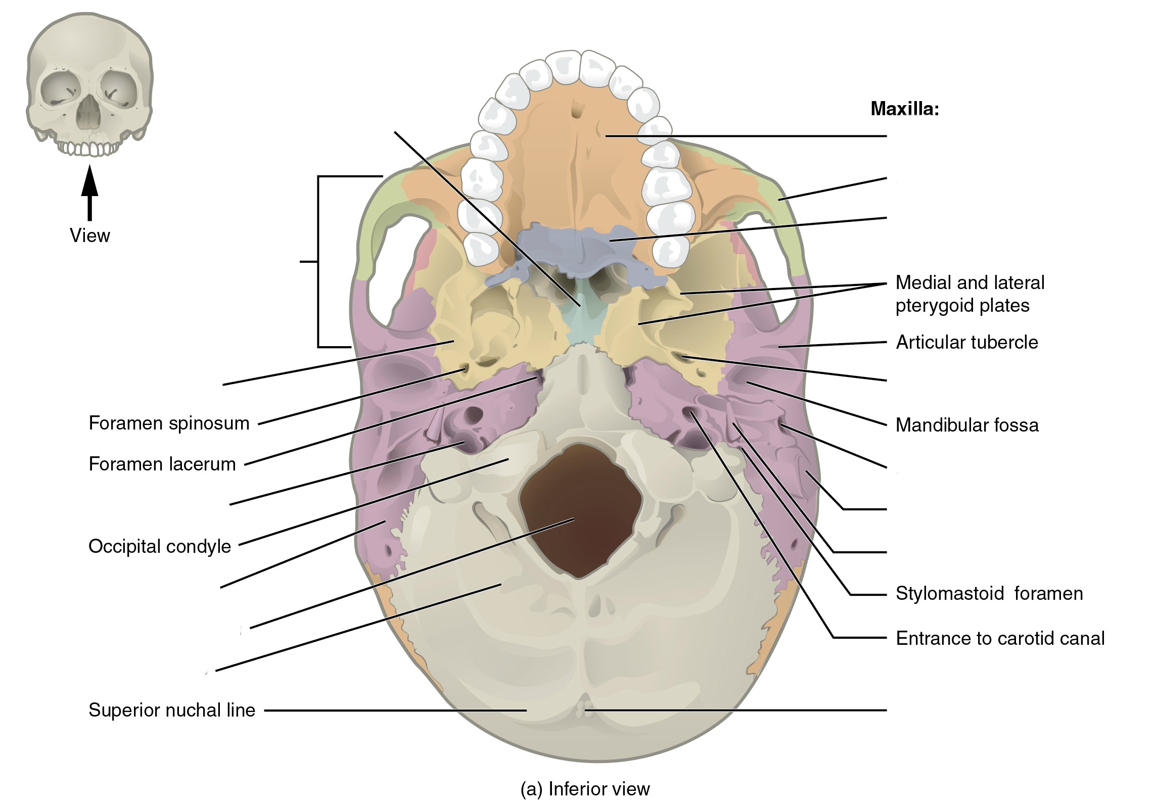
Label the cranial bones and special features. (0.5 points)
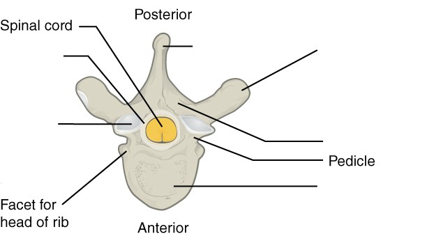
Label the distinctive parts of the vertebra. (0.5 points)
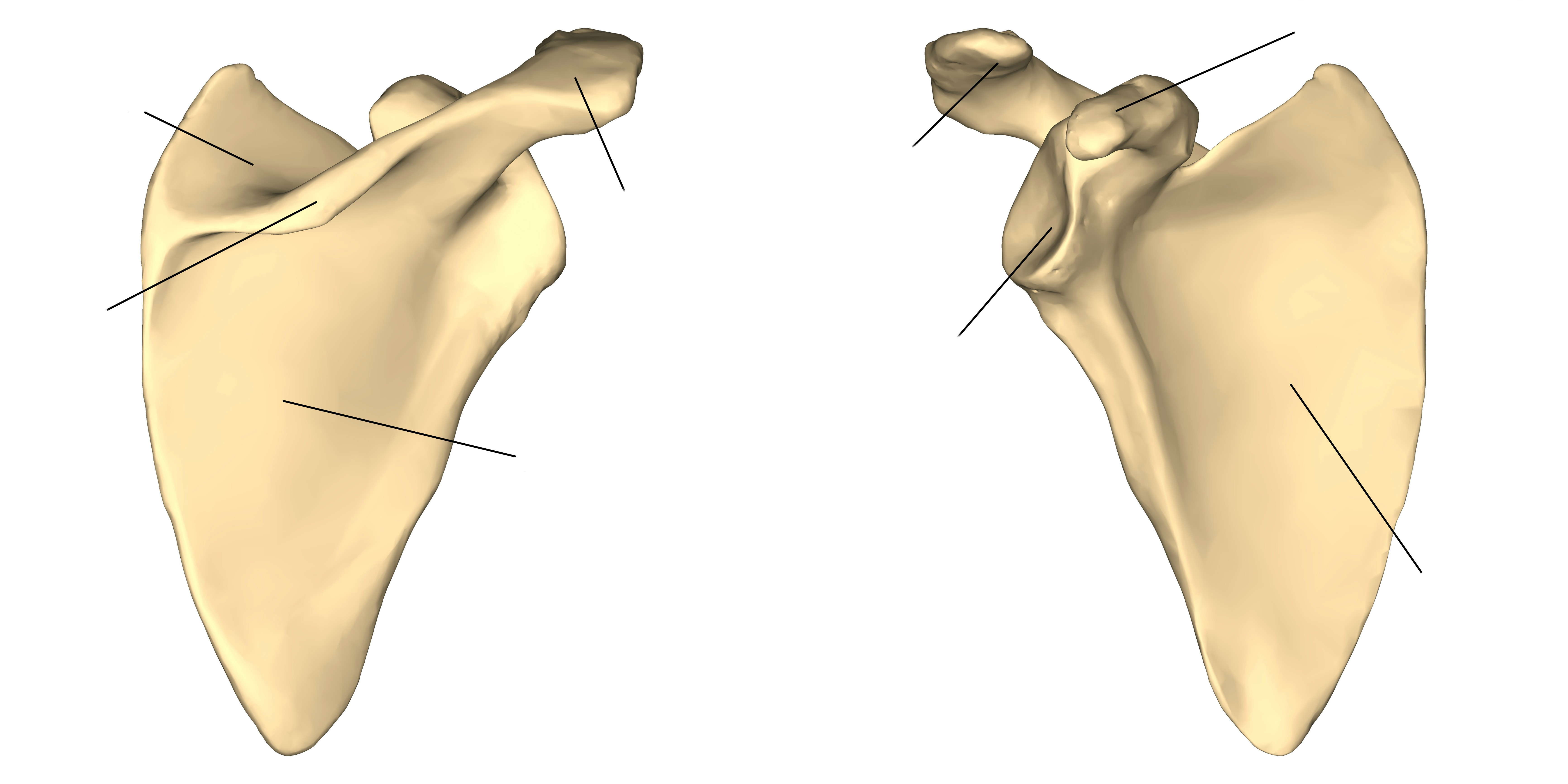
Label the features of the scapula. (0.5 point)
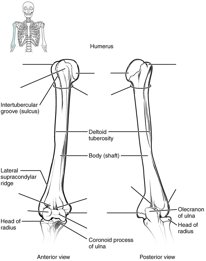
Label the features of the humerus. (0.5 points)
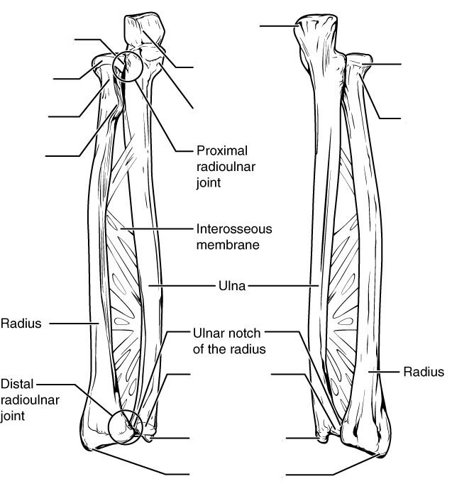
Label the features of the radius and ulna. (0.5 point)
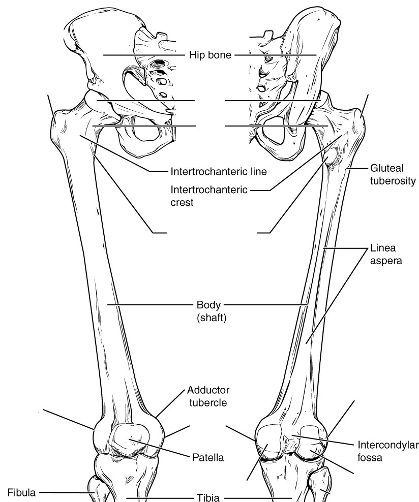
Label the features of the femur. (0.5 points)
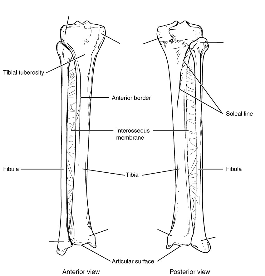
Label the features of the tibia and fibula. (0.5 points)
A list of words is provided below that you are expected to identify, learn, and label on the models provided. Note that not all models will have some of the organs/structures, so be sure to find them on an alternate model. You must use all the words provided. Using the colored tape provided, write the number that corresponds to the organ/structure and place them on your model. When complete, notify your TA so they may check your work.
For each additional station, directions will be provided for the activity.
Label the models of this station with the number that corresponds to the appropriate structure of the peripheral nervous system using the colored tape. When you are finished, ask your TA to check your labeling. Before leaving the station, remove all the labels you have placed on the model.
Note: For the following structures, be able to differentiate between left and right halves when applicable.
#1 frontal bone | #5 ethmoid bone | #9 zygomatic bone | #13 superior nasal conchae |
#2 parietal bone | #6 sphenoid bone | #10 nasal bone | #14 middle nasal conchae |
#3 temporal bone | #7 palatine bone | #11 vomer | #15 inferior nasal conchae |
#4 occipital bone | #8 maxilla | #12 lacrimal bone | #16 mandible |
#18 external auditory meatus | #20 styloid process | #22 cribriform plate of ethmoid bone | #24 zygomatic process of temporal bone |
#19 mastoid process | #21 external occipital protuberance | #23 olfactory foramina | #25 temporal process of zygomatic bone |
#26 foramen magnum | #28 foramen ovale | #30 coronal suture | #32 lambdoid suture |
#27 jugular foramen | #29 sella turcica | #31 sagittal suture |
Label the models of this station with the number that corresponds to the appropriate structure of the peripheral nervous system using the colored tape. When you are finished, ask your TA to check your labeling. Before leaving the station, remove all the labels you have placed on the model.
Note: For the following structures, be able to differentiate between left and right halves when applicable.
#1 hyoid Bone |
#2 vertebrae | #4 thoracic region | #6 sacrum | #8 intervertebral foramen |
#3 cervical region | #5 lumbar region | #7 coccyx | #9 intervertebral disc |
#10 body | #12 lamina | #14 transverse process | #16 inferior articular process | #18 facet of inferior articular process |
#11 vertebral foramen | #13 spinous process | #15 superior articular process | #17 facet of superior articular process |
#19 bifid spinous process | #21 atlas | #23 dens |
#20 transverse foramen | #22 axis |
#24 sternum | #26 sternal body | #28 ribs |
#25 manubrium | #27 xiphoid process | #29 costal cartilage |
In this station, you will be given a bucket filled with random bones some of which you will use to assemble an arm and a leg. Note below which bucket you are working with. Your assignment is to lay out the bones of each limb in their correct positions relative to each other and determine which bones do not belong to either limb. Additionally, you will need to determine whether each limb is a right or left limb; circle your results below. When you are finished, ask your TA to check whether you have assembled and identified your limbs correctly.
Bucket # ________
Upper limb: Left / Right
Lower limb: Left / Right
Sketch the slides available for today’s lab and specify the magnitude at which you are observing/ sketching. Be sure to identify and label your sketch with the corresponding structures listed beneath each slide.
 | |
Compact Bone | Spongy Bone |
Label the models of this station with the number that corresponds to the appropriate structure of the peripheral nervous system using the colored tape. When you are finished, ask your TA to check your labeling. Before leaving the station, remove all the labels you have placed on the model.
Note: For the following structures, be able to differentiate between left and right halves when applicable.
#1 acromial end of clavicle | #2 sternal end of clavicle |
#3 glenoid cavity | #5 coracoid process | #7 supraspinous fossa | #9 subscapular fossa |
#4 acromion | #6 spine of scapula | #8 infraspinous fossa |
#10 head | #13 lesser tubercle | #16 coronoid fossa | #19 lateral epicondyle |
#11 neck | #14 trochlea | #17 radial fossa | #20 olecranon fossa |
#12 greater tubercle | #15 capitulum | #18 medial epicondyle |
#21 head | #23 trochlear notch | #25 radial notch |
#22 olecranon | #24 coronoid process | # 26 styloid process |
#27 head | #29 radial tuberosity |
#28 neck | #30 styloid process |
#31 carpals (8) | #33 phalanges | #35 middle phalanges |
#32 metacarpals | #34 proximal phalanges | #36 distal phalanges |
Label the models of this station with the number that corresponds to the appropriate structure of the peripheral nervous system using the colored tape. When you are finished, ask your TA to check your labeling. Before leaving the station, remove all the labels you have placed on the model.
Note: For the following structures, be able to differentiate between left and right halves when applicable.
#1 ilium | #3 ischium | #5 pubis | #7 acetabulum |
#2 iliac crest | #4 ischial spine | #6 pubic symphysis |
#8 head | #11 lesser trochanter | #14 medial condyle |
#9 neck | #12 medial epicondyle | #15 lateral condyle |
#10 greater trochanter | #13 lateral epicondyle | #16 intercondylar fossa |
# 17 patella |
#18 lateral condyle | #19 medial condyle | #20 medial malleolus |
#21 head | #22 lateral malleolus |
#23 tarsals (7) | #25 metatarsals | #27 proximal phalanges | #29 distal phalanges |
#24 calcaneus | #26 phalanges | #28 middle phalanges |
(3 points)
Last Name: _______________________ First Name: _______________________
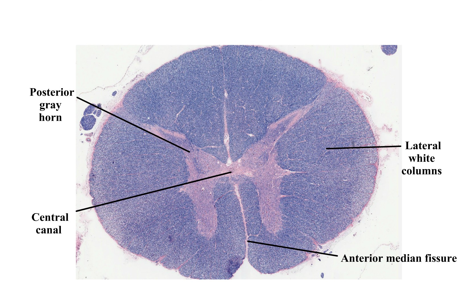
The spinal cord is made of white matter encompassed by gray matter with a central canal running through it that serves as a path for cerebrospinal fluid (CSF). The gray matter is divided into posterior (dorsal) grey horns which contain sensory neurons, and lateral and anterior (ventral) horns that contain the cell bodies of motor neurons. The surrounding white matter is divided into anterior (ventral) white columns, lateral white columns, and posterior (dorsal) white columns. The grey commissure is the gray matter posterior to the central canal where the neurons from either side of the spinal cord crossover. The same principle applies to the white commissure which lies anteriorly to the gray matter.
The spinal cord has several layers to protect it from damage. Beginning superficially and working our way deeper, the vertebral column encases the spinal cord and provides a hard shell for protection. Deep to the vertebrae are the meninges, consisting of the dura mater, arachnoid mater, and pia mater. Extensions from the pia mater, the denticulate ligaments, suspend the spinal cord in CSF and act as a shock absorber.
The spinal cord begins at the terminal end of the brain stem and extends to approximately the L1 vertebra adults and L2 vertebrae in children; it is located within the vertebral foramen and is divided into 4 distinct regions. The cervical segment extends from C1 to the C7 vertebrae. The thoracic segment extends from T1 to the T8 vertebrae. The lumbar segment corresponds with T9-T11 vertebrae. Finally, the sacral segment extends from T12 to L2. The cervical enlargement, C4-T1, is a bulbous structure from which many neurons of the upper extremities invaginate. Likewise, the lumbar enlargement, T9-T12, is a bulbous structure from which neurons that innervate the lower limbs originate.
Note: do not confuse the regions of the spine with the regions of the spinal cord, they are not the same.
There are 31 pairs of spinal nerves: 8 cervical pairs 12 thoracic pairs, 5 lumbar pairs, 5 sacral pairs and 1 coccygeal pair. However, nerves from every other area along the spinal cord do not do this; they first converge in a network called a plexus. With the exception of the thoracic region, nerves of the cervical, brachial, lumbar and sacral regions of the spinal cord branch from a network of nerves known as plexuses.
Vocabulary for Spinal Cord and Spinal Nerves can be found on page(s) 171-172.
A list of words is provided below that you are expected to identify, learn, and label on the models provided. Note that not all models will have some of the organs/structures, so be sure to find them on an alternate model. You must use all the words provided. Using the colored tape provided, write the number that corresponds to the organ/structure and place them on your model. When complete, notify your TA so they may check your work.
For each additional station, directions will be provided for the activity.
.
Label the models of this station with the # that corresponds to the appropriate structure of the spinal cord and its protective structures using the colored tape. When you have finished, have your TA check your labeling. Before leaving the station, remove all of the labels you have placed on the model.
Note: For the following structures, be able to differentiate left and right halves when applicable.
#1 vertebral column | #4 dura mater | #7 subarachnoid space | #10 denticulate ligaments | #13 filum terminale |
#2 spinal meninges | #5 subdural space | #8 cerebrospinal fluid | #11 spinal cord | #14 cauda equina |
#3 epidural space | #6 arachnoid mater | #9 pia mater | #12 conus medullaris |
#15 anterior median fissure | #19 posterior white columns | #23 anterior white commissure | #27 thoracic innervation segment | #31 lumbar enlargement |
#16 posterior median sulcus | #20 anterior gray horns | #24 posterior gray commissure | #28 lumbar innervation segment | |
#17 anterior white columns | #21 lateral gray horns | #25 central canal | #29 sacral innervation segment | |
#18 lateral white columns | #22 posterior gray horns | #26 cervical innervation segment | #30 cervical enlargement |
Label the models of this station with the number that corresponds to the appropriate structure of the peripheral nervous system using the colored tape. When you are finished, ask your TA to check your labeling. Before leaving the station, remove all the labels you have placed on the model.
Note: For the following structures, be able to differentiate between left and right halves when applicable.
#1 cervical nerve one (C1) | #9 thoracic nerve one (T1) | #17 thoracic nerve nine (T9) | #25 lumbar nerve five (L5) |
#2 cervical nerve two (C2) | #10 thoracic nerve two (T2) | #18 thoracic nerve ten (T10) | #26 sacral nerve one (S1) |
#3 cervical nerve three (C3) | #11 thoracic nerve three (T3) | #19 thoracic nerve eleven (T11) | #27 sacral nerve two (S2) |
#4 cervical nerve four (C4) | #12 thoracic nerve four (T4) | #20 thoracic nerve twelve (T12) | #28 sacral nerve three (S3) |
#5 cervical nerve five (C5) | #13 thoracic nerve five (T5) | #21 lumbar nerve one (L1) | #29 sacral nerve four (S4) |
#6 cervical nerve six (C6) | #14 thoracic nerve six (T6) | #22 lumbar nerve two (L2) | #30 sacral nerve five (S5) |
#7 cervical nerve seven (C7) | #15 thoracic nerve seven (T7) | #23 lumbar nerve three (L3) | #31 coccygeal nerve one (Coc1) |
#8 cervical nerve eight (C8) | #16 thoracic nerve eight (T8) | #24 lumbar nerve four (L4) |
Note: When labeling the nerves that exit the cervical plexus, focus on their location, the connections between the nerves of the plexus, and what they innervate. Also note any interesting characteristics you find, for example, which is the longest nerve? Make use of your textbook and atlas during this time.
#32 lesser occipital nerve | #34 transverse cervical nerve | #36 superior root of Ansa cervicalis nerve | #38 phrenic nerve |
#33 great auricular nerve | #35 supraclavicular | #37 inferior root of Ansa cervicalis nerve | #39 segmental branches |
Label the models of this station with the number that corresponds to the appropriate structure of the peripheral nervous system using the colored tape. When you are finished, ask your TA to check your labeling. Before leaving the station, remove all the labels you have placed on the model.
Note: When labeling the nerves that exit the brachial plexus, focus on their location, the connections between the nerves of the plexus and what they innervate. Also note any interesting characteristics you find, for example, which is the longest nerve? Make use of your textbook and atlas during this time.
#1 dorsal scapular nerve | #5 musculocutaneous nerve | #9 lower subscapular nerve | #13 medial pectoral nerve |
#2 long thoracic nerve | #6 lateral pectoral nerve | #10 axillary nerve | #14 medial cutaneous nerve of arm |
#3 nerve to subclavius | #7 upper subscapular nerve | #11 median nerve | #15 medial cutaneous nerve of forearm |
#4 suprascapular nerve | #8 thoracodorsal nerve | #12 radial nerve | #16 ulnar nerve |
Sketch the slides available for today’s lab and specify the magnitude at which you are observing/ sketching. Be sure to identify and label your sketch with the corresponding structures listed beneath each slide.
| |
Spinal cord | Sympathetic ganglion
|
Label the models of this station with the number that corresponds to the appropriate structure of the peripheral nervous system using the colored tape. When you are finished, ask your TA to check your labeling. Before leaving the station, remove all the labels you have placed on the model.
Note: When labeling the nerves that exit the lumbar plexus, focus on their location, the connections between the nerves of the plexus and what they innervate. Also note any interesting characteristics you find, for example, which is the longest nerve? Make use of your textbook and atlas during this time.
#1 iliohypogastric nerve | #3 genitofemoral nerve | #5 femoral nerve |
#2 ilioinguinal nerve | #4 lateral cutaneous nerve of thigh | #6 obturator nerve |
Label the models of this station with the number that corresponds to the appropriate structure of the peripheral nervous system using the colored tape. When you are finished, ask your TA to check your labeling. Before leaving the station, remove all the labels you have placed on the model.
Note: When labeling the nerves that exit the sacral plexus, focus on their location, the connections between the nerves of the plexus and what they innervate. Also note any interesting characteristics you find, for example, which is the longest nerve? Make use of your textbook and atlas during this time.
#1 superior gluteal nerve | #4 nerve to quadratus | #7 posterior cutaneous nerve of thigh | #10 tibial median plantar nerve | #13 deep common fibular nerve |
#2 inferior gluteal nerve | #5 nerve to obturator internus and superior gemellus | #8 pudenal nerve | #11 tibial lateral plantar nerve | |
#3 nerve to piriformis | #6 perforating cutaneous nerve | #9 sciatic nerve | #12 superficial common fibular nerve |
(2 points)
Last Name: _______________________ First Name: _______________________
The central nervous system entails all neurons of the brain and spinal cord. The brain is the central processing organ of the body and contains 100 billion neurons and a remarkable 1 trillion glial cells. It is estimated that cortical neurons alone consume around 5 billion ATP molecules per second. Whats more, some neurons can have axons that extend several feet. Unlike the spinal cord, the gray and white matter in the brain are arranged in three segments. From deep to superficial, the innermost region is made of gray matter which is surrounded by the myelinated axons of the white matter. The thin layer of the cerebral cortex responsible for higher order cognition is the outermost layer of gray matter. The brain is divided into four major regions, the brainstem, diencephalon, cerebellum, and cerebrum. The brainstem contains the medulla oblongata, pons, and midbrain (which houses the pineal gland). Caudal to the forebrain is the diencephalon, a region which contains the epithalamus, hypothalamus, thalamus and third ventricle.
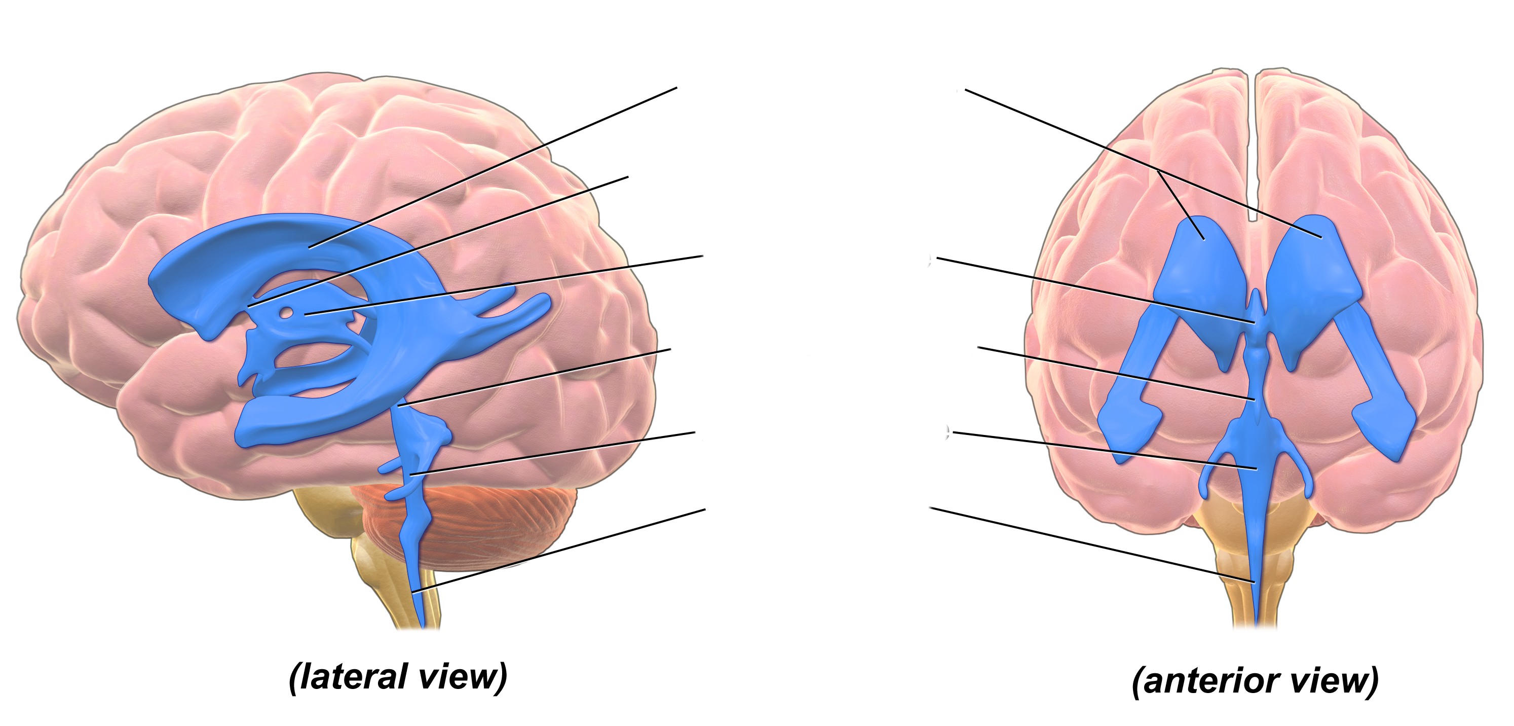
There are four cavities in the brain called ventricles; here cerebrospinal fluid (CSF) is produced and circulated by ependymal cells and the choroid plexuses. The two largest ventricles lie within each cerebral hemispheres and are known as the lateral ventricles. Cerebrospinal fluid drains from the lateral ventricles, through the interventricular foramen and into the third ventricle. The third ventricle lies between the halves of the thalamus. From here, it flows through the cerebral aqueduct (aqueduct of sylvius) and into the fourth ventricle, which lies between the cerebellum and the pons. Cerebrospinal fluid drains from the fourth ventricle, into the lateral and median apertures and down through the central canal of the spinal cord. Cerebrospinal fluid leaks out through foramina into the subarachnoid space where it is reabsorbed by veins on the surface of the brain and spinal cord.
Like the spinal cord, the brain is protected by three meninx, the dura, arachnoid and pia mater. Unlike the spinal meninges, the cranial dura mater is subdivided into two distinct layers; the periosteal layer, which is the superficial mot layer, and the inner meningeal dura mater. The two dural layers form the superior sagittal sinus which collectively channels venous blood from the brain. The falx cerebri divides the cerebrum into left and right hemispheres, the falx cerebelli divides the cerebellum into left and right hemispheres, and the tentorium cerebelli forms a physical barrier between the cerebrum and the cerebellum.
Vocabulary for the Brain and Cranial Nerves on page(s) 162-163.
(5 points)
Last Name: _______________________ First Name: _______________________
Fill in the table below with the appropriate terms. For the remaining exercises, label the designated structures.
(1 point)
| Name of a structure | is | directional term | to | Name of the second structure |
|---|---|---|---|---|
| pons | is | anterior | to | cerebellum |
| corpus callosum | is | superior | to | |
| is | inferior | to | hypothalamus | |
| precentral gyrus | is | anterior | to | |
| is | superficial | to | diencephalon | |
| interthalamic adhesion | is | medial | to | |
| is | superior | to | pons |
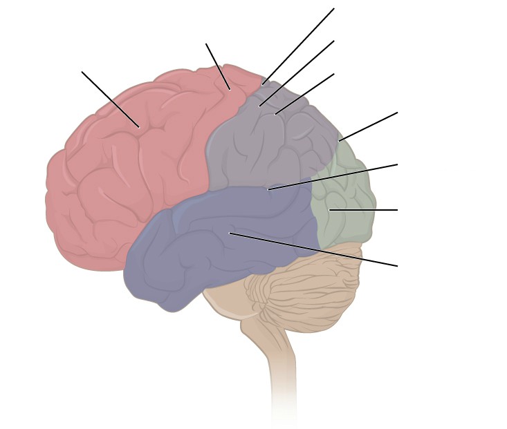
Label the sulci, gyri, and lobes of the cerebrum. (1 point)
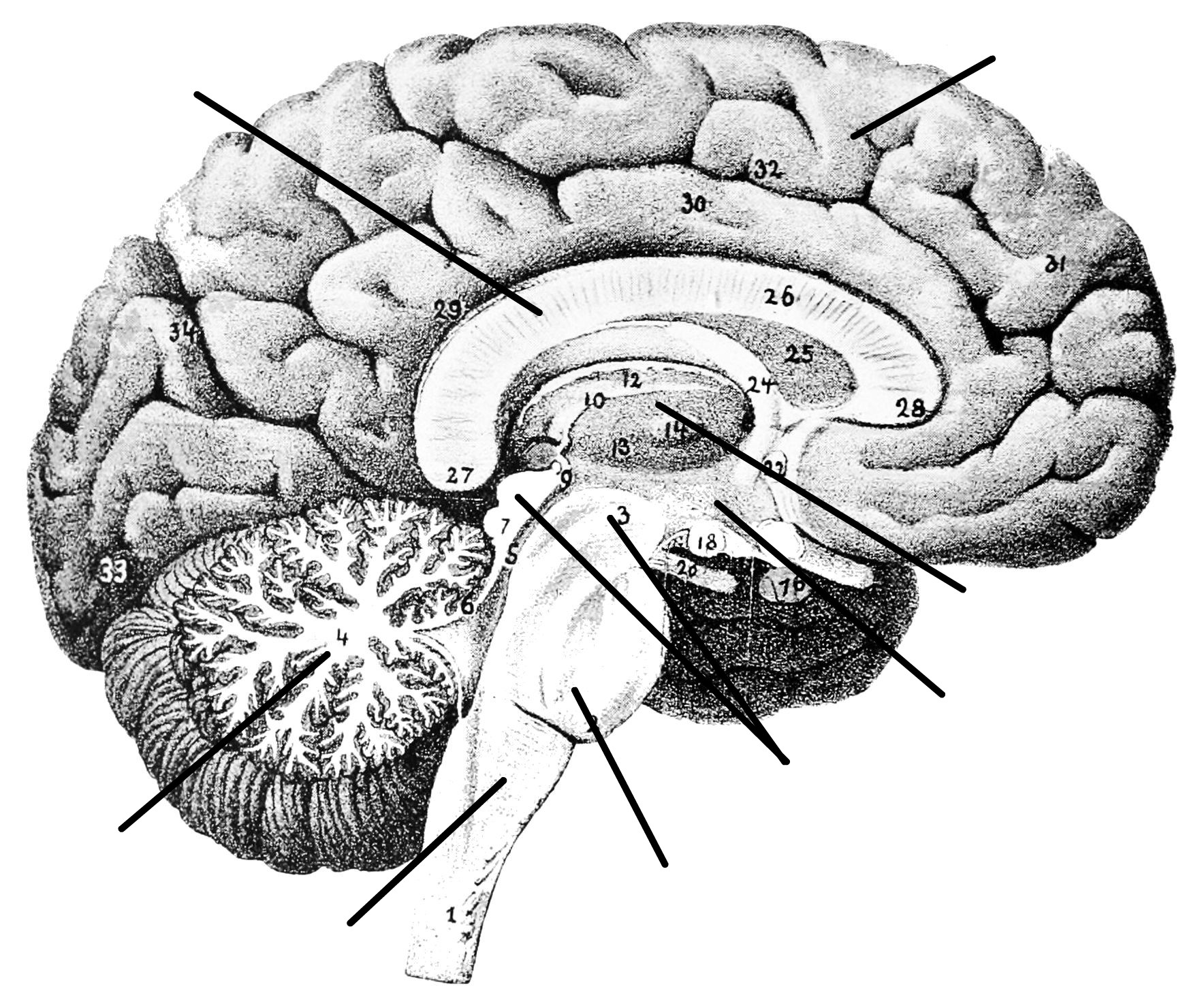
Label the major structures of the brain. (1 point)
Label the ventricles and passageway of CSF through the brain. (1 point)
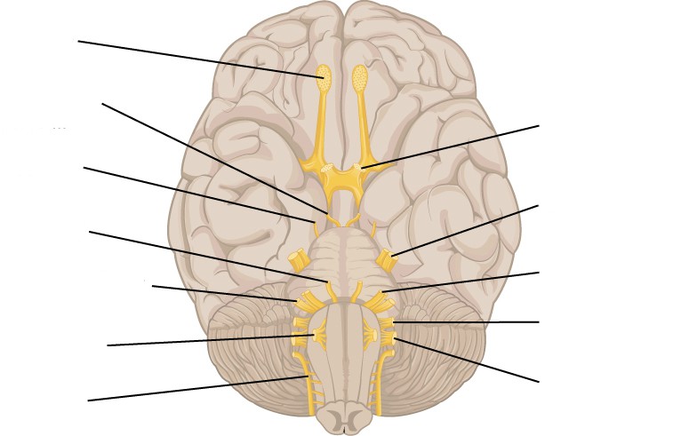
Label the cranial nerves. (1 point)
A list of words is provided below that you are expected to identify, learn, and label on the models provided. Note that not all models will have some of the organs/structures, so be sure to find them on an alternate model. You must use all the words provided. Using the colored tape provided, write the number that corresponds to the organ/structure and place them on your model. When complete, notify your TA so they may check your work.
For each additional station, directions will be provided for the activity.
Label the models of this station with the number that corresponds to the appropriate structure of the peripheral nervous system using the colored tape. When you are finished, ask your TA to check your labeling. Before leaving the station, remove all the labels you have placed on the model.
Note: For the following structures, be able to differentiate between left and right halves when applicable.
#1 cerebral cortex | #3 temporal lobes | #5 occipital lobe |
#2 frontal lobe | #4 parietal lobes | #6 insula |
#7 thalamus | #9 mammillary bodies | #11 pineal glands |
#8 hypothalamus | #10 epithalamus |
#12 midbrain | #14 superior colliculi | #16 cerebral peduncles | #18 medulla oblongata |
#13 tectum (corpora quadrigemina) | #15 inferior colliculi | #17 pons |
#19 arbor vitae | #21 vermis |
#20 folia | #22 cerebellar peduncles |
#23 basal nuclei | #25 fornix | #27 pituitary gland | #29 optic chiasm |
#24 corpus callosum | #26 cingulate gyrus | #28 infundibulum |
Label the models of this station with the number that corresponds to the appropriate structure of the peripheral nervous system using the colored tape. When you are finished, ask your TA to check your labeling. Before leaving the station, remove all the labels you have placed on the model.
Note: For the following structures, be able to differentiate between left and right halves when applicable.
#1 gray matter | #2 white matter |
#3 gyri (convulsions) | #5 sulci | #7 postcentral gyrus | #9 central sulcus | #11 transverse fissure |
#4 fissures | #6 precentral gyrus | #8 lateral cerebral sulcus | #10 parieto-occiptal sulcus | #12 longitudinal fissure |
These features may not be shown on models, but it is important to be able to identify them in diagrams and on the brains that you will dissect.
#13 dura mater | #15 falx cerebelli | #17 arachnoid mater |
#14 falx cerebri | #16 tentorium cerebelli | #18 pia mater |
Using the terms in the table below, determine the pathway of cerebrospinal fluid.
#19 lateral ventricles | #21 interventricular foramen | #23 cerebral aqueduct (aqueduct of midbrain) | #25 choroid plexuses |
#20 septum pellucidum | #22 third ventricles | #24 fourth ventricles | #26 cerebrospinal fluid |
Label the models of this station with the number that corresponds to the appropriate structure of the peripheral nervous system using the colored tape. When you are finished, ask your TA to check your labeling. Before leaving the station, remove all the labels you have placed on the model.
While learning the names, corresponding numbers and location of each of the cranial nerves, be sure to connect these to their functions and the structures they innervate.
#1 olfactory nerve (I) | #4 trochlear nerve (IV) | #7 facial nerve (VII) | #10 vagus nerve (X) |
#2 optic nerve (II) | #5 trigeminal nerve (V) | #8 vestibulocochlear/ acoustic nerve (VIII) | #11 Accessory/spinal nerve (XI) |
#3 oculomotor nerve (III) | #6 abducens nerve (VI) | #9 glossopharyngeal nerve (IX) | #12 hypoglossal nerve (XII) |
Sketch the slides available for today’s lab and specify the magnitude at which you are observing/ sketching. Be sure to identify and label your sketch with the corresponding structures listed beneath each slide.
| |
| Cerebrum | Cerebellum |
*If you are the last table to use this station, be sure to clean off the dissection kits in the lab sink.
*If you are the last table to use this station, be sure to clean off the dissection kits in the lab sink.
(3 points)
Last Name: _______________________ First Name: _______________________
In anatomy, special senses are the senses that have organs specifically devoted to them such as vision, gustation, olfaction, audition, and equilibrioception. These senses have specialized organs that detect and process stimuli and send signals to the brain which lead to the perception of that stimulus. These specialized organs include the tongue, the nose, the eyes and the ears.
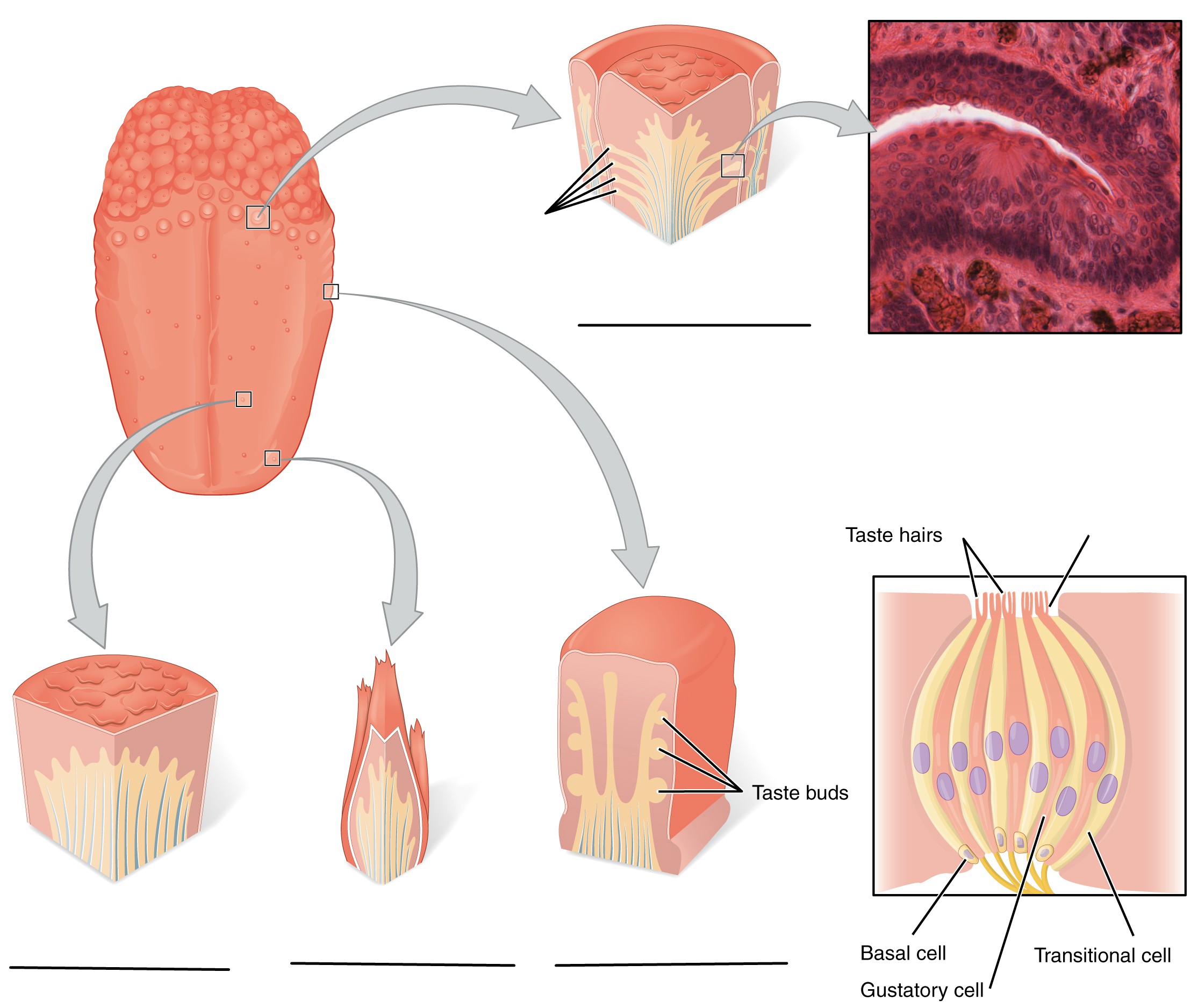
The tongue is a crucial organ in mechanical digestion and taste. Taste buds contain taste receptor cells which are the smallest functional unit in gustation. Taste buds can be found throughout the length of the upper digestive tract. On the surface of the tongue are protrusions called papillae. Circumvallate papillae are arranged in a v shape pattern on toward the base of the tongue, on the dorsal aspect, and contain more than 100 taste buds each. The fungiform papillae are found all over the dorsal aspect of the tongue and contain only about 5 taste buds each. The foliate papillae are found on the lateral aspects of the tongue and only contain taste buds during childhood. Finally, there are the filiform papillae which, like the fungiform papillae, are found all over the tongue, however, they do not contain taste buds. Instead, their barbed shape provides the friction for moving food around during mastication.
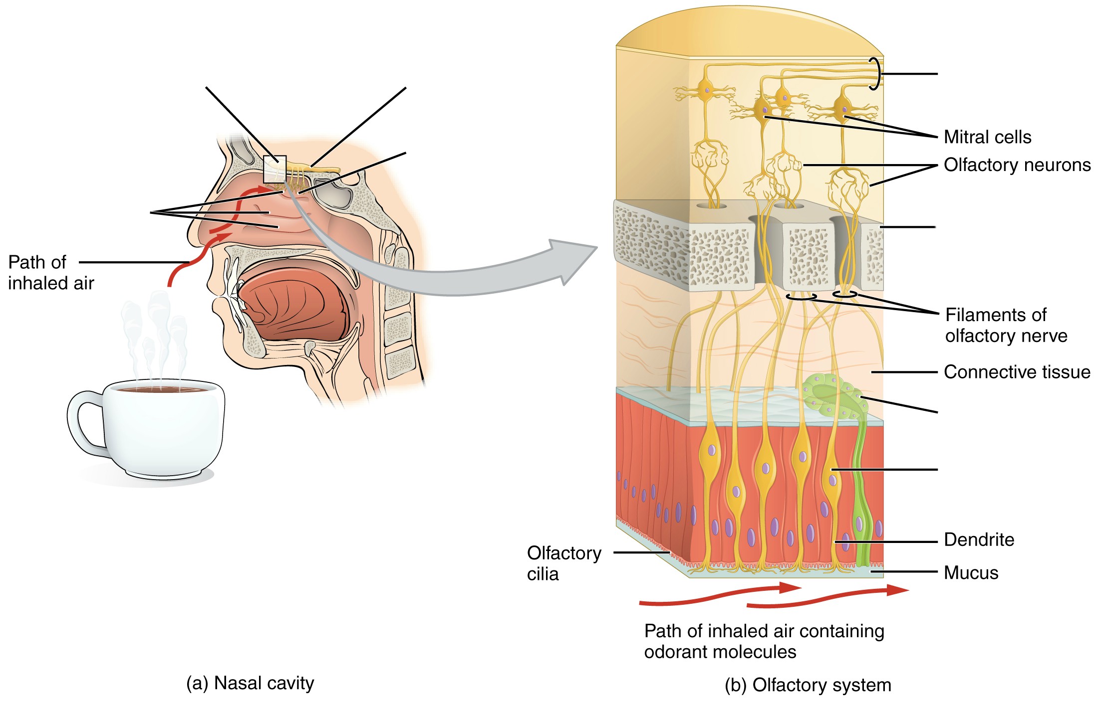
The olfactory epithelium is easily discernable on most models. Unlike any of the following special senses, neurons from the olfactory bulb bypass the thalamus and synapse directly with the olfactory cortex.
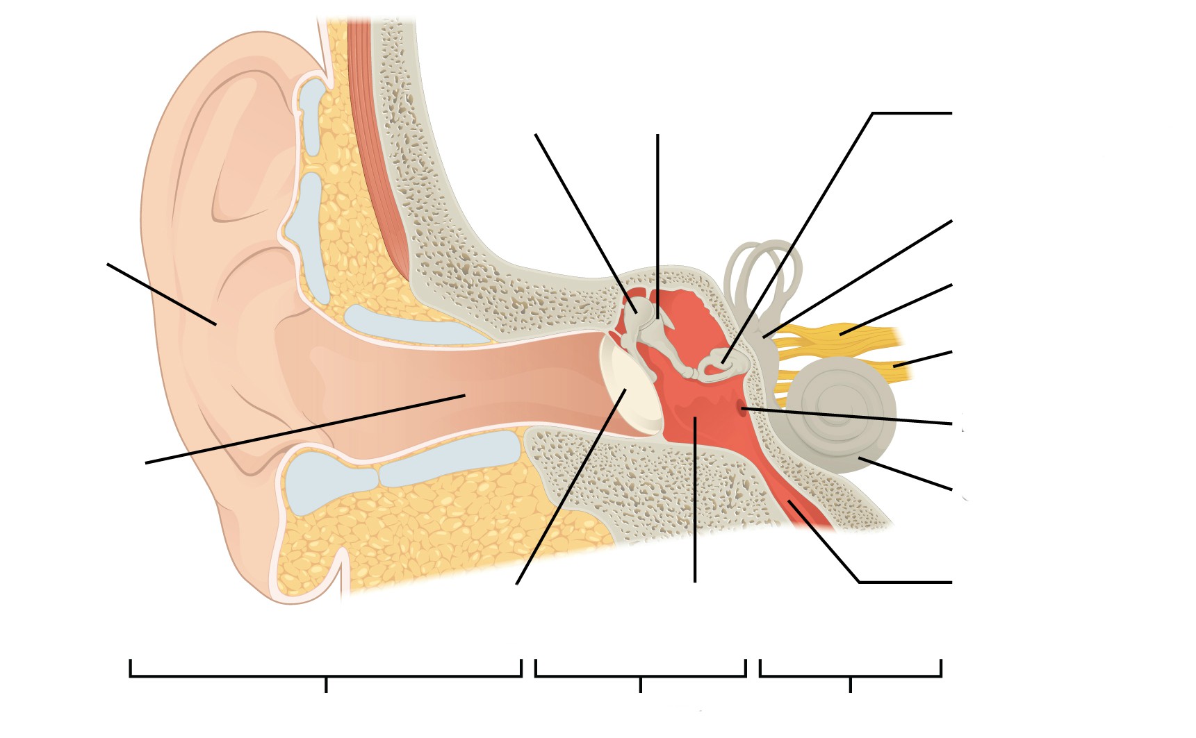
The ear is a complex organ which houses special structures that allow us to hear, balance and orientate ourselves. Sound waves are collected by the auricle and funneled into the external acoustic meatus. The ear is divided into three sections, the outer, middle, and inner ear. The outer ear consists of the auricle which extends through the external auditory canal and terminates at the tympanic membrane. The main structures of the middle ear are the auditory ossicles, Eustachian tube, oval window and round window. The auditory ossicles inward from the tympanic membrane, are the malleus, incus, and stapes. The base of the stapes covers the oval window which allows sound waves to pass from the tympanic membrane, into the cochlea of the inner ear. The inner ear is the innermost region of the ear where the cochlea, vestibule, and semicircular canals are. The cochlea, vestibule, and semicircular canals are responsible for hearing, static and dynamic equilibrium respectively. The vestibulocochlear nerve branches, into the cochlear branch, which innervates the cochlea, and the vestibular branch which innervates the vestibule and semicircular canals.
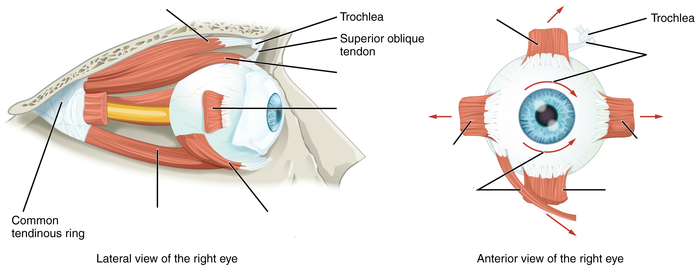
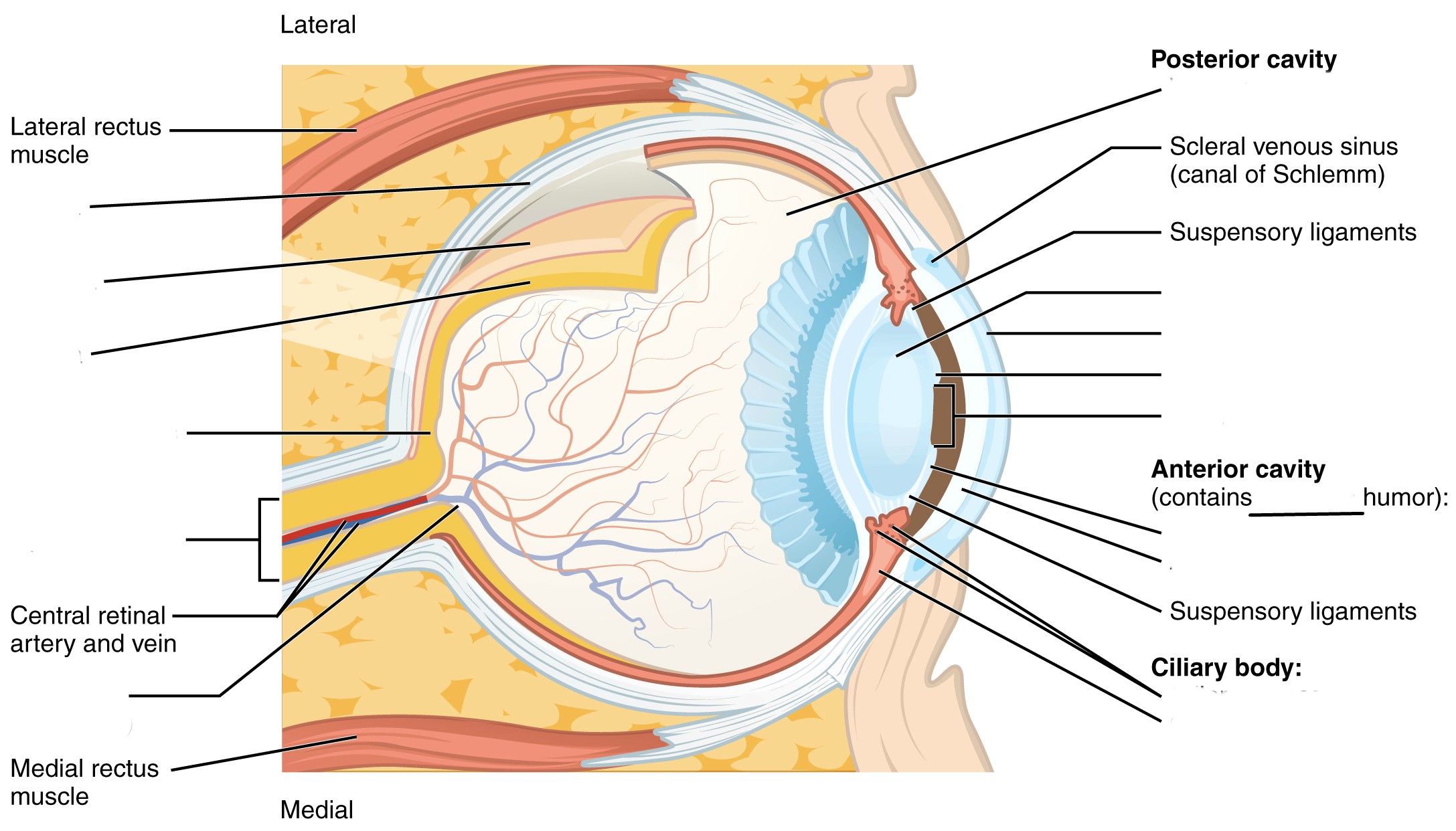
The eye is the specialized organ of sight which has three principal layers, the fibrous tunic, the vascular tunic and the neural tunic. Furthermore, there are two main chambers, the anterior chamber, containing aqueous humor and the posterior chamber, that contains vitreous humor. In the neural tunic of the retina, light propagates from the ganglionic cells through the bipolar cells to the rods and cons, which, somewhat paradoxically hyperpolarize opposite the direction of light.
The lacrimal apparatus frames the eye and coats the sclera and cornea in lacrimal fluid, a bacteriacide, which lubricates and protects them. The lacrimal apparatus is made of the lacrimal gland, lacrimal canaliculi, lacrimal sac and nasolacrimal duct. This network of structures allows tears produced by the lacrimal gland to cover the eye, drain through the lacrimal puncta into the lacrimal canaliculi, collect in the lacrimal sac, travel down the nasolacrimal duct and finally empty into the nose. This is why crying leads to a runny nose.
Vocabulary for Special Senses can be found on page(s) 169-171.
(5 points)
Last Name: _______________________ First Name: _______________________
Fill in the table below with the appropriate terms. For the remaining exercises, label the structures accordingly.
(1 point)
| Name of a structure | is | directional term | to | Name of the second structure |
|---|---|---|---|---|
| retina | is | posterior | to | lens |
| middle nasal conchae | is | superior | to | |
| is | inferior | to | cribriform plate | |
| cornea | is | anterior | to | |
| is | distal | to | tympanic membrane | |
| medial rectus | is | medial | to | |
| is | lateral | to | tongue |
Label the structures of the olfactory epithelium and olfactory pathway. (1 point)
Label the types of papillae and parts of the taste buds. (1 point)
Label the regions and structures of the ear. (1 point)
Label the muscles of the eye. (0.5 points)
Label the structures and regions of the eye. (0.5 points)
A list of words is provided below that you are expected to identify, learn, and label on the models provided. Note that not all models will have some of the organs/structures, so be sure to find them on an alternate model. You must use all the words provided. Using the colored tape provided, write the number that corresponds to the organ/structure and place them on your model. When complete, notify your TA so they may check your work.
For each additional station, directions will be provided for the activity.
Note: if your lab does not permit the use of food items in the lab, leave the room before conducting this experiment.
Subject #1: ____________________________
Number of taste buds: ___________________
Tasting abilities: ________________________
Subject #2: ____________________________
Number of taste buds: ___________________
Tasting abilities: ________________________
Label the models of this station with the number that corresponds to the appropriate structure of the peripheral nervous system using the colored tape. When you are finished, ask your TA to check your labeling. Before leaving the station, remove all the labels you have placed on the model.
Note: For the following structures, be able to differentiate between left and right halves when applicable.
#1 lingual tonsils | #4 fungiform papillae | #7 circumvallate papillae |
#2 palatine tonsils | #5 filiform papillae | #8 taste bud |
#3 lingual papillae | #6 foliate papillae | #9 taste pore |
#10 facial nerve (CN VII) | #12 vagus nerve (CN X) | #14 primary gustatory area |
#11 glossopharyngeal nerve (CN IX) | #13 thalamus |
#15 superior nasal conchae | #19 middle nasal meatus | #23 cribriform plate of ethmoid bone |
#16 middle nasal conchae | #20 inferior nasal meatus | #24 olfactory foramina |
#17 inferior nasal conchae | #21 olfactory epithelium | |
#18 superior nasal meatus | #22 olfactory glands |
#25 olfactory epithelium | #27 olfactory nerve (CN I) | #29 olfactory tract |
#26 olfactory receptors | #28 olfactory bulb | #30 primary olfactory area of the cerebral cortex |
Label the models of this station with the number that corresponds to the appropriate structure of the peripheral nervous system using the colored tape. When you are finished, ask your TA to check your labeling. Before leaving the station, remove all the labels you have placed on the model.
Note: For the following structures, be able to differentiate between left and right halves when applicable.
#1 auricle (pinna) | #3 lobule | #5 external auditory canal | #7 tympanic membrane |
#2 helix | #4 external auditory meatus | #6 ceruminous glands |
#8 auditory ossicles | #10 incus | #12 Eustachian tube | #14 round window |
#9 malleus | #11 stapes | #13 oval window |
#15 bony labyrinth | #18 cochlea | #21 utricle |
#16 semicircular canals | #19 membranous labyrinth | #22 saccule |
#17 vestibule | #20 semicircular canals | #23 organ of corti |
#24 vestibulocochlear nerve (CN VIII) | #25 primary auditory area of the cerebral cortex |
Sketch the slides available for today’s lab and specify the magnitude at which you are observing/ sketching. Be sure to identify and label your sketch with the corresponding structures listed beneath each slide.
| |
Retina | Tongue |
*If you are the last table to use this station, be sure to clean off the dissection kits in the lab sink.
Label the models of this station with the number that corresponds to the appropriate structure of the peripheral nervous system using the colored tape. When you are finished, ask your TA to check your labeling. Before leaving the station, remove all the labels you have placed on the model.
#1 sclera | #2 cornea |
#3 iris | #5 lens | #7 ciliary body |
#4 pupil | #6 choroid |
#8 retina | #12 pigmented layer | #16 bipolar cells |
#9 optic disc | #13 neural layer | #17 horizontal cells |
#10 macula lutea | #14 rods | #18 ganglion cells |
#11 fovea centralis | #15 cones |
#20 optic nerve | #22 optic tract |
#21 optic chiasm | #23 primary visual area of the cerebral cortex |
#24 anterior chamber | #26 posterior chamber |
#25 aqueous humor | #27 vitreous humor (body) |
#28 levator palpebrae superioris | #30 inferior rectus | #32 medial rectus | #34 inferior oblique |
#29 superior rectus | #31 lateral rectus | #33 superior oblique |
#35 lacrimal gland | #36 superior lacrimal canaliculi | #38 lacrimal sac |
#36 lacrimal puncta | #37 inferior lacrimal canaliculi | #39 nasolacrimal duct |
#40 palpebral conjunctiva | #41 bulbar conjunctiva |
(3 point)
Last Name: _______________________ First Name: _______________________
Match the following structures with their corresponding descriptions. (1 point)
| Name of Structure | Descriptions | No. of Structure |
|---|---|---|
| 1. Optic disc | an area where odorants bind to receptors to produce a sensation that will be perceived as smell | |
| 2. Round window | contains approximately 100 taste buds | |
| 3. Fungiform papillae | location of no visual activity, known as the “blind spot” | |
| 4. Vitreous humor | contains the organs that sense dynamic equilibrium | |
| 5. Olfactory epithelium | contains the organs that sense static equilibrium | |
| 6. Filiform papillae | jelly-like mass that provides stability and structure to the eye | |
| 7. Semicircular canals | provide friction, contains no taste buds | |
| 8. Retina | contains approximately 5 taste buds | |
| 9. Auditory ossicles (malleus, incus, and stapes) | the smallest bones in the body; transmits vibrations that are key to hearing | |
| 10. Circumvallate papillae | possess the following layers to allow for the transmission of stimuli to the optic nerve; pigmented layer, photoreceptor layer, outer synaptic layer, bipolar cell layer, inner synaptic layer, ganglion layer | |
| 11. Vestibule | membrane between the inner and middle ear to allow for pressure changes to equilibrate |
The respiratory system is responsible for the gas exchange of oxygen and carbon dioxide. The main specialized organs of this process are the lungs which house clusters of sac-like structures known as alveoli. There are from 480 to 790 million alveoli which increase the efficiency of gas exchange by increasing surface area to around 118m2 in men and 91m2 in women. The respiratory system consists of the nasal cavity, pharynx, larynx, trachea, lungs, bronchi, bronchioles, and alveoli, along with their accessory structures. These structures are divided into the upper and lower respiratory systems, with the lower portion beginning at the larynx. The primary function of this system is to exchange oxygen and carbon dioxide between the body and the environment. Functionally, the respiratory system can be divided into the conducting zone, terminating at the terminal bronchioles; then air flows into the respiratory zone, where the actual gas exchange occurs.
Though we view each system individually in this lab, it is important to keep in mind that all organ systems overlap and work together in such a way that scientist are constantly discovering new connections. One such example is the nose. Not only is it the primary entrance and exit for respiration, but it also contains the olfactory epithelium, the primary structure of one of the special senses, olfaction. Likewise, the pharynx is a structure shared by both the respiratory and digestion systems.
Although both lungs functionally participate in respiration, they differ physically in various ways. The right lung is shorter and wider than the left lung, and the left lung occupies a smaller volume than the right. Another distinction between the two lungs is that the left lung contains the cardiac notch, which makes space for the heart. Furthermore, whereas the right lung has three lobes, the left lung has only two.
Though not visible on every model, each lung is surrounded by the pleura, which consists of two layers called the visceral and parietal pleurae. They are important because they lubricate the lungs and reduce friction during inhalation and exhalation.
Vocabulary for Respiratory System can be found on page(s) 169.
A list of words is provided below that you are expected to learn and label on the models provided. Note that not all models will have some of the organs/structures, so be sure to find them on an alternate model. You must use all the words provided. Using the colored tape provided, write the number that corresponds to the organ/structure and place it on your model. When complete, notify your TA so they may check your work.
For each additional station, directions will be provided for the activity.
Label the models of this station with the number that corresponds to the appropriate structure of the peripheral nervous system using the colored tape. When you are finished, ask your TA to check your labeling. Before leaving the station, remove all the labels you have placed on the model.
#1 nose | #6 septal nasal cartilage | #12 nasal conchae* | #16 laryngopharynx | #21 soft palate |
#2 root | #7 major alar cartilages | #12 nasal meatuses* | #17 lingual tonsils | #22 uvula |
#3 bridge | #8 minor cartilages | #13 pharynx | #18 palatine tonsils | |
#4 apex | #9 external naris | #14 nasopharynx | #19 pharyngeal tonsil (adenoid) | |
#5 lateral nasal cartilages | #10 nasal cavity | #15 oropharynx | #20 hard palate |
*There are Superior, Middle, and Inferior parts to these structures.
Label the models of this station with the number that corresponds to the appropriate structure of the peripheral nervous system using the colored tape. When you are finished, ask your TA to check your labeling. Before leaving the station, remove all the labels you have placed on the model.
#1 larynx | #8 corniculate cartilages | #15 esophagus | #22 alveolar sacs | #29 middle lobe |
#2 epiglottis | #9 cuneiform cartilage | #16 carina | #23 alveoli | #30 cardiac notch |
#3 vestibular folds | #10 cricothyroid ligament | #17 primary (main) bronchi | #24 L/R lungs | #31 horizontal fissure |
#4 vocal folds | #11 cricoid cartilage | #18 secondary (lobar) bronchi | #25 apex of lung | #32 oblique fissure |
#5 thyrohyoid membrane | #12 cricotracheal ligament | #19 tertiary (segmental) bronchi | #26 base of lung | #33 hilum |
#6 thyroid cartilage | #13 tracheal cartilages | #20 respiratory bronchioles | #27 superior lobe | |
#7 arytenoid cartilages | #14 trachea | #21 alveolar ducts | #28 inferior lobe |
Label the models of this station with the number that corresponds to the appropriate structure of the peripheral nervous system using the colored tape. When you are finished, ask your TA to check your labeling. Before leaving the station, remove all the labels you have placed on the model.
#1 diaphragm | #2 external intercostals | #3 scalenes | #4 sternocleidomastoid |
*Make note of which muscles are the primary muscles of inhalation, and which are the accessory muscles.
#5 internal intercostals | #6 external oblique | #7 internal oblique | #8 transverse abdominis | #9 rectus abdominis |
Sketch the slides available for today’s lab and specify the magnitude at which you are observing/ sketching. Be sure to identify and label your sketch with the corresponding structures listed beneath each slide.
Lung
Terminal bronchioles, Respiratory bronchioles, Alveolar ducts, Alveolar sacs, Alveoli
*If you are the last table to use this station, be sure to clean off the dissection kits in the lab’s sink.
As a group, determine the path that oxygen travels starting from the nostrils to the alveoli. Be sure to identify where along that path each of the structures on the vocabulary list is located.
As a group, determine the route of pulmonary circulation. Be mindful of the fact that several structures are directly connected to the heart. Label the models/posters of this station with the # that corresponds to the appropriate vessels involved in pulmonary circulation using the colored tape. When you have finished, have your TA check your labeling. Before leaving the station, remove all of the labels you have placed on the models/posters.
#1 pulmonary trunk | #2 pulmonary arteries | #3 pulmonary capillaries | #4 pulmonary veins |
(2 points)
Last Name: _______________________ First Name: _______________________
1) Write a C if the listed structure is part of the conducting zone and an R if it is part of the respiratory zone. Also, label whether the structure is part of the upper respiratory (U) or lower respiratory system (L). (0.5 point)
Example: Larynx C , L
Alveoli ________________
Trachea ________________
Nasal cavity ________________
Bronchi ________________
Respiratory bronchioles ________________
Pharynx ________________
Alveolar ducts ________________
Terminal bronchioles ________________
2) Write the route that oxygen takes from when you inhale to the point of gas exchange with carbon dioxide. (0.5 point)
3) Give two unique characteristics of the pulmonary artery and vein. (0.5 point)
4) Describe the route of pulmonary circulation. (0.5 point)
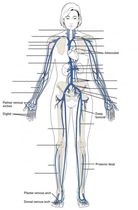
The cardiovascular system is responsible for the circulation of blood and transport of nutrients. Large multicellular organisms developed such a system as a means of actively transporting nutrients to the cells of the body. The heart is the organ of focus in this lab. It is divided into four distinct chambers, which in concert work to circulate blood. When the heart beats, it pumps blood into two different circuits: pulmonary and systemic. Pulmonary circulation carries blood from the right side of the heart to the alveoli of the lungs and back to the left side of the heart, while the systemic circulation carries blood from the left side of the heart to all the organs and tissues of the body, then back to the right side of the heart. If it were possible to stretch out all of the blood vessels in the body, they would measure 60,000 to 100,000 miles, enough circle the earth roughly four times. The heart is an incredible organ capable, on average, of circulating roughly 2,000 gallons worth of blood each day. Furthermore, the heart is one of the few organs capable of operating entirely apart from the central nervous system which makes it one of the hardest working organs.
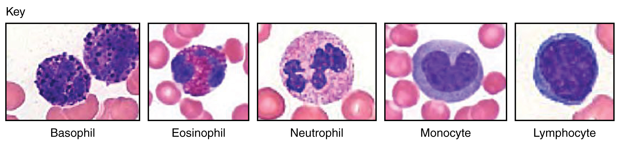
Blood is classified as liquid connective tissue and is vital in its roles of transportation, regulation, and protection. It is made of distinct types of cells, mostly derived from bone marrow, and helps maintain homeostasis. Plasma and cellular elements are the two main components of blood, where plasma makes up 55% of blood and cellular elements make up 45%. Plasma is mostly water but contains proteins and other solutes as well. The vast majority of cell elements are erythrocytes with less than 1% comprising of leukocytes and platelets.
In this lab we will focus on the major blood vessels of the cardiovascular system. Arteries are blood vessels that always carry blood away from the heart; the blood they carry is oxygenated (exception: pulmonary arteries). They generally have thicker walls than veins, the other major blood vessels in the cardiovascular system. Veins carry blood toward the heart and carry deoxygenated blood (exception: pulmonary veins). Both vessel types are formed by the tunica intima, tunica media, and tunica adventitia.
Vocabulary for the Cardiovascular System can be found on page(s) 163-165.
(5 points)
Last Name: _______________________ First Name: _______________________
Fill in the table below with the appropriate terms. For the remaining exercises, label the structures accordingly.
(1 point)
| Name of a structure | is | directional term | to | Name of the second structure |
|---|---|---|---|---|
| pulmonary vein | is | proximal | to | right ventricle |
| auricle | is | superior | to | |
| is | inferior | to | heart’s base | |
| anterior interventricular sulcus | is | anterior | to | |
| is | distal | to | ascending aorta | |
| heart | is | medial | to | |
| is | lateral | to | left ventricle |
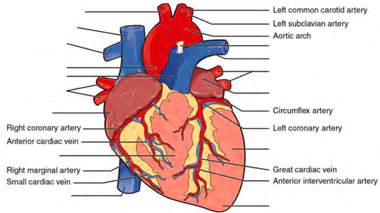
Label the prominent coronary surface vessels. (0.5 points)
Label the prominent coronary surface vessels. (0.5 points)
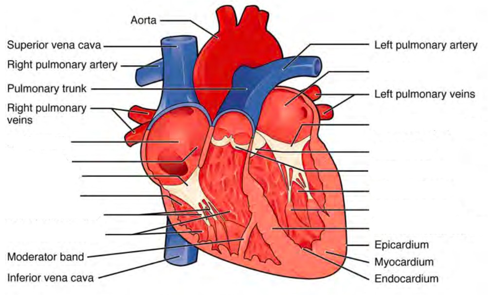
Label the internal formations of the heart. (1 point)
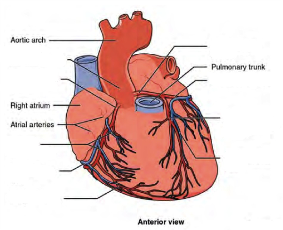
Label the surface features of the anterior aspect of the heart. (0.5 points)
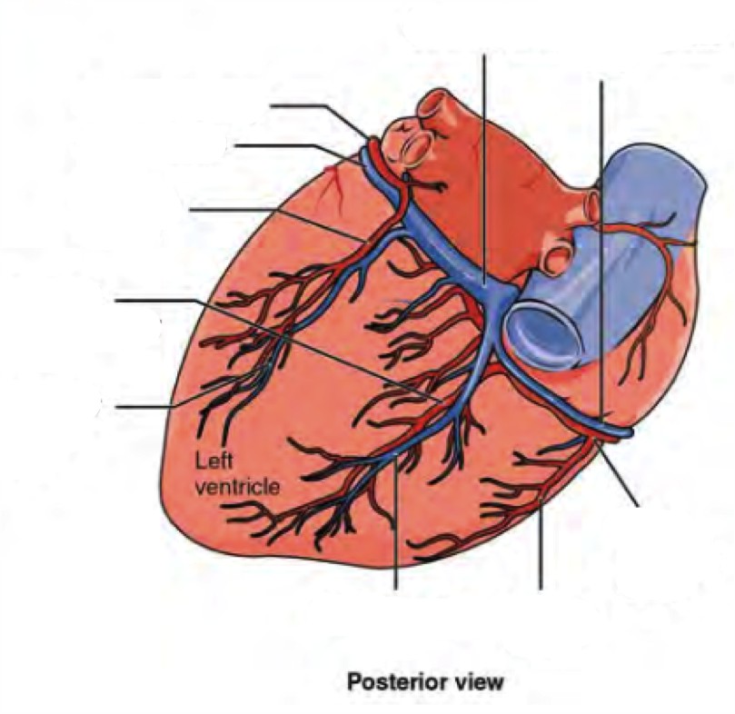
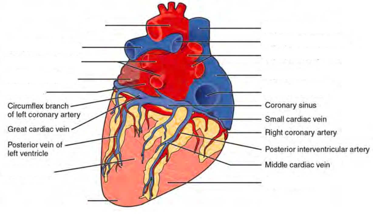
Label the surface features on the posterior aspect of the heart. (0.5 points)
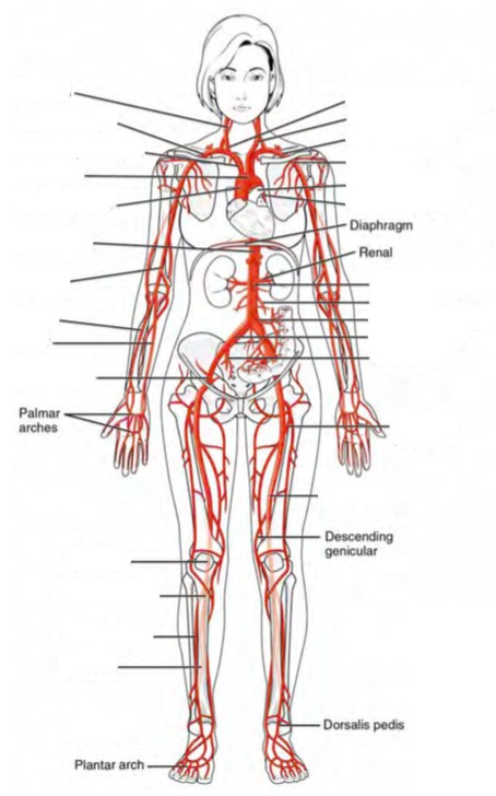
Label the major systemic arteries of the body. (0.5 points)
Label the major systemic veins of the body. (0.5 points)
A list of words is provided below that you are expected to identify, learn, and label on the models provided. Note that not all models will have some of the organs/structures, so be sure to find them on an alternate model. You must use all the words provided. Using the colored tape provided, write the number that corresponds to the organ/structure and place them on your model. When complete, notify your TA so they may check your work.
For each additional station, directions will be provided for the activity.
Label the models of this station with the number that corresponds to the appropriate structure of the peripheral nervous system using the colored tape. When you are finished, ask your TA to check your labeling. Before leaving the station, remove all the labels you have placed on the model.
Note: For the following structures, be able to differentiate between left and right halves when applicable.
#1 apex | #2 base |
#3 pericardium | #4 epicardium | #5 endocardium | #6 myocardium |
#7 superior vena cava | #10 left pulmonary artery | #13 ascending aorta | #16 posterior interventricular sulcus |
#8 right pulmonary artery | #11 coronary sulcus | #14 descending aorta | #17 epicardial fat |
#9 inferior vena cava | #12 arch of aorta | #15 anterior interventricular sulcus | #18 auricles |
#19 papillary muscles | #23 tricuspid valve | #27 right atrium | #31 left ventricle |
#20 pectinate muscles | #24 bicuspid valve | #28 left atrium | #32 interventricular septum |
#21 chordae tendineae | #25 pulmonary valve | #29 interatrial septum | #33 right bundle branches |
#22 trabeculae carneae | #26 aortic valve | #30 right ventricle | #34 left bundle branches |
#35 coronary arteries | #37 posterior interventricular branch | #39 circumflex branch | #41 branch of left coronary artery |
#36 marginal branches | #38 right pulmonary artery | #40 anterior interventricular branch | #42 middle cardiac |
#43 coronary sinus | #45 great cardiac | #47 left pulmonary |
#44 anterior cardiac | #46 small cardiac | #48 right pulmonary |
Label the models of this station with the number that corresponds to the appropriate structure of the peripheral nervous system using the colored tape. When you are finished, ask your TA to check your labeling. Before leaving the station, remove all the labels you have placed on the model.
Note: For the following structures, be able to differentiate between left and right halves when applicable.
#1 brachiocephalic trunk | #6 vertebral arteries | #11 anterior cerebral artery | #16 thoracic aorta |
#2 common carotid arteries | #7 basilar artery | #12 anterior communicating artery | #17 abdominal aorta |
#3 internal carotid arteries | #8 posterior cerebral artery | #13 axillary arteries | |
#4 external carotid arteries | #9 posterior communicating artery | #14 radial arteries | |
#5 subclavian arteries | #10 middle cerebral artery | #15 ulnar arteries |
#18 brachiocephalic veins | #22 axillary veins | #26 medial cubital veins | #30 azygos vein |
#19 internal jugular veins | #23 brachial veins | #27 radial veins | #31 hemiazygos vein |
#20 subclavian veins | #24 cephalic veins | #28 ulnar veins | #32 accessory hemiazygos vein |
#21 external jugular veins | #25 basilic veins | #29 median antebrachial veins |
Label the models of this station with the number that corresponds to the appropriate structure of the peripheral nervous system using the colored tape. When you are finished, ask your TA to check your labeling. Before leaving the station, remove all the labels you have placed on the model.
Note: For the following structures, be able to differentiate between left and right halves when applicable.
#1 suprarenal arteries | #6 celiac trunk | #11 external iliac arteries | #16 anterior tibial arteries |
#2 renal arteries | #7 common hepatic artery | #12 internal iliac arteries | #17 posterior tibial arteries |
#3 gonadal arteries | #8 splenic artery | #13 femoral arteries | #18 fibular arteries |
#4 inferior mesenteric artery | #9 lumbar arteries | #14 deep femoral arteries | |
#5 superior mesenteric artery | #10 common iliac arteries | #15 popliteal arteries |
#19 ascending lumbar veins | #24 hepatic portal veins | #30 common iliac veins | #35 popliteal veins |
#20 gonadal veins | #25 inferior mesenteric vein | #31 internal iliac veins | #36 small saphenous veins |
#21 renal veins | #26 splenic vein | #32 external iliac veins | #37 anterior tibial veins |
#22 suprarenal veins | #27 superior mesenteric vein | #33 femoral veins | #38 fibular veins |
#23 hepatic veins | #28 inferior phrenic vein | #34 great saphenous veins |
Sketch the slides available for today’s lab and specify the magnitude at which you are observing/ sketching. Be sure to identify and label your sketch with the corresponding structures listed beneath each slide.
| |
| Vein | Artery |
| |
| Leukocyte | Thrombocyte |
Erythrocyte | |
| Basophil | Eosinophil |
| |
| Neutrophil | Lymphocyte |
Monocyte
Cardiac muscle
Note: Platlets may not be visible at this magnification
*If you are the last table to use this station, be sure to clean off the dissection kits in the lab’s sink.
As a group, determine the flow of blood through the various structures and vessels of the heart. Be sure to identify where along that path each of the structures on the vocabulary list is located. Use the rest of this page to draw out the pathway.
As a group, determine the different blood type in this station. Follow the procedure below in order to do so.
(3 points)
Last Name: _______________________ First Name: _______________________
Correctly match the term with the correct order of blood flow through the heart. (0.5 point)
Venous blood enters the ______ from the ________ and ________ as well as the coronary sinus, which converge into the the ________. From there blood passes the _______ valves and enters the _______. The venous blood passes through the _______ and from there branches off into the _______ and ______ before circulating through the _____. After being oxygenatated, the blood re-enters the heart through the ________ which converge into the _______. Then the blood flows through the _______ into the ________. From here, blood is ejected through the ________ into the ______ before entering the _______ and finally systemic circulation.
What is the anatomical significance of the pericardium and epicardial fat? The visceral layer of the pericardium is also known as the _________________? (0.5 point)
An individual who cannot coagulate properly is at risk of bleeding out with any significant lesion. A reduction in what type of cell might cause this in such an individual? How does this affect the composition of their blood? (0.5 point)
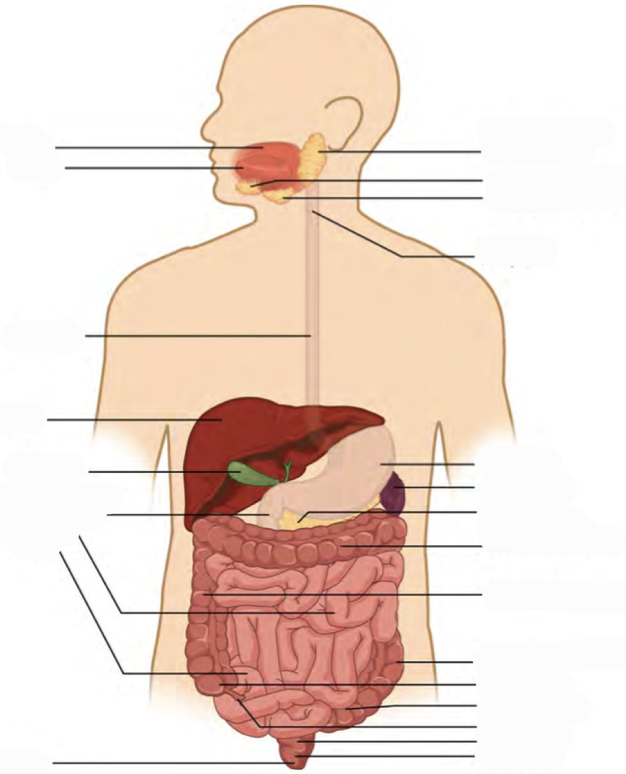
The digestive system consists of the gastrointestinal tract (also known as the alimentary canal), a hollow muscular tube extending from the mouth to the anus, and accessory organs, including the liver and pancreas. Technically, until food is absorbed in the intestines it is considered to be outside of the body. To promote absorption, the intestines have villi which contain hair-like structures called microvilli. Like the alveoli of the lungs, microvilli substantially increase the surface area of the intestines to between 180 to 300 m2 (the size of the average American home). Major structures of the gastrointestinal tract include the oral cavity, pharynx, esophagus, stomach, small intestine, large intestine, rectum, and anus. These structures and organs form a hollow space from mouth to anus and function to chemically and mechanically catabolize and absorb nutrients. Along the way organs such as the salivary glands, liver, gallbladder and pancreas release enzymes to aid digestion and are known collectively as accessory structures.
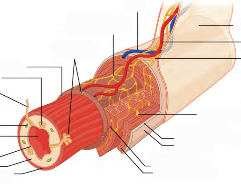
The organs of the GI tract are made from four layers, the inner lining or mucosa, the submucosa containing blood vessels and lymphatics, the muscularis or smooth muscle layer, and the outermost layer or serosa/adventitia. Each layer plays a vital role in the digestive system ranging in their capacity to form a protective barrier from the highly acidic contents of the stomach to supplying hormones, producing muscle contractions and draining lymph. Furthermore, specialized cells such as the foveolar, chief cells of the stomach are supporting cells which produce a protective layer of mucus and gastric acid for digestion. Other supporting cells, such as the gastric parietal cells of the stomach and the ductal and acinar cells of the pancreas release zymogens, inactive forms of digestive enzymes.
The peritoneum is a large serous membrane which lines the abdominal cavity and coverers most of the digestive organs. some organs are only partially covered by the peritoneum while others are entirely uncovered. These organs are referred to as being retroperitoneal. Formed by the double folding of the peritoneum is a continuous set of tissues known as the mesentery. This organ was relatively recently reclassified as an organ after discovering its complex constitution. The mesentery houses lymphatic vessels as well as providing a conduit for the blood vessels for the small and large intestines.
Vocabulary for Digestive System can be found on page(s) 165-166.
(5 points)
Last Name: _______________________ First Name: _______________________
Fill in the table below with the appropriate terms. For the remaining exercises, label the structures accordingly.
(1 point)
| Name of a structure | is | directional term | to | Name of the second structure |
|---|---|---|---|---|
| gallbladder | is | posterior | to | liver |
| transverse colon | is | superior | to | |
| is | inferior | to | small intestine | |
| liver | is | anterior | to | |
| is | distal | to | duodenum | |
| jejunum | is | medial | to | |
| is | lateral | to | left lobe of liver |
Label all digestive organs of the GI tract. (1 point)
Label the elements of the alimentary canal. (0.5 points)
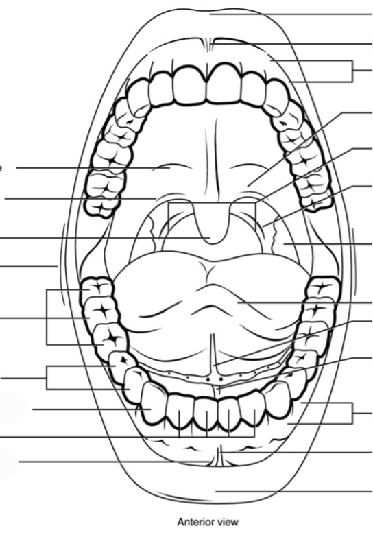
Label the different aspects of the mouth. (0.5 points)
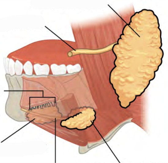
Label the major salivary glands and ducts. (0.5 points)
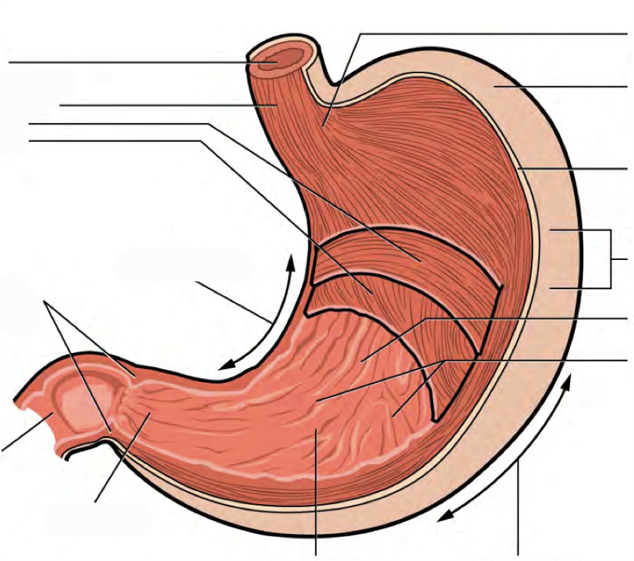
Label the aspects of the stomach accordingly. (0.5 points)
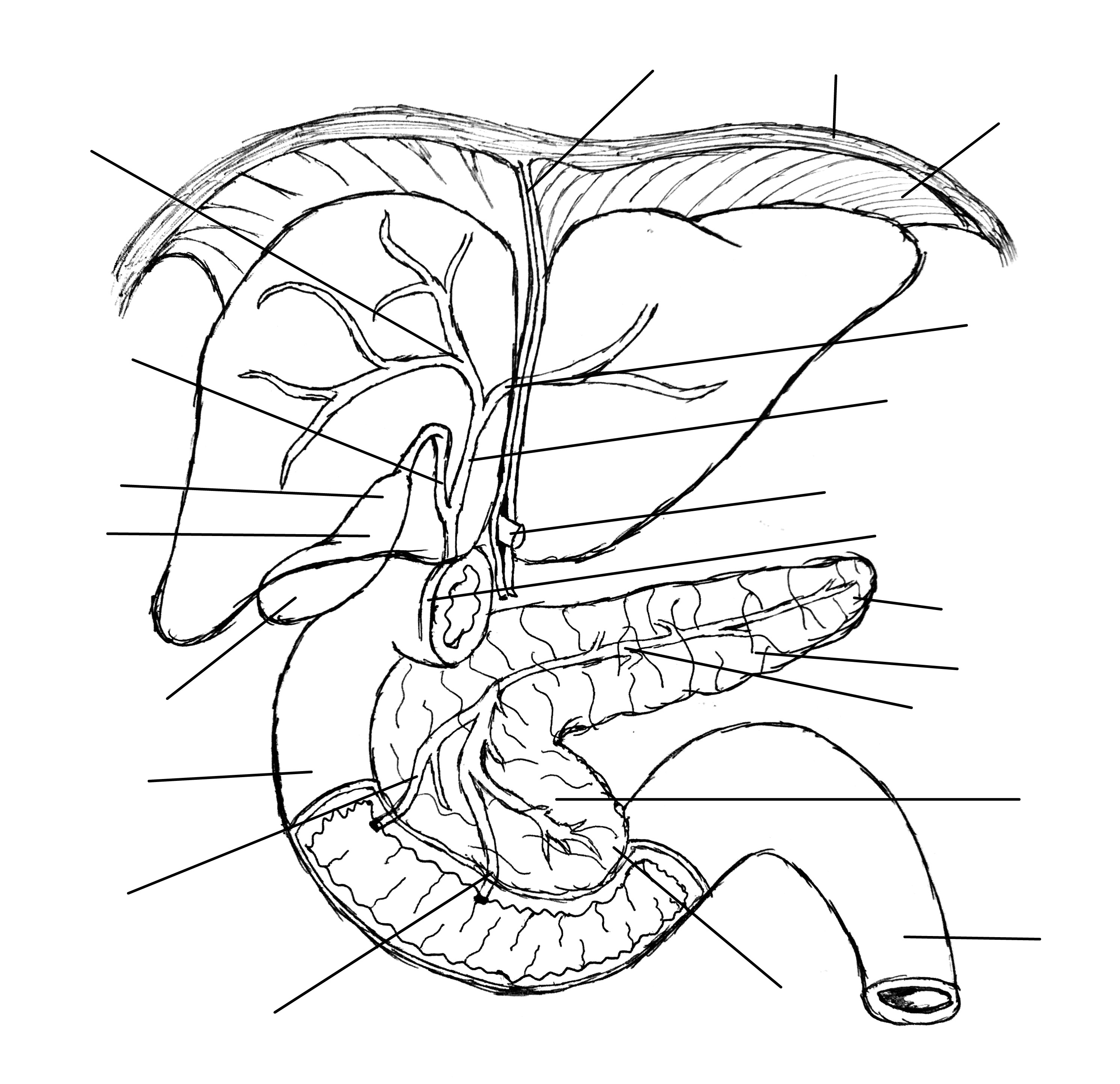
Label the accessory organs, structures, and ducts of the digestive system. (0.5 points)
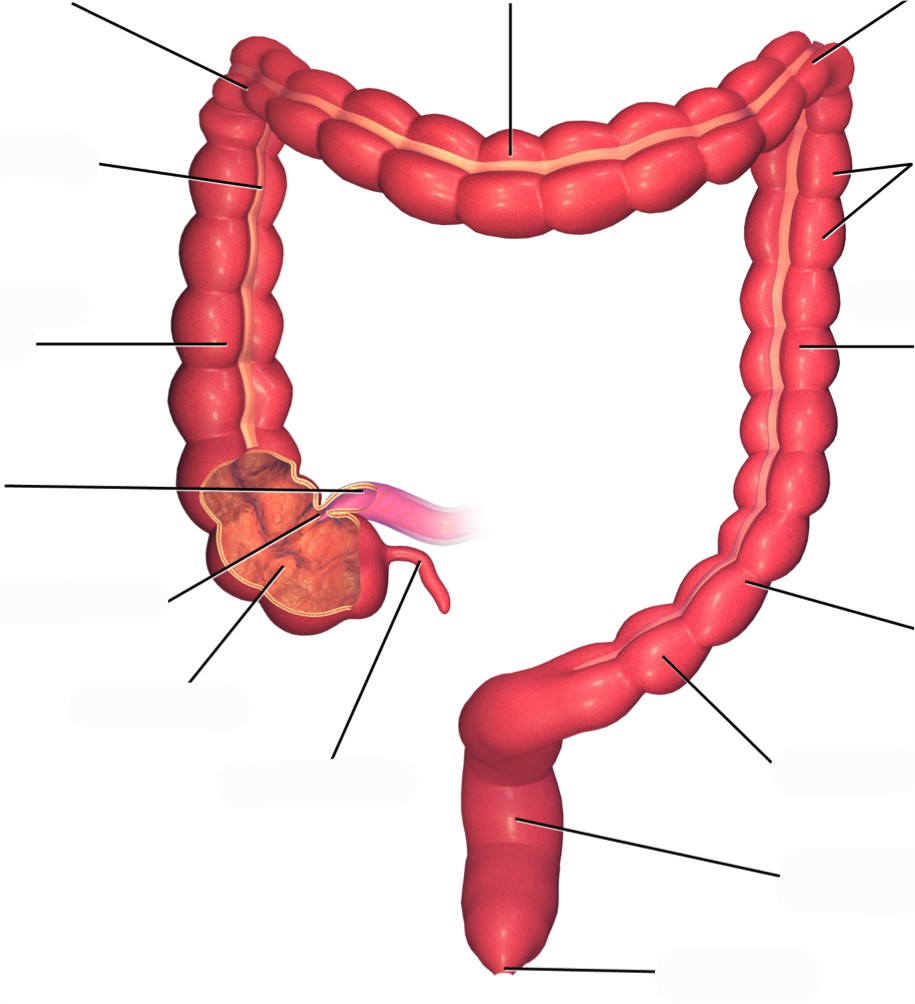
Label the structures and features of the large intestine. (0.5 points)
A list of words is provided below that you are expected to identify, learn, and label on the models provided. Note that not all models will have some of the organs/structures, so be sure to find them on an alternate model. You must use all the words provided. Using the colored tape provided, write the number that corresponds to the organ/structure and place them on your model. When complete, notify your TA so they may check your work.
For each additional station, directions will be provided for the activity.
Label the models of this station with the number that corresponds to the appropriate structure of the peripheral nervous system using the colored tape. When you are finished, ask your TA to check your labeling. Before leaving the station, remove all the labels you have placed on the model.
Note: For the following structures, be able to differentiate between left and right halves when applicable.
#1 labial frenulum | #3 hard palate | #5 uvula |
#2 fauces | #4 soft palate |
#6 tongue | #9 fungiform papillae | #11 circumvallate papillae | #13 taste pore |
#7 lingual frenulum | #10 filiform papillae | #12 taste bud | #14 base |
#8 apex |
#15 incisor | #18 molar | #21 root | #24 pulp cavity | #27 cementum |
#16 canine | #19 crown | #22 enamel | #25 pulp | #28 periodontal ligament |
#17 premolar | #20 neck | #23 dentin | #26 apical foramen | #29 gingiva |
#30 submandibular | #31 parotid | #32 sublingual |
Label the models of this station with the number that corresponds to the appropriate structure of the peripheral nervous system using the colored tape. When you are finished, ask your TA to check your labeling. Before leaving the station, remove all the labels you have placed on the model.
#1 upper esophageal sphincter | #2 lower esophageal sphincter |
#3 gastric pits | #6 cardia | #9 pylorus | #12 circular muscle layer |
#4 gastric glands | #7 gastric body | #10 pyloric sphincter | #13 oblique muscle layer |
#5 fundus | #8 rugae | #11 longitudinal muscle layer |
Label the models of this station with the number that corresponds to the appropriate structure of the peripheral nervous system using the colored tape. When you are finished, ask your TA to check your labeling. Before leaving the station, remove all the labels you have placed on the model.
Note: For the following structures, be able to differentiate between left and right halves when applicable.
#1 right lobe of liver | #3 right hepatic duct | #5 common hepatic duct | #7 hepatic canaliculi |
#2 left lobe of liver | #4 left hepatic duct | #6 hepatic lobule | #8 falciform ligament |
#9 fundus of gallbladder | #11 neck of gallbladder | #13 common bile duct |
#10 body of gallbladder | #12 cystic duct |
#14 acinar cells | #16 islets of Langerahans | #18 pancreatic head | #20 uncinate process | #22 pancreatic duct |
#15 endocrine cells | #17 pancreatic tail | #19 pancreatic body | #21 accessory duct |
Sketch the slides available for today’s lab and specify the magnitude at which you are observing/ sketching. Be sure to identify and label your sketch with the corresponding structures listed beneath each slide.
| |
Tooth | Parotid Gland |
| |
Tongue | Esophagus Mucosa, Submucosa, Muscularis externa |
|
|
Pancreas | Liver |
|
|
Duodenum | Large Intestine Lumen, Crypts of Lieberkühn, Mucosa, Submucosa, Muscularis externa, Serosa |
Vermiform appendix
Label the models of this station with the number that corresponds to the appropriate structure of the peripheral nervous system using the colored tape. When you are finished, ask your TA to check your labeling. Before leaving the station, remove all the labels you have placed on the model.
Note: For the following structures, be able to differentiate between left and right halves when applicable.
#1 microvilli | #4 submucosa | #8 enterocytes | #11 ampulla of Vater | #14 ileum |
#2 vili | #6 muscularis | #9 plicae circulares | #12 sphincter of Oddi | #15 ileocecal valve |
#3 mucosa | #7 serosa | #10 duodenum | #13 jejunum |
#16 crypts of Lieberkühn | #20 serosa | #24 right colic flexure | #28 sigmoid colon | #32 rectum |
#17 mucosa | #21 cecum | #25 transverse colon | #29 teniae coli | #33 anal canal |
#18 submucosa | #22 vermiform appendix | #26 left colic flexure | #30 haustra | #34 anal sphincter |
#19 muscularis | #23 ascending colon | #27 descending colon | #31 epiploic appendices | #35 anus |
#36 peritoneum | #38 greater omentum | #40 mesoappendix |
#37 mesentery of transverse colon | #39 lesser omentum |
As a group, determine the route boluses take through the various organs of the digestive tract. Be sure to identify the location of each structure on the vocabulary list of this lab section.
(3 points)
Last Name: _______________________ First Name: _______________________
Describe the pathway food takes upon ingesting it, making sure to include all accessory structures. (0.5 point)
Explain the differences between the layers of the gastrointestinal tract. (0.5 points)
Match the terms with their corresponding descriptions. (0.5 points)
| Name of Structure | Description | No. of Structure |
|---|---|---|
| 1. Ileum | the largest salivary glands that produce approximately 25% of the saliva produced daily | |
| 2. Gallbladder | passageway for liquids, foods, AND air | |
| 3. Fauces | the modified muscularis of the large intestine | |
| 4. Parotid glands | structures on the tongue that provide friction, allowing the tongue to move food in the oral cavity during mastication efficiently | |
| 5. Filiform papillae | the terminal portion of the small intestine | |
| 6. Pulp cavity | where bile made in the liver joins the bile stored in the gallbladder | |
| 7. Pharynx | the opening between the oral cavity and the oropharynx | |
| 8. Rugae | inner part of the tooth containing nerves and blood vessels | |
| 9. Common bile duct | storage area for bile | |
| 10. Teniae coli | folds of the inner wall of the stomach |
List each type of tooth. How do they function during mastication? (0.5)
List the accessory and primary structures of the GI tract. Why would accessory structures not be classified as primary organs/structures of the digestive system? (0.5 point)
A patient with cancerous growths in their salivary glands undergoes surgery to have them removed. How might this affect the digestive processes? (0.5 points)
The urinary system is one of excretion, elimination and reabsorption. It is made from four organs, only one of which produces urine (the kidney). Nephrons, the smallest functional unit of the kidneys, are found in numbers of one to two million within the kidney and can filter up to 400 gallons of cycled blood, daily. The kidneys receive more blood than the heart, liver, or even the brain and have vital functions such as the regulation of pH, blood pressure, concentration of blood solutes and concentration of red blood cells. The remaining three organs (ureters, urinary bladder, and urethra) facilitate urine storage and secretion. Of these organs, only the urethra is anatomically distinct between males and females.
The reproductive system is designed to propagate a species and therefore has two primary functions: the production of gametes (n) and sex hormones. Male gametes are referred to as sperm cells, whereas female gametes are called ova. Reproduction is very metabolically taxing especially for the female. To illustrate, mature ovum can contain as many as 600,000 mitochondria; to reference, liver cells and cardiac muscles cells contain 2,000 and 5,000 mitochondria respectively. The role of the male reproductive system is to produce sperm and transfer them to the female reproductive tract. Although they originate from similar primordial tissues, the female and male reproductive systems differ in gonad type, ducts, accessory glands, and external genitalia. Male gonads are referred to as testes while the female gonads as ovaries; both are the sites of their respective gametogenesis. The hormones produced by the gonads are crucial to the reproductive system and sexual development, including primary and secondary sexual development, tissue regeneration, and production of gametes.
Humans are a sexually dimorphic species, which mean that there are distinguishing secondary sex characteristics. The hormones that influence male primary and secondary sexual development are called androgens. The hormones that influence female primary secondary sexual development are called estrogens. In females, this entails the development of breasts which are specialized sweat glands. Males also have mammary tissue but their development is arrested early. Similarly, the thyroid cartilage is enlarged and commonly referred to as an Adam’s apple in males but not so in females.
A developing fetus remains anatomically undifferentiated a will either develop characteristically male or female anatomy. At some point of gestation, the fetus will develop both Wolffian and Müllerian ducts, anlagen of the male and female reproductive systems. As a result, there are several elements of the male and female reproductive systems which are homologous. Such structures share developmental and evolutionary origins but are not necessarily similar in function. The following are the homologous structures of the male and female reproductive system: labia majora – male scrotum; labia minora – shaft of penis; clitoris – glans penis; paraurethral gland – prostate gland; greater vestibular gland – bulbourethral gland.
Vocabulary for the Urinary and Reproductive systems on page(s) 172 and 168.
(5 points)
Last Name: _______________________ First Name: _______________________
Fill in the table below with the appropriate terms. For the remaining exercises, label the structures accordingly.
(1 point)
| Name of a structure | is | directional term | to | Name of the second structure |
|---|---|---|---|---|
| scrotum | is | posterior | to | penis |
| kidneys | is | superior | to | |
| is | inferior | to | urinary bladder | |
| pubic symphysis | is | anterior | to | |
| is | distal | to | prostate gland | |
| uterus | is | medial | to | |
| is | lateral | to | urethra |
Label the structures and regions of the left kidney. (1 point)
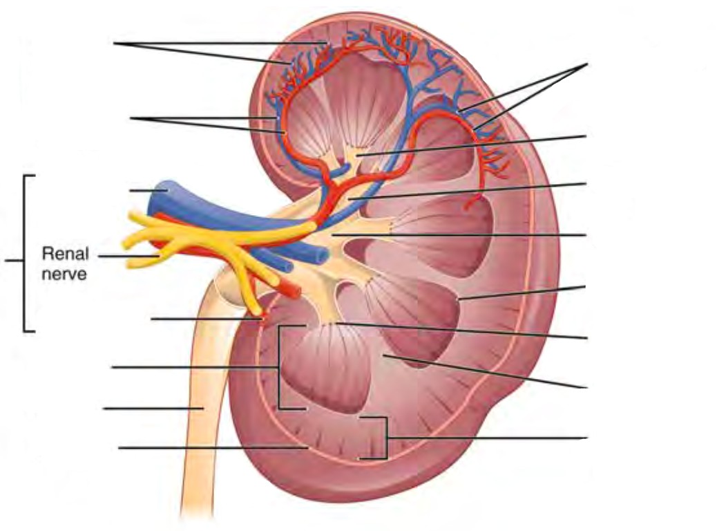
Label the structures of the nephron. (0.5 points)
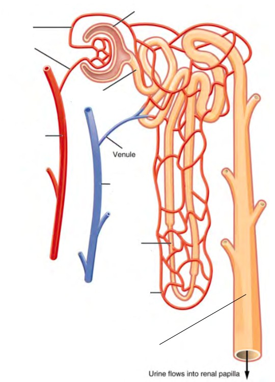
Label the structures of the bladder. (0.5 points)
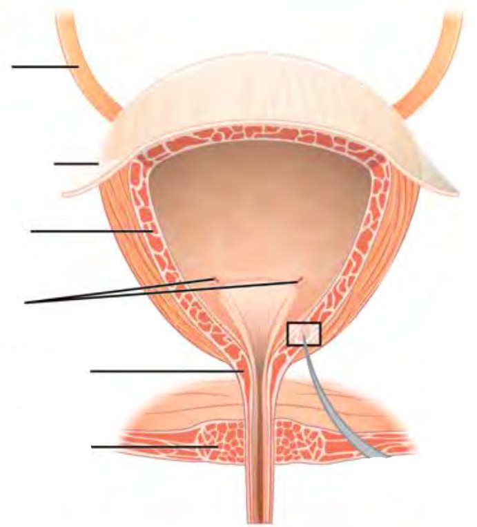
Label the parts of the male urinary/reproductive systems. (0.5 points)
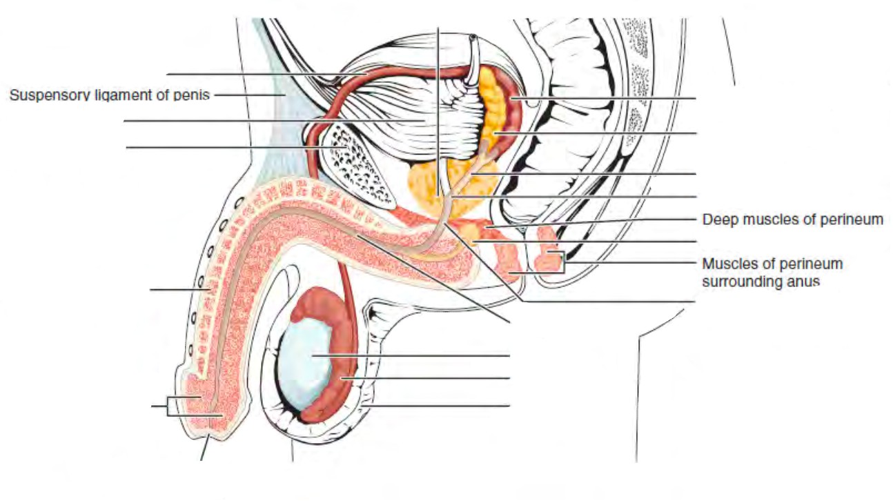
| Label the structures of the male reproductive system. (0.25 points) | Label the structures of the male reproductive system. (0.25 points) |
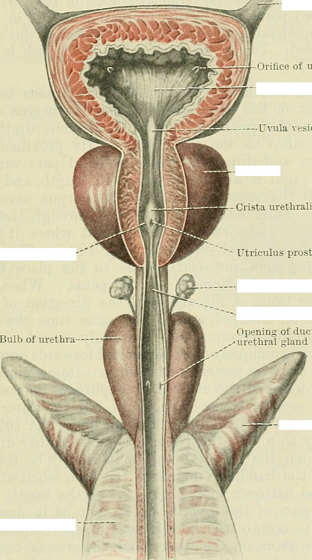 | 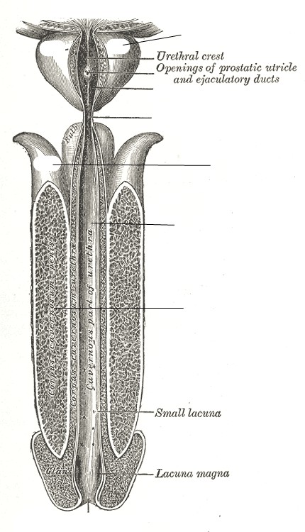 |
Label the parts of the female urinary/reproductive system. (0.5 points)
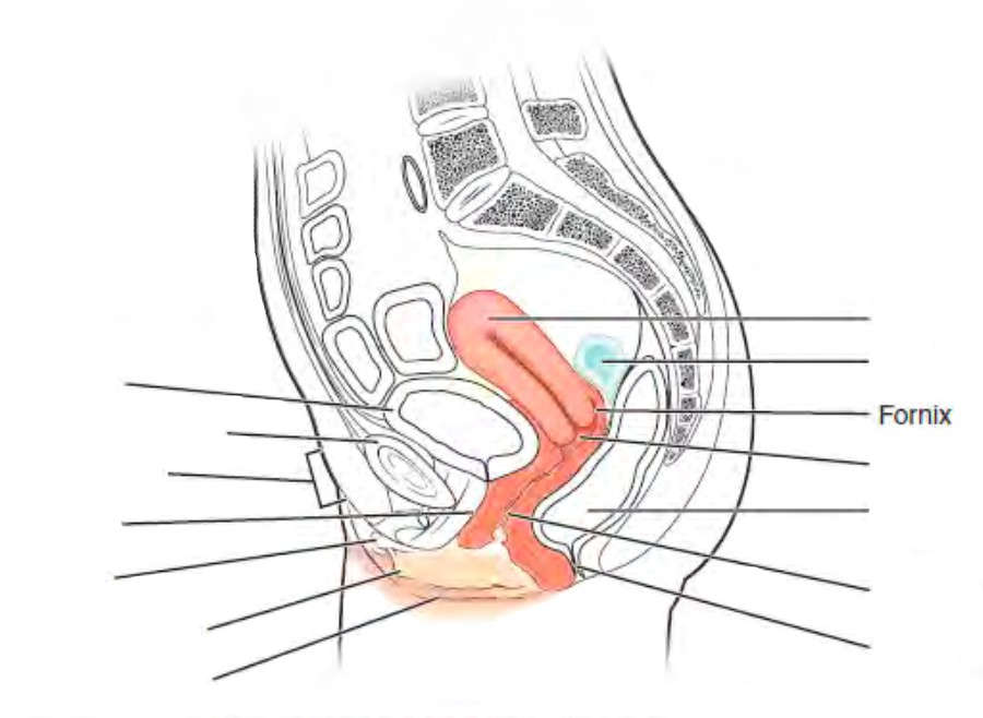
Label the structures of the breasts. (0.25 points)
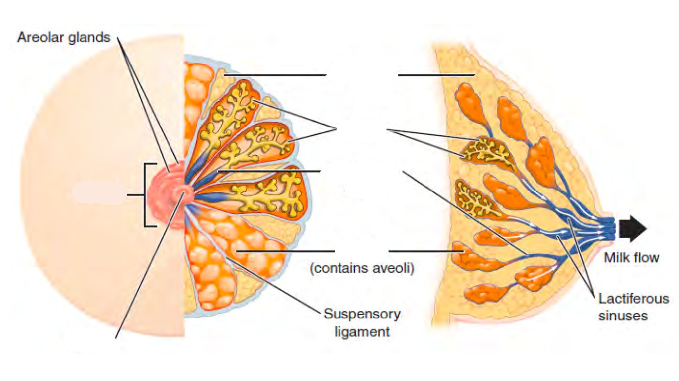
Label the structures of the uterus. (0.25 points)
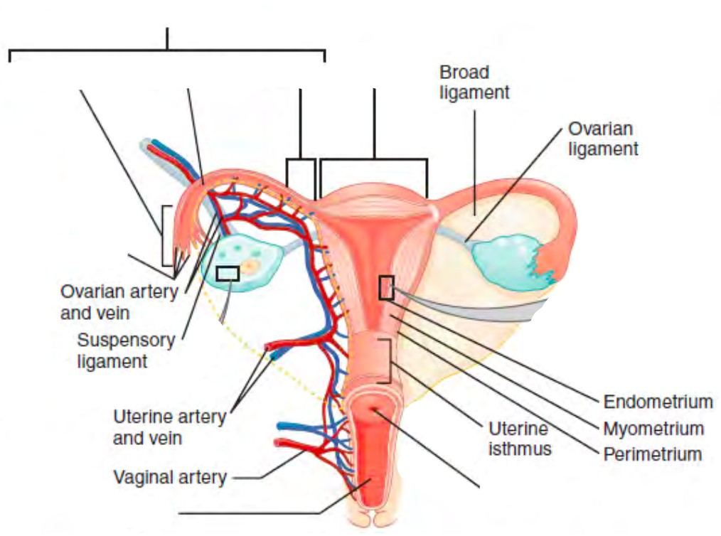
A list of words is provided below that you are expected to identify, learn, and label on the models provided. Note that not all models will have some of the organs/structures, so be sure to find them on an alternate model. You must use all the words provided. Using the colored tape provided, write the number that corresponds to the organ/structure and place them on your model. When complete, notify your TA so they may check your work.
For each additional station, directions will be provided for the activity.
Label the models of this station with the number that corresponds to the appropriate structure of the peripheral nervous system using the colored tape. When you are finished, ask your TA to check your labeling. Before leaving the station, remove all the labels you have placed on the model.
#1 renal fascia | #11 renal pelvis | #21 external urethral orifice | #31 proximal convoluted tubule |
#2 adipose capsule | #12 renal hilum | #22 cortical nephron | #32 descending loop of Henle |
#3 renal capsule | #13 ureter | #23 juxtamedullary nephron | #33 ascending loop of Henle |
#4 renal cortex | #14 urinary bladder | #24 juxtaglomerular apparatus | #35 distal convoluted tubule |
#5 renal medulla | #15 detrusor muscle | #25 renal corpuscle | #36 collecting duct |
#6 renal lobe | #16 rugae | #26 glomerulus | #37 papillary duct |
#7 renal pyramid | #17 urinary trigone | #27 podocyte | #38 minor calyx |
#8 renal columns | #18 internal urethral sphincter | #28 bowman’s capsule | #39 major calyx |
#9 renal papilla | #19 external urethral sphincter | #29 capsular space | |
#10 renal sinus | #20 urethra | #30 renal tubules |
#40 renal artery | #43 arcuate arteries | #46 glomerular capillaries | #49 cortical radiate veins | #52 renal vein |
#41 segmental arteries | #44 cortical radiate arteries | #47 efferent arterioles | #50 arcuate veins | |
#42 interlobar arteries | #45 afferent arterioles | #48 peritubular capillaries | #51 interlobar veins |
Label the models of this station with the number that corresponds to the appropriate structure of the peripheral nervous system using the colored tape. When you are finished, ask your TA to check your labeling. Before leaving the station, remove all the labels you have placed on the model.
#1 pubic symphysis | #11 head of sperm | #21 seminal vesicles | #31 prepuce of penis |
#2 dartos muscle | #12 midpiece of sperm | #22 bulbourethral (Cowper’s) glands | #32 external urethral orifice |
#3 cremaster muscle | #13 tail of sperm | #23 ejaculatory ducts | #33 root of penis |
#4 scrotum | #14 seminiferous tubules | #24 prostatic urethra | #34 bulb of penis |
#5 scrotal septum | #15 straight tubule | #25 intermediate urethra | #35 crus of penis |
#6 testis | #16 rete testis | #26 spongy urethra | #36 suspensory ligament of penis |
#7 lobules | #17 epididymis | #27 penis | #37 spermatic cord |
#8 leydig cells | #18 ductus (vas) deferens | #28 corpus cavernosum | #38 deep muscle of perineum |
#9 sertoli cells | #19 ampulla of ductus deferens | #29 corpus spongiosum | |
#10 sperm | #20 prostate glands | #30 glans penis |
Label the models of this station with the number that corresponds to the appropriate structure of the peripheral nervous system using the colored tape. When you are finished, ask your TA to check your labeling. Before leaving the station, remove all the labels you have placed on the model.
#1 pubic symphysis | #10 isthmus of uterine tube | #19 fundus of uterus | #28 vaginal orifice | #37 breast |
#2 placenta | #11 broad ligament | #20 body of uterus | #29 mons pubis | #36 areola |
#3 ovary | #12 round ligament | #21 isthmus | #30 vulva | #39 nipple |
#4 ova | #13 uterosacral ligament | #22 cervix | #31 labia majora | #40 mammary glands |
#5 ovarian ligament | #14 uterus | #23 external os | #32 labia minora | #41 lobule |
#6 uterine (Fallopian) tube | #15 endometrium | #24 internal os | #33 vestibule | #42 lactiferous ducts |
#7 fimbriae of uterine tube | #16 myometrium | #25 vagina | #34 clitoris | #43 lactiferous sinus |
#8 infundibulum of uterine tube | #17 perimetrium | #26 fornix | #35 external urethral orifice | #44 mammary ducts |
#9 ampulla of uterine tube | #18 uterine cavity | #27 rugae | #36 vestibular glands | #45 mammary alveoli |
Sketch the slides available for today’s lab and specify the magnitude at which you are observing/ sketching. Be sure to identify and label your sketch with the corresponding structures listed beneath each slide.
Urinary

Kidney
Bowman’s capsule (renal corpuscle), Glomerulus
Male Reproductive
| |
Testis | Epididymis |
Human Sperm
Head, Midpiece, Tail
Female Reproductive
| |
| Mammary gland Mammary alveoli, Lobule, Ducts | Uterus Endometrium, Myometrium, Perimetrium |
| |
| Placenta | Ovary Mature (Graafian) follicle |
*If you are the last table to use this station, be sure to clean off the dissection kits in the lab’s sink.
As a group, determine the route of urine through the various ducts of the kidney, originating at the glomerulus and ending with the urethra. Be sure to identify where along that path each of the structures on the vocabulary list is located.
As a group, determine the course of blood through the vessels of the kidney.
Note: The following three pages are left blank for the purpose of drawing out these two pathways.
(2 points)
Last Name: _______________________ First Name: _______________________
| Name of Structure | Description | No. of structure |
|---|---|---|
| 1. ureters | Area where renal vessels and ureters converge | |
| 2. kidney | urination | |
| 3. renal capsule | nephrons located deep in the renal medullas | |
| 4. micturition | smooth muscle of the bladder | |
| 5. rugae of the mucosa | organ of urine production | |
| 6. hilum | includes Bowman’s capsule and glomerulus | |
| 7. collecting duct | folds in the bladder when empty | |
| 8. detrusor | a structure where nephrons drain urine into | |
| 9. renal capsule | collagen membrane around the kidney | |
| 10. juxtamedullary nephrons | tubules that conduct urine from the kidney to bladder |
Match the structure with the corresponding description. (0.5 point)
| Name of Structure | Description | No. of structure |
|---|---|---|
| 1. Ductus- vas- deferens | produce sperm and testosterone | |
| 2. Areola | small convoluted tubules and site of spermatogenesis | |
| 3. Testes | conduct sperm to the urethra during ejaculation | |
| 4. Mammary gland | produces an ovum, estrogen, and progesterone | |
| 5. Fimbriae | a gland in mammals that produces milk | |
| 6. Corpus cavernosum and spongiosum | ducts that carry milk from the mammary glands to the nipple | |
| 7. Labia major | pigmented area around the nipple | |
| 8. Seminiferous tubules | the larger outer folds of the vulva surrounding the inner folds; contain adipose tissue and hair | |
| 9. Ovaries | erectile tissues that form the bulk of the penis | |
| 10. Lactiferous ducts | small fingerlike projections at the end of the fallopian tubes |
The body’s first line of defense against pathogens and other microbes is the skin. The skin is multi-layered and it functions to maintain homeostasis, retain water, synthesize vitamin D and regulate body temperature (thermoregulation). It is made of two chief layers: the epidermis, made of closely packed epithelial cells, and the dermis, made of dense, irregular connective tissue which houses blood vessels, hair follicles, sweat glands, and other structures. Beneath the dermis lies the hypodermis, which is composed mainly of loose connective and fatty tissues. One of skin’s accessory structures, nails, are considered to be specialized structures of the epidermis found at the tips of fingers and toes. Other accessory structures, sudoriferous glands, produce sweat which cools the body by evaporation. Skin is the largest continuous organ of the body, encompassing approximately 16 percent of our body weight.
The muscular system is an intricate network of contractile tissue which works antagonistically in order to move the body. The action of walking requires roughly 200 different muscles alone. Besides skeletal muscles, there are also cardiac muscle and smooth muscle. Cardiac muscle is found uniquely in the heart and is responsible for pumping blood through the circulatory system. Smooth muscle is the type of muscle involved in involuntary movements such as peristalsis which propel boluses through the GI tract. Skeletal muscle is also known as striated muscle, as is cardiac muscle. As you approach the muscles in this lab, make note of which muscles may be named after their shape and which ones may be named after their location or their attachments to the skeleton. Individually, all cells, with the exception of sperm, are unable to move on their own. Nevertheless, with bones as there scaffold, muscles are able to produce movent through a complex series of metabolic reactions.
Vocabulary for Muscles and Integumentary systems can be found on page(s) 166-167 and 166.
(5 points)
Last Name: _______________________ First Name: _______________________
Fill in the table below with the appropriate terms. For the remaining exercises, label the structures accordingly.
(1 point)
| Name of a structure | is | directional term | to | Name of the second structure |
|---|---|---|---|---|
| trapezius | is | proximal | to | pectoralis major |
| diaphragm | is | superior | to | |
| is | inferior | to | scalenes | |
| rectus abdominis | is | anterior | to | |
| is | distal | to | biceps femoris | |
| pectoralis minor | is | medial | to | |
| is | lateral | to | external oblique |
Label the major muscles. (0.5 points)
This is the ( ventral /dorsal ) aspect of the body. (circle one)
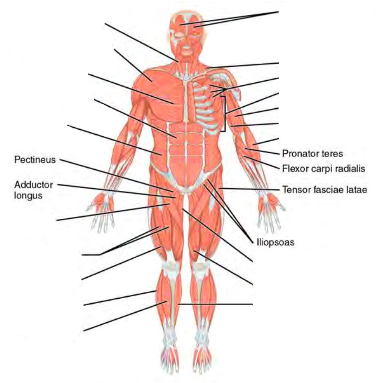
Label the major muscles of the body. (0.5 points)
This is the ( ventral /dorsal ) aspect of the body. (circle one)
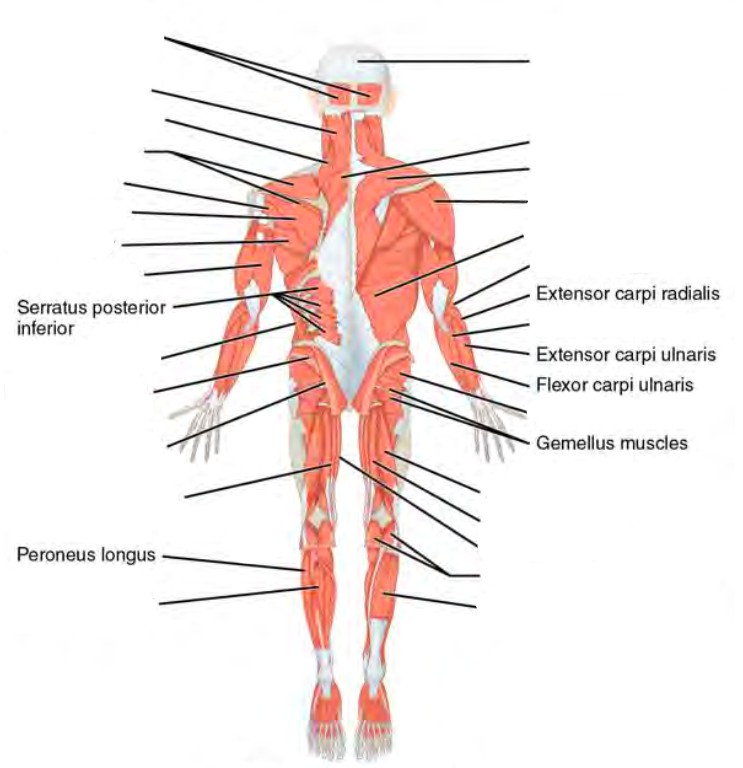
Label the muscles of the head. (0.25 points)
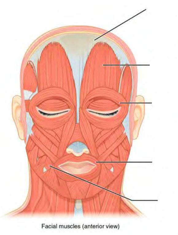
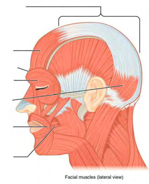
Label the muscles of the eye. (0.25 points)

Label the major abdominal muscles. (0.25 points)
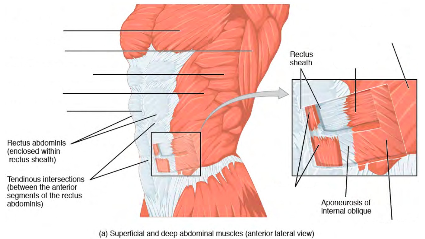
Label the major muscles of the lower leg. (0.25 points)
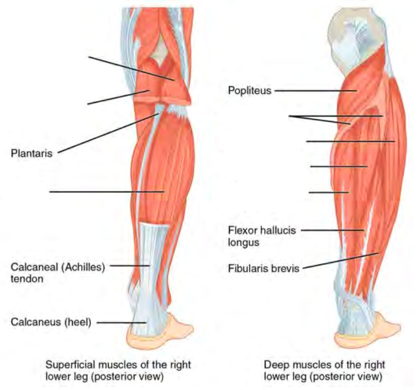
Label the layers of the epidermis. (0.5 points)
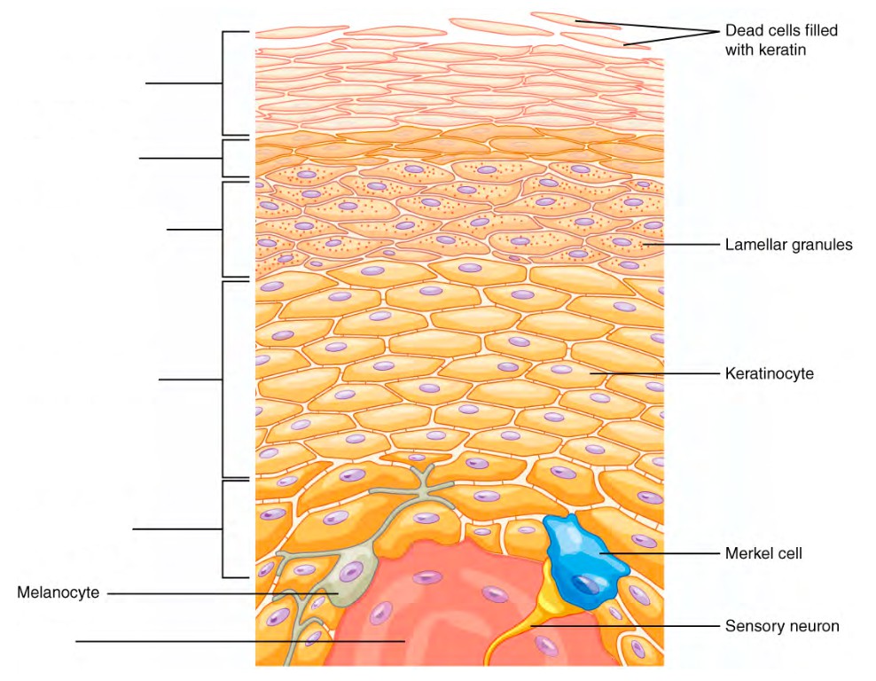
Label the layers of integument and accessory structures. (0.5 points)
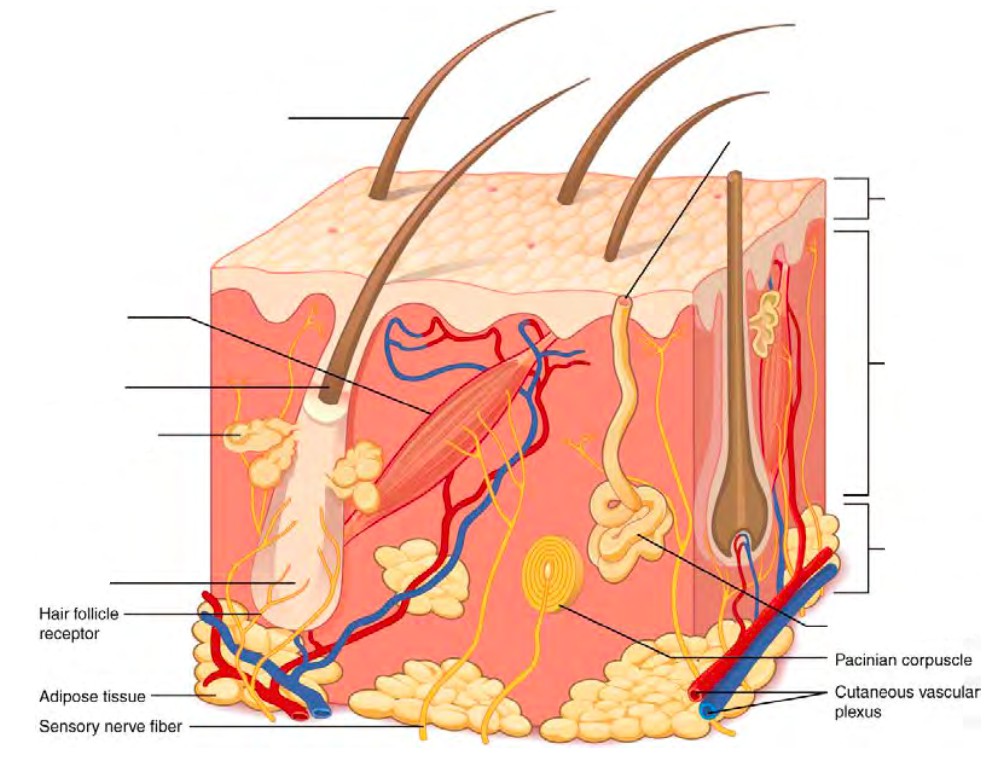
A list of words is provided below that you are expected to identify, learn, and label on the models provided. Note that not all models will have some of the organs/structures, so be sure to find them on an alternate model. You must use all the words provided. Using the colored tape provided, write the number that corresponds to the organ/structure and place them on your model. When complete, notify your TA so they may check your work.
For each additional station, directions will be provided for the activity.
Label the models of this station with the number that corresponds to the appropriate structure of the peripheral nervous system using the colored tape. When you are finished, ask your TA to check your labeling. Before leaving the station, remove all the labels you have placed on the model.
Note: For the following structures, be able to differentiate between left and right halves when applicable.
#1 epicranial aponeurosis | #7 nasalis | #13 zygomaticus minor | #19 sternocleidomastoid |
#2 front belly of occipitofrontalis | #8 orbicularis oculi | #14 zygomaticus major | #20 platysma |
#3 occipital belly of occipitofrontalis | #9 levator labii superioris | #15 buccinator | #21 sternohyoid |
#4 temporalis | #10 levator anguli oris | #16 risorius | #22 scalenes |
#5 auricularis superior | #11 depressor anguli oris | #17 orbicularis oris | |
#6 procerus | #12 depressor labii inferioris | #18 mentalis |
#23 levator palpebrae superioris | #25 medial rectus | #27 inferior recuts | #29 superior oblique |
#24 lateral recuts | #26 superior rectus | #28 inferior oblique | #30 trochlea |
#31 deltoid | #33 clavicular part of deltoid | #35 coracobrachialis | #37 biceps brachii | #39 brachioradialis |
#32 acromial part of deltoid | #34 spinal part of deltoid | #36 triceps brachii | #38 brachialis | #40 extensor digitorum |
Label the models of this station with the number that corresponds to the appropriate structure of the peripheral nervous system using the colored tape. When you are finished, ask your TA to check your labeling. Before leaving the station, remove all the labels you have placed on the model.
Note: For the following structures, be able to differentiate between left and right halves when applicable.
#1 trapezius | #7 teres major | #13 pectoralis minor | #19 internal intercostals |
#2 levator scapulae | #8 teres minor | #14 serratus anterior | #20 external intercostals |
#3 splenius capitis | #9 rhomboid major | #15 recuts abdominis | #21 diaphragm |
#4 supraspinatus | #10 rhomboid minor | #16 external oblique | |
#5 infraspinatus | #11 latissimus dorsi | #17 internal oblique | |
#6 subscapularis | #12 pectoralis major | #18 transversus abdominis |
Label the models of this station with the number that corresponds to the appropriate structure of the peripheral nervous system using the colored tape. When you are finished, ask your TA to check your labeling. Before leaving the station, remove all the labels you have placed on the model.
Note: For the following structures, be able to differentiate between left and right halves when applicable.
#1 gluteus maximus | #7 vastus lateralis | #13 gracilis | #19 extensor digitorum longus |
#2 gluteus medius | #8 vastus intermedius | #14 adductor longus | #20 fibularis longus |
#3 gluteus minimus | #9 hamstrings | #15 pectineus | #21 tibialis anterior |
#4 quadriceps | #10 biceps femoris | #16 sartorius | #22 flexor digitorum longus |
#5 rectus femoris | #11 semitendionosus | #17 gastrocnemius | #23 tibialis posterior |
#6 vastus medialis | #12 semimembranosus | #18 soleus |
Sketch the slides available for today’s lab and specify the magnitude at which you are observing/ sketching. Be sure to identify and label your sketch with the corresponding structures listed beneath each slide.
| |
| Striated muscle | Cardiac muscle |
Sketch the slides available for today’s lab and specify the magnitude at which you are observing/ sketching. Be sure to identify and label your sketch with the corresponding structures listed beneath each slide.
| |
Skin of palm | Human scalp w/hair follicle |
Squamous epithelium
Label the models of this station with the number that corresponds to the appropriate structure of the peripheral nervous system using the colored tape. When you are finished, ask your TA to check your labeling. Before leaving the station, remove all the labels you have placed on the model.
#1 epidermis | #5 stratum spinosum | #9 papillary layer | #13 hypodermis superficial fascia |
#2 stratum corneum | #6 stratum basale | #10 reticular layer | #14 lamellated corpuscles |
#3 stratum lucidum | #7 epidermal ridges | #11 dermal papillae | #15 thin (hairy) skin |
#4 stratum granulosum | #8 dermis | #12 subcutaneous layer (Hypodermis) | #16 thick (hairless) skin |
#17 squamous epithelium |
#18 pili | #20 hair root | #22 bulb |
#19 hair shaft | #21 hair follicle | #23 arrector pili muscles |
#24 sebaceous | #26 eccrine sweat | #28 ceruminous |
#25 sudoriferous | #27 aprocrine |
#29 nail body (nail plate) | #31 lunula | #33 nail bed |
#30 free edge | #32 eponychium (cuticle) |
(2 points)
Last Name: _______________________ First Name: _________________________
Mavs Open Press (https://library.uta.edu/scholcomm/mavs-open-press)
Creative Commons licenses (https://creativecommons.org/licenses/)
OER (https://library.uta.edu/scholcomm/open-education/oer)
Pressbooks Accessibility Policy (https://pressbooks.org/blog/2018/05/01/our-accessibility-policy-and-forthcoming-accessibility-improvements)
Open Education at UTA (http://libguides.uta.edu/utacares)
OER Adoption Form (https://uta.qualtrics.com/jfe/form/SV_8HTkgCym5Q6Mk7j)
BCcampus Open Education (https://open.bccampus.ca/)
Images are individually licensed as noted below.
“Planes of body” by OpenStax College is licensed under CC BY 3.0.
“Abdominal Quadrant Regions” by OpenStax College is licensed under CC BY 3.0.
“Directional Terms” by OpenStax College is licensed under CC BY 3.0.
“Regions of Human Body” by OpenStax College is licensed under CC BY 4.0.
“1916 Leukocyte Key” by OpenStax College is licensed under CC by 3.0.
“Compact bone” by Lord of Konrad is in the public domain. It was accessed via Wikimedia Commons.
“1313 Spinal Cord Cross Section” by OpenStax College is licensed under CC BY 4.0.
“Renal corpuscle” by Ed Uthman is licensed under CC BY-SA 2.0.
“705 Lateral View of Skull-01” by OpenStax College is licensed under CC BY 3.0.
“707 Superior-Inferior View of Skull Base-01” by OpenStax College is licensed under CC BY 3.1.
“Vertebra Superior View” by BodyParts3D/Anatomography is licensed under CC BY-SA 3.0.
“Right scapula – close-up – anterior view” by Anatomography is licensed under CC BY-SA 2.1 JP.
“Right scapula – close-up – posterior view” by Anatomography is licensed under CC BY-SA 2.1 JP.
“804 Humerus and Elbow” by OpenStax College is licensed under CC BY 3.0.
“805 Ulna and Radius” by OpenStax College is licensed under CC BY 3.0.
“810 Femur and Patella” by OpenStax College is licensed under CC BY 3.0.
“811 Tibia and fibula” by OpenStax College is licensed under CC BY 3.0.
N/A
“1306 Lobes of Cerebral CortexN” by OpenStax College is licensed under CC BY 4.0.
“PSM V46 D168 Mesial view of the human brain” is in the public domain. It was accessed via Wikimedia Commons.
“Blausen 0896 Ventricles Brain” by BruceBlaus is licensed under a CC BY 3.0 Unported license.
“1320 The Cranial Nerves” by OpenStax College is licensed under CC BY 4.0.
“1402 The Tongue” by OpenStax College is licensed under CC BY 4.0.
“1403 Olfaction” by OpenStax College is licensed under CC BY 4.0.
“1404 The Structures of the Ear” by OpenStax College is licensed under CC BY 4.0.
“1412 Extraocular Muscles” by OpenStax College is licensed under CC BY 3.0.
“1413 Structure of the Eye” by OpenStax College is licensed under CC BY 3.0.
N/A
“2014ab Coronary Blood Vessels” by OpenStax is licensed under CC BY 3.0.
“2008 Internal Anatomy of the HeartN” by OpenStax is licensed under CC BY 3.0.
“2005 Surface Anatomy of the Heart” by OpenStax is licensed under CC BY 3.0.
“2120 Major Systemic Artery” by OpenStax is licensed under CC BY 3.0.
“2131 Major Systemic Veins” by OpenStax is licensed under CC BY 3.0.
“2401 Components of the Digestive System” by OpenStax is licensed under CC BY 3.0.
“2402 Layers of the Gastrointestinal Tract” by OpenStax is licensed under CC BY 3.0.
“2406 Structures of the Mouth” by OpenStax is licensed under CC BY 3.0.
“2408 Salivary Glands” by OpenStax is licensed under CC BY 3.0.
“2414 Stomach” by OpenStax is licensed under CC BY 3.0 .
“Liver Gallbladder SI” by Andrea Campo-Velez is licensed under CC BY-SA 4.0.
“Blausen 0604 LargeIntestine2” by OpenStax is licensed under CC BY 3.0.
“2610 The Kidney” by OpenStax is licensed under CC BY 3.0.
“2611 Blood Flow in the Nephron” by OpenStax is licensed under CC BY 3.0.
“2605 The Bladder” by OpenStax is licensed under CC BY 3.0.
“Figure 28 01 01” by OpenStax is licensed under CC BY 3.0.
“Image from page 1340 of ‘Cunningham’s Text -book of anatomy’ (1914)” is in the public domain. It was accessed via the Internet Archive Book Images Flikr.
“Image from ‘Anatomy of the Human Body’ (1918) ” Is in the public domain. It was accessed via Wikimedia Commons.
“Figure 28 02 01” by OpenStax is licensed under CC BY 3.0.
“Figure 28 02 09” by OpenStax is licensed under CC BY 3.0.
“1105 Anterior and Posterior View of Muscles” by OpenStax is licensed under CC BY 4.0.
“1106 Front and Side Views of the Muscles of Facial Expressions” by OpenStax is licensed under CC BY 4.0.
“1107 The Extrinsic Eye Muscles” by OpenStax is licensed under CC BY 4.0.
“1112 Muscles of the Abdomen” by OpenStax is licensed under CC BY 4.0 .
“1123 Muscles of the Leg that Move the Foot and Toes” by OpenStax is licensed under CC BY 4.0.
“502 Layers of epidermis” by OpenStax is licensed under CC BY 3.0.
“501 Structure of the skin” by OpenStax is licensed under CC BY 3.0.