Building a Medical Terminology Foundation

Building a Medical Terminology Foundation

Kimberlee Carter and Marie Rutherford

eCampus Ontario

Toronto, Ontario

Contents

1

Introduction

Welcome to Building a Medical Terminology Foundation. Medical terminology is a language that is used in health care settings. Medical terms are built from Greek and Latin word parts and in addition include acronymns, eponyms, and modern-day language terms.

Learning a new language can be a daunting task. In this resource, we offer a method for breaking down medical words that takes that daunting task and makes it manageable. What is required from you is a commitment to memorizing the word parts, learning the rules, and identifying the rebels. Once you meet that commitment we will show you how to apply the rules to the word parts you have memorized. As you memorize the language components of medical terminology it is important to support that learning with the context of anatomy and physiology.  Consider where in the body the medical term is referencing and then how it works within the body. This will build a medical terminology foundation that you can continue to grow in your future health-care courses.

How this open educational resource (OER) works.

The introductory anatomy and physiology content of this OER has been adapted from the OpenStax Anatomy and Physiology OER by Betts, et al., which is licensed under a Creative Commons Attribution 4.0 International License. Following OpenStax’s leadership and in the spirit of OPEN education we have licensed this OER with the same license.

Students: this OER is different than many traditional medical terminology textbooks. The interactive content is built into each chapter. In this resource you will work through each body system that includes word parts, whole medical terms, and common abbreviations associated with that particular body system. At the end of each body system chapter is a vocabulary list of associated terms related to that body system. The interactive reinforcement activities require you to click, drag and drop, listen and repeat, flip, and test yourself.

Faculty and teaching staff: while this OER was curated and created for Health Office Administration and Health Services students in the first year of college, our hope is that you will take this OER and customize it for your program and share again.


Anatomy and Physiology Book Citation:

Betts, J.G., Young, K.A., Wise, J.A., Johson, E., Poe, B., Kruse, D. H., Korol, O., Johnson, J.E., Womble, M. & DeSaix, P. (2013). Anatomy and Physiology. OpenStax. http://cnx.org/content/col11496/latest/

Cover image: Medical Diagram. From Spratt, A., 2018. Licensed for reuse under UnSplash’s licence.

2

Acknowledgements

In the Spring of 2018, I (Kimberlee) attended a workshop on building OER (Open Educational Resources) for high impact in first year courses. I was moved by the student speaker’s plea over the rising costs of textbooks and motivated to learn more about the ability to customize OER. In health office administration programs, customization is important because we are combination of health and business. I attended a Pressbooks webinar, signed up for an account, and started plugging away. I will be honest the task was daunting to do alone.

Marie and I met in the Winter of 2019 as participants in the Ontario Extend eCampus Ontario mOOC and went on to become Empowered Educators. Through this program we learned about creative commons licensing and were inspired to create OER. In the fall of 2019, I shared what I had been working on with Marie and this led to a collaborative partnership that snowballed into this OER. We advocated for support, found collaborators, and ultimately crowd-sourced this OER. Please read below to learn about the amazing collaborative support we had for this OER, for which we are truly grateful.

Kimberlee Carter B.Ed., M.A., Conestoga College ITAL

Thank you Kimberlee for inviting me to partake and share this adventure with you. The opportunity to create a resource for learners in this OPEN format has been a wonderful and collaborative endeavour.

Marie Rutherford, Dip., Nursing, LD., BGS., Georgian College CAAT

Thanks and Gratitude

We wish to thank Gary Hallam, Vice President of Research & Executive Dean of the School of Business at Conestoga College ITAL, for sharing his dream for OER and championing our project to other business heads in the Ontario College System. His encouragement and resource support led to the expansive collaboration team that saw this project through to publishing.

We also extend our gratitude to Pavla Kazda, Associate Dean from Georgian College CAAT, for her support, encouragement and resource allocation.

We wish to recognize the work of our students whose dedication, creation of activities, and commitment to reviewing interactive activities throughout development of this resource was invaluable. Additional thanks for examining content and providing feedback from the student perspective. Thank you for paying it forward for future students.

To our amazing subject matter experts who continued to trek up the mountain carrying a backpack full of extra responsibilities during the COVID-19 pandemic of 2020 to author and assist with crowd-sourcing this OER.

A special thank you to Jesslyn Wilkinson (OCT. M.Ed candidate, BEd, Hons. BA) Educational Technology Officer – Teaching & Learning at Conestoga College ITAL for her infectious enthusiasm for OER, support for H5P technology, and asking the question, “Are you ready to Sprint?”.

A big shout out to Holly Ashbourne, our copy editor, and the Library team for their tireless work in copy editing, copyright proofing, accessibility compliance, and their continuous championing of OER. You are, truly, the quiet leaders that make students’ lives better.

This OER was greatly enhanced by the leadership, support and contributions of the following colleagues:

Special thanks to eCampus Ontario for the work that they do putting collaborators together. Supporting OER through the Open library, Open Publishing Infrastructure, Ontario Extend Professional Learning for Educators, and answering countless Pressbooks questions. Thank you to:


1

Identifying Word Parts in Medical Terms

Word Parts

Medical terms are built from word parts. Those word parts are prefix, word root, suffix, and combining form vowel. When a word root is combined with a combining form vowel the word part is referred to as a combining form.

An interactive or media element has been excluded from this version of the text. You can view it online here:
https://ecampusontario.pressbooks.pub/medicalterminology/?p=5

Identifying Word Parts in Medical Terms

By the end of this resource, you will have identified hundreds of word parts within medical terms.  Let’s start with some common medical terms that many non-medically trained people may be familiar with.

Examples

Osteoarthritis
Oste/o/arthr/itis – Inflammation of bone and joint.

Oste/o is a combining form that means bone
arthr/o is a combining form that means joint
-itis is a suffix that means inflammation

Intravenous
Intra/ven/ous – Pertaining to within a vein.
Intra- is a prefix that means within
ven/o – is a combining form that means vein
-ous is a suffix that means pertaining to

Notice, when breaking down words that you place slashes between word parts and a slash on each side of a combining form vowel.

Language Rules

Language rules are a good place to start when building a medical terminology foundation.  Many medical terms are built from word parts and can be translated literally. At first, literal translations sound awkward. Once you build a medical vocabulary and become proficient at using it, the awkwardness will slip away. For example, suffixes will no longer be stated and will be assumed. The definition of intravenous then becomes within the vein.

Since you are at the beginning of building your medical terminology foundation stay literal when applicable. It should be noted that as with all language rules there are always exceptions and we refer to those as rebels. So let’s begin by analyzing the language rules for medical terminology.

2

Medical Language Rules

Language Review

Before we begin analyzing the rules let’s complete a short language review that will assist with pronunciation and spelling. In class, you will practice pronunciation with your Instructor.

Short Vowels
a, e, i, o, u, and sometimes y are indicated by lower case.

Long Vowels
A, E, I, O, U are indicated by upper case.

Consonants
Consonants are all of the other letters in the alphabet. b, c, d, f, g, h, j, k, l, m, n, p, q, r, s, t, v, w, x, and z.

Language Rules for Building Medical Terms

  1. When combining two combining forms you keep the combining form vowel.
  2. When combining a combining form with a suffix that begins with a consonant you keep the combining form vowel.

    Examples

    Gastr/o/enter/o/logy – The study of the stomach and the intestines

    • Following rule 1, when we join combining form gastr/o (meaning stomach) with the combining form enter/o (meaning intestines) we keep the combining form vowel o.
    • Following rule 2, when we join the combining form enter/o (meaning intestines) with the suffix -logy (that starts with a suffix and means the study of) we keep the combining form vowel o.
  3. When combining a combining form with a suffix that begins with a vowel you drop the combining form vowel.
  4.  A prefix goes at the beginning of the word and no combining form vowel is used.

    Examples

    Intra/ven/ous – Pertaining to within the vein

    • Following rule 3, notice that when combining the combining form ven/o (meaning vein) with the suffix -ous ( that starts with a vowel and means pertaining to) we drop the combining form vowel o.
    • Following rule 4, the prefix intra- (meaning within) is at the beginning of the medical term with no combining form vowel used.
  5. When defining a medical word, start with the suffix first and then work left to right stating the word parts. You may need to add filler words. As long as the filler word does not change the meaning of the word you may use it for the purpose of building a medical vocabulary. Once you start to apply the word in the context of a sentence it will be easier to decide which filler word(s) to choose.

    Examples

    Intra/ven/ous – Pertaining to within the vein or Pertaining to within a vein.

    • Following rule 5, notice that I start with the suffix -ous (that means pertaining to) then we work left to right starting with the prefix Intra- (meaning within) and the combining form ven/o (meaning vein).
    • Notice that we have used two different definitions that mean the same thing.
    • In these examples we do not have the context of a full sentence. For the purpose of building a medical terminology foundation either definition is accepted.

3

Prefix

Prefixes are located at the beginning of a medical term. The prefix alters the meaning of the medical term. It is important to spell and pronounce prefixes correctly.

Many prefixes that you find in medical terms are common to English language prefixes. A good technique to help with memorization is the following:

PREFIX MEANING EXAMPLE OF USE IN MEDICAL TERMS
a-, an No, not, without, negates the meaning atypical, anoxia
ab- away from; from abduction
ad- toward, at, increase, on, toward adduction
ante- before antepartum
anti- against, opposing antipsychotic
PREFIX MEANING EXAMPLE OF USE IN MEDICAL TERMS
bi-,bin- two, twice, double bilateral, binocular
brady- slow bradycardia
PREFIX MEANING EXAMPLE OF USE IN MEDICAL TERMS
de- from, down, away from dehydrate
dia- through, complete dialysis
dys- painful, abnormal, difficult, laboured dysphagia
PREFIX MEANING EXAMPLE OF USE IN MEDICAL TERMS
endo- within, in endotracheal
epi- on, upon, over epidermis
eu- normal, good eupnea
PREFIX MEANING EXAMPLE OF USE IN MEDICAL TERMS
hemi- half hemicolectomy
hyper- above, excessive hyperthyroidism
hypo- below, incomplete, deficient, under hypoglycemia
PREFIX MEANING EXAMPLE OF USE IN MEDICAL TERMS
inter- between intercostal
intra- within, in intramuscular
PREFIX MEANING EXAMPLE OF USE IN MEDICAL TERMS
macro- large, long macrocephalus
meta- after, beyond, change metacarpal bones
micro- small microscope
multi- many multipara
PREFIX MEANING EXAMPLE OF USE IN MEDICAL TERMS
neo- new neonate
nulli- none nulligravida
PREFIX MEANING EXAMPLE OF USE IN MEDICAL TERMS
pachy- thick, thickening, thickened pachyderma
pan- all, total pancytopenia
para- beside, beyond, around, abnormal parathyroid glands
per- through percutaneous
peri- surrounding (outer) peripheral vision
poly- many, much polymyositis
post- after postpartum
pre- before prenatal
pro- before prognosis
PREFIX MEANING EXAMPLE OF USE IN MEDICAL TERMS
sub- below, under subcutaneous
supra- above suprascapular
sym-, syn- together, joined symphysis
PREFIX MEANING EXAMPLE OF USE IN MEDICAL TERMS
tachy- fast, rapid tachycardia
trans- through, across, beyond transdermal
tri- three triceps
PREFIX MEANING EXAMPLE OF USE IN MEDICAL TERMS
uni- one unilateral

4

Suffix

Suffixes are word parts that are located at the end of words. Suffixes can alter the meaning of medical terms. It is important to spell and pronounce suffixes correctly.

Suffixes in medical terms are common to English language suffixes. Suffixes are not always explicitly stated in the definition of a word. It is common that suffixes will not be explicitly stated when defining a medical term in the workplace. However, when transcribing or reading medical reports the suffix is always clearly written. In order to properly spell and pronounce medical terms, it is helpful to learn the suffixes.


SUFFIX MEANING EXAMPLE OF USE IN MEDICAL TERMS
-a noun ending, no meaning leukoderma
-ac pertaining to cardiac
-ad toward dorsad
-algia pain myalgia
-amnios amnion, amniotic fluid oligohydramnios
-apheresis removal plasmapheresis
-ar pertaining to appendicular
-ary pertaining to coronary
– asthenia weakness Myasthenia gravis
SUFFIX MEANING EXAMPLE OF USE IN MEDICAL TERMS
-carcinoma cancerous tumour adenocarcinoma
-cele hernia, protrusion, swelling hydrocele
-centesis surgical puncture to aspirate fluid amniocentesis
-crine to secrete exocrine
-cyesis pregnancy pseudocyesis
-cyte cell leukocyte
SUFFIX MEANING EXAMPLE OF USE IN MEDICAL TERMS
-desis surgical fixation, fusion arthrodesis
-drome run, running syndrome
SUFFIX MEANING EXAMPLE OF USE IN MEDICAL TERMS
-e noun ending, no meaning neonate
-eal pertaining to esophageal
-ectasis stretching out, dilation, expansion bronchiectasis
-ectomy excision, surgical removal, cut out gastrectomy
-emesis vomiting hematemesis
-emia in the blood anemia
-esis condition diuresis
SUFFIX MEANING EXAMPLE OF USE IN MEDICAL TERMS
-gen substance or agent that produces or causes teratogen
-genic producing, originating, causing carcinogenic
-gram the record, radiographic image electrocardiogram
-graph instrument used to record; the record electrocardiograph
-graphy process of recording, radiographic imaging electrocardiography
SUFFIX MEANING EXAMPLE OF USE IN MEDICAL TERMS
-ia condition of, diseased state, abnormal state tachycardia
-iasis condition choledocholithiasis
-iatrist specialist psychiatrist
-iatry specialty, treatment psychiatry
-ic pertaining to cardiac
-ictal seizure, attack postictal
-ior pertaining to anterior
-ism state of hyperthyroidism
-itis inflammation colitis
SUFFIX MEANING EXAMPLE OF USE IN MEDICAL TERMS
-logist specialist who studies and treats oncologist
-logy study of cardiology
-lysis separating, loosening, dissolution thrombolysis
SUFFIX MEANING EXAMPLE OF USE IN MEDICAL TERMS
-malacia softening chondromalacia
-megaly enlarged, enlargement gastromegaly
-meter instrument used to measure thermometer
-metry measuring, process of measuring spirometry
SUFFIX MEANING EXAMPLE OF USE IN MEDICAL TERMS
-oid resembling lipoid
-oma tumour, swelling melanoma
-opia vision diplopia
-opsy viewing, process of viewing biopsy
-osis abnormal condition, increased number (blood) erythrocytosis
-ous pertaining to intravenous
SUFFIX MEANING EXAMPLE OF USE IN MEDICAL TERMS
-paresis slight paralysis hemiparesis
-pathy disease polyneuropathy
-penia abnormal reduction in number erythrocytopenia
-pepsia digestion dyspepsia
-pexy surgical fixation, suspension colpopexy
-phagia swallowing, eating dysphagia
-phobia aversion, abnormal fear photophobia
-physis growth symphysis
-plasia formation, development, growth hyperplasia
-plasm growth, substance, formation cytoplasm
-plasty surgical repair rhinoplasty
-plegia paralysis ophthalmoplegia
-pnea breathing dyspnea
-poiesis formation leukopoiesis
-ptosis prolapse, drooping nephroptosis
SUFFIX MEANING EXAMPLE OF USE IN MEDICAL TERMS
-rrhage excessive bleeding hemorrhage
-rrhagia excessive bleeding, rhinorrhagia
-rrhaphy repairing, suturing cholecystorrhaphy
-rrhea discharge, flow rhinorrhea
-rrhexis rupture amniorrhexis
SUFFIX MEANING EXAMPLE OF USE IN MEDICAL TERMS
-salpinx fallopian tube, uterine tube pyosalpinx
-sarcoma malignant tumour rhabdomyosarcoma
-schisis split, fissure cranioschisis
-sclerosis hardening arteriosclerosis
-scope instrument used for visual examination hysteroscope
-scopic pertaining to visual examination pelviscopic
-scopy process of visually examining gastroscopy
-spasm sudden, involuntary contraction of muscle vasospasm
-stasis stop, control, standing hemostasis
-stenosis constriction, narrowing ureterostenosis
-stomy creation of artificial opening nephrostomy
SUFFIX MEANING EXAMPLE OF USE IN MEDICAL TERMS
-thorax chest cavity, chest hemothorax
-tocia labour, birth dystocia
-tome instrument used to cut dermatome
-tomy incision, cut into laparotomy
-tripsy surgical crushing cholecystolithotripsy
-trophy nourishment, development hypertrophy
SUFFIX MEANING EXAMPLE OF USE IN MEDICAL TERMS
-um no meaning endocardium
-uria urine, urination nocturia
-us no meaning microcephalus


5

Medical Language Within the Context of Anatomy and Physiology

Learning Objectives

  • Connect medical language learning to the context of anatomy and physiology
  • Introduce the basic architecture and levels of organization of the human body
  • Evaluate the anatomical position, regional terms, directional terms, body planes, and body quadrants for anatomical positioning
  • Describe body cavities and the functions of associated membranes


As you memorize the language components of medical terminology it is important to support that learning within the context of anatomy and physiology. Proceeding through the body system chapters you will learn word parts, whole medical terms, and common abbreviations. It is important to put into context where in the body the medical term is referencing, and then consider how it works within the body.

Anatomy focuses on structure and physiology focuses on function. Much of the study of physiology centers on the body’s tendency toward homeostasis .

Consider the structures of the body in terms of fundamental levels of organization that increase in complexity: subatomic particles, atoms, molecules, organelles, cells, tissues, organs, organ systems, organisms, and biosphere (Figure 5.1).

Levels of structural organization of the human body. Image description available.
Figure 5.1 Levels of Structural Organization of the Human Body. The organization of the body often is discussed in terms of six distinct levels of increasing complexity, from the smallest chemical building blocks to a unique human organism. From Betts, et al., 2013. Licensed under CC BY 4.0. [Image description.]

The Levels of Organization

All matter in the universe is composed of one or more unique pure substances called elements, familiar examples are hydrogen, oxygen, carbon, nitrogen, calcium, and iron.

An organ system is a group of organs that work together to perform major functions or meet the physiological needs of the body.

Did you know?

  • Organs are very collaborative and work with multiple body systems.
  • For example, the heart (cardiovascular system) and lungs (respiratory system) work together to deliver oxygen throughout the body and remove carbon dioxide from the body.


Consider the breakdown into eleven distinct organ systems in the human body (Figure 5.2 and Figure 5.3). Assigning organs to organ systems can be imprecise since organs that “belong” to one system can also have functions integral to another system. In fact, most organs contribute to more than one system.

Organ systems of the human body. Image description available.
Figure 5.2. Organ Systems of the Human Body. Organs that work together are grouped into organ systems. From Betts, et al., 2013. Licensed under CC BY 4.0 [Image description.]
Organ Systems of the Human Body continued from Figure 5.2. Image description available.
Figure 5.3. Organ Systems of the Human Body (continued). Organs that work together are grouped into organ systems. From Betts, et al., 2013. Licensed under CC BY 4.0 [Image description.]

The organism level is the highest level of organization. An organism is a living being that has a cellular structure and that can independently perform all physiologic functions necessary for life. In multicellular organisms, including humans, all cells, tissues, organs, and organ systems of the body work together to maintain the life and health of the organism.

Watch this video:

Thumbnail for the embedded element "Introduction to Anatomy & Physiology: Crash Course A&P #1"

A YouTube element has been excluded from this version of the text. You can view it online here: https://ecampusontario.pressbooks.pub/medicalterminology/?p=1382

Media 5.1. Introduction to Anatomy & Physiology: Crash Course A&P #1 [Online video]. Copyright 2015 by CrashCourse.

Did you know?

Terminology is used for the purpose of precision and reduction of medical error.

Anatomical Position

Anatomists and health care providers use terminology for the purpose of precision and to reduce medical errors. For example, is a scar “above the wrist” located on the forearm two or three inches away from the hand? Or is it at the base of the hand? Is it on the palm-side or back-side? By using precise anatomical terminology, we eliminate ambiguity. Anatomical terms derive from ancient Greek and Latin words.

To further increase precision, anatomists standardize the way in which they view the body. Just as maps are normally oriented with north at the top, the standard body “map,” or anatomical position, is that of the body standing upright, with the feet at shoulder width and parallel, toes forward. The upper limbs are held out to each side, and the palms of the hands face forward as illustrated.

Using this standard position reduces confusion. It does not matter how the body being described is oriented, the terms are used as if it is in anatomical position. For example, a scar in the “anterior (front) carpal (wrist) region” would be present on the palm side of the wrist. The term “anterior” would be used even if the hand were palm down on a table.

Regions of the human body. Image description available.
Figure 5.4. Regions of the Human Body. The human body is shown in anatomical position in an (a) anterior view and a (b) posterior view. The regions of the body are labeled in boldface. From Betts, et al., 2013. Licensed under CC BY 4.0. [Image description.]

A body that is lying down is described as either prone or supine.  These terms are sometimes used in describing the position of the body during specific physical examinations or surgical procedures.

Regional Terms

The human body’s numerous regions have specific terms to help increase precision. Notice that the term “brachium” or “arm” is reserved for the “upper arm” and “antebrachium” or “forearm” is used rather than “lower arm.” Similarly, “femur” or “thigh” is correct, and “leg” or “crus” is reserved for the portion of the lower limb between the knee and the ankle. You will be able to describe the body’s regions using the terms from the anatomical position.

Directional Terms

Directional terms are essential for describing the relative locations of different body structures. For instance, an anatomist might describe one band of tissue as “inferior to” another or a physician might describe a tumor as “superficial to” a deeper body structure. Commit these terms to memory to avoid confusion when you are studying or describing the locations of particular body parts.

  • Anterior (or ventral) describes the front or direction toward the front of the body. The toes are anterior to the foot.
  • Posterior (or dorsal) describes the back or direction toward the back of the body. The popliteus is posterior to the patella.
  • Superior (or cranial) describes a position above or higher than another part of the body proper. The orbits are superior to the oris.
  • Inferior (or caudal) describes a position below or lower than another part of the body proper; near or toward the tail (in humans, the coccyx, or lowest part of the spinal column). The pelvis is inferior to the abdomen.
  • Lateral describes the side or direction toward the side of the body. The thumb (pollex) is lateral to the digits.
  • Medial describes the middle or direction toward the middle of the body. The hallux is the medial toe.
  • Proximal describes a position in a limb that is nearer to the point of attachment or the trunk of the body. The brachium is proximal to the antebrachium.
  • Distal describes a position in a limb that is farther from the point of attachment or the trunk of the body. The crus is distal to the femur.
  • Superficial describes a position closer to the surface of the body. The skin is superficial to the bones.
  • Deep describes a position farther from the surface of the body. The brain is deep to the skull.
Directional terms applied to the human body. Image description available.
Figure 5.5. Directional Terms Applied to the Human Body. Paired directional terms are shown as applied to the human body. From Betts, et al., 2013. Licensed under CC BY 4.0. [Image description.]

Practice these directional terms.

An interactive or media element has been excluded from this version of the text. You can view it online here:
https://ecampusontario.pressbooks.pub/medicalterminology/?p=1382


Concept Check

  • Find a partner and take turns choosing two body parts on your or your partner’s body.
  • Using directional terms, describe the location of those body parts relative to one another.

Body Planes

A section is a two-dimensional surface of a three-dimensional structure that has been cut. Modern medical imaging devices enable clinicians to obtain “virtual sections” of living bodies. We call these scans. Body sections and scans can be correctly interpreted, however, only if the viewer understands the plane along which the section was made. A plane is an imaginary two-dimensional surface that passes through the body. There are three planes commonly referred to in anatomy and medicine:

  • The sagittal plane is the plane that divides the body or an organ vertically into right and left sides. If this vertical plane runs directly down the middle of the body, it is called the midsagittal or median plane. If it divides the body into unequal right and left sides, it is called a parasagittal plane or less commonly a longitudinal section.
  • The frontal plane is the plane that divides the body or an organ into an anterior (front) portion and a posterior (rear) portion. The frontal plane is often referred to as a coronal plane. (“Corona” is Latin for “crown.”)
  • The transverse plane is the plane that divides the body or organ horizontally into upper and lower portions. Transverse planes produce images referred to as cross sections.

Can you locate the planes?

An interactive or media element has been excluded from this version of the text. You can view it online here:
https://ecampusontario.pressbooks.pub/medicalterminology/?p=1382

Body Cavities and Serous Membranes

The body maintains its internal organization by means of membranes, sheaths, and other structures that separate compartments. The dorsal (posterior) cavity and the ventral (anterior) cavity are the largest body compartments (Figure 5.6). These cavities contain and protect delicate internal organs, and the ventral cavity allows for significant changes in the size and shape of the organs as they perform their functions. The lungs, heart, stomach, and intestines, for example, can expand and contract without distorting other tissues or disrupting the activity of nearby organs.

Dorsal and ventral body cavities with labels. Image description available.
Figure 5.6. Dorsal and Ventral Body Cavities. The ventral cavity includes the thoracic and abdominopelvic cavities and their subdivisions. The dorsal cavity includes the cranial and spinal cavities. From Betts, et al., 2013. Licensed under CC BY 4.0. [Image description.]

Subdivisions of the Posterior (Dorsal) and Anterior (Ventral) Cavities

The posterior (dorsal) and anterior (ventral) cavities are each subdivided into smaller cavities:

The posterior (dorsal) cavity has two main subdivisions:

  • In the posterior (dorsal) cavity, the cranial cavity houses the brain
    • Protected by the bones of the skulls and cerebrospinal fluid
  • The spinal cavity (or vertebral cavity) encloses the spinal cord.
    • Protected by the vertebral column and cerebrospinal fluid

The anterior (ventral) cavity has two main subdivisions:

  • The thoracic cavity is the more superior subdivision of the anterior cavity, and it is enclosed by the rib cage.
    • The thoracic cavity contains the lungs and the heart, which is located in the mediastinum.
    • The diaphragm forms the floor of the thoracic cavity and separates it from the more inferior abdominopelvic cavity.
  • The abdominopelvic cavity is the largest cavity in the body.
    • No membrane physically divides the abdominopelvic cavity.
    • The abdominal cavity houses the digestive organs, the pelvic cavity, and the reproductive organs.

Practice locating cavities.

An interactive or media element has been excluded from this version of the text. You can view it online here:
https://ecampusontario.pressbooks.pub/medicalterminology/?p=1382

Abdominal Regions and Quadrants

To promote clear communication, for instance about the location of a patient’s abdominal pain or a suspicious mass, health care providers typically divide up the cavity into either nine regions or four quadrants.

Practice locating the quadrants.

An interactive or media element has been excluded from this version of the text. You can view it online here:
https://ecampusontario.pressbooks.pub/medicalterminology/?p=1382

Tissue Membranes

A tissue membrane is a thin layer or sheet of cells that covers the outside of the body (for example, skin), the organs (for example, pericardium), internal passageways that lead to the exterior of the body (for example, abdominal mesenteries), and the lining of the movable joint cavities. There are two basic types of tissue membranes: connective tissue and epithelial membranes (Figure 5.7).

Tissue membranes in the human body with labels. Image description available.
Figure 5.7. Tissue Membranes. The two broad categories of tissue membranes in the body are (1) connective tissue membranes, which include synovial membranes, and (2) epithelial membranes, which include mucous membranes, serous membranes, and the cutaneous membrane, in other words, the skin. From Betts, et al., 2013. Licensed under CC BY 4.0. [Image description.]

Did you know?

Synovial membranes line cavities which hold synovial fluid.

Synovial fluid lubricates the joints for movement.


Connective Tissue Membranes

  • The connective tissue membrane is formed solely from connective tissue.
    • These membranes encapsulate organs, such as the kidneys, and line our movable joints.
  • A synovial membrane is a type of connective tissue membrane that lines the cavity of a freely movable joint.
    • For example, synovial membranes surround the joints of the shoulder, elbow, and knee.

Epithelial Membranes

  • The epithelial membrane is composed of epithelium attached to a layer of connective tissue.
    • For example, your skin.
  • The mucous membrane is also a composite of connective and epithelial tissues.
    • Sometimes called mucosae, these epithelial membranes line the body cavities and hollow passageways that open to the external environment, and include the digestive, respiratory, excretory, and reproductive tracts.
    • Mucus, produced by the epithelial exocrine glands, covers the epithelial layer.
    • The underlying connective tissue, called the lamina propria (literally “own layer”), help support the fragile epithelial layer.
  • The skin is an epithelial membrane also called the cutaneous membrane.
    • It is a stratified squamous epithelial membrane resting on top of connective tissue. The apical surface of this membrane is exposed to the external environment and is covered with dead, keratinized cells that help protect the body from desiccation and pathogens.

Membranes of the Anterior (Ventral) Body Cavity

  • A serous membrane (also referred to as serosa) is an epithelial membrane composed of mesodermally derived epithelium called the mesothelium that is supported by connective tissue.  These membranes line the coelomic cavities of the body and they cover the organs located within those cavities. They are essentially membranous bags, with mesothelium lining the inside and connective tissue on the outside.
    • Parietal layers: line the walls of the body cavity.
    • Visceral layer:  covers the organs (the viscera).
    • Between the parietal and visceral layers is a very thin, fluid-filled serous space.
Serous membrane of the heart. Image description available.
Figure 5.8. Serous Membrane. Serous membrane lines the pericardial cavity and reflects back to cover the heart—much the same way that an underinflated balloon would form two layers surrounding a fist. From Betts, et al., 2013. Licensed under CC BY 4.0. [Image description.]

There are three serous cavities and their associated membranes. Serous membranes provide additional protection to the viscera they enclose by reducing friction that could lead to inflammation of the organs.

  • Pleura: surrounds the lungs in the pleural cavity and reduces friction between the lungs and the body wall.
  • Pericardium:  surrounds the heart in the pericardial cavity and reduces friction between the heart and the wall of the pericardium.
  • Peritoneum: surrounds several organs in the abdominopelvic cavity. The peritoneal cavity reduces friction between the abdominal and pelvic organs and the body wall.

Test Yourself

An interactive or media element has been excluded from this version of the text. You can view it online here:
https://ecampusontario.pressbooks.pub/medicalterminology/?p=1382

References

[CrashCourse]. (2015, January 6). Introduction to anatomy & physiology: Crash course A&P #1 [Video]. YouTube. https://youtu.be/uBGl2BujkPQ


Image Descriptions

Figure 5.1 image description: This illustration shows biological organization as a pyramid. The chemical level is at the apex of the pyramid where atoms bond to form molecules with three dimensional structures. An example is shown with two white hydrogen atoms bonding to a red oxygen atom to create water. The next level down on the pyramid is the cellular level, as illustrated with a long, tapered, smooth muscle cell. At this level, a variety of molecules combine to form the interior fluid and organelles of a body cell. The next level down is the tissue level. A community of similar cells forms body tissue. The example given here is a section of smooth muscle tissue, which contains many smooth muscle cells closely bound side by side. The next level down is the organ level, as illustrated with the bladder and urethra. The bladder contains smooth muscle while the urethra contains skeletal muscle. These are both examples of muscle tissues. The next level down is the organ system level, as illustrated by the entire urinary system containing the kidney, ureters, bladder and urethra. At this level, two or more organs work closely together to perform the functions of a body system. At the base of the pyramid is the organismal level illustrated with a woman drinking water. At this level, many organ systems work harmoniously together to perform the functions of an independent organism. [Return to Figure 5.1].

Figure 5.2 image description: This illustration shows eight silhouettes of a human female, each showing the components of a different organ system. The integumentary system encloses internal body structures and is the site of many sensory receptors. The integumentary system includes the hair, skin, and nails. The skeletal system supports the body and, along with the muscular system, enables movement. The skeletal system includes cartilage, such as that at the tip of the nose, as well as the bones and joints. The muscular system enables movement, along with the skeletal system, but also helps to maintain body temperature. The muscular system includes skeletal muscles, as well as tendons that connect skeletal muscles to bones. The nervous system detects and processes sensory information and activates bodily responses. The nervous system includes the brain, spinal cord, and peripheral nerves, such as those located in the limbs. The endocrine system secretes hormones and regulates bodily processes. The endocrine system includes the pituitary gland in the brain, the thyroid gland in the throat, the pancreas in the abdomen, the adrenal glands on top of the kidneys, and the testes in the scrotum of males as well as the ovaries in the pelvic region of females. The cardiovascular system delivers oxygen and nutrients to the tissues as well as equalizes temperature in the body. The cardiovascular system includes the heart and blood vessels.[Return to Figure 5.2].

Figure 5.3 image description: The lymphatic system returns fluid to the blood and defends against pathogens. The lymphatic system includes the thymus in the chest, the spleen in the abdomen, the lymphatic vessels that spread throughout the body, and the lymph nodes distributed along the lymphatic vessels. The respiratory system removes carbon dioxide from the body and delivers oxygen to the blood. The respiratory system includes the nasal passages, the trachea, and the lungs. The digestive system processes food for use by the body and removes wastes from undigested food. The digestive system includes the stomach, the liver, the gall bladder (connected to the liver), the large intestine, and the small intestine. The urinary system controls water balance in the body and removes and excretes waste from the blood. The urinary system includes the kidneys and the urinary bladder. The reproductive system of males and females produce sex hormones and gametes. The male reproductive system is specialized to deliver gametes to the female while the female reproductive system is specialized to support the embryo and fetus until birth and produce milk for the infant after birth. The male reproductive system includes the two testes within the scrotum as well as the epididymis which wraps around each testis. The female reproductive system includes the mammary glands within the breasts and the ovaries and uterus within the pelvic cavity. [Return to Figure 5.3]

Figure 5.4 image description: This illustration shows an anterior and posterior view of the human body. The cranial region encompasses the upper part of the head while the facial region encompasses the lower half of the head beginning below the ears. The eyes are referred to as the ocular region. The cheeks are referred to as the buccal region. The ears are referred to as the auricle or otic region. The nose is referred to as the nasal region. The chin is referred to as the mental region. The neck is referred to as the cervical region. The trunk of the body contains, from superior to inferior, the thoracic region encompassing the chest, the mammary region encompassing each breast, the abdominal region encompassing the stomach area, the coxal region encompassing the belt line, and the pubic region encompassing the area above the genitals. The umbilicus, or naval, is located at the center of the abdomen. The pelvis and legs contain, from superior to inferior, the inguinal or groin region between the legs and the genitals, the pubic region surrounding the genitals, the femoral region encompassing the thighs, the patellar region encompassing the knee, the crural region encompassing the lower leg, the tarsal region encompassing the ankle, the pedal region encompassing the foot and the digital/phalangeal region encompassing the toes. The great toe is referred to as the hallux. The regions of the upper limbs, from superior to inferior, are the axillary region encompassing the armpit, the brachial region encompassing the upper arm, the antecubital region encompassing the front of the elbow, the antebrachial region encompassing the forearm, the carpal region encompassing the wrist, the palmar region encompassing the palm, and the digital/phalangeal region encompassing the fingers. The thumb is referred to as the pollux. The posterior view contains, from superior to inferior, the cervical region encompassing the neck, the dorsal region encompassing the upper back and the lumbar region encompassing the lower back. The regions of the back of the arms, from superior to inferior, include the cervical region encompassing the neck, acromial region encompassing the shoulder, the brachial region encompassing the upper arm, the olecranal region encompassing the back of the elbow, the antebrachial region encompasses the back of the arm, and the manual region encompassing the palm of the hand. The posterior regions of the legs, from superior to inferior, include the gluteal region encompassing the buttocks, the femoral region encompassing the thigh, the popliteus region encompassing the back of the knee, the sural region encompassing the back of the lower leg, and the plantar region encompassing the sole of the foot. Some regions are combined into larger regions. These include the trunk, which is a combination of the thoracic, mammary, abdominal, naval, and coxal regions. The cephalic region is a combination of all of the head regions. The upper limb region is a combination of all of the arm regions. The lower limb region is a combination of all of the leg regions. [Return to Figure 5.4].

Figure 5.5 image description: This illustration shows two diagrams: one of a side view of a female and the other of an anterior view of a female. Each diagram shows directional terms using double-sided arrows. The cranial-distal arrow runs vertically behind the torso and lower abdomen. The cranial arrow is pointing toward the head while the caudal arrow is pointing toward the tail bone. The posterior/anterior arrow is running horizontally through the back and chest. The posterior or dorsal arrow is pointing toward the back while the anterior, or ventral arrow, is pointing toward the abdomen. On the anterior view, the proximal/distal arrow is on the right arm. The proximal arrow is pointing up toward the shoulder while the distal arrow is pointing down toward the hand. The lateral-medial arrow is a horizontal arrow on the abdomen. The medial arrow is pointing toward the navel while the lateral arrow is pointing away from the body to the right. Right refers to the right side of the woman’s body from her perspective while left refers to the left side of the woman’s body from her perspective. [Return to Figure 5.5].

Figure 5.6 image description: This illustration shows a lateral and anterior view of the body and highlights the body cavities with different colors. The cranial cavity is a large, bean-shaped cavity filling most of the upper skull where the brain is located. The vertebral cavity is a very narrow, thread-like cavity running from the cranial cavity down the entire length of the spinal cord. Together the cranial cavity and vertebral cavity can be referred to as the dorsal body cavity. The thoracic cavity consists of three cavities that fill the interior area of the chest. The two pleural cavities are situated on both sides of the body, anterior to the spine and lateral to the breastbone. The superior mediastinum is a wedge-shaped cavity located between the superior regions of the two thoracic cavities. The pericardial cavity within the mediastinum is located at the center of the chest below the superior mediastinum. The pericardial cavity roughly outlines the shape of the heart. The diaphragm divides the thoracic and the abdominal cavities. The abdominal cavity occupies the entire lower half of the trunk, anterior to the spine. Just under the abdominal cavity, anterior to the buttocks, is the pelvic cavity. The pelvic cavity is funnel shaped and is located inferior and anterior to the abdominal cavity. Together the abdominal and pelvic cavity can be referred to as the abdominopelvic cavity while the thoracic, abdominal, and pelvic cavities together can be referred to as the ventral body cavity. [Return to Figure 5.6].

Figure 5.7 image description: This illustrations shows the silhouette of a human female from an anterior view. Several organs are showing in her neck, thorax, abdomen left arm and right leg. Text boxes point out and describe the mucous membranes in several different organs. The topmost box points to the mouth and trachea. It states that mucous membranes line the digestive, respiratory, urinary and reproductive tracts. They are coated with the secretions of mucous glands. The second box points to the outside edge of the lungs as well as the large intestine and states that serous membranes line body cavities that are closed to the exterior of the body, including the peritoneal, pleural and pericardial cavities. The third box points to the skin of the hand. It states that cutaneous membrane, also known as the skin, covers the body surface. The fourth box points to the right knee. It states that synovial membranes line joint cavities and produce the fluid within the joint.[Return to Figure 5.7]

Figure 5.8 image description: This diagram shows the pericardium on the left next to an analogy of a hand punching a balloon on the right. The pericardium is a two-layered sac that surrounds the entire heart except where the blood vessels emerge on the heart’s superior side. The pericardium has two layers because it folds over itself in the shape of the letter U. The inner layer that borders the heart is the visceral pericardium while the outer layer is the parietal pericardium. The space between the two layers is called the pericardial cavity. The heart sits in the cavity much like a fist punching into a balloon. The balloon surrounds the lower part of the fist with a two-layered sac, with the top of the balloon, where it contacts the fist, being analogous to the visceral pericardium. The bottom of the balloon, where it is tied off, is analogous to the parietal pericardium. The air within the balloon is analogous to the pericardial cavity. [Return to Figure 5.8].

Unless otherwise indicated, this chapter contains material adapted from Anatomy and Physiology (on OpenStax), by Betts, et al. and is used under a a CC BY 4.0 international license. Download and access this book for free at https://openstax.org/books/anatomy-and-physiology/pages/1-introduction.

6

Integumentary System

Learning Objectives

  • Identify the anatomy of the integumentary system
  • Describe the main functions of the integumentary system
  • Spell the integumentary system medical terms and use correct abbreviations
  • Identify the medical specialties associated with the integumentary system
  • Explore common diseases, disorders, and procedures related to the integumentary system

Integumentary System Word Parts

Click on prefixes, combining forms, and suffixes to reveal a list of word parts to memorize for the Integumentary System.

An interactive or media element has been excluded from this version of the text. You can view it online here:
https://ecampusontario.pressbooks.pub/medicalterminology/?p=245

Introduction to the Integumentary System

Did You Know?

The skin and accessory structures are the largest organ system in the human body.

The integumentary system refers to the skin and its accessory structures. In the adult human body, the skin makes up about 16 percent of body weight and covers an area of 1.5 to 2 m2.

In fact, the skin and accessory structures are the largest organ system in the human body. The skin protects your inner organs and it is in need of daily care and protection to maintain its health.

Watch this video:

Thumbnail for the embedded element "The Integumentary System, Part 1 - Skin Deep: Crash Course A&P #6"

A YouTube element has been excluded from this version of the text. You can view it online here: https://ecampusontario.pressbooks.pub/medicalterminology/?p=245

Media 6.1. The Integumentary System, Part 1 – Skin Deep: Crash Course A&P #6 [Video]. Copyright 2015 by CrashCourse.

Practice integumentary system medical terms.

An interactive or media element has been excluded from this version of the text. You can view it online here:
https://ecampusontario.pressbooks.pub/medicalterminology/?p=245

Anatomy (Structures) of the Integumentary System

The skin and its accessory structures make up the integumentary system, which provides the body with overall protection. The skin is made of multiple layers of cells and tissues, which are held to underlying structures by connective tissue. The deeper layer of skin is well vascularized . It also has numerous sensory, and autonomic and sympathetic nerve fibers ensuring communication to and from the brain.

The skin is composed of two main layers:

  1. The epidermis
  2. The dermis
    1. Beneath the dermis lies the hypodermis
Layers of the skin. Image description available.
Figure 6.1 Layers of Skin. From Betts, et al., 2013. Licensed under CC BY 4.0. [Image description.]

Concept Check

  • On the diagram above find the two layers of the skin; epidermis and dermis.
  • The literal breakdown for hypodermis is below the dermis. On the diagram above where can you locate it?
  • Can you find a hair follicle, hair root and hair shaft?
  • Keep reading to find out what the arrector pili muscle does when you are frightened.

​​​​Epidermis

The epidermis is composed of keratinized, stratified squamous epithelium. It is made of four or five layers of epithelial cells, depending on its location in the body. It is avascular.

  • Thin skin has four layers of cells. From deep to superficial, these layers are the stratum basale, stratum spinosum, stratum granulosum, and stratum corneum. Most of the skin can be classified as thin skin.
  • Thick skin is found only on the palms of the hands and the soles of the feet. It has a fifth layer, called the stratum lucidum, located between the stratum corneum and the stratum granulosum (see Figure 6.2).
Thin skin versus thick skin in photographs. Image description available.
Figure 6.2 Thin Skin versus Thick Skin. These slides show cross-sections of the epidermis and dermis of (a) thin and (b) thick skin. Note the significant difference in the thickness of the epithelial layer of the thick skin. From top, LM × 40, LM × 40. (Micrographs provided by the Regents of University of Michigan Medical School © 2012). From Betts, et al., 2013. Licensed under CC BY 4.0. [Image description.]

The cells in all of the layers except the stratum basale are called keratinocytesKeratin is an intracellular fibrous protein that gives hair, nails, and skin their hardness and water-resistant properties. The keratinocytes in the stratum corneum are dead and regularly slough away, being replaced by cells from the deeper layers (see Figure 6.3).

The epidermis. Image description available.
Figure 6.3 Epidermis. The epidermis is epithelium composed of multiple layers of cells. The basal layer consists of cuboidal cells, whereas the outer layers are squamous, keratinized cells, so the whole epithelium is often described as being keratinized stratified squamous epithelium. LM × 40. (Micrograph provided by the Regents of University of Michigan Medical School © 2012). From Betts, et al., 2013. Licensed under CC BY 4.0. [Image description.]

Dermis

The dermis contains blood and lymph vessels, nerves, and other structures, such as hair follicles and sweat glands. The dermis is made of two layers (papillary layer and reticular layer) of connective tissue that compose an interconnected mesh of elastin and collagenous fibers, produced by fibroblasts (see Figure 6.4).
Layers of the dermis. Image description available.
Figure 6.4 Layers of the Dermis. This stained slide shows the two components of the dermis—the papillary layer and the reticular layer. Both are made of connective tissue with fibers of collagen extending from one to the other, making the border between the two somewhat indistinct. The dermal papillae extending into the epidermis belong to the papillary layer, whereas the dense collagen fiber bundles below belong to the reticular layer. LM × 10. (credit: modification of work by “kilbad”/Wikimedia Commons).  From Betts, et al., 2013. Licensed under CC BY 4.0. [Image description.]

Papillary Layer

The papillary layer is made of loose, areolar connective tissue, which means the collagen and elastin fibers of this layer form a loose mesh. This superficial layer of the dermis projects into the stratum basale of the epidermis to form finger-like dermal papillae (see Figure 6.4). Within the papillary layer are fibroblasts, a small number adipocytes, and an abundance of small blood vessels. In addition, the papillary layer contains phagocytes, that help fight bacteria or other infections that have breached the skin. This layer also contains lymphatic capillaries, nerve fibers, and  Meissner corpuscles.

Reticular Layer

Underlying the papillary layer is the much thicker reticular layer, composed of dense, irregular connective tissue. This layer is well vascularized and has a rich sensory and sympathetic nerve supply. The reticular layer appears reticulated due to a tight meshwork of fibers. Elastin fibers provide some elasticity to the skin, enabling movement. Collagen fibers provide structure and tensile strength, with strands of collagen extending into both the papillary layer and the hypodermis. In addition, collagen binds water to keep the skin hydrated. Collagen injections and Retin-A creams help restore skin turgor by either introducing collagen externally or stimulating blood flow and repair of the dermis, respectively.

Hypodermis

The hypodermis serves to connect the skin to the underlying fascia of the bones and muscles. It is not strictly a part of the skin, although the border between the hypodermis and dermis can be difficult to distinguish. The hypodermis consists of well-vascularized, loose, areolar connective tissue and adipose tissue, which functions as a mode of fat storage and provides insulation and cushioning for the integument.

Practice labeling the layers of the skin.

An interactive or media element has been excluded from this version of the text. You can view it online here:
https://ecampusontario.pressbooks.pub/medicalterminology/?p=245


Physiology (Function) of the Integumentary System

The skin and accessory structures perform a variety of essential functions, such as protecting the body from invasion by microorganisms, chemicals, and other environmental factors; preventing dehydration; acting as a sensory organ; modulating body temperature and electrolyte balance; and synthesizing vitamin D. The underlying hypodermis has important roles in storing fats, forming a “cushion” over underlying structures, and providing insulation from cold temperatures.

Protection

The skin protects the body from wind, water, and UV sunlight. It acts as a protective barrier against water loss and it also is the first line of defense against abrasive activity such as grit, microbes, or harmful chemicals. Sweat excreted from sweat glands deters microbes from over-colonizing the skin surface by generating dermicidin, which has antibiotic properties.

Sensory Function

The skin acts as a sense organ because the epidermis, dermis, and the hypodermis contain specialized sensory nerve structures that detect touch, surface temperature, and pain. These receptors are more concentrated on the tips of the fingers, which are most sensitive to touch, especially the Meissner corpuscle, which responds to light touch, and the Pacinian corpuscle , which responds to vibration. Merkel cells, seen scattered in the stratum basale, are also touch receptors. In addition to these specialized receptors, there are sensory nerves connected to each hair follicle, pain and temperature receptors scattered throughout the skin, and motor nerves innervate the arrector pili muscles and glands. This rich innervation helps us sense our environment and react accordingly,

Thermoregulation

The integumentary system helps regulate body temperature through its tight association with the sympathetic nervous system. The sympathetic nervous system is continuously monitoring body temperature and initiating appropriate motor responses.

  1.  When the body becomes warm sweat glands, accessory structures to the skin, secrete water, salt, and other substances to cool the body.
    1. Even when the body does not appear to be noticeably sweating, approximately 500 mL of sweat are secreted a day.
  2. If the body becomes excessively warm due to high temperatures, vigorous activity, or a combination of the two, sweat glands will be stimulated by the sympathetic nervous system to produce large amounts of sweat.
    1. When the sweat evaporates from the skin surface, the body is cooled as body heat is dissipated.
    2. In addition to sweating, arterioles in the dermis dilate so that excess heat carried by the blood can dissipate through the skin and into the surrounding environment (Figure 2b).
    3. This accounts for the skin redness that many people experience when exercising.
  3. When body temperatures drop, the arterioles constrict to minimize heat loss, particularly in the ends of the digits and tip of the nose.
    1. This reduced circulation can result in the skin taking on a whitish hue.
    2. Although the temperature of the skin drops as a result, passive heat loss is prevented, and internal organs and structures remain warm.
    3. If the temperature of the skin drops too much (such as environmental temperatures below freezing), the conservation of body core heat can result frostbite .
Thermoregulation. Image description available.
Figure 6.5 Thermoregulation. During strenuous physical activities, such as skiing (a) or running (c), the dermal blood vessels dilate and sweat secretion increases (b). These mechanisms prevent the body from overheating. In contrast, the dermal blood vessels constrict to minimize heat loss in response to low temperatures (b). (credit a: “Trysil”/flickr; credit c: Ralph Daily). From Betts, et al., 2013. Licensed under CC BY 4.0. [Image description.]

Concept Check

Can you describe the thermoregulation process between the integumentary system and the sympathetic system?

  • When body temperature is too warm.
  • When body temperature is too cold.

Vitamin D Synthesis

Did You Know?

Vitamin D is essential for general immunity against bacterial, viral and fungal infections.

The epidermal layer of human skin synthesizes Vitamin D when exposed to UV radiation. In the presence of sunlight, a form of Vitamin D3 called cholecalciferol is synthesized from a derivative of the steroid cholesterol in the skin. The liver converts cholecalciferol to calcidiol, which is then converted to calcitriol (the active chemical form of the vitamin) in the kidneys.

  • Vitamin D is essential for normal absorption of calcium and phosphorous, which are required for healthy bones.
  • The absence of sun exposure can lead to a lack of vitamin D in the body, in children this can cause rickets.  Vitamin D deficiency in elderly individuals may lead to osteomalacia.
  • In present day society, Vitamin D is added as a supplement to many foods, including milk and orange juice, compensating for the need for sun exposure. In addition to its essential role in bone health, Vitamin D is essential for general immunity against bacterial, viral, and fungal infections.

Watch this video:

Thumbnail for the embedded element "The Integumentary System, Part 2 - Skin Deeper: Crash Course A&P #7"

A YouTube element has been excluded from this version of the text. You can view it online here: https://ecampusontario.pressbooks.pub/medicalterminology/?p=245

Media 6.2. The Integumentary System, Part 2 – Skin Deeper: Crash Course A&P #7 [Online video]. Copyright 2015 by CrashCourse.

Accessory Structures

Accessory structures of the skin include hair, nails, sweat glands, and sebaceous glands. These structures embryologically originate from the epidermis and can extend down through the dermis into the hypodermis.

Hair

Hair is a keratinous filament growing out of the epidermis. It is primarily made of dead, keratinized cells. Strands of hair originate in an epidermal penetration of the dermis called the hair follicle. The hair shaft is the part of the hair not anchored to the follicle, and much of this is exposed at the skin’s surface. The rest of the hair, which is anchored in the follicle, lies below the surface of the skin and is referred to as the hair root. The hair root ends deep in the dermis at the hair bulb, and includes a layer of mitotically active basal cells called the hair matrix. The hair bulb surrounds the hair papilla, which is made of connective tissue and contains blood capillaries and nerve endings from the dermis (see Figure 6.6).

Cross section of a hair follicle. Image description available.
Figure 6.6 Hair. Hair follicles originate in the epidermis and have many different parts. From Betts, et al., 2013. Licensed under CC BY 4.0. [Image description.]

Hair Function

Did You Know?

When frightened, the arrector pili muscle is responsible for your hair standing on end. The same is true when  a cat’s fur is raised.

Hair serves a variety of functions, including protection, sensory input, thermoregulation, and communication. For example:

  • Hair on the head protects the skull from the sun.
  • Hair in the nose and ears, and around the eyes (eyelashes) defends the body by trapping and excluding dust particles that may contain allergens and microbes.
  • Hair of the eyebrows prevents sweat and other particles from dripping into and bothering the eyes.

Hair also has a sensory function due to sensory innervation by a hair root plexus surrounding the base of each hair follicle. Hair is extremely sensitive to air movement or other disturbances in the environment, much more so than the skin surface. This feature is also useful for the detection of the presence of insects or other potentially damaging substances on the skin surface.

Each hair root is connected to a smooth muscle called the arrector pili that contracts in response to nerve signals from the sympathetic nervous system, making the external hair shaft “stand up.” The primary purpose for this is to trap a layer of air to add insulation. This is visible in humans as goose bumps and even more obvious in animals, such as when a frightened cat raises its fur. Of course, this is much more obvious in organisms with a heavier coat than most humans, such as dogs and cats.

Hair Growth, Loss and Colour

Hair grows and is eventually shed and replaced by new hair.  Hair typically grows at the rate of 0.3 mm per day. On average, 50 hairs are lost and replaced per day. Hair loss occurs if there is more hair shed than what is replaced and can happen due to hormonal or dietary changes. Hair loss can also result from the aging process, or the influence of hormones. Similar to the skin, hair gets its colour from the pigment melanin, produced by melanocytes in the hair papilla. Different hair color results from differences in the type of melanin. As a person ages, the melanin production decreases, and hair tends to lose its color and becomes gray and/or white.

Nails

The nail bed is a specialized structure of the epidermis that is found at the tips of our fingers and toes. The nail body is formed on the nail bed, and protects the tips of our fingers and toes as they are the farthest extremities and the parts of the body that experience the maximum mechanical stress (see Figure 6.7). The nail body forms a back-support for picking up small objects with the fingers. The nail body is composed of densely packed dead keratinocytes.

The epidermis in this part of the body has evolved a specialized structure upon which nails can form. The nail body forms at the nail root, which has a matrix of proliferating cells from the stratum basale that enables the nail to grow continuously. The lateral nail fold overlaps the nail on the sides, helping to anchor the nail body. The nail fold that meets the proximal end of the nail body forms the nail cuticle, also called the eponychium.

The nail bed is rich in blood vessels, making it appear pink, except at the base, where a thick layer of epithelium over the nail matrix forms a crescent-shaped region called the lunula (the “little moon”). The area beneath the free edge of the nail, furthest from the cuticle, is called the hyponychium. It consists of a thickened layer of stratum corneum.

Anatomy of the fingernail. Image description available.
Figure 6.7 Nails. The nail is an accessory structure of the integumentary system. From Betts, et al., 2013. Licensed under CC BY 4.0. [Image description.]

Sweat Glands

Sudoriferous Glands

When the body becomes warm, sudoriferous glands produce sweat to cool the body. Sweat glands develop from epidermal projections into the dermis and are classified as merocrine glands; that is, the secretions are excreted by exocytosis through a duct without affecting the cells of the gland. There are two types of sweat glands, each secreting slightly different products.

An eccrine sweat gland is type of gland that produces a hypotonic sweat for thermoregulation as described previously. These glands are found all over the skin’s surface, but are especially abundant on the palms of the hand, the soles of the feet, and the forehead (Figure 6.8). They are coiled glands lying deep in the dermis, with the duct rising up to a pore on the skin surface, where the sweat is released. This type of sweat, released by exocytosis, is hypotonic and composed mostly of water, with some salt, antibodies, traces of metabolic waste, and dermicidin, an antimicrobial peptide. Eccrine glands are a primary component of thermoregulation in humans and thus help to maintain homeostasis .

Eccrine sweat gland. Image description available.
Figure 6.8 Eccrine Gland. Eccrine glands are coiled glands in the dermis that release sweat that is mostly water. From Betts, et al., 2013. Licensed under CC BY 4.0. [Image description.]

An apocrine sweat gland is usually associated with hair follicles in densely hairy areas, such as armpits and genital regions. Apocrine sweat glands are larger than eccrine sweat glands and lie deeper in the dermis, sometimes even reaching the hypodermis, with the duct normally emptying into the hair follicle. In addition to water and salts, apocrine sweat includes organic compounds that make the sweat thicker and subject to bacterial decomposition and subsequent smell. The release of this sweat is under both nervous and hormonal control, and plays a role in the poorly understood human pheromone response. Most commercial antiperspirants use an aluminum-based compound as their primary active ingredient to stop sweat. When the antiperspirant enters the sweat gland duct, the aluminum-based compounds precipitate due to a change in pH and form a physical block in the duct, which prevents sweat from coming out of the pore.

Did You Know?

Aluminum-based compounds due to a change in pH form a physical block in the sweat gland duct. This prevents sweating.

Sebaceous Glands

A sebaceous gland is a type of oil gland that is found all over the body and helps to lubricate and waterproof the skin and hair. Most sebaceous glands are associated with hair follicles. They generate and excrete sebum, a mixture of lipids, onto the skin surface, thereby naturally lubricating the dry and dead layer of keratinized cells of the stratum corneum, keeping it pliable. The fatty acids of sebum also have antibacterial properties, and prevent water loss from the skin in low-humidity environments. The secretion of sebum is stimulated by hormones, many of which do not become active until puberty. Thus, sebaceous glands are relatively inactive during childhood.

Words not Easily Broken into Word Parts

An interactive or media element has been excluded from this version of the text. You can view it online here:
https://ecampusontario.pressbooks.pub/medicalterminology/?p=245

Common Integumentary System Abbreviations

Many terms and phrases related to the integumentary system are abbreviated.  Learn these common abbreviations by expanding the list below.

An interactive or media element has been excluded from this version of the text. You can view it online here:
https://ecampusontario.pressbooks.pub/medicalterminology/?p=245

Changes Due to Aging

All systems in the body accumulate subtle and some not-so-subtle changes as a person ages. Among these changes are reductions in cell division, metabolic activity, blood circulation, hormonal levels, and muscle strength (see Figure 6.9). In the skin, these changes are reflected in decreased mitosis in the stratum basale, leading to a thinner epidermis. The dermis, which is responsible for the elasticity and resilience of the skin, exhibits a reduced ability to regenerate, which leads to slower wound healing. The hypodermis, with its fat stores, loses structure due to the reduction and redistribution of fat, which in turn contributes to the thinning and sagging of skin.

Young and older woman on the telephone. Image description available.
Figure 6.9 Aging. Generally, skin, especially on the face and hands, starts to display the first noticeable signs of aging, as it loses its elasticity over time. (credit: Janet Ramsden). From Betts, et al., 2013. Licensed under CC BY 4.0. [Image description.]

Did You Know?

A reduced sweating ability can cause some elderly to be intolerant to extreme heat.

The accessory structures also have lowered activity, generating thinner hair and nails, and reduced amounts of sebum and sweat. A reduced sweating ability can cause some elderly to be intolerant to extreme heat. Other cells in the skin, such as melanocytes and dendritic cells, also become less active, leading to a paler skin tone and lowered immunity. Wrinkling of the skin occurs due to breakdown of its structure, which results from decreased collagen and elastin production in the dermis, weakening of muscles lying under the skin, and the inability of the skin to retain adequate moisture.

Disease and Disorders

The integumentary system is susceptible to a variety of diseases, disorders, and injuries. These range from annoying but relatively benign bacterial or fungal infections that are categorized as disorders, to skin cancer and severe burns, which can be fatal. In this section, you will learn several of the most common skin conditions.

One of the most talked about diseases is skin cancer. Most cancers are identified by the organ or tissue in which the cancer originates. One common form of cancer is skin cancer.

In general, cancers result from an accumulation of DNA mutations. These mutations can result in cell populations that do not die when they should and uncontrolled cell proliferation that leads to tumors. Although many tumors are benign, some metastasize. Cancers are characterized by their ability to metastasize.

Sun Damage

It requires about 10 days after initial sun exposure for melanin synthesis to peak, which is why pale-skinned individuals tend to suffer sunburns of the epidermis initially. Dark-skinned individuals can also get sunburns, but are more protected than are pale-skinned individuals. Too much sun exposure can eventually lead to wrinkling due to the destruction of the cellular structure of the skin, and in severe cases, can cause sufficient DNA damage to result in skin cancer. When there is an irregular accumulation of melanocytes in the skin, freckles appear. Moles are larger masses of melanocytes, and although most are benign, they should be monitored for changes that might indicate the presence of cancer (see Figure 6.10).

Five photos of moles. Image description available.
Figure 6.10 Moles. Moles range from benign accumulations of melanocytes to melanomas. These structures populate the landscape of our skin. (credit: the National Cancer Institute). From Betts, et al., 2013. Licensed under CC BY 4.0. [Image description.]

Basal Cell Carcinoma (BCC)

Image of basal cell carcinoma
Figure 6.11 Basal Cell Carcinoma. Basal cell carcinoma can take several different forms. Similar to other forms of skin cancer, it is readily cured if caught early and treated. (credit: John Hendrix, MD). From Betts, et al., 2013. Licensed under CC BY 4.0.

Basal cell carcinoma is a form of cancer that affects the mitotically active stem cells in the stratum basale of the epidermis. It is the most common of all cancers that occur in the United States and is frequently found on the head, neck, arms, and back, which are are as that are most susceptible to long-term sun exposure. Although UV rays are the main culprit, exposure to other agents, such as radiation and arsenic, can also lead to this type of cancer. Wounds on the skin due to open sores, tattoos, burns, etc. may be predisposing factors. Basal cell carcinomas start in the stratum basale and usually spread along this boundary. At some point, they begin to grow toward the surface and become an uneven patch, bump, growth, or scar on the skin surface (see Figure 6.11). Like most cancers, basal cell carcinomas respond best to treatment when caught early. Treatment options include surgery, freezing (cryosurgery), and topical ointments.

Squamous Cell Carcinoma (SCC)

Image of squamous cell carcinoma on a person's nose
Figure 6.12 Squamous Cell  Carcinoma Squamous cell carcinoma presents here as a lesion on a nose. (credit: the National Cancer Institute). From Betts, et al., 2013. Licensed under CC BY 4.0.

Squamous cell carcinoma is a cancer that affects the keratinocytes of the stratum spinosum and presents as lesions commonly found on the scalp, ears, and hands (see Figure 6.12). It is the second most common skin cancer. The American Cancer Society reports that two of 10 skin cancers are squamous cell carcinomas, and it is more aggressive than basal cell carcinoma. If not removed, these carcinomas can metastasize. Surgery and radiation are used to cure squamous cell carcinoma.

Melanoma

Image of a melanoma
Figure 6.13 Melanoma. Melanomas typically present as large brown or black patches with uneven borders and a raised surface. (credit: the National Cancer Institute). From Betts, et al., 2013. Licensed under CC BY 4.0.

A melanoma is a cancer characterized by the uncontrolled growth of melanocytes, the pigment-producing cells in the epidermis. Typically, a melanoma develops from a mole. It is the most fatal of all skin cancers, as it is highly metastatic and can be difficult to detect before it has spread to other organs. Melanomas usually appear as asymmetrical brown and black patches with uneven borders and a raised surface (see Figure 6.13). Treatment typically involves surgical excision and immunotherapy.

ABCDE for Early Diagnosis

Doctors often give their patients the following ABCDE mnemonic to help with the diagnosis of early-stage melanoma. If you observe a mole on your body displaying these signs, consult a doctor.

Asymmetry – the two sides are not symmetrical
Borders – the edges are irregular in shape
Color – the color is varied shades of brown or black
Diameter – it is larger than 6 mm (0.24 in)
Evolving – its shape has changed

Some specialists cite the following additional signs for the most serious form, nodular melanoma:

Elevated – it is raised on the skin surface
Firm – it feels hard to the touch
Growing – it is getting larger

 Albinism

Albinism is a genetic disorder that affects (completely or partially) the coloring of skin, hair, and eyes. This is primarily due to the inability of melanocytes to produce melanin. Individuals with albinism tend to appear white or very pale due to the lack of melanin in their skin and hair. Recall that melanin helps protect the skin from the harmful effects of UV radiation. Individuals with albinism tend to need more protection from UV radiation, as they are more prone to sunburns and skin cancer. They also tend to be more sensitive to light and have vision problems due to the lack of pigmentation on the retinal wall (Betts, et al., 2013)

Treatment of this disorder usually involves addressing the symptoms, such as limiting UV light exposure to the skin and eyes. In vitiligo, the melanocytes in certain areas lose their ability to produce melanin, possibly due to an autoimmune reaction. This leads to a loss of color in patches (see Figure 6.14). Neither albinism nor vitiligo directly affects the lifespan of an individual (Betts, et al., 2013)

This photo shows the back of a man’s neck. There is a large, discolored patch of skin at the base of his hairline. The discolored area extends over the ears onto the cheeks, toward the front of the face. The man’s head and facial hair are mostly gray, but white patches of hair are seen above the discolored skin.
Figure 6.14 Vitiligo. Individuals with vitiligo experience depigmentation that results in lighter colored patches of skin. The condition is especially noticeable on darker skin. (credit: Klaus D. Peter). From Betts, et al., 2013. Licensed under CC BY 4.0.

Changes in Skin Colouration

Other changes in the appearance of skin colouration can be indicative of diseases associated with other body systems.

  • Liver disease or liver cancer can cause the accumulation of bile and the yellow pigment bilirubin, leading to the skin appearing yellow or jaundiced.
  • Tumors of the pituitary gland can result in the secretion of large amounts of melanocyte-stimulating hormone (MSH), which results in a darkening of the skin.
  • Addison’s disease can stimulate the release of excess amounts of adrenocorticotropic hormone (ACTH), which can give the skin a deep bronze color
  • A sudden drop in oxygenation can affect skin color, causing the skin to initially turn ashen (white).
  • A prolonged reduction in oxygen levels, dark red deoxyhemoglobin becomes dominant in the blood, making the skin appear blue, a condition referred to as cyanosis. This happens when the oxygen supply is restricted, as when someone is experiencing difficulty in breathing because of asthma or a heart attack. However, in these cases the effect on skin color has nothing do with the skin’s pigmentation (Betts, et al., 2013)

Skin Disorders

Two common skin disorders are eczema and acne. Eczema is an inflammatory condition and occurs in individuals of all ages. Acne involves the clogging of pores, which can lead to infection and inflammation, and is often seen in adolescents. Other disorders, include seborrheic dermatitis (on the scalp), psoriasis, fungal infections, cold sores, impetigo, scabies, hives, and warts (Betts, et al., 2013).

Eczema

Person with eczema on their forearms.
Figure 6.15 Eczema. Eczema is a common skin disorder that presents as a red, flaky rash. (credit: “Jambula”/Wikimedia Commons). From Betts, et al., 2013. Licensed under CC BY 4.0.

Eczema is an allergic reaction that manifests as dry, itchy patches of skin that resemble rashes (see Figure 6.15). It may be accompanied by swelling of the skin, flaking, and in severe cases, bleeding. Symptoms are usually managed with moisturizers, corticosteroid creams, and immunosuppressants (Betts, et al., 2013).

Acne

The three stages of acne plugged follicle, mild inflammation and marked inflammation
Figure 6.16. Acne. Acne is a result of over-productive sebaceous glands, which leads to formation of blackheads and inflammation of the skin. From Betts, et al., 2013. Licensed under CC BY 4.0.

Acne is a skin disturbance that typically occurs on areas of the skin that are rich in sebaceous glands (face and back). It is most common along with the onset of puberty due to associated hormonal changes, but can also occur in infants and continue into adulthood. Hormones, such as androgens, stimulate the release of sebum. An overproduction and accumulation of sebum along with keratin can block hair follicles. This plug is initially white. The sebum, when oxidized by exposure to air, turns black. Acne results from infection by acne-causing bacteria (Propionibacterium and Staphylococcus), which can lead to redness and potential scarring due to the natural wound healing process (see Figure 6.16) (Betts, et al., 2013).

Ringworm

Tinea or dermatophytosis is often referred to as ringworm. Ringworm presents as a circular rash that is itchy and red and can be found on various parts of the body. It is referred to by the location that it is found:

    • Tinea Pedis – feet or commonly referred to as athlete’s feet
    • Tinea Capitis – scalp
    • Tinea barbae – beard
    • Tinea manuum – hands
    • Tinea unguium – Toenails and fingernails also called onychomycosis
    • Tinea corporis – Body parts such as arms and legs (Center for Disease Control and Prevention, 2018a)

To learn more about ringworm, visit the Center for Disease Control and Prevention’s web page on fungal infections. 

Psoriasis

Psoriasis is a chronic autoimmune disorder that results in patches of thick red skin with the appearance of silvery scales. These patches can be found on elbows, knees, scalp, low back, face, feet, fingernails, toenails and even the mouth. Psoriasis can be confused with other skin disease so a dermatologist is the best physician to diagnosis psoriasis. Treatments may include creams, ointments, ultraviolet light therapy and medication (Center for Disease Control and Prevention, 2018).  To learn more, visit the Center for Disease Control and Prevention’s web page on psoriasis.

Injuries

Because the skin is the part of our bodies that meets the world most directly, it is especially vulnerable to injury. Injuries include burns, wounds, as well as scars and calluses. They can be caused by sharp objects, heat, or excessive pressure or friction to the skin (Betts, et al., 2013).

Skin injuries set off a healing process that occurs in several overlapping stages.

  • The first step to repairing damaged skin is the formation of a blood clot that helps stop the flow of blood and scabs over with time. Many different types of cells are involved in wound repair, especially if the surface area that needs repair is extensive.
  • Before the basal stem cells of the stratum basale can recreate the epidermis, fibroblasts mobilize and divide rapidly to repair the damaged tissue by collagen deposition, forming granulation tissue.
  • Blood capillaries follow the fibroblasts and help increase blood circulation and oxygen supply to the area.
  • Immune cells, such as macrophages, roam the area and engulf any foreign matter to reduce the chance of infection (Betts, et al., 2013).

Burns

A burn results when the skin is damaged by intense heat, radiation, electricity, or chemicals. The damage results in the death of skin cells, which can lead to a massive loss of fluid. Dehydration, electrolyte imbalance, and renal and circulatory failure follow, which can be fatal. Burn patients are treated with intravenous fluids to offset dehydration, as well as intravenous nutrients that enable the body to repair tissues and replace lost proteins. Another serious threat to the lives of burn patients is infection. Burned skin is extremely susceptible to bacteria and other pathogens, due to the loss of protection by intact layers of skin (Betts, et al., 2013).

Burn Classification

Burns are sometimes measured in terms of the size of the total surface area affected. This is referred to as the rule of nines, which associates specific anatomical areas with a percentage that is a factor of nine (see Figure 6.17) (Betts, et al., 2013).

Calculating the size of a burn. Image description available.
Figure 6.17 Calculating the Size of a Burn. The size of a burn will guide decisions made about the need for specialized treatment. Specific parts of the body are associated with a percentage of body area. From Betts, et al., 2013. Licensed under CC BY 4.0. [Image description.]

Burns are also classified by the degree of their severity.

  • A first-degree burn is a superficial burn that affects only the epidermis. Although the skin may be painful and swollen, these burns typically heal on their own within a few days. Mild sunburn fits into the category of a first-degree burn.
  • A second-degree burn goes deeper and affects both the epidermis and a portion of the dermis. These burns result in swelling and a painful blistering of the skin. It is important to keep the burn site clean and sterile to prevent infection. If this is done, the burn will heal within several weeks.
  • A third-degree burn fully extends into the epidermis and dermis, destroying the tissue and affecting the nerve endings and sensory function. These are serious burns that may appear white, red, or black; they require medical attention and will heal slowly without it.
  • A fourth-degree burn is even more severe, affecting the underlying muscle and bone.

Oddly, third and fourth-degree burns are usually not as painful because the nerve endings themselves are damaged. Full-thickness burns cannot be repaired by the body, because the local tissues used for repair are damaged and require debridement, or amputation in severe cases, followed by grafting of the skin from an unaffected part of the body, or from skin grown in tissue culture for grafting purposes. Skin grafts are required when the damage from trauma or infection cannot be closed with sutures or staples (Betts et al., 2013).

Scars and Keloids

Most cuts or wounds, with the exception of ones that only scratch the epidermis, lead to scar formation. Scarring occurs in cases in which there is repair of skin damage, but the skin fails to regenerate the original skin structure. Fibroblasts generate scar tissue in the form of collagen, and the bulk of repair is due to the basket-weave pattern generated by collagen fibers and does not result in regeneration of the typical cellular structure of skin. Instead, the tissue is fibrous in nature and does not allow for the regeneration of accessory structures, such as hair follicles, sweat glands, or sebaceous glands (Betts, et al., 2013).

Sometimes, there is an overproduction of scar tissue, because the process of collagen formation does not stop when the wound is healed; this results in a keloid. In contrast, scars that result from acne and chickenpox have a sunken appearance and are called atrophic scars (Betts, et al., 2013)

Scarring of skin after wound healing is a natural process and does not need to be treated further. Application of mineral oil and lotions may reduce the formation of scar tissue. However, modern cosmetic procedures, such as dermabrasion, laser treatments, and filler injections have been invented as remedies for severe scarring. All of these procedures try to reorganize the structure of the epidermis and underlying collagen tissue to make it look more natural (Betts, et al., 2013).

Bedsores and Stretch Marks

Skin and its underlying tissue can be affected by excessive pressure. One example of this is called a bedsore. Bedsores, also called decubitus ulcers, are caused by constant, long-term, unrelieved pressure on certain body parts that are bony, reducing blood flow to the area and leading to necrosis . Bedsores are most common in elderly patients who have debilitating conditions that cause them to be immobile. Most hospitals and long-term care facilities have the practice of turning the patients every few hours to prevent the incidence of bedsores. If left untreated bedsores can be fatal if they become infected (Betts, et al., 2013)

The skin can also be affected by pressure associated with rapid growth. A stretch mark results when the dermis is stretched beyond its limits of elasticity, as the skin stretches to accommodate the excess pressure. Stretch marks usually accompany rapid weight gain during puberty and pregnancy. They initially have a reddish hue, but lighten over time. Other than for cosmetic reasons, treatment of stretch marks is not required. They occur most commonly over the hips and abdomen (Betts, et al., 2013).

Calluses

When you wear shoes that do not fit well and are a constant source of abrasion on your toes, you tend to form a callus at the point of contact. This occurs because the basal stem cells in the stratum basale are triggered to divide more often to increase the thickness of the skin at the point of abrasion to protect the rest of the body from further damage. This is an example of a minor or local injury, and the skin manages to react and treat the problem independent of the rest of the body. Calluses can also form on your fingers if they are subject to constant mechanical stress, such as long periods of writing, playing string instruments, or video games. A corn is a specialized form of callus. Corns form from abrasions on the skin that result from an elliptical-type motion (Betts, et al., 2013).

Medical Terms in Context

An interactive or media element has been excluded from this version of the text. You can view it online here:
https://ecampusontario.pressbooks.pub/medicalterminology/?p=245

An interactive or media element has been excluded from this version of the text. You can view it online here:
https://ecampusontario.pressbooks.pub/medicalterminology/?p=245

Medical Specialties and Procedures Related to the Integumentary System

A dermatologist is a medical doctor with specialized training in treating diseases, disorders and injuries related to the integumentary system and its accessory structures. There are many dermatologic subspecialties such as cosmetic dermatology, dermatopathology and pediatric dermatology. To learn more visit the Dermatology and Subspecialties section of the Canadian Dermatology Association website.

Dermatologists can be specially trained to perform a procedure called Mohs surgery. Mohs surgery excises skin cancers in thin layers until all cancer is removed from the tissue (Mayo Clinic Staff, 2017).

Integumentary System Vocabulary

Adipocytes

Fat cells.

Adipose tissue

Fat tissue.

Autonomic nerve fibers

Unconsciously regulates communication to and from the brain.

Avascular

Without blood vessels.

Benign

Noncancerous, harmless.

Cancer

A process where abnormal cells in the body divide uncontrollably.

Cyanosis

Abnormal condition of blue (bluish colour, lips and nail beds). Typically caused by low oxygenation.

Debridement

Excision of damaged tissue or foreign object.

Dehydration

Loss of fluids/water is greater than what is taken in.

Dermatologic

Pertaining to dermatology.

Dermatopathology

Study of diseases of the skin.

Dermis

The layer of skin that is made of dense, irregular connective tissue that houses blood vessels, hair follicles, sweat glands, and other structures.

Epidermis

Outer layer of skin, made of closely packed epithelial cells.

Excises

Remove by cutting out.

Exocytosis

Active transport of molecules out of the cell.

Fascia

Fibrous tissue.

Frostbite

Conservation of core body heat results in the skin actually freezing.

Hypodermis

Literally means below the dermis. The layer of skin below the dermis that is composed mainly of loose connective and fatty tissues.

Infection

Invasion by disease-causing organisms.

Intravenous

Pertaining to within the vein.

Jaundiced

Yellow-coloured.

Keloid

Formation of a raised or hypertrophic scar.

Keratinocytes

Cells that manufacture and store the protein keratin.

Meissner corpuscle

Tactile corpuscle that responds to light and touch, touch receptor.

Meissner corpuscles

Tactile corpuscle that responds to light and touch, touch receptors.

Melanocytes

Specialized cells that produce melanin which is a dark pigment responsible for colouration of skin and hair.

Metastasize

Production of cells that can mobilize and establish tumors in other organs of the body.

Necrosis

Tissue death.

Osteomalacia

Softening of the bones.

Pacinian corpuscle

Lamellated corpuscle that responds to vibration.

Pathogens

Disease-causing agents.

Phagocytes

Cells that engulf and absorb bacteria and cell particles.

Reticulated

Net like.

Rickets

A painful condition in children where bones are misshapen due to a lack of calcium, causing bow leggedness.

Scar

Collagen-rich skin formed after the process of wound healing that differs from normal skin.

Stratum Basale

Deepest layer of the epidermal.

Sympathetic nerve fibers

Flight or fight response determines communication to and from the brain.

Sympathetic Nervous System

Responsible for fight or flight responses.

Vascularized

Has numerous blood vessels.

Test Yourself

An interactive or media element has been excluded from this version of the text. You can view it online here:
https://ecampusontario.pressbooks.pub/medicalterminology/?p=245

References

Centers for Disease Control and Prevention. (2018, October 25). Psoriasis. Centers for Disease Control and Prevention: Fungal Diseases. https://www.cdc.gov/psoriasis/

Centers for Disease Control and Prevention. (2018a, August 6). Ringworm.  Centers for Disease Control and Prevention: Fungal Diseases.  https://www.cdc.gov/fungal/diseases/ringworm/definition.html

CrashCourse. (2015, January 6). The Integumentary system, part 1 – skin deep: Crash Course A&P #6 [Video]. YouTube. https://youtu.be/Orumw-PyNjw

CrashCourse. (2015, February 16). The Integumentary system, part 2 – skin deeper: Crash Course A&P #7 [Video]. YouTube. https://youtu.be/EN-x-zXXVwQ

Mayo Clinic Staff. (2017, September 6). Mohs surgery. Mayo Clinic. https://www.mayoclinic.org/tests-procedures/mohs-surgery/about/pac-20385222#:~:text=Mohs%20surgery%20is%20a%20precise,known%20as%20Mohs%20micrographic%20surgery.

Image Descriptions

Figure 6.1 image description: This illustration shows a cross section of skin tissue. The outermost layer is called the epidermis, and occupies one fifth of the cross section. Several hairs are emerging from the surface. The epidermis dives around one of the hairs, forming a follicle. The middle layer is called the dermis, which occupies four fifths of the cross section. The dermis contains an erector pilli muscle connected to one of the follicles. The dermis also contains an eccrine sweat gland, composed of a bunch of tubules. One tubule travels up from the bunch, through the epidermis, opening onto the surface a pore. There are two string-like nerves travelling vertically through the dermis. The right nerve is attached to a Pacinian corpuscle, which is a yellow structure consisting of concentric ovals similar to an onion. The lowest level of the skin, the hypodermis, contains fatty tissue, arteries, and veins. Blood vessels travel from the hypodermis and connect to hair follicles and erector pilli muscle in the dermis. [Return to Figure 6.1].

Figure 6.2 image description: Part A is a micrograph showing a cross section of thin skin. The topmost layer is a thin, translucent layer with irregular texture and areas where cells are sloughing off. The deepest layer is dark purple and extends into the third layer with finger like projections. The third light purple layer contains thin bands of fibers and small, dark cells. The fourth, and deepest layer, is darker than the third layer, but is still light purple. It contains thick fiber bands that are loosely packed. Part B is a magnified view of the epidermis of thick skin. It shows the topmost layer is five times thicker than the topmost layer of thin skin. The topmost layer of thick skin is also denser and less translucent than the topmost layer of thin skin. [Return to Figure 6.2].

Figure 6.3 image description: The outer layer of cells in this micrograph is the thinnest layer and stained deep purple due to full keratinization of dead cells. The next layer occupies one quarter of the micrograph, is lightly stained, and is a dense collection of cells. The third layer from the top is mostly white, with lightly stained, loosely-packed strands radiating in random directions. The bottom-most layer is densely-packed, with thick bands of highly organized muscle tissue that are darkly stained. [Return to Figure 6.3].

Figure 6.4 image description: This micrograph shows layers of skin in a cross section. The papillary layer of the dermis extends between the downward fingers of the darkly stained epidermis. The papillary layer appears finer than the reticular layer, consisting of smaller, densely-packed fibers. The reticular layer is three times thicker than the papillary layer and contains larger, thicker fibers. The fibers seem more loosely packed than those of the papillary layer, with some separated by empty spaces. Both layers of the dermis contain cells with darkly stained nuclei. [Return to Figure 6.4].

Figure 6.5 image description: Part A is a photo of a man skiing with several snow-covered trees in the background. Part B is a diagram with a right and left half. The left half is titled “ Heat is retained by the body,” while the right half is titled “Heat loss through radiation and convection.” Both show blood flowing from an artery through three capillary beds within the skin. The beds are arranged vertically, with the topmost bed located along the boundary of the dermis and epidermis. The bottommost bed is located deep in the hypodermis. The middle bed is evenly spaced between the topmost and bottommost beds. In each bed, oxygenated blood (red) enters the bed on the left and deoxygenated blood (blue) leaves the bed on the right. The left diagram shows a picture of snowflakes above the capillary beds, indicating that the weather is cold. Blood is only flowing through the deepest of the three capillary beds, as the upper beds are closed off to reduce heat loss from the outer layers of the skin. The right diagram shows a picture of the sun above the capillary beds, indicating that the weather is hot. Blood is flowing through all three capillary beds, allowing heat to radiate out of the blood, increasing heat loss. Part C is a photo of a man running through a forested trail on a summer day. [Return to Figure 6.5].

Figure 6.6 image description: A cross section of the skin containing a hair follicle. The follicle is teardrop shaped. Its enlarged base, labeled the hair bulb, is embedded in the hypodermis. The outermost layer of the follicle is the epidermis, which invaginates from the skin surface to envelope the follicle. Within the epidermis is the outer root sheath, which is only present on the hair bulb. It does not extend up the shaft of the hair. Within the outer root sheath is the inner root sheath. The inner root sheath extends about half of the way up the hair shaft, ending midway through the dermis. The hair matrix is the innermost layer. The hair matrix surrounds the bottom of the hair shaft where it is embedded within the hair bulb. The hair shaft, in itself, contains three layers: the outermost cuticle, a middle layer called the cortex, and an innermost layer called the medulla. [Return to Figure 6.6].

Figure 6.7 image description: The anatomy of the fingernail region. The top image shows a dorsal view of a finger. The proximal nail fold is the part underneath where the skin of the finger connects with the edge of the nail. The eponychium is a thin, pink layer between the white proximal edge of the nail (the lunula), and the edge of the finger skin. The lunula appears as a crescent-shaped white area at the proximal edge of the pink-shaded nail. The lateral nail folds are where the sides of the nail contact the finger skin. The distal edge of the nail is white and is called the free edge. An arrow indicates that the nail grows distally out from the proximal nail fold. The lower image shows a lateral view of the nail bed anatomy. In this view, one can see how the edge of the nail is located just proximal to the nail fold. This end of the nail, from which the nail grows, is called the nail root. [Return to Figure 6.7].

Figure 6.8 image description: An illustration of an eccrine sweat gland embedded in a cross section of skin tissue. The eccrine sweat gland is a bundle of white tubes embedded in the dermis. A single white tube travels up from the bundle and opens on to the surface of the epidermis. The opening is called a pore. There are several pores on the small block of skin portrayed in this diagram. [Return to Figure 6.8].

Figure 6.9 image description: This figure consists of two photos. One photo shows a young woman on the phone. Her skin is smooth and unwrinkled. The other photo shows an elderly women in the same posture while on the phone. The skin of her hands and forearms is wrinkled. [Return to Figure 6.9].

Figure 16.10 image description: Five photos of moles. The three upper photos show moles that are small, flat, and dark brown. The bottom left photo shows a dark black mole that is raised above the skin. The bottom right photo shows a large, raised, reddish mole with protruding hairs. [Return to Figure 6.10].

Figure 16.17 image description: This diagram depicts the percentage of the total body area burned when a victim suffers complete burns to regions of the body. Complete burning of the face, head and neck account for 19% of the total body area. Burning of the chest, abdomen and entire back above the waist accounts for 36% of the total body area. Anterior and posterior surfaces of the arms and hands account for 18% of the total body area (9% for each arm). The anterior and posterior surface of both legs, along with the buttocks, accounts for 36% of the total body area (18% for each leg). Finally, the anterior and posterior surfaces of the genitalia account for 1% of the total body area. [Return to Figure 6.17].

Unless otherwise indicated, this chapter contains material adapted from Anatomy and Physiology (on OpenStax), by Betts, et al. and is used under a a CC BY 4.0 international license. Download and access this book for free at https://openstax.org/books/anatomy-and-physiology/pages/1-introduction.

7

Respiratory System

Learning Objectives

  • Identify the anatomy of the respiratory System
  • Describe the main functions of the respiratory System
  • Spell the respiratory system medical terms and use correct abbreviations
  • Identify the medical specialties associated with the respiratory system
  • Explore common diseases, disorders, and procedures related to the respiratory system

Respiratory System Word Parts

Click on prefixes, combining forms, and suffixes to reveal a list of word parts to memorize for the Respiratory System.

An interactive or media element has been excluded from this version of the text. You can view it online here:
https://ecampusontario.pressbooks.pub/medicalterminology/?p=247

Introduction to the Respiratory System

Did You Know?

If you hold your breath for longer than 3 minutes your autonomic nervous system will take control.

How long you can hold your breath as you continue reading… How long can you do it? Chances are you are feeling uncomfortable already. A typical human cannot survive without breathing for more than three minutes, and even if you wanted to hold your breath longer, your autonomic nervous system would take control. Although oxygen is critical for cells, it is the accumulation of carbon dioxide that primarily drives your need to breathe.

The major structures of the respiratory system function primarily to provide oxygen to body tissues for cellular respiration, remove the waste product carbon dioxide, and help to maintain acid-base balance. Portions of the respiratory system are also used for non-vital functions, such as sensing odors, speech production, and for straining, such as coughing.


Major respiratory structures. Image description available.
Figure 7.1 Major Respiratory Structures. The major respiratory structures span the nasal cavity to the diaphragm. From Betts, et al., 2013. Licensed under CC BY 4.0. [Image description.]

Watch this video:

Thumbnail for the embedded element "Respiratory System, Part 1: Crash Course A&P #31"

A YouTube element has been excluded from this version of the text. You can view it online here: https://ecampusontario.pressbooks.pub/medicalterminology/?p=247

Media 7.1. Respiratory System, Part 1: Crash Course A&P #31 [Online video]. Copyright 2015 by CrashCourse.

Respiratory System Medical Terms

An interactive or media element has been excluded from this version of the text. You can view it online here:
https://ecampusontario.pressbooks.pub/medicalterminology/?p=247

Anatomy (Structures) of the Respiratory System

The Nose and its Adjacent Structures

The major entrance and exit for the respiratory system is through the nose. When discussing the nose, it is helpful to divide it into two major sections:

  •  external nose
  •  internal nose

The nares open into the nasal cavity, which is separated into left and right sections by the nasal septum (Figure 7.2). The nasal septum is formed anteriorly by a portion of the septal cartilage  and posteriorly by the perpendicular plate of the ethmoid bone and the thin vomer bones.

Each lateral wall of the nasal cavity has three bony projections the inferior conchae are separate bones and the superior and middle conchae are portions of the ethmoid bone. Conchae increase the surface area of the nasal cavity, disrupt the flow of air as it enters the nose, causing air to bounce along the epithelium, where it is cleaned and warmed. The conchae and meatuses  trap water during exhalation preventing dehydration.

The floor of the nasal cavity is composed of the hard palate and the soft palate. Air exits the nasal cavities via the internal nares and moves into the pharynx.

Diagram of the upper airway. Image description available.
Figure 7.2 Upper Airway. From Betts, et al., 2013. Licensed under CC BY 4.0[Image description.]

Paranasal sinuses, serve to warm and humidify incoming air and are lined with a mucosa which produces mucus. Paranasal sinuses are named for their associated bone:

  • frontal sinus
  • maxillary sinus
  • sphenoidal sinus
  • ethmoidal sinus

The nares and anterior portion of the nasal cavities are lined with mucous membranes, containing sebaceous glands and hair follicles that serve to prevent the passage of large debris, such as dirt, through the nasal cavity. An olfactory epithelium used to detect odors is found deeper in the nasal cavity.

Did You Know?

Cold air slows the movement of cilia that may result in the accumulation of mucus leading to rhinorrhea during cold weather.

The conchae, meatuses, and paranasal sinuses are lined by respiratory epithelium composed of pseudostratified ciliated columnar epithelium (Figure 7.3). The epithelium contains specialized epithelial cells that produce mucus to trap debris. The cilia of the respiratory epithelium help to remove mucus and debris with a constant beating motion, sweeping materials towards the throat to be swallowed.

This moist epithelium functions to warm and humidify incoming air. Capillaries located just beneath the nasal epithelium warm the air by convection. Serous and mucus-producing cells also secrete defensins, immune cells patrol the connective tissue providing additional protection.

Pseudostratified Ciliated Columnar Epithelium. Image description available.
Figure 7.3 Pseudostratified Ciliated Columnar Epithelium. Respiratory epithelium is pseudostratified ciliated columnar epithelium. Seromucous glands provide lubricating mucus. LM × 680. (Micrograph provided by the Regents of University of Michigan Medical School © 2012). From Betts, et al., 2013. Licensed under CC BY 4.0. [Image description.]

Pharynx

The pharynx is divided into three major regions: the nasopharynx, the oropharynx, and the laryngopharynx (see Figure 7.4).

Divisions of the pharynx. Image description available.
Figure 7.4 Divisions of the Pharynx. The pharynx is divided into three regions: the nasopharynx, the oropharynx, and the laryngopharynx. From Betts, et al., 2013. Licensed under CC BY 4.0. [Image description.]

At the top of the nasopharynx are the pharyngeal tonsils. The function of the pharyngeal tonsil is not well understood, but it contains a rich supply of lymphocytes and is covered with ciliated epithelium that traps and destroys invading pathogens that enter during inhalation. The pharyngeal tonsils are large in children, but tend to regress with age and may even disappear. The uvula and soft palate move like a pendulum during swallowing, swinging upward to close off the nasopharynx to prevent ingested materials from entering the nasal cavity. Auditory (Eustachian) tubes that connect to each middle ear cavity open into the nasopharynx. This connection is why colds often lead to ear infections.

The oropharynx is bordered superiorly by the nasopharynx and anteriorly by the oral cavity.  The oropharynx contains two distinct sets of tonsils:

  • The palatine tonsils.
    • A palatine tonsil is one of a pair of structures located laterally in the oropharynx in the area of the fauces.
  • The lingual tonsils.
    • The lingual tonsil is located at the base of the tongue.

Similar to the pharyngeal tonsil, the palatine and lingual tonsils are composed of lymphoid tissue, and trap and destroy pathogens entering the body through the oral or nasal cavities.

The laryngopharynx is inferior to the oropharynx and posterior to the larynx. It continues the route for ingested material and air until its inferior end, where the digestive and respiratory systems diverge. The stratified squamous epithelium of the oropharynx is continuous with the laryngopharynx. Anteriorly, the laryngopharynx opens into the larynx, whereas posteriorly, it enters the esophagus.

Larynx

The structure of the larynx is formed by several pieces of cartilage. Three large cartilage pieces form the major structure of the larynx.

  • Thyroid cartilage (anterior):
    • The thyroid cartilage is the largest piece of cartilage that makes up the larynx. The thyroid cartilage consists of the laryngeal prominence, or “Adam’s apple,” which tends to be more prominent in males.
  • Epiglottis (superior):
    • Three smaller, paired cartilages—the arytenoids, corniculates, and cuneiforms—attach to the epiglottis and the vocal cords and muscle that help move the vocal cords to produce speech.
  • Cricoid cartilage (inferior):
    • The thick cricoid cartilage forms a ring, with a wide posterior region and a thinner anterior region.
Anterior and right lateral view of the larynx. Image description available.
Figure 7.5 Larynx. The larynx extends from the laryngopharynx and the hyoid bone to the trachea. From Betts, et al., 2013. Licensed under CC BY 4.0. [Image description.]

Did You Know?

Folds of the true vocal cords differ between individuals resulting in voices with different pitches.


When the epiglottis is in the “closed” position, the unattached end of the epiglottis rests on the glottis.  A vestibular fold, or false vocal cord, is one of a pair of folded sections of mucous membrane. A true vocal cord is one of the white, membranous folds attached by muscle to the thyroid and arytenoid cartilages of the larynx on their outer edges. The inner edges of the true vocal cords are free, allowing oscillation to produce sound.

The act of swallowing causes the pharynx and larynx to lift upward, allowing the pharynx to expand and the epiglottis of the larynx to swing downward, closing the opening to the trachea. These movements produce a larger area for food to pass through, while preventing food and beverages from entering the trachea.

Cross-section of the vocal cords. Image description available.
Figure 7.6 Vocal Cords. The true vocal cords and vestibular folds of the larynx are viewed inferiorly from the laryngopharynx. From Betts, et al., 2013. Licensed under CC BY 4.0. [Image description.]

Similar to the nasal cavity and nasopharynx, this specialized epithelium produces mucus to trap debris and pathogens as they enter the trachea. The cilia beat the mucus upward towards the laryngopharynx, where it can be swallowed down the esophagus.

Trachea

The trachea is formed by 16 to 20 stacked, C-shaped pieces of hyaline cartilage that are connected by dense connective tissue. The trachealis muscle and elastic connective tissue together form the fibroelastic membrane. The fibroelastic membrane allows the trachea to stretch and expand slightly during inhalation and exhalation, whereas the rings of cartilage provide structural support and prevent the trachea from collapsing. The trachealis muscle can be contracted to force air through the trachea during exhalation. The trachea is lined with pseudostratified ciliated columnar epithelium, which is continuous with the larynx. The esophagus borders the trachea posteriorly.

Trachea and lungs. Image description available.
Figure 7.7 Trachea. (a) The tracheal tube is formed by stacked, C-shaped pieces of hyaline cartilage. (b) The layer visible in this cross-section of tracheal wall tissue between the hyaline cartilage and the lumen of the trachea is the mucosa, which is composed of pseudostratified ciliated columnar epithelium that contains goblet cells. LM × 1220. (Micrograph provided by the Regents of University of Michigan Medical School © 2012). From Betts, et al., 2013. Licensed under CC BY 4.0. [Image description.]

Bronchial Tree

The trachea branches into the right and left primary bronchi at the carina. These bronchi are also lined by pseudostratified ciliated columnar epithelium containing mucus-producing goblet cells (Figure 7.7b). The carina is a raised structure that contains specialized nervous tissue that induces violent coughing if a foreign body, such as food, is present. Rings of cartilage, similar to those of the trachea, support the structure of the bronchi and prevent their collapse. The primary bronchi enter the lungs at the hilum. The bronchi continue to branch into bronchial a tree. A bronchial tree (or respiratory tree) is the collective term used for these multiple-branched bronchi. The main function of the bronchi, like other conducting zone structures, is to provide a passageway for air to move into and out of each lung. The mucous membrane traps debris and pathogens.

A bronchiole branches from the tertiary bronchi. Bronchioles, which are about 1 mm in diameter, further branch until they become the tiny terminal bronchioles, which lead to the structures of gas exchange. There are more than 1000 terminal bronchioles in each lung. The muscular walls of the bronchioles do not contain cartilage like those of the bronchi. This muscular wall can change the size of the tubing to increase or decrease airflow through the tube.

Respiratory Zone

In contrast to the conducting zone, the respiratory zone includes structures that are directly involved in gas exchange. The respiratory zone begins where the terminal bronchioles join a respiratory bronchiole, the smallest type of bronchiole (see Figure 7.8), which then leads to an alveolar duct, opening into a cluster of alveoli.

The respiratory zone. Image description available.
Figure 7.8 Respiratory Zone. Bronchioles lead to alveolar sacs in the respiratory zone, where gas exchange occurs. From Betts, et al., 2013. Licensed under CC BY 4.0. [Image description.]

Alveoli

An alveolar duc opens into a cluster of alveoli. An alveolus is one of the many small, grape-like sacs that are attached to the alveolar ducts. An alveolar sac is a cluster of many individual alveoli that are responsible for gas exchange. An alveolus is approximately 200 μm in diameter with elastic walls that allow the alveolus to stretch during air intake, which greatly increases the surface area available for gas exchange. Alveoli are connected to their neighbors by alveolar pores, which help maintain equal air pressure throughout the alveoli and lung (see Figure 7.9).

Structures of the respiratory zone. Image description available.
Figure 7.9 Structures of the Respiratory Zone. (a) The alveolus is responsible for gas exchange. (b) A micrograph shows the alveolar structures within lung tissue. LM × 178. (Micrograph provided by the Regents of University of Michigan Medical School © 2012). From Betts, et al., 2013. Licensed under CC BY 4.0. [Image description.]

Concept Check

  • What are the components of the bronchial tree?
  • What is the purpose of cilia?
  • Where does gas exchange take place?


Gross Anatomy of the Lungs

The lungs are pyramid-shaped, paired organs that are connected to the trachea by the right and left bronchi; on the inferior surface, the lungs are bordered by the diaphragm. The lungs are enclosed by the pleurae, which are attached to the mediastinum. The right lung is shorter and wider than the left lung, and the left lung occupies a smaller volume than the right. The cardiac notch  allows space for the heart (see Figure 7.10). The apex of the lung is the superior region, whereas the base is the opposite region near the diaphragm. The costal surface of the lung borders the ribs. The mediastinal surface faces the mid line.

Gross anatomy of the lungs. Image description available.
Figure 7.10 Gross Anatomy of the Lungs. From Betts, et al., 2013. Licensed under CC BY 4.0. [Image description.]

Each lung is composed of smaller units called lobes. Fissures separate these lobes from each other. The right lung consists of three lobes: the superior, middle, and inferior lobes. The left lung consists of two lobes: the superior and inferior lobes.  A pulmonary lobule is a subdivision formed as the bronchi branch into bronchioles. Each lobule receives its own large bronchiole that has multiple branches. An interlobular septum is a wall, composed of connective tissue, which separates lobules from one another.

Can you correctly label the respiratory system structures?

An interactive or media element has been excluded from this version of the text. You can view it online here:
https://ecampusontario.pressbooks.pub/medicalterminology/?p=247

Physiology (Function) of the Respiratory System

Blood Supply

The major function of the lungs is to perform gas exchange, which requires blood from the pulmonary circulation.

  • This blood supply contains deoxygenated blood and travels to the lungs where erythrocytes pick up oxygen to be transported to tissues throughout the body.
  • The pulmonary artery carries deoxygenated, arterial blood to the alveoli.
  • The pulmonary artery branches multiple times as it follows the bronchi, and each branch becomes progressively smaller in diameter.
  • One arteriole and an accompanying venule supply and drain one pulmonary lobule. As they near the alveoli, the pulmonary arteries become the pulmonary capillary network.
  • The pulmonary capillary network consists of tiny vessels with very thin walls that lack smooth muscle fibers.
  • The capillaries branch and follow the bronchioles and structure of the alveoli. It is at this point that the capillary wall meets the alveolar wall, creating the respiratory membrane.
  • Once the blood is oxygenated, it drains from the alveoli by way of multiple pulmonary veins, which exit the lungs through the hilum.

Nervous Innervation

The blood supply of the lungs plays an important role in gas exchange and serves as a transport system for gases throughout the body. Innervation by the both the parasympathetic and sympathetic nervous systems provides an important level of control through dilation and constriction of the airway.

  • The parasympathetic system causes bronchoconstriction.
  • The sympathetic nervous system stimulates bronchodilation.

Reflexes such as coughing, and the ability of the lungs to regulate oxygen and carbon dioxide levels, also result from autonomic nervous system control. Sensory nerve fibers arise from the vagus nerve, and from the second to fifth thoracic ganglia. The pulmonary plexus is a region on the lung root formed by the entrance of the nerves at the hilum. The nerves then follow the bronchi in the lungs and branch to innervate muscle fibers, glands, and blood vessels.

Pleura of the Lungs

Each lung is enclosed within a cavity that is surrounded by the pleura. The pleura (plural = pleurae) is a serous membrane that surrounds the lung. The right and left pleurae, which enclose the right and left lungs, respectively, are separated by the mediastinum.

The pleurae consist of two layers:

  1. The visceral pleura is the layer that is superficial to the lungs, and extends into and lines the lung fissures (see Figure 7.11).
  2. The parietal pleura is the outer layer that connects to the thoracic wall, the mediastinum, and the diaphragm.

The visceral and parietal pleurae connect to each other at the hilum. The pleural cavity is the space between the visceral and parietal layers.

Parietal and visceral pleurae of the lungs. Image description available.
Figure 7.11 Parietal and Visceral Pleurae of the Lungs. From Betts, et al., 2013. Licensed under CC BY 4.0. [Image description.]

The pleurae perform two major functions:

  1. Produce pleural fluid that that lubricates surfaces, reduces friction to prevent trauma during breathing, and creates surface tension that helps maintain the position of the lungs against the thoracic wall. This adhesive characteristic of the pleural fluid causes the lungs to enlarge when the thoracic wall expands during ventilation, allowing the lungs to fill with air.
  2. The pleurae also create a division between major organs that prevents interference due to the movement of the organs, while preventing the spread of infection.

Pulmonary Ventilation

The difference in pressures drives pulmonary ventilation because air flows down a pressure gradient, that is, air flows from an area of higher pressure to an area of lower pressure.

  • Air flows into the lungs largely due to a difference in pressure; atmospheric pressure is greater than intra-alveolar pressure, and intra-alveolar pressure is greater than intrapleural pressure.
  • Air flows out of the lungs during expiration based on the same principle; pressure within the lungs becomes greater than the atmospheric pressure.

Pulmonary ventilation comprises two major steps: inspiration and expiration. Inspiration is the and expiration (Figure 7.12). A respiratory cycle is one sequence of inspiration and expiration.

Two muscle groups are used during normal inspiration the diaphragm and the external intercostal muscles. Additional muscles can be used if a bigger breath is required.

  • The diaphragm contracts, it moves inferiorly toward the abdominal cavity, creating a larger thoracic cavity and more space for the lungs.
  • The external intercostal muscles contract and moves the ribs upward and outward, causing the rib cage to expand, which increases the volume of the thoracic cavity.

Due to the adhesive force of the pleural fluid, the expansion of the thoracic cavity forces the lungs to stretch and expand as well. This increase in volume leads to a decrease in intra-alveolar pressure, creating a pressure lower than atmospheric pressure. As a result, a pressure gradient is created that drives air into the lungs.

Inspiration and expiration process diagram. Image description available.
Figure 7.12 Inspiration and Expiration. Inspiration and expiration occur due to the expansion and contraction of the thoracic cavity, respectively. From Betts, et al., 2013. Licensed under CC BY 4.0. [Image description.]

The process of normal expiration is passive, meaning that energy is not required to push air out of the lungs.

  • The elasticity of the lung tissue causes the lung to recoil, as the diaphragm and intercostal muscles relax following inspiration.
  • The thoracic cavity and lungs decrease in volume, causing an increase in interpulmonary pressure. The interpulmonary pressure rises above atmospheric pressure, creating a pressure gradient that causes air to leave the lungs.

There are different types, or modes, of breathing that require a slightly different process to allow inspiration and expiration:

  • Quiet breathing, also known as eupnea, is a mode of breathing that occurs at rest and does not require the cognitive thought of the individual. During quiet breathing, the diaphragm and external intercostals must contract.
  • Diaphragmatic breathing, also known as deep breathing, requires the diaphragm to contract. As the diaphragm relaxes, air passively leaves the lungs.
  • Costal breathing, also known as a shallow breath, requires contraction of the intercostal muscles. As the intercostal muscles relax, air passively leaves the lungs.
  • Forced breathing, also known as hyperpnea, is a mode of breathing that can occur during exercise or actions that require the active manipulation of breathing, such as singing.
    • During forced breathing, inspiration and expiration both occur due to muscle contractions. In addition to the contraction of the diaphragm and intercostal muscles, other accessory muscles must also contract.
      • During forced inspiration, muscles of the neck contract and lift the thoracic wall, increasing lung volume.
      • During forced expiration, accessory muscles of the abdomen contract, forcing abdominal organs upward against the diaphragm. This helps to push the diaphragm further into the thorax, pushing more air out. In addition, accessory muscles help to compress the rib cage, which also reduces the volume of the thoracic cavity.

Concept Check

  • Breathing normally, place your hand on your stomach take in one full respiratory cycle.
    • What type of breathing are you doing?
  • Keeping your hand on your stomach, take in one large breath and exhale.
    • What type of breathing are you doing?
  • Complete 10 jumping jacks, once completed, place your hand on your stomach and take in one full respiratory cycle.
    • What type of  breathing are you doing?


Respiratory Rate and Control of Ventilation

Did You Know?

Respiratory rate is the total number of breaths that occur each minute.

Breathing usually occurs without thought, although at times you can consciously control it, such as when you swim under water, sing a song, or blow bubbles. The respiratory rate is the total number of breaths that occur each minute. Respiratory rate can be an important indicator of disease, as the rate may increase or decrease during an illness or in a disease condition. The respiratory rate is controlled by the respiratory center located within the medulla oblongata in the brain, which responds primarily to changes in carbon dioxide, oxygen, and pH levels in the blood.

The normal respiratory rate of a child decreases from birth to adolescence:

  • A child under 1 year of age has a normal respiratory rate between 30 and 60 breaths per minute.
  • By the time a child is about 10 years old, the normal rate is closer to 18 to 30.
  • By adolescence, the normal respiratory rate is similar to that of adults, 12 to 18 breaths per minute.

Watch this video:

Thumbnail for the embedded element "Respiratory System, Part 2: Crash Course A&P #32"

A YouTube element has been excluded from this version of the text. You can view it online here: https://ecampusontario.pressbooks.pub/medicalterminology/?p=247

Media 7.1. Respiratory System, Part 2: Crash Course A&P #32 [Online video]. Copyright 2015 by CrashCourse.

Medical Terms not Easily Broken into Word Parts

An interactive or media element has been excluded from this version of the text. You can view it online here:
https://ecampusontario.pressbooks.pub/medicalterminology/?p=247

Common Respiratory Abbreviations

An interactive or media element has been excluded from this version of the text. You can view it online here:
https://ecampusontario.pressbooks.pub/medicalterminology/?p=247

Diseases and Disorders

A variety of diseases can affect the respiratory system, such as asthma, emphysema, chronic obstruction pulmonary disorder (COPD), and lung cancer. All of these conditions affect the gas exchange process and result in labored breathing and other difficulties.  (Betts, et al., 2013).

The Effects of Second-Hand Tobacco Smoke

The burning of a tobacco cigarette creates multiple chemical compounds that are released through mainstream smoke, which is inhaled by the smoker, and through sidestream smoke, which is the smoke that is given off by the burning cigarette. Second-hand smoke, which is a combination of sidestream smoke and the mainstream smoke that is exhaled by the smoker, has been demonstrated by numerous scientific studies to cause disease. At least 40 chemicals in sidestream smoke have been identified that negatively impact human health, leading to the development of cancer or other conditions, such as immune system dysfunction, liver toxicity, cardiac arrhythmias, pulmonary edema, and neurological dysfunction. 
Tobacco and second-hand smoke are considered to be carcinogenic. Exposure to second-hand smoke can cause lung cancer in individuals who are not tobacco users themselves.
  • It is estimated that the risk of developing lung cancer is increased by up to 30 percent in nonsmokers who live with an individual who smokes in the house, as compared to nonsmokers who are not regularly exposed to second-hand smoke.
  • Children who live with an individual who smokes inside the home have a larger number of lower respiratory infections, which are associated with hospitalizations, and higher risk of sudden infant death syndrome (SIDS). Second-hand smoke in the home has also been linked to a greater number of ear infections in children, as well as worsening symptoms of asthma (Betts, et al., 2013).

Chronic Obstructive Pulmonary Disease (COPD)

COPD is a term used to represent a number of respiratory diseases including chronic bronchitis and emphysema. COPD is a chronic condition with most symptoms appearing in people in their middle 50s. Symptoms include shortness of breath, cough, and sputum production. Symptoms during flare ups or times of exacerbation, may include green or brown mucous, increase in the viscosity or amount of mucus, chest pain, fever, swollen ankles, headaches, dizziness, and blue lips or fingers  There is no cure for COPD. Shortness of breath may be controlled with bronchodilators. The best plan is to avoid triggers and getting sick. Clients with COPD are advised to avoid people who are sick, get the flu shot and reduce their exposure to pollution and cigarette smoke. While there are several risk factors 80% of cases are associated with cigarette smoking (Government of Canada, 2018). To learn more about COPD visit the Public Health Agency of Canada’s web page on COPD.

Asthma

Asthma is a common chronic condition that affects all age groups. In 2011/2012 there were 3.8 million Canadians diagnosed with asthma and a disproportionate number of children and youth (Government of Canada, 2018). To learn more, visit the Asthma in Canada Data Blog . Asthma is a chronic disease characterized by inflammation, edema of the airway, and bronchospasms which can inhibit air from entering the lungs. Bronchospasms can lead to an “asthma attack.” An attack may be triggered by environmental factors such as dust, pollen, pet hair, or dander, changes in the weather, mold, tobacco smoke, and respiratory infections, or by exercise and stress (Betts, et al., 2013).

Symptoms of an asthma attack involve coughing, shortness of breath, wheezing, and tightness of the chest. Symptoms of a severe asthma attack require immediate medical attention and may include dyspnea that results in cyanotic lips or face, confusion, drowsiness, a rapid pulse, sweating, and severe anxiety. The severity of the condition, frequency of attacks, and identified triggers influence the type of medication that an individual may require. Longer-term treatments are used for those with more severe asthma. Short-term, fast-acting drugs that are used to treat an asthma attack are typically administered via an inhaler. For young children or individuals who have difficulty using an inhaler, asthma medications can be administered via a nebulizer (Betts, et al., 2013.

Lung Cancer

Lung cancer is a leading cause of cancer death among both males and females in Canada with 98% occurring in adults over 50. Symptoms often appear in the late stages with 50% being diagnosed at STAGE IV (Government of Canada, 2019a). Symptoms may include shortness of breath, wheezing, blood in the mucus, chronic chest infections, dysphagia, pleural effusion, and enlarged lymph nodes. There are two types of lung cancer, small cell lung cancer (SCLC) linked to cigarette smoking, grows quickly and metastasizes. Non-small cell lung cancer (NSCLC) is more common and grows slowly. Changes in lung cells may lead to benign tumours or malignant tumours. Cancers that start in other parts of the body may metastasize to the lungs. Risk factors include smoking, air pollution, family history exposure to second-hand smoke, exposure to radon gas, and exposure to carcinogens (Government of Canada, 2019). Treatment will depend on the type of lung cancer and the stage at diagnosis. Treatments may include surgery, chemotherapy, targeted therapy, immunotherapy, and radiation therapy (Government of Canada, 2019a).

Sleep Apnea

Sleep apnea is a chronic disorder that occurs in children and adults. It is characterized by the cessation of breathing during sleep. These episodes may last for several seconds or several minutes, and may differ in the frequency with which they are experienced. Sleep apnea leads to poor sleep, symptoms include fatigue, evening napping, irritability, memory problems, morning headaches, and excessive snoring. A diagnosis of sleep apnea is usually done during a sleep study, where the patient is monitored in a sleep laboratory for several nights. Treatment of sleep apnea commonly includes the use of a device called a continuous positive airway pressure (CPAP) machine during sleep. The CPAP machine has a mask that covers the nose, or the nose and mouth, and forces air into the airway at regular intervals. This pressurized air can help to gently force the airway to remain open, allowing more normal ventilation to occur (Betts, et al., 2013).

Medical Terms in Context

An interactive or media element has been excluded from this version of the text. You can view it online here:
https://ecampusontario.pressbooks.pub/medicalterminology/?p=247

An interactive or media element has been excluded from this version of the text. You can view it online here:
https://ecampusontario.pressbooks.pub/medicalterminology/?p=247

An interactive or media element has been excluded from this version of the text. You can view it online here:
https://ecampusontario.pressbooks.pub/medicalterminology/?p=247

Medical Specialties and Procedures Related to the Respiratory System

Respiratory Medicine (Respirology)

Respiratory medicine is concerned with the diagnosis and treatment of diseases related to the respiratory system. Respiratory medicine requires in-depth knowledge of internal medicine.  A physician who specializes in respirology is called a respirologist. Physicians specialize with three years in either adult or pediatric respiratory medicine in addition to three-years core training in internal medicine or pediatric medicine (Canadian Medical Association, 2018). For more information, visit the Canadian Medical Association’s information page (PDF file) on respirology.

Respiratory Therapists (RTs)

Respiratory Therapists (RTs) are health care professionals that monitor, assess and treat people who are having problems breathing. RTs are regulated which means they must be a member of the College of Respiratory Therapists of Ontario to work as an RT in Ontario. RTs are trained in ventilation and airway management, cardiopulmonary resuscitation, oxygen and aerosol therapy. They care for patients during cardiac stress-testing, pulmonary function testing, smoking cessation, high-risk births, rehabilitation, and surgery. They treat patients with asthma, bronchitis, COPD, emphysema, heart disease, and pneumonia (College of Respiratory Therapists of Ontario, n.d.). For more information, visit the College of Respiratory Therapist’s What is a Respiratory Therapist? web page.

Thoracic Surgeon

A thoracic surgeon refers to a surgeon who has specialized in either thoracic (chest) surgery or cardiothoracic (heart and chest) surgery and care or perform surgery for patients with serious conditions of the chest (London Health Sciences Centre, 2020). To learn more, visit the London Health Science Centre’s Welcome to Thoracic Surgery web page.

Spirometry Testing

Spirometry testing is used to find out how well lungs are working by measuring air volume.

  • Respiratory volume, describes the amount of air in a given space within the lungs, or which can be moved by the lung, and is dependent on a variety of factors.
  • Tidal volume, refers to the amount of air that enters the lungs during quiet breathing, whereas inspiratory reserve volume is the amount of air that enters the lungs when a person inhales past the tidal volume.
  • Expiratory reserve volume, is the extra amount of air that can leave with forceful expiration, following tidal expiration.
  • Residual volume, is the amount of air that is left in the lungs after expelling the expiratory reserve volume.
  • Respiratory capacity, is the combination of two or more volumes.
  • Anatomical dead space, refers to the air within the respiratory structures that never participates in gas exchange, because it does not reach functional alveoli.
  • Respiratory rate, is the number of breaths taken per minute, which may change during certain diseases or conditions.

Both respiratory rate and depth are controlled by the respiratory centers of the brain, which are stimulated by factors such as chemical and pH changes in the blood. These changes are sensed by central chemoreceptors, which are located in the brain, and peripheral chemoreceptors, which are located in the aortic arch and carotid arteries. A rise in carbon dioxide or a decline in oxygen levels in the blood stimulates an increase in respiratory rate and depth (Betts, et al., 2013).

Watch this video:

Thumbnail for the embedded element "Spirometry"

A YouTube element has been excluded from this version of the text. You can view it online here: https://ecampusontario.pressbooks.pub/medicalterminology/?p=247

Media 7.3. Spirometry [Online video]. Copyright 2009 by freshwaterl.

Respiratory System Vocabulary

Alveolar Duc

A tube composed of smooth muscle and connective tissue.

Anteriorly

Pertaining to front.

Autonomic

Unconsciously regulates.

Benign

Non-cancerous.

Bronchodilators

Substance that dilates the bronchi and bronchioles.

Carcinogenic

Causing cancer.

Cardiac Notch

The cardiac notch is an indentation on the surface of the left lung.

Carina

The carina is a ridge of cartilage that separates the two main bronchi.

Cessation

Stop or stopping.

Chronic

A condition the lasts over a long time with periods of exacerbation and periods of remission.

Conducting Zone

The major functions of the conducting zone are to provide a route for incoming and outgoing air, remove debris and pathogens from the incoming air, and warm and humidify the incoming air. Several structures within the conducting zone perform other functions as well. The epithelium of the nasal passages, for example, is essential to sensing odors, and the bronchial epithelium that lines the lungs can metabolize some airborne carcinogens.

Cyanotic

Pertaining to abnormal colour of blue (bluish colour, lips and nail beds) caused by deoxygenation.

Defensins

The lysozyme enzyme and proteins which have antibacterial properties.

Diaphragm

A flat, dome shaped muscle located at the base of the lungs and thoracic cavity.

Dyspnea

Difficulty breathing.

Epiglottis

The epiglottis, attached to the thyroid cartilage, is a very flexible piece of elastic cartilage that covers the opening of the trachea.

Erythrocytes

Red blood cells.

Eupnea

Normal breathing.

Expiration

Exhalation or the process of causing air to leave the lungs.

External nose

The external nose consists of the surface and skeletal structures that result in the outward appearance of the nose and contribute to its numerous functions.

Fauces

The fauces is the opening at the connection between the oral cavity and the oropharynx.

Fibroelastic Membrane

A fibroelastic membrane is a flexible membrane that closes the posterior surface of the trachea, connecting the C-shaped cartilages.

Glottis

The glottis is composed of the vestibular folds, the true vocal cords, and the space between these folds.

Hard Palate

The hard palate is located at the anterior region of the nasal cavity and is composed of bone.

Hilum

The hilum is a ridge of cartilage that separates the two main bronchi. A concave region where blood vessels, lymphatic vessels, and nerves also enter the lungs.

Hyperpnea

Forced breathing or breathing that is excessive.

Inferior

Pertaining to below.

Inspiration

Inhalation or process of breathing air into the lungs.

Laryngeal

Pertaining to the larynx.

Laryngopharynx

The laryngopharynx borders the oropharynx, trachea, and esophagus.

Larynx

The larynx is a cartilaginous structure inferior to the laryngopharynx that connects the pharynx to the trachea and helps regulate the volume of air that enters and leaves the lungs. Also known as the voice box.

Lingual

Pertaining to the tongue.

Lymphocytes

Lymphocytes are lymph cells, a type of white blood cell.

Malignant

Cancerous.

Nasopharynx

The nasopharynx serves as an airway and is continuous with the nasal cavity.

Oropharynx

The oropharynx is a passageway for both air and food and borders the nasopharynx and the oral cavity.

Pharyngeal

Pertaining to the pharynx.

Pharyngeal Tonsils

A pharyngeal tonsil, also called an adenoid, is an aggregate of lymphoid reticular tissue similar to a lymph node that lies at the superior portion of the nasopharynx.

Pharynx

The pharynx is a tube formed by skeletal muscle and lined by mucous membrane that is continuous with that of the nasal cavities. Also known as the throat.

Posterior

Pertaining to behind.

Pulmonary Artery

The pulmonary artery is the artery that arises from the pulmonary trunk.

Respiratory Zone

The respiratory zone includes structures that are directly involved in gas exchange.

Rhinorrhea

Excessive flow or discharge from the nasal cavity (runny nose).

Septal Cartilage

The flexible portion you can touch with your fingers.

Soft Palate

The soft palate is located at the posterior portion of the nasal cavity and consists of muscle tissue.

Sympathetic

Flight or fight response.

Trachea

The trachea (windpipe) extends from the larynx toward the lungs.

Uvula

The uvula is a small bulbous, teardrop-shaped structure located at the apex of the soft palate.


Test Yourself

An interactive or media element has been excluded from this version of the text. You can view it online here:
https://ecampusontario.pressbooks.pub/medicalterminology/?p=247

References

Canadian Cancer Society. (2020). Treatments for non–small cell lung cancer. Cancer Information. https://www.cancer.ca/en/cancer-information/cancer-type/lung/treatment/?region=on

Canadian Medical Association. (2018, August). Respirology profile. Canadian Specialty Profiels. https://www.cma.ca/sites/default/files/2019-01/respirology-e.pdf

College of Respiratory Therapists of Ontario. (n.d.). What is a respiratory therapist?. https://www.crto.on.ca/public/what-is-respiratory-therapy/

CrashCourse. (2015, August 24). Respiratory system, part 1: crash course A&P #31 [Video]. YouTube. https://youtu.be/bHZsvBdUC2I

CrashCourse. (2015, August 31). Respiratory system, part 2: crash course A&P #32 [Video]. YouTube. https://youtu.be/Cqt4LjHnMEA

[freshwaterl]. (2009, September 11). Spirometry [Video]. YouTube. https://youtu.be/y9eiVqddVVo

Government of Canada. (2018, May 1). Asthma in Canada. Data Blog, Government of Canada. https://health-infobase.canada.ca/datalab/asthma-blog.html

Government of Canada. (2019, October 21). Lung cancer. Public Health Agency of Canada. https://www.canada.ca/en/public-health/services/chronic-diseases/cancer/lung-cancer.html

Government of Canada. (2019a, October 21). Lung cancer in Canada. Public Health Agency of Canada. https://www.canada.ca/en/public-health/services/publications/diseases-conditions/lung-cancer.html

London Health Sciences Centre. (2020). Welcome to thoracic surgery. https://www.lhsc.on.ca/thoracic-surgery/welcome-to-thoracic-surgery


Image Descriptions

Figure 7.1 image description: This figure shows the upper half of the human body. The major organs in the respiratory system are labeled. [Return to Figure 7.1].

Figure 7.2 image description: This figure shows a cross section view of the nose and throat. The major parts are labeled. [Return to Figure 7.2].

Figure 7.3 image description: This figure shows a micrograph of pseudostratified epithelium. [Return to Figure 7.3].

Figure 7.4 image description: This figure shows the side view of the face. The different parts of the pharynx are color-coded and labeled (from the top): nasal cavity, hard palate, soft palate, tongue, epiglotis, larynx, esophagus, trachea. [Return to Figure 7.4].

Figure 7.5 image description: The top panel of this figure shows the anterior view of the larynx, and the bottom panel shows the right lateral view of the larynx. [Return to Figure 7.5].

Figure 7.6 image description: This diagram shows the cross section of the larynx. The different types of cartilages are labeled (clockwise from top): pyriform fossa, true vocal cord, epiglottis, tongue, glottis, vestibular fold, trachea, esophagus. [Return to Figure 7.6].

Figure 7.7 image description: The top panel of this figure shows the trachea and its organs. The major parts including the larynx, trachea, bronchi, and lungs are labeled. [Return to Figure 7.7].

Figure 7.8 image description: This image shows the bronchioles and alveolar sacs in the lungs and depicts the exchange of oxygenated and deoxygenated blood in the pulmonary blood vessels. [Return to Figure 7.8].

Figure 7.9 image description: This figure shows the detailed structure of the alveolus. The top panel shows the alveolar sacs and the bronchioles. The middle panel shows a magnified view of the alveolus, and the bottom panel shows a micrograph of the cross section of a bronchiole. [Return to Figure 7.9].

Figure 7.10 image description: Diagram of the lungs with the major parts labelled (from top, clockwise): trachea, superior lobe, main bronchus, lobar bronchus, segmental bronchus, inferior lobe, inferior lobe, middle lobe, superior lobe of the left lung. [Return to Figure 7.10].

Figure 7.11 image description: This figure shows the lungs and the chest wall, which protects the lungs, in the left panel. In the right panel, a magnified image shows the pleural cavity and a pleural sac. [Return to Figure 7.11].

Figure 7.12 image description: The left panel of this image shows a person inhaling air and the location of the chest muscles. The right panel shows the person exhaling air and the contraction of the thoracic cavity. [Return to Figure 7.12].


Unless otherwise indicated, this chapter contains material adapted from Anatomy and Physiology (on OpenStax), by Betts, et al. and is used under a a CC BY 4.0 international license. Download and access this book for free at https://openstax.org/books/anatomy-and-physiology/pages/1-introduction.

8

Urinary System

Learning Objectives

  • Identify the anatomy of the urinary system
  • Describe the main functions of the urinary system
  • Spell the urinary system medical terms and use correct abbreviations
  • Identify the medical specialties associated with the urinary system
  • Explore common diseases, disorders, and procedures related to the urinary system

Urinary System Word Parts

Click on prefixes, combining forms, and suffixes to reveal a list of word parts to memorize for the urinary system.

An interactive or media element has been excluded from this version of the text. You can view it online here:
https://ecampusontario.pressbooks.pub/medicalterminology/?p=249

Introduction to the Urinary System

The urinary system has roles you may be well aware of. Cleansing the blood and ridding the body of wastes probably come to mind. However, there are additional, equally important functions, played by the system. Take, for example, regulation of pH, a function shared with the lungs and the buffers in the blood. Additionally, the regulation of blood pressure is a role shared with the heart and blood vessels. What about regulating the concentration of solutes in the blood? Did you know that the kidney is important in determining the concentration of red blood cells? Eighty-five percent of the erythropoietin (EPO) produced to stimulate red blood cell production is produced in the kidneys. The kidneys also perform the final synthesis step of vitamin D production, converting calcidiol to calcitriol, the active form of vitamin D. If the kidneys fail, these functions are compromised or lost altogether, with devastating effects on homeostasis.

Watch this video:

Thumbnail for the embedded element "Urinary System, Part 1: Crash Course A&P #38"

A YouTube element has been excluded from this version of the text. You can view it online here: https://ecampusontario.pressbooks.pub/medicalterminology/?p=249

Media 8.1. Urinary System, Part 1: Crash Course A&P #38 [Online video]. Copyright 2015 by CrashCourse.

Urinary System Medical Terms

An interactive or media element has been excluded from this version of the text. You can view it online here:
https://ecampusontario.pressbooks.pub/medicalterminology/?p=249

Anatomy (Structures) of the Urinary System

Kidney(s)

The kidneys lie on either side of the spine in the retroperitoneal space between the parietal peritoneum and the posterior abdominal wall, well protected by muscle, fat, and ribs. They are roughly the size of your fist. The male kidney is typically a bit larger than the female kidney. The kidneys are well vascularized, receiving about twenty-five percent of the cardiac output at rest. Figure 8.1 displays the location of the kidneys.

Diagram of a human torso showing the location of the kidneys within the torso.
Figure 8.1 Kidneys. The kidneys are slightly protected by the ribs and are surrounded by fat for protection (not shown). From Betts, et al., 2013. Licensed under CC BY 4.0.

Did You Know?

The right kidney is smaller than the left. It also sits slightly lower to make room for the liver located on the right side of your body.

Kidneys’ Internal Structure

A frontal section through the kidney reveals an outer region called the renal cortex and an inner region called the medulla (see Figure 8.2). The renal columns are connective tissue extensions that radiate downward from the cortex through the medulla to separate the most characteristic features of the medulla, the renal pyramids and renal papillae. The papillae are bundles of collecting ducts that transport urine made by nephrons to the calyces of the kidney for excretion. The renal columns also serve to divide the kidney into 6–8 lobes and provide a supportive framework for vessels that enter and exit the cortex. The pyramids and renal columns taken together constitute the kidney lobes.

Left kidney diagram. Image description available.
Figure 8.2 Left Kidney. From Betts, et al., 2013. Licensed under CC BY 4.0. [Image description.]

Renal Hilum

The renal hilum is the entry and exit site for structures servicing the kidneys: vessels, nerves, lymphatics, and ureters. The medial-facing hila are tucked into the sweeping convex outline of the cortex. Emerging from the hilum is the renal pelvis, which is formed from the major and minor calyxes in the kidney. The smooth muscle in the renal pelvis funnels urine via peristalsis into the ureter. The renal arteries form directly from the descending aorta, whereas the renal veins return cleansed blood directly to the inferior vena cava. The artery, vein, and renal pelvis are arranged in an anterior-to-posterior order.

Nephrons and Vessels

The renal artery first divides into segmental arteries, followed by further branching to form interlobar arteries that pass through the renal columns to reach the cortex (see Figure 8.3). The interlobar arteries, in turn, branch into arcuate arteries, cortical radiate arteries, and then into afferent arterioles. The afferent arterioles service about 1.3 million nephrons in each kidney.

This figure shows the network of blood vessels and the blood flow in the kidneys.
Figure 8.3 Blood Flow in the Kidney. From Betts, et al., 2013. Licensed under CC BY 4.0.

Nephrons are the “functional units” of the kidney; they cleanse the blood and balance the constituents of the circulation. The afferent arterioles form a tuft of high-pressure capillaries about 200 µm in diameter, the glomerulus. The rest of the nephron consists of a continuous sophisticated tubule whose proximal end surrounds the glomerulus in an intimate embrace—this is Bowman’s capsule. The glomerulus and Bowman’s capsule together form the renal corpuscle. As mentioned earlier, these glomerular capillaries filter the blood based on particle size. After passing through the renal corpuscle, the capillaries form a second arteriole, the efferent arteriole (see Figure 8.4). These will next form a capillary network around the more distal portions of the nephron tubule, the peritubular capillaries and vasa recta, before returning to the venous system. As the glomerular filtrate progresses through the nephron, these capillary networks recover most of the solutes and water, and return them to the circulation. Since a capillary bed (the glomerulus) drains into a vessel that in turn forms a second capillary bed, the definition of a portal system is met. This is the only portal system in which an arteriole is found between the first and second capillary beds. (Portal systems also link the hypothalamus to the anterior pituitary, and the blood vessels of the digestive viscera to the liver.

This image shows the blood vessels and the direction of blood flow in the nephron.
Figure 8.4. Blood Flow in the Nephron. The two capillary beds are clearly shown in this figure. The efferent arteriole is the connecting vessel between the glomerulus and the peritubular capillaries and vasa recta. From Betts, et al., 2013. Licensed under CC BY 4.0.

Ureter(s)

The kidneys and ureters are completely retroperitoneal, and the bladder has a peritoneal covering only over the dome. As urine is formed, it drains into the calyces of the kidney, which merge to form the funnel-shaped renal pelvis in the hilum of each kidney. The hilum narrows to become the ureter of each kidney. As urine passes through the ureter, it does not passively drain into the bladder but rather is propelled by waves of peristalsis. The ureters are approximately 30 cm long. The inner mucosa is lined with transitional epithelium and scattered goblet cells that secrete protective mucus. The muscular layer of the ureter consists of longitudinal and circular smooth muscles that create the peristaltic contractions to move the urine into the bladder without the aid of gravity. Finally, a loose adventitial layer composed of collagen and fat anchors the ureters between the parietal peritoneum and the posterior abdominal wall.

Bladder

The urinary bladder collects urine from both ureters ( see Figure 8.5). The bladder lies anterior to the uterus in females, posterior to the pubic bone and anterior to the rectum. During late pregnancy, its capacity is reduced due to compression by the enlarging uterus, resulting in increased frequency of urination. In males, the anatomy is similar, minus the uterus, and with the addition of the prostate inferior to the bladder. The bladder is partially retroperitoneal (outside the peritoneal cavity) with its peritoneal-covered “dome” projecting into the abdomen when the bladder is distended with urine.

Cross-section of the bladder. Image description available.
Figure 8.5 Bladder. (a) Anterior cross section of the bladder. (b) The detrusor muscle of the bladder (source: monkey tissue) LM × 448. (Micrograph provided by the Regents of the University of Michigan Medical School © 2012). From Betts, et al., 2013. Licensed under CC BY 4.0. [Image description.]

Urethra

The urethra transports urine from the bladder to the outside of the body for disposal. The urethra is the only urologic organ that shows any significant anatomic difference between males and females; all other urine transport structures are identical (see Figure 8.6).

Diagrams of female and male genitalia. Image descriptions available.
Figure 8.6. Female and Male Urethras. The urethra transports urine from the bladder to the outside of the body. This image shows (a) a female urethra and (b) a male urethra. From Betts, et al., 2013. Licensed under CC BY 4.0. [Image description.]

The urethra in both males and females begins inferior and central to the two ureteral openings forming the three points of a triangular-shaped area at the base of the bladder called the trigone (Greek tri- = “triangle” and the root of the word “trigonometry”). The urethra tracks posterior and inferior to the pubic symphysis (see Figure 8.6). In both males and females, the proximal urethra is lined by transitional epithelium, whereas the terminal portion is a nonkeratinized, stratified squamous epithelium. In the male, pseudostratified columnar epithelium lines the urethra between these two cell types. Voiding is regulated by an involuntary autonomic nervous system-controlled internal urinary sphincter, consisting of smooth muscle and voluntary skeletal muscle that forms the external urinary sphincter below it.

Did You Know?

A healthy adult bladder can store up to 455 millilitres of urine for between two to five hours.

Micturition Reflex

Micturition is a less-often used, but proper term for urination or voiding. It results from an interplay of involuntary and voluntary actions by the internal and external urethral sphincters. When bladder volume reaches about 150 mL, an urge to void is sensed but is easily overridden. Voluntary control of urination relies on consciously preventing relaxation of the external urethral sphincter to maintain urinary continence. As the bladder fills, subsequent urges become harder to ignore. Ultimately, voluntary constraint fails with resulting incontinence, which will occur as bladder volume approaches 300 to 400 ml.

Nerves involved in the control of urination include the hypogastric, pelvic, and pudendal. Voluntary micturition requires an intact spinal cord and functional pudendal nerve arising from the sacral micturition center. Since the external urinary sphincter is voluntary skeletal muscle, actions by cholinergic neurons maintain contraction (and thereby continence) during filling of the bladder. At the same time, sympathetic nervous activity via the hypogastric nerves suppresses contraction of the detrusor muscle. With further bladder stretch, afferent signals traveling over sacral pelvic nerves activate parasympathetic neurons. This activates efferent neurons to release acetylcholine at the neuromuscular junctions, producing detrusor contraction and bladder emptying.

Concept Check

  • Describe two organs or structures essential to the urinary system.
  • Identify the structure within the kidneys which filters blood.
  • Name a commonly used term for the micturition reflex.

Anatomy Labeling Activity

An interactive or media element has been excluded from this version of the text. You can view it online here:
https://ecampusontario.pressbooks.pub/medicalterminology/?p=249

Physiology (Function) of the Urinary System

Having reviewed the anatomy of the urinary system now is the time to focus on physiology. You will discover that different parts of the nephron utilize specific processes to produce urine: filtration, reabsorption, and secretion. You will learn how each of these processes works and where they occur along the nephron and collecting ducts. The physiologic goal is to modify the composition of the plasma and, in doing so, produce the waste product urine.

Nephrons: The Functional Unit

Nephrons take a simple filtrate of the blood and modify it into urine. Many changes take place in the different parts of the nephron before urine is created for disposal. The term “forming urine” will be used hereafter to describe the filtrate as it is modified into true urine. The principal task of the nephron population is to balance the plasma to homeostatic set points and excrete potential toxins in the urine. They do this by accomplishing three principle functions—filtration, reabsorption, and secretion. They also have additional secondary functions that exert control in three areas: blood pressure (via the production of renin), red blood cell production (via the hormone EPO), and calcium absorption (via the conversion of calcidiol into calcitriol, the active form of vitamin D).

Loop of Henle

The descending and ascending portions of the loop of Henle (sometimes referred to as the nephron loop) are, of course, just continuations of the same tubule. They run adjacent and parallel to each other after having made a hairpin turn at the deepest point of their descent. The descending loop of Henle consists of an initial short, thick portion and long, thin portion, whereas the ascending loop consists of an initial short, thin portion followed by a long, thick portion. The descending and ascending thin portions consist of simple squamous epithelium. Different portions of the loop have different permeabilities for solutes and water.

Collecting Ducts

The collecting ducts are continuous with the nephron but are not technically part of it. In fact, each duct collects filtrate from several nephrons for final modification. Collecting ducts merge as they descend deeper in the medulla to form about 30 terminal ducts, which empty at a papilla.

Glomerular Filtration Rate (GFR)

The volume of filtrate formed by both kidneys per minute is termed the glomerular filtration rate (GFR). The heart pumps about 5 L blood per min under resting conditions. Approximately 20 percent or one liter enters the kidneys to be filtered. On average, this liter results in the production of about 125 mL/min filtrate produced in men (range of 90 to 140 mL/min) and 105 mL/min filtrate produced in women (range of 80 to 125 mL/min). This amount equates to a volume of about 180 L/day in men and 150 L/day in women. Ninety-nine percent of this filtrate is returned to the circulation by reabsorption so that only about 1–2 liters of urine are produced per day.

GFR is influenced by the hydrostatic pressure and colloid osmotic pressure on either side of the capillary membrane of the glomerulus. Recall that filtration occurs as pressure forces fluid and solutes through a semipermeable barrier with the solute movement constrained by particle size. Hydrostatic pressure is the pressure produced by a fluid against a surface. If you have fluid on both sides of a barrier, both fluids exert pressure in opposing directions. The net fluid movement will be in the direction of the lower pressure. Osmosis is the movement of solvent (water) across a membrane that is impermeable to a solute in the solution. This creates osmotic pressure which will exist until the solute concentration is the same on both sides of a semipermeable membrane. As long as the concentration differs, water will move. Glomerular filtration occurs when glomerular hydrostatic pressure exceeds the luminal hydrostatic pressure of Bowman’s capsule. There is also an opposing force, the osmotic pressure, which is typically higher in the glomerular capillary. To understand why this is so, look more closely at the microenvironment on either side of the filtration membrane.

You will find osmotic pressure exerted by the solutes inside the lumen of the capillary as well as inside of Bowman’s capsule. Since the filtration membrane limits the size of particles crossing the membrane, the osmotic pressure inside the glomerular capillary is higher than the osmotic pressure in Bowman’s capsule. Recall that cells and the medium-to-large proteins cannot pass between the podocyte processes or through the fenestrations of the capillary endothelial cells. This means that red and white blood cells, platelets, albumins, and other proteins too large to pass through the filter remain in the capillary, creating an average colloid osmotic pressure of 30 mm Hg within the capillary. The absence of proteins in Bowman’s space (the lumen within Bowman’s capsule) results in an osmotic pressure near zero. Thus, the only pressure moving fluid across the capillary wall into the lumen of Bowman’s space is hydrostatic pressure. Hydrostatic (fluid) pressure is sufficient to push water through the membrane despite the osmotic pressure working against it. The sum of all of the influences, both osmotic and hydrostatic, results in a net filtration pressure (NFP) of about 10 mm Hg (see Figure 8.7).

Net filtration pressure calculation. Image description available.
Figure 8.7 Net Filtration Pressure. The NFP is the sum of osmotic and hydrostatic pressures. From Betts, et al., 2013. Licensed under CC BY 4.0. [Image description.]

A proper concentration of solutes in the blood is important in maintaining osmotic pressure both in the glomerulus and systemically. There are disorders in which too much protein passes through the filtration slits into the kidney filtrate. This excess protein in the filtrate leads to a deficiency of circulating plasma proteins. In turn, the presence of protein in the urine increases its osmolarity; this holds more water in the filtrate and results in an increase in urine volume. Because there is less circulating protein, principally albumin, the osmotic pressure of the blood falls. Less osmotic pressure pulling water into the capillaries tips the balance towards hydrostatic pressure, which tends to push it out of the capillaries. The net effect is that water is lost from the circulation to interstitial tissues and cells. This “plumps up” the tissues and cells, a condition termed systemic edema.

Reabsorption and Secretion

The renal corpuscle filters the blood to create a filtrate that differs from blood mainly in the absence of cells and large proteins. From this point to the ends of the collecting ducts, the filtrate or forming urine is undergoing modification through secretion and reabsorption before true urine is produced. Here, some substances are reabsorbed, whereas others are secreted. Note the use of the term “reabsorbed.” All of these substances were “absorbed” in the digestive tract—99 percent of the water and most of the solutes filtered by the nephron must be reabsorbed. Water and substances that are reabsorbed are returned to the circulation by the peritubular and vasa recta capillaries.

It is vital that the flow of blood through the kidney is at a suitable rate to allow for filtration. This rate determines how much solute is retained or discarded, how much water is retained or discarded, and ultimately, the osmolarity of blood and the blood pressure of the body.

Urinalysis

Urinalysis (urine analysis) often provides clues to renal disease. Normally, only traces of protein are found in urine, and when higher amounts are found, damage to the glomeruli is the likely basis. Unusually large quantities of urine may point to diseases like diabetes mellitus or hypothalamic tumors that cause diabetes insipidus. The color of urine is determined mostly by the breakdown products of red blood cell destruction (see Figure 8.8). The “heme” of hemoglobin is converted by the liver into water-soluble forms that can be excreted into the bile and indirectly into the urine. This yellow pigment is urochrome. Urine color may also be affected by certain foods like beets, berries, and fava beans. A kidney stone or a cancer of the urinary system may produce sufficient bleeding to manifest as pink or even bright red urine. Diseases of the liver or obstructions of bile drainage from the liver impart a dark “tea” or “cola” hue to the urine. Dehydration produces darker, concentrated urine that may also possess the slight odor of ammonia. Most of the ammonia produced from protein breakdown is converted into urea by the liver, so ammonia is rarely detected in fresh urine. The strong ammonia odor you may detect in bathrooms or alleys is due to the breakdown of urea into ammonia by bacteria in the environment. About one in five people detect a distinctive odor in their urine after consuming asparagus; other foods such as onions, garlic, and fish can impart their own aromas! These food-caused odors are harmless.

Hydration chart. Image description available.
Figure 8.8 Urine Color. From Betts, et al., 2013. Licensed under CC BY 4.0. [Image description.]

Urine volume varies considerably. The normal range is one to two liters per day.  The kidneys must produce a minimum urine volume of about 500 mL/day to rid the body of wastes. Output below this level may be caused by severe dehydration or renal disease and is termed oliguria. The virtual absence of urine production is termed anuria. Excessive urine production is polyuria, which may be due to diabetes mellitus or diabetes insipidus. In diabetes mellitus, blood glucose levels exceed the number of available sodium-glucose transporters in the kidney, and glucose appears in the urine. The osmotic nature of glucose attracts water, leading to its loss in the urine. In the case of diabetes insipidus, insufficient pituitary antidiuretic hormone (ADH) release or insufficient numbers of ADH receptors in the collecting ducts means that too few water channels are inserted into the cell membranes that line the collecting ducts of the kidney. Insufficient numbers of water channels (aquaporins) reduce water absorption, resulting in high volumes of very dilute urine.

Concept Check

  • Contrast the following terms: oliguria, anuria and polyuria. What are the differences between these terms as they describe urinary output?
  • Explain how urine colour varies based on food consumed and/or hydration levels.

Endocrine Urinary Function

Several hormones have specific, important roles in regulating kidney function. They act to stimulate or inhibit blood flow. Some of these are endocrine, acting from a distance, whereas others are paracrine, acting locally.

Renin–Angiotensin–Aldosterone

Renin is an enzyme that is produced by the granular cells of the afferent arteriole. It enzymatically converts angiotensinogen (made by the liver, freely circulating) into angiotensin I. Its release is stimulated by prostaglandins to decreased extracellular fluid volume.

Angiotensin II is a potent vasoconstrictor that plays an immediate role in the regulation of blood pressure. It acts systemically to cause vasoconstriction as well as constriction of both the afferent and efferent arterioles of the glomerulus. In instances of blood loss or dehydration, it reduces both GFR and renal blood flow, thereby limiting fluid loss and preserving blood volume. Its release is usually stimulated by decreases in blood pressure, and so the preservation of adequate blood pressure is its primary role.

Aldosterone is often called the “salt-retaining hormone,” is released from the adrenal cortex in response to angiotensin II or directly in response to increased plasma potassium. It promotes sodium reabsorption by the nephron, promoting the retention of water.

Antidiuretic Hormone (ADH)

Diuretics are drugs that can increase water loss by interfering with the recapture of solutes and water from the forming urine. They are often prescribed to lower blood pressure. Coffee, tea, and alcoholic beverages are familiar diuretics. ADH, released by the posterior pituitary, works to do the exact opposite. It promotes the recovery of water, decreases urine volume, and maintains plasma osmolarity and blood pressure. It does so by stimulating the movement of aquaporin proteins into the apical cell membrane of principal cells of the collecting ducts to form water channels, allowing the transcellular movement of water from the lumen of the collecting duct into the interstitial space in the medulla of the kidney by osmosis. From there, it enters the vasa recta capillaries to return to the circulation. Water is attracted by the high osmotic environment of the deep kidney medulla.

Parathyroid Hormone

Parathyroid hormone (PTH) is produced by the parathyroid glands in response to decreased circulating calcium levels.

Maintaining Homeostasis

Homeostasis requires that volume and osmolarity be preserved. Blood volume is important in maintaining sufficient blood pressure, and there are nonrenal mechanisms involved in its preservation, including vasoconstriction, which can act within seconds of a drop in pressure. Thirst mechanisms are also activated to promote the consumption of water lost through respiration, evaporation, or urination. Hormonal mechanisms are activated to recover volume while maintaining a normal osmotic environment. These mechanisms act principally on the kidney.

Diuretics and Fluid Volume

A diuretic is a compound that increases urine volume. Three familiar drinks contain diuretic compounds: coffee, tea, and alcohol. The caffeine in coffee and tea works by promoting vasodilation in the nephron, which increases GFR. Alcohol increases GFR by inhibiting ADH release from the posterior pituitary, resulting in less water recovery by the collecting duct. In cases of high blood pressure, diuretics may be prescribed to reduce blood volume and, thereby, reduce blood pressure. The most frequently prescribed anti-hypertensive diuretic is hydrochlorothiazide.

Regulation of Nitrogen Wastes

Nitrogen wastes are produced by the breakdown of proteins during normal metabolism. Proteins are broken down into amino acids, which in turn are deaminated by having their nitrogen groups removed. Deamination converts the amino (NH2) groups into ammonia (NH3), ammonium ion (NH4+), urea, or uric acid (Figure 8.9). Ammonia is extremely toxic, so most of it is very rapidly converted into urea in the liver. Human urinary wastes typically contain primarily urea with small amounts of ammonium and very little uric acid.

This figure shows the chemical structure of ammonia, urea, and uric acid.
Figure 8.9 Nitrogen Wastes. From Betts, et al., 2013. Licensed under CC BY 4.0.

Elimination of Drugs and Hormones

Water-soluble drugs may be excreted in the urine and are influenced by one or all of the following processes: glomerular filtration, tubular secretion, or tubular reabsorption. Drugs that are structurally small can be filtered by the glomerulus with the filtrate. Large drug molecules such as heparin or those that are bound to plasma proteins cannot be filtered and are not readily eliminated. Some drugs can be eliminated by carrier proteins that enable secretion of the drug into the tubule lumen. There are specific carriers that eliminate basic (such as dopamine or histamine) or acidic drugs (such as penicillin or indomethacin). As is the case with other substances, drugs may be both filtered and reabsorbed passively along a concentration gradient.

Watch this video:

Thumbnail for the embedded element "Urinary System, Part 2: Crash Course A&P #39"

A YouTube element has been excluded from this version of the text. You can view it online here: https://ecampusontario.pressbooks.pub/medicalterminology/?p=249

Media 8.2. Urinary System, Part 2: Crash Course A&P #39 [Online video]. Copyright 2015 by CrashCourse.

Urinary System Medical Terms not Easily Broken into Word Parts

An interactive or media element has been excluded from this version of the text. You can view it online here:
https://ecampusontario.pressbooks.pub/medicalterminology/?p=249

Urinary System Abbreviations

Many terms and phrases related to the urinary system are abbreviated.
Learn these common abbreviations by expanding the list below.

An interactive or media element has been excluded from this version of the text. You can view it online here:
https://ecampusontario.pressbooks.pub/medicalterminology/?p=249

Diseases and Disorders

Diabetic Nephropathy

Diabetic nephropathy impacts the kidneys as a result of having diabetes mellitus type 1 or 2.  Higher levels of blood sugar can lead to high blood pressure and this additional pressure exerted on the kidneys causes destruction of the small filtering structures within the kidney. (Mayo Clinic Staff, 2019). To learn more about diabetic nephropathy visit the Mayo Clinic’s Diabetic Nephropathy web page.

Glomerulonephritis

Glomerulonephritis refers to acute or chronic nephritis that involves inflammation of the capillaries of the renal glomeruli. It has various causes, and is noted especially by blood or protein in the urine and by edema. If untreated, it could lead to kidney failure.

Hydronephrosis

Hydronephrosis is a condition whereby the kidneys begin to swell because of the retention of urine. Several conditions can cause hydronephrosis, such as a kidney stone or blood clot. Treatment will vary, depending on the cause (Cleveland Clinic, 2019). To learn more about hydronephrosis the Cleveland Clinic’s web page on hydronephrosis.

Polycystic Kidney Disease

Polycystic kidney disease (PKD) is a genetic disease where cysts grow inside the kidneys. The kidneys enlarge from the cystic collections and damage to the filtering structures of the kidneys can occur. As the disease progresses it may lead to chronic kidney disease (American Kidney Fund, 2020).  To learn more, visit the Kidney Fund’s PKD web page.

Renal Cell Carcinoma

Renal cell carcinoma is a cancer occurring in the kidney tubes where urine is produced or collected. This one of the most common cancers found within the kidneys. Removal of the cancerous lesions is the typical approach from a treatment perspective (Innovation for Patient Care, 2018). To learn more, visit Innovation for Patient Care’s web page on renal cell carcinoma.

Renal Failure

Renal failure occurs when kidneys suddenly or gradually become unable to filter waste products from blood. When kidneys stop filtering, high level of wastes may build. Two types exist acute kidney failure and chronic kidney failure (Mayo Clinic Staff, 2019a). To learn more about kidney failure visit the Mayo Clinic’s page on Chronic Kidney Failure.

Cystitis

Cystitis is inflammation of the urinary bladder, often caused by an infection. A chronic form of this condition is known as interstitial cystitis. Symptoms of cystitis include bladder pressure, voiding frequently, and pain (Mayo Clinic Staff, 2019b). To learn more about cystitis visit the Mayo Clinic’s page on Interstitial Cystitis.

Urinary Tract Infection

A urinary tract infection (UTI) is an infection caused by bacteria, or sometimes, fungi. The exact type of bacterial growth is determined by conducting urine for culture and sensitivity (C&S) testing. In rare cases a UTI may be caused by a virus (Lights & Boskey, 2019). For more information, visit Healthline’s web page on Urinary Tract Infections.

Urinary Incontinence

Urinary incontinence is a loss of bladder control. Those afflicted with the condition will experience urine leakage from the bladder. Weak bladder muscles are a risk factor for developing this condition (Kim & O’Connell, 2017). To learn more about this condition visit Healthline’s webpage Urologic Diseases.

Medical Terms in Context

An interactive or media element has been excluded from this version of the text. You can view it online here:
https://ecampusontario.pressbooks.pub/medicalterminology/?p=249

An interactive or media element has been excluded from this version of the text. You can view it online here:
https://ecampusontario.pressbooks.pub/medicalterminology/?p=249

Medical Specialties and Procedures Related to the Urinary System

Urology is specialty that “addresses the medical and surgical treatment of disorders and diseases of the female urinary tract and the male urogenital system” (Canadian Medical Association, 2018). This specialty focuses on diagnosis, treatment, and surgical repair. Common clinical visits involve kidney stones, kidney failure and bladder dysfunction. To learn more about urology as a specialty visit the Urology Profile (PDF file) authored by the Canadian Medical Association.

Urologist

A urologist is a medical specialist involved in the diagnosis and treatment of urinary and male genitourinary system conditions, disorders, and diseases such as prostate disease, renal and bladder dysfunctions, and others (Canadian Medical Association, 2018).

Procedures and Testing

Urinalysis

A urinalysis is microscopic group of urine testing. This test detects and measures several substances in the urine such as  products of normal and abnormal metabolism and bacteria (Lab Tests Online, 2020). To learn more about urinalysis visit Lab Tests Online’s Urinalysis web page.

Urine for C&S

Urine for culture and sensitivity. Urine produced by the kidneys is analyzed by way of a urine culture test which can detect and identify bacteria in the urine, which may be causing a urinary tract infection (UTI). If harmful bacteria is found a sensitivity report is generated.  This report lists antibiotics sensitive in the treatment of the bacteria present (Lab Tests Online, 2020a). To learn more about Urine for C&S, visit Lab Tests Online’s Urine Culture web page.

24 Hour Urine Collection

This is a test whereby all urinary output is collected over a 24-hour period of time. The analysis of urinary output over this extended period of time provides a greater indication of normal or abnormal kidney function (Lab Tests Online, 2017). To learn more a, visit Lab Tests Online’s 24-hour Urine Sample article.

CT Scan of Kidney

Computed tomography is a diagnostic imaging procedure that uses a combination of x-rays and computer technology to produce a variety of images. It provides detailed images of the kidney looking for disease, cancer, obstructions and other kidney conditions (Johns Hopkins Medicine, n.d.) . To learn more about a CT scan of the kidney visit  Johns Hopkins Medicine’s page on Computed Tomography (CT or CAT) Scan of the Kidney.

Cystoscopy

A cystoscopy is a  procedure allowing a physician to check for bladder or ureteral problems, such as bladder cancer.  An endoscope, also known as a cystoscope, containing a camera at the end of it is used (Canadian Cancer Society, 2020). To learn more about cystoscopy visit the Canadian Cancer Society’s Cystoscopy and Ureteroscopy web page .

Dialysis

Dialysis is a treatment that removes waste products from the blood when the kidneys are not fully functioning. This type of therapy is available at home or in a hospital or clinic and there are two main types: peritoneal dialysis and hemodialysis (Kidney Foundation, 2020). To learn more about dialysis visit the Kidney Foundation’s Dialysis web page.

Intravenous Pyelogram

An intravenous pyelogram (IVP) is a specialized x-ray designed to produce views of the entire urinary tract. A dye is used to secure the enhanced imaging. The x-rays can also show how well the urinary tract is functioning and any identify any blockages (Canadian Cancer Society, 2020a). To learn more about IVP visit the Canadian Cancer Society’s IVP web page.

Kidney Scan

A kidney scan is an imaging test which views the kidneys. It is considered a nuclear imaging test as it uses radioactive tracers to pick up hot or cold spots within the kidney. These variation are are considered abnormal.

Kidney Transplant

When kidneys fail or when a person is in end stage chronic kidney disease, a surgical procedure is performed in the form of a kidney transplant. This procedure involves harvesting a donor kidney which is transplanted into the recipient in need of a functioning kidney to support vital function of the urinary system.

Urinary System Vocabulary

Adventitial

The outermost layer of the wall of a blood vessel.

Apical

Relating to or denoting an apex.

Autonomic

Involuntary or unconscious.

Calyces

A cuplike cavity or structure.

Deamination

The removal of an amino group from a molecule.

Detrusor

A muscle which forms a layer of the wall of the bladder.

Excretion

Waste is eliminated from an organism. In vertebrates this is primarily carried out by the lungs, kidneys, and skin.

Homeostasis

A biological process that results in stable equilibrium.

Hydrostatic

Relating to the equilibrium of liquids and the pressure exerted by liquid at rest.

Hypothalamic

A region of the forebrain below the thalamus.

Lethargy

Periods of weakness.

Mitochondria

An organelle found in large numbers in most cells.

Osmosis

A process by which molecules of a solvent tend to pass through a membrane from a less concentrated solution into a more concentrated one.

pH

pH is a measure of how acidic or alkaline a substance is, as determined by the number of free hydrogen ions in the substance.

Prostaglandins

Any of a group of compounds with varying hormone-like effects.

Pseudostratified

Consisting of closely packed cells which appear to be arranged in layers.

Solutes

The minor component in a solution.

Voiding

Excrete (waste matter).

Test Yourself

An interactive or media element has been excluded from this version of the text. You can view it online here:
https://ecampusontario.pressbooks.pub/medicalterminology/?p=249

References

American Kidney Fund. (2020, June 17). Polycystic kidney disease. https://www.kidneyfund.org/kidney-disease/other-kidney-conditions/polycystic-kidney-disease.html

Canadian Medical Association. (2018, August). Urology profile. Canadian Medical Association Speciatly Profiles. https://www.cma.ca/sites/default/files/2019-01/urology-e.pdf

Canadian Cancer Society. (2020). Cystoscopy and ureteroscopy. Canadian Cancer Society: Cancer Information. https://www.cancer.ca/en/cancer-information/diagnosis-and-treatment/tests-and-procedures/cystoscopy/?region=on

Canadian Cancer Society. (2020a). Intravenous pyelogram. Canadian Cancer Society: Cancer Information. https://www.cancer.ca/en/cancer-information/diagnosis-and-treatment/tests-and-procedures/intravenous-pyelogram/?region=on

Cleveland Clinic. (2019, May 22). Hydronephrosis. https://my.clevelandclinic.org/health/diseases/15417-hydronephrosis

[CrashCourse]. (2015, October 12). Urinary system, part 1: Crash course A&P #38 [Video]. YouTube. https://www.youtube.com/watch?v=l128tW1H5a8

[CrashCourse]. (2015, June 22). Urinary system, part 2: Crash course A&P #39 [Video]. YouTube. https://youtu.be/DlqyyyvTI3k

Innovation for Patient Care. (2020, October 16). Renal cell carcinoma: The most common form of kidney cancer in adults. https://www.ipsen.ca/therapeutic-areas/oncology/renal-cell-carcinoma/

Johns Hopkins Medicine. (n.d.). Computed tomography (CT or CAT) scan of the kidney. John Hopkins Medicine: Health. https://www.hopkinsmedicine.org/health/treatment-tests-and-therapies/ct-scan-of-the-kidney

Kidney Foundation of Canada. (2020). Dialysis. Kidney Foundation. https://kidney.ca/Kidney-Health/Living-With-Kidney-Failure/Dialysis

Lab Tests Online. (2017, July 10). 24-hour urine sample. https://labtestsonline.org/glossary/urine-24

Lab Tests Online. (2020, June 16). Urinalysis. https://labtestsonline.org/tests/urinalysis

Lab Tests Online. (2020a, January 30). Urine culture. https://labtestsonline.org/tests/urinalysis

Lights, V., & Boskey, E. (2019, March 21). Everything you need to know about urinary tract infection. Healthline. https://www.healthline.com/health/urinary-tract-infection-adults

Mayo Clinic Staff. (2019a, August 15). Chronic kidney disease. Mayo Clinic. https://www.mayoclinic.org/diseases-conditions/chronic-kidney-disease/symptoms-causes/syc-20354521

Mayo Clinic Staff. (2019, September 19). Diabetic nephropathy. Mayo Clinic. https://www.mayoclinic.org/diseases-conditions/diabetic-nephropathy/symptoms-causes/syc-20354556

Mayo Clinic Staff. (2019b, September 14). Interstitial cystitis. Mayo Clinic. https://www.mayoclinic.org/diseases-conditions/interstitial-cystitis/symptoms-causes/syc-20354357

Image Descriptions

Figure 8.2 image description: LeftThe left panel of this figure shows the location of the kidneys in the abdomen. The right panel shows the cross section of the kidney. [Return to Figure 8.2].

Figure 8.5 image description: The left panel of this figure shows the cross section of the bladder and the major parts are labeled. The right panel shows a micrograph of the bladder. [Return to Figure 8.5].

Figure 8.6 image description: Diagrams of the (a) female and (b) male genitalia highlighting the respective urethras. [Return to Figure 8.6].

Figure 8.7 image description: This figure shows the different pressures acting across the glomerulus including blood hydrostatic pressure, blood colloid osmotic pressure, capsular hydrostatic pressure. [Return to Figure 8.7].

Figure 8.8 image description: This color chart shows 8 different shades of yellow and associates each shade with stages of hydration (lightest 3 shades) or dehydration (remaining 5 darker shades). [Return to Figure 8.8].

Unless otherwise indicated, this chapter contains material adapted from Anatomy and Physiology (on OpenStax), by Betts, et al. and is used under a a CC BY 4.0 international license. Download and access this book for free at https://openstax.org/books/anatomy-and-physiology/pages/1-introduction.

9

Male Reproductive System

Learning Objectives

  • Identify the anatomy of the male reproductive system
  • Describe the main functions of the male reproductive system
  • Spell the male reproductive system medical terms and use correct abbreviations
  • Identify the medical specialities associated with the male reproductive system
  • Explore common diseases, disorders, and procedures related to the male reproductive system


Male Reproductive System Word Parts

Click on prefixes, combining forms, and suffixes to reveal a list of word parts to memorize for the Male Reproductive System.

An interactive or media element has been excluded from this version of the text. You can view it online here:
https://ecampusontario.pressbooks.pub/medicalterminology/?p=251

Introduction to the Male Reproductive System

Gametes are the reproductive cells that combine to form a fetus. Organs called gonads produce the gametes, along with the hormones that regulate human reproduction. The male gametes are called sperm. Spermatogenesis occurs within the seminiferous tubules that make up most of the testis. The scrotum is a sac that holds the testes outside of the body cavity.

Watch this video:

Thumbnail for the embedded element "Reproductive System, Part 2 - Male Reproductive System: Crash Course A&P 41"

A YouTube element has been excluded from this version of the text. You can view it online here: https://ecampusontario.pressbooks.pub/medicalterminology/?p=251

Media 9.1. Reproductive System, Part 2 – Male Reproductive System: Crash Course A&P 41 [Online video]. Copyright 2015 by CrashCourse.

Male Reproductive Medical Terms

An interactive or media element has been excluded from this version of the text. You can view it online here:
https://ecampusontario.pressbooks.pub/medicalterminology/?p=251

Anatomy (Structures) of the Male Reproductive System

The structures of the male reproductive system include the testes, the epididymis, the penis, and the ducts and glands that produce and carry semen. Sperm exit the scrotum through the vas deferens. The spermatic cord is an enclosed sheath which includes the vas deferens, arteries, veins and nerves. The seminal vesicles and prostate gland add fluids to the sperm to create semen .

The male reproductive system. Image description available.
Figure 9.1. Male Reproductive System. From Betts, et al., 2013. Licensed under CC BY 4.0. [Image description.]

Physiology (Function) of the Male Reproductive System

Did You Know?

Sperm counts slowly decline after age 35, and some studies suggest that smoking can lower sperm counts irrespective of age.

Spermatogenesis

Spermatogenesis occurs in the seminiferous tubules that form the bulk of each testis. The process begins at puberty, after which time sperm are produced constantly throughout a man’s life. One production cycle takes approximately 64 days. One production cycle is considered  from spermatogonia through to formed sperm. A new cycle starts approximately every 16 days, although this timing is not synchronous across the seminiferous tubules.

Sperm

Sperm are smaller than most cells in the body; in fact, the volume of a sperm cell is 85,000 times less than that of the female gamete. Approximately 100 to 300 million sperm are produced each day, whereas women typically ovulate only one oocyte   per month as is true for most cells in the body, the structure of sperm cells speaks to their function. Sperm have a distinctive head, mid-piece, and tail region (see Figure 9.2).

Structure of the sperm cell. Image description available.
Figure 9.2. Structure of Sperm. Sperm cells are divided into a head, containing DNA; a mid-piece, containing mitochondria; and a tail, providing motility. The acrosome is oval and somewhat flattened. From Betts, et al., 2013. Licensed under CC BY 4.0. [Image description.]

Sperm Transport

To fertilize an egg, sperm must be moved from the seminiferous tubules in the testes, through the epididymis, and—later during ejaculation—along the length of the penis and out into the female reproductive tract. It takes an average of 12 days for sperm to move through the coils of the epididymis, with the shortest recorded transit time in humans being one day.

Epididymis

Sperm enter the head of the epididymis and are moved by the contraction of smooth muscles lining the epididymal tubes. As the sperm mature they acquire the ability to move under their own power. Once inside the female reproductive tract, they will use this ability to move independently toward the unfertilized egg. The more mature sperm are then stored in the tail of the epididymis until ejaculation occurs.

Ducts

During ejaculation, sperm exit the tail of the epididymis and are pushed by smooth muscle contraction to the vas deferens (also called the ductus deferens). The vas deferens is a thick, muscular tube that is bundled together inside the scrotum with connective tissue, blood vessels, and nerves into a structure called the spermatic cord. From each epididymis, each vas deferens extends through the inguinal canal in the abdominal wall and continues to a region called the ampulla. The sperm is mixed with fluid from the paired seminal vesicles and moves into its associated ejaculatory duct. The ejaculatory ducts transport the seminal fluid to the prostate gland.

Prostate Gland

The prostate gland secretes an alkaline, milky fluid to the passing seminal fluid (referred to as semen) to first coagulate and then decoagulate the semen following ejaculation. The temporary thickening of semen helps retain it within the female reproductive tract and once decoagulated the sperm can pass farther into the female reproductive tract.

Bulbourethral Glands

Bulbourethral glands release a thick, salty fluid that lubricates the end of the urethra and vagina, and helps to clean urine residues from the penile urethra.

Concept Check

  • Write or draw out the components of the pathway that sperms takes from beginning until the end.
  • Consider fertility challenges that may be experienced if a large number of defective sperm are produced.


Anatomy Labeling Activity

An interactive or media element has been excluded from this version of the text. You can view it online here:
https://ecampusontario.pressbooks.pub/medicalterminology/?p=251

Male Reproductive Terms not Easily Broken into Word Parts

An interactive or media element has been excluded from this version of the text. You can view it online here:
https://ecampusontario.pressbooks.pub/medicalterminology/?p=251

Common Male Reproductive System Abbreviations

An interactive or media element has been excluded from this version of the text. You can view it online here:
https://ecampusontario.pressbooks.pub/medicalterminology/?p=251

Diseases and Disorders

Erectile Dysfunction Disorder (EDD) 

Erectile dysfunction (ED) is a condition in which a male has difficulty either initiating or maintaining an erection. The combined prevalence of minimal, moderate, and complete ED is approximately 40% in men at age 40 and reaches nearly 70% by 70 years of age. In addition to aging, ED is associated with diabetes, vascular disease, psychiatric disorders, prostate disorders, the use of some drugs such as certain antidepressants, and problems with the testes resulting in low testosterone concentrations. These physical and emotional conditions can lead to disruptions in the vasodilation pathway and result in an inability to achieve an erection  (Betts, et al., 2013).

Cancer

Prostate Cancer

According to the Centers for Disease Control and Prevention (CDC), prostate cancer is the second most common cancer occurring in men. However, some forms of prostate cancer grow very slowly and may not require treatment. Aggressive forms of prostate cancer, in contrast, involve metastasis to organs like the lungs and brain. There is no link between Benign Prostatic Hyperplasia and prostate cancer, but the symptoms are similar. Prostate cancer is detected by medical history, a blood test, and a digital rectal exam that allows physicians to palpate the prostate and check for unusual masses. If a mass is detected, the cancer diagnosis is confirmed by biopsy of the cells (Betts, et al., 2013).

Did You Know?

Family history is a common risk factor for testicular cancer.

Testicular Cancer

Testicular cancer begins in the testicle or testis. It is most often found in men age 15 to 44 years, although it can be diagnosed at any age (Canadian Cancer Society, 2020). Testicular cancer is rare and treatable when diagnosed early. Common symptoms are a painless lump in the testicle, swelling, a heavy feeling in the scrotum or abdomen, amongst others. Sometimes, testicular cancer is found during infertility testing. An orchiectomy  is the most common procedure for diagnosing and treating testicular cancer (Canadian Cancer Society, 2020). To learn more about testicular cancer, diagnosis and treatments please go to the Canadian Cancer Society’s web page on testicular cancer.

Sexually Transmitted Infections (STIs)

The terms for sexually transmitted infections (STI) and sexuality transmitted diseases (STD) are often used interchangeably. Sexuality transmitted disease (STD) implies the disease was acquired through sexual transmission. A disease is a disorder of structure or function in a human, which produces specific signs or symptoms. A disease must be managed, as with the case of human immunodeficiency virus (which can also be acquired through the transmission of other bodily fluids; thus not solely sexual transmission). The treatment may include antiretrovirals or anti-virals (Urology Care Foundation, 2019).

Chlamydia (CT)

Chlamydia is one of the most common sexually transmitted infections (STIs) caused by bacteria that infect the cervix, urethra and other reproductive organs. Chlamydia is easy to treat and can be cured. Many people with chlamydia do not have any symptoms and unknowingly pass the infection to their sexual partner(s). If symptoms develop, they usually appear two to six weeks after sexual contact with an infected person. Males may have penial discharge and itching around the urethra. The urethra is the opening in the penis. Males may also experience dysuria , polyuria , urethral pain and urethritis (Ontario Agency for Health Protection and Promotion , 2019; Region of Peel, 2007).

Chlamydia spreads through unprotected oral, anal or vaginal sex with an infected person. Chlamydia can be spread to the eyes via the hands with direct contact of infected fluids. Until a patient finishes their treatment, they continue to have the infection and can continue to pass it to others. Chlamydia is treated with antibiotic pills. If the patient has epididymitis, they may need to be hospitalized and be treated with intravenous (IV) antibiotics. All sexual partners within the past 60 days should be examined, treated, and informed that having no symptoms does not mean there is no infection (Ontario Agency for Health Protection and Promotion, 2019; Region of Peel, 2007).

Gonorrhea (Gonococcus) – (GC)

Gonorrhea is a sexually transmitted infection (STI) caused by bacteria that infects the cervix, urethra and other reproductive organs. Infections can also infect the throat and anus. Gonorrhea can be treated and cured. Many people infected with gonorrhea have no symptoms and can unknowingly pass the infection on to their sexual partner(s). If symptoms develop, they may appear two to seven days after sexual contact with an infected person. Symptoms vary depending on which part of the body is infected. Males may have yellowish-white discharge from the penis. They may also have dysuria , polyuria , testicular pain and testitis . Gonorrhea infection from oral sex may lead to sore throat and swollen glands. Gonorrhea infection from anal sex may cause itchiness and discharge from the anus. Gonorrhea is spread through unprotected oral, vaginal or anal sex with an infected person.  Until the patient finishes their treatment, they continue to have the infection and can pass it to others (Ontario Agency for Health Protection and Promotion, 2019a; Region of Peel, 2007).

Gonorrhea is treated with oral antibiotics in combination with an intramuscular (IM) injection. It is important that one completes the treatment and abstain from unprotected sexual activity for at least seven days following treatment. If the patient develops epididymitis, the patient may need to go to a hospital and be treated with intravenous antibiotics.
All sexual partners within the past 60 days should be examined, treated and informed that having no symptoms does not mean there is no infection (Ontario Agency for Health Protection and Promotion, 2019a; Region of Peel, 2007).

Reportable Diseases

Both chlamydia and gonorrhea are reportable diseases to the Ministry of Health and Long Term Care. Therefore, the local health department will be calling the doctor’s office or patient to ensure correct treatment was received and sexual partners have been followed up with testing and treatment (Ontario Agency for Health Protection and Promotion, 2019a; Region of Peel, 2007). To learn more about STIs and STDs such as chlamydia and gonorrhea please go to the Public Health Ontario website.

Human Papillomavirus- HPV

HPV is another common sexually transmitted infection (STI). Both males and females can be infected with HPV. Around three quarters of sexually active individuals have been exposed to HPV during their lifetime. There are over 100 strains of HPV and some strains of HPV can cause visible genital warts. The warts are usually painless but may be itchy, uncomfortable and hard to treat. Some strains of HPV cause genital, anal, throat and cervical cancers. HPV spreads through sexual activity and skin-to-skin contact in the genital area with an infected person. Since some people are asymptomatic they don’t know they have the virus and consequently pass the virus to their sexual partners. Treatments are available for genital warts but there is no cure for HPV (York Region Health Connect, n.d.). To learn more about HPV symptoms, treatments, and prognosis visit the York Region Fact Sheet (PDF file) on HPV. 

HPV Vaccine

A vaccine called Gardasil® 9 is available for 9 HPV strains. This vaccine assists the immune system in protecting the body against infections and diseases caused by HPV (York Region Health Connection, n.d.). To learn more about Gardasil® 9 treatments, please visit the Gardasil® 9 website.

Herpes Simplex Virus (HSV)

Genital herpes is a sexually transmitted infection (STI) that is caused by a virus called herpes simplex virus (HSV). There are two types of herpes simplex viruses:

These viruses are very similar and either type can cause genital herpes or cold sores. Symptoms might include dysuria, enlarged glands, myalgia, arthralgia and fever. Once a patient is infected with HSV, the virus remains in their body even after the symptoms are gone and can cause recurring outbreaks. When the virus becomes active again, the symptoms return but are usually less painful and heal faster. Recurring outbreaks vary from person-to-person, however they can be triggered by emotional or physical stress, exposure to sunlight, hormonal changes, poor nutrition, sexual intercourse, lack of sleep or a low immune system.

Herpes is spread through direct contact with the sores or blisters of an infected person. Contact (and transfer of the virus) can occur from genitals-to-genitals, mouth-to-genitals or mouth-to-mouth. Herpes can also be passed to the anal area. Herpes spreads easily during sexual contact while symptoms are present, or just before an outbreak of symptoms. An infected person may spread herpes even when they have no symptoms; this is called asymptomatic shedding. One can spread the herpes virus to other parts of their body after touching the sores; autoinoculation. The fingers, eyes and other body areas can accidentally become infected in this way. Hand washing after touching sores and blisters is recommended to prevent spreading the virus.

There is no cure for herpes. Antiviral pills help to reduce symptoms and speed the healing of blisters or sores and are prescribed by a doctor. Treatment of symptoms may be managed with medication for pain, bath salts, cold compresses and urinating in water may help to relieve discomfort. Keep the infected area clean and dry, wear cotton underwear and loose clothing to reduce discomfort. All sexual partner(s) should be informed. The only way to reduce the risk of transmission of herpes is to avoid direct contact with the sores and to use condoms. Condoms will reduce but not eliminate risk as the virus can be present and shed from the skin in the genital area (Ontario Ministry of Health and Long-Term Care, 2015).

To learn more about the symptoms, complications, treatments and prognosis of HSV please visit the Ontario Ministry of Health and Long-Term Care’s Sexually Transmitted Diseases : Genital Herpes website or Public Health Ontario’s Testing Index.

STI Medical Abbreviations

An interactive or media element has been excluded from this version of the text. You can view it online here:
https://ecampusontario.pressbooks.pub/medicalterminology/?p=251

Medical Terms in Context

An interactive or media element has been excluded from this version of the text. You can view it online here:
https://ecampusontario.pressbooks.pub/medicalterminology/?p=251

Medical Specialties and Procedures related to the Male Reproductive System

Vasectomy

Watch the Animated Dissection of Anatomy for Medicine’s (A.D.A.M.) video to learn about a vasectomy. As described in this video, a vasectomy is a procedure in which a small section of the ductus (vas) deferens is removed from the scrotum. This cuts off the path taken by sperm through the ductus deferens. (as cited in Betts, et al., 2013).

No-Scalpel Vasectomy (NSV)

An alternative to a traditional vasectomy is the no-scalpel vasectomy (NSV). This is a minimally invasive procedure and an added benefit is that the recovery time is shorter. All vasectomies are completed by a urologist (Gentle Procedures Clinic, n.d.). To learn more about the NSV procedure, visit No-Scalpel Vasectomy Procedure Info by the Gentle Procedures Clinic in Toronto, Ontario.

Urology

Urology is a surgical sub specialty in which the surgeon has additional training in the treatments of diseases and disorders of the male and female urogenital systems (Canadian Medical Association, 2018). To learn more about urology and the training involved to become a urologist visit the Canadian Medical Association’s Urology Profile.

Male Reproductive Vocabulary

Arthralgia

Joint pain.

Bulbourethral glands

(Also, Cowper’s glands) glands that secrete a lubricating mucus that cleans and lubricates the urethra prior to and during ejaculation.

Corpus cavernosum

Either of two columns of erectile tissue in the penis that fill with blood during an erection.

Corpus spongiosum

(Plural = corpora cavernosa) column of erectile tissue in the penis that fills with blood during an erection and surrounds the penile urethra on the ventral portion of the penis.

Ductus deferens

(also, vas deferens) duct that transports sperm from the epididymis through the spermatic cord and into the ejaculatory duct; also referred as the vas deferens.

Dysuria

Painful urination.

Ejaculatory duct

Duct that connects the ampulla of the ductus deferens with the duct of the seminal vesicle at the prostatic urethra.

Epididymis

(plural = epididymides) coiled tubular structure in which sperm start to mature and are stored until ejaculation.

Epididymitis

Inflammation/swelling of the epididymis.

Gamete

Haploid reproductive cell that contributes genetic material to form an offspring.

Glans penis

Bulbous end of the penis that contains a large number of nerve endings.

Gonadotropin-releasing hormone (GnRH)

Hormone released by the hypothalamus that regulates the production of follicle-stimulating hormone and luteinizing hormone from the pituitary gland.

Gonads

Reproductive organs (testes in men and ovaries in women) that produce gametes and reproductive hormones.

Inguinal canal

Opening in abdominal wall that connects the testes to the abdominal cavity.

Leydig cells

Cells between the seminiferous tubules of the testes that produce testosterone, a type of interstitial cell.

Myalgia

Muscle pain.

Penis

Male organ of copulation.

Polyuria

Frequent urination.

Prepuce

(Also, foreskin) flap of skin that forms a collar around, and thus protects and lubricates, the glans penis; also referred as the foreskin.

Prostate gland

Doughnut-shaped gland at the base of the bladder surrounding the urethra and contributing fluid to semen during ejaculation.

Scrotum

External pouch of skin and muscle that houses the testes.

Semen

Ejaculatory fluid composed of sperm and secretions from the seminal vesicles, prostate, and bulbourethral glands.

Seminal vesicle

Gland that produces seminal fluid, which contributes to semen.

Seminiferous tubules

Tube structures within the testes where spermatogenesis occurs.

Sertoli cells

Cells that support germ cells through the process of spermatogenesis; a type of sustentacular cell.

Sperm

(Also, spermatozoon) male gamete.

Spermatic cord

Bundle of nerves and blood vessels that supplies the testes; contains ductus deferens.

Spermatid

Immature sperm cells produced by meiosis II of secondary spermatocytes.

Spermatocyte

Cell that results from the division of spermatogonium and undergoes meiosis I and meiosis II to form spermatids.

Spermatogenesis

Formation of new sperm, occurs in the seminiferous tubules of the testes.

Spermatogonia

Diploid precursor cells that become sperm (singular = spermatogonium).

Spermiogenesis

Transformation of spermatids to spermatozoa during spermatogenesis.

Testes

Male gonads (singular = testis).

Testitis

Inflammation of the testicles.

Urethritis

Inflammation of the urethra.

Test Yourself

An interactive or media element has been excluded from this version of the text. You can view it online here:
https://ecampusontario.pressbooks.pub/medicalterminology/?p=251


References

Canadian Cancer Society. (2020). Symptoms of testicular cancer. https://www.cancer.ca/en/cancer-information/cancer-type/testicular/signs-and-symptoms/?region=on

Canadian Medical Association. (2018). Urology Profile. https://www.cma.ca/sites/default/files/2019-01/urology-e.pdf

CrashCourse. (2015, November 9). Reproductive system, part 2 – Male reproductive system: Crash course A&P 41. YouTube. https://youtu.be/-XQcnO4iX_U

Gentle Procedures Clinic. (n.d). No-Scalpel vasectomy procedure info. https://gentleprocedurestoronto.ca/vasectomy/no-scalpel-no-needle/

Ontario Agency for Health Protection and Promotion. (2019). Chlamydia. Public Health Ontario. https://www.publichealthontario.ca/en/diseases-and-conditions/infectious-diseases/sexually-transmitted-infections/chlamydia

Ontario Agency for Health Protection and Promotion. (2019a). Gonorrhea. Public Health Ontario. https://www.publichealthontario.ca/en/diseases-and-conditions/infectious-diseases/sexually-transmitted-infections/gonorrhea

Ontario Ministry of Health and Long-Term Care. (2015). Sexually transmitted diseases: Genital herpes (hur-peez). Publications. http://www.health.gov.on.ca/en/public/publications/std/herpes.aspx

Region of Peel. (2007). Chlamydia and Gonorrhea. https://www.peelregion.ca/health/talk-to-me/download/lesson-plans/lesson6-pdf/lesson6i.pdf

Urology Care Foundation. (2019). What are sexually transmitted infections (STIs) or diseases (STDs)?. https://www.urologyhealth.org/urologic-conditions/sexually-transmitted-infections#Acquired_Immune_Deficiency_Syndrome_(AIDS)

York Region Health Connection. (n.d.). Human Papillomavirus: https://www.york.ca/wps/wcm/connect/yorkpublic/b5158069-a667-4f43-bb25-e0449ba22caa/6052+Human+Papilloma+Virus+Fact+Sheet.pdf?MOD=AJPERES&CACHEID=b5158069-a667-4f43-bb25-e0449ba22caa

Image Descriptions

Figure 9.1 image description: This figure shows the different organs in the male reproductive system. The top panel shows the side view of a man and an uncircumcised and a circumcised penis. The bottom panel shows the lateral view of the male reproductive system and the major parts are labeled. [Return to Figure 9.1].

Figure 9.2 image description: This diagram shows the structure of sperm; the major parts are labeled (from left to right): head section (acrosome, plasma membrane, nucleaus), mid-piece (centriole, mitochondria, flagellum), tail (flagellum, axial filament), end piece (end piece). [Return to Figure 9.2].

Unless otherwise indicated, this chapter contains material adapted from Anatomy and Physiology (on OpenStax), by Betts, et al. and is used under a a CC BY 4.0 international license. Download and access this book for free at https://openstax.org/books/anatomy-and-physiology/pages/1-introduction.


10

Female Reproductive System

Learning Objectives

  • Identify the anatomy of the female reproductive system
  • Describe the main functions of the female reproductive system
  • Spell the medical terms of the female reproductive system and use correct abbreviations
  • Identify the medical specialties associated with the female reproductive system
  • Explore common diseases, disorders, and procedures related to the female reproductive system

Female Reproductive System Word Parts

Click on prefixes, combining forms, and suffixes to reveal a list of word parts to memorize for the female reproductive system.

An interactive or media element has been excluded from this version of the text. You can view it online here:
https://ecampusontario.pressbooks.pub/medicalterminology/?p=253

Introduction to the Female Reproductive System

The female reproductive system produces gametes and reproductive hormones. In addition, the female reproductive system supports the developing fetus and delivers it to the outside world. The female reproductive system is located primarily inside the pelvic cavity. The female gonads are called ovaries and the gamete they produce is called an oocyte.

Female reproductive system. Image description available.
Figure 10.1 Female Reproductive System. The major organs of the female reproductive system are located inside the pelvic cavity. From Betts, et al., 2013. Licensed under CC BY 4.0. [Image description.]

Watch this video:

Thumbnail for the embedded element "Reproductive System, Part 1 - Female Reproductive System: Crash Course A&P #40"

A YouTube element has been excluded from this version of the text. You can view it online here: https://ecampusontario.pressbooks.pub/medicalterminology/?p=253

Media 10.1. Reproductive System, Part 1 – Female Reproductive System: Crash Course A&P #40 [Online video]. Copyright 2015 by CrashCourse.

Female Reproductive System Medical Terms

An interactive or media element has been excluded from this version of the text. You can view it online here:
https://ecampusontario.pressbooks.pub/medicalterminology/?p=253

Anatomy (Structures) of the Female Reproductive System

External Female Genitals

The external female reproductive structures are referred to collectively as the vulva and they include:

The vulva. Image description available.
Figure 10.2. The Vulva. The external female genitalia are referred to collectively as the vulva. From Betts, et al., 2013. Licensed under CC BY 4.0. [Image description.]

Internal Female Reproductive Organs

Vagina

The vagina is a muscular canal (approximately 10 cm long) that is the entrance to the reproductive tract. It also serves as the exit from the uterus during menses and childbirth. The outer walls of the anterior and posterior vagina are columns with ridges. The superior fornix meets the uterine cervix. The cervix is the opening to the uterus.

The walls of the vagina are lined with:

Together, the middle and inner layers allow the expansion of the vagina to accommodate intercourse and childbirth. The thin, perforated hymen can partially surround the opening to the vaginal orifice. The Bartholin’s glands  and the lesser vestibular glands (located near the clitoris) secrete mucus, which keeps the vestibular area moist.

The vagina has a normal population of microorganisms that help to protect against infection. There is both pathogenic bacteria, and yeast in the vagina. In a healthy woman, the most predominant type of vaginal bacteria is from the genus Lactobacillus, which secretes lactic acid. the lactic acid protects the vagina by maintaining an acidic pH (below 4.5).

Lactic acid, in combination with other vaginal secretions, makes the vagina a self-cleansing organ. However, douching can disrupt the normal balance of healthy microorganisms, and increase a woman’s risk for infections and irritation. It is recommend that women do not douche and that they allow the vagina to maintain its normal healthy population of protective microbial flora.  

Ovaries

The ovaries are the female gonads. There are two, one at each entrance to the fallopian tube. They are each about 2 to 3 cm in length, about the size of an almond. The ovaries are located within the pelvic cavity. The ovary itself is attached to the uterus via the ovarian ligament. The ovarian stroma forms the bulk of the adult ovary. Oocytes develop within the outer layer of this stroma, each surrounded by supporting cells. This grouping of an oocyte and its supporting cells is called a follicle.

The Fallopian Tubes

Did You Know?

Fallopian tubes are not connected to the ovaries. Instead fimbriae catch the oocyte like a baseball in a glove.

The fallopian tubes are the conduit of the oocyte from the ovary to the uterus. Each of the two fallopian tubes is close to, but not directly connected to, the ovary.

  • The isthmus is the narrow medial end of each uterine tube that is connected to the uterus.
  • The wide distal infundibulum flares out with slender, finger-like projections called fimbriae.
  • The middle region of the tube, called the ampulla, is where fertilization often occurs.

The fallopian tubes have three layers:

  • An outer serosa
  • A middle smooth muscle layer
  • An inner mucosal layer
    • In addition to its mucus-secreting cells, the inner mucosa contains ciliated cells that beat in the direction of the uterus, producing a current that will be critical to moving the oocyte .

The Uterus and Cervix

The uterus is the muscular organ that nourishes and supports the growing embryo. Its average size is approximately 5 cm wide by 7 cm long  and it has three sections.

  • The portion of the uterus superior to the opening of the uterine tubes is called the fundus.
  • The middle section of the uterus is called the body of uterus (or corpus).
  • The cervix is the narrow inferior portion of the uterus that projects into the vagina.
    • The cervix produces mucus secretions that become thin and stringy under the influence of high systemic plasma estrogen concentrations, and these secretions can facilitate sperm movement through the reproductive tract.

The wall of the uterus is made up of three layers:

  • Perimetrium: the most superficial layer and serous membrane.
  • Myometrium: a thick layer of smooth muscle responsible for uterine contractions.
  • Endometrium: the innermost layer containing a connective tissue lining covered by epithelial tissue that lines the lumen. It provides the site of implantation for a fertilized egg, and sheds during menstruation if no egg is fertilized.

Concept Check

  • Write or draw out the components of the pathway that an oocyte takes from beginning to end.
  • Why do you think the fallopian tubes are not connected to the ovaries?


Physiology (Function) of the Female Reproductive System-Ovulation

Following ovulation, the Fallopian tube receives the oocyte. Oocytes lack flagella, and therefore cannot move on their own.

  • High concentrations of estrogen that occur around the time of ovulation induce contractions of the smooth muscle along the length of the Fallopian tube.
  • These contractions occur every 4 to 8 seconds, causing the oocyte to flow towards the uterus, through the coordinated beating of the cilia that line the outside and lumen of the length of the Fallopian tube which pulls the oocyte into the interior of the tube.
  • Once inside, the muscular contractions and beating cilia move the oocyte slowly toward the uterus.
  • When fertilization does occur, sperm typically meet the egg while it is still moving through the ampulla.

Watch this video:

Watch this video on ovulation from MedLine Plus to observe ovulation and its initiation in response to the release of FSH and LH from the pituitary gland.
Media 10.2. Ovulation. From Betts, et al., 2013. Licensed under CC BY 4.0.

The Menstrual Cycle

The three phases of the menstrual cycle are:

  1. The menses phase of the menstrual cycle is the phase during which reproductive hormone levels are low, the woman menstruates, and the lining is shed. The menses phase lasts between 2 – 7 days with an average of 5 days.
  2. The proliferative phase is when menstrual flow ceases and the endometrium begins to proliferate . During this phase reproductive hormones are working in homeostasis to trigger ovulation on approximately day 14 of a typical 28-day menstrual cycle. Ovulation marks the end of the proliferative phase.
  3. The secretory phase the endometrial lining prepares for implantation of a fertilized egg. If no pregnancy occurs within approximately 10- 12 days the endometrium will grow thinner and shed starting the first day of the next cycle.

Anatomy Labeling Activity

An interactive or media element has been excluded from this version of the text. You can view it online here:
https://ecampusontario.pressbooks.pub/medicalterminology/?p=253

Female Reproductive System Terms not Easily Broken into Word Parts

An interactive or media element has been excluded from this version of the text. You can view it online here:
https://ecampusontario.pressbooks.pub/medicalterminology/?p=253

Female Reproductive System Medical Abbreviations

An interactive or media element has been excluded from this version of the text. You can view it online here:
https://ecampusontario.pressbooks.pub/medicalterminology/?p=253

Diseases and Disorders of the Female Reproductive System

Cancer

Breast Cancer

Breast cancer starts in the cells that line the ducts or the lobule of the breast. Some warning signs include a new lump in the breast or axilla, thickening or swelling, irritation or dimpling of the breast skin, redness or flaky skin, pain, discharge, all in the breast or nipple area, and change in breast size. Risk factors include family history, obesity, hormonal treatment and changes in breast cancer-related genes (BRCA1 or BRCA2) (Centers for Disease Control and Prevention, n.d.; Cancer Care Ontario, n.d.). 

Treatment options include chemotherapy, radiation and surgical interventions such as mastectomy, biopsy, incision and drainage and mammoplasty (Centers for Disease Control and Prevention, n.d.; Cancer Care Ontario, n.d.). To learn more about breast cancer, view the Cancer Care Ontario: Breast Cancer web page.

Cervical Cancer

Cervical cancer is typically slow-growing cancer and is highly curable when found and treated early. Advanced cervical cancer may cause abnormal bleeding or discharge from the vagina such as bleeding after sex. It is diagnosed during a Papanicolaou test (or Pap smear) which looks for precancers, cell changes, on the cervix. The Pap test can find cervical cancer early, when treatment is most effective. The Pap test only screens for cervical cancer (Centers for Disease Control and Prevention, 2019).

The HPV (Human papillomavirus) test looks for HPV strains which is the virus that can cause precancerous cell changes. Almost all cervical cancers are caused by HPV. HPV is a common virus that is passed from one person to another during sexual contact. In Canada, there is the HPV vaccine. The age of administration varies between the provinces and territories. See below under HPV for more information about the HPV vaccine (York Region Health Connect, n.d.). To learn more about cervical cancer please visit the Centers for Disease Control and Prevention’s cervical cancer factsheet (PDF file).

Endometriosis

Endometriosis is an abnormal condition of the endometrium. Endometriosis occurs when this tissue grows and implants outside the uterus. The female hormone estrogen causes these implants to grow, bleed, and break down. They are implanted outside the uterus have no way to leave the body. They become painful, inflamed, and swollen. The inflammation causes scar tissue around nearby organs which can interfere with their normal functioning and cause pain (Canadian Women’s Health Network, 2012).

Endometriosis generally appears between the ages of 15 and 50.  Signs and symptoms may include dysmenorrhea, lumbago, dyspareunia, menstrual irregularity and infertility. One-third of women diagnosed with endometriosis have no symptoms at all.Diagnosis may include laparoscopy and endometrial biopsy. Treatment may include medication, surgical interventions such as hysterectomy and oophorectomy.  The cause of endometriosis is unknown (Canadian Women’s Health Network, 2012). To learn more about endometriosis visit the Endometriosis FAQ article on Canadian Women’s Health Network.

PCOS

Polycystic Ovary Syndrome (PCOS) has no known etiology but researchers have linked it to excessive insulin production. Excessive insulin in the body can release extra male hormones in women. Since the ovaries produce high levels of androgens this causes the eggs to develop into cysts and instead of releasing during ovulation, the cysts build up and enlarge. The most common symptoms of PCOS include oligomenorrhea, amenorrhea, polymenorrhea, enlarged ovaries with multiple small painless cysts or follicles that form in the ovary, acrochordons, acanthosis nigricans, hirsuitism, thinning hair, acne, weight gain, anxiety, depression, hyperglycemia, and infertility (Canadian Women’s Health Network, 2012a).

Treatments like medications such as birth control pills or antiandrogens can help balance the hormones in your body and relieve some of the symptoms (Canadian Women’s Health Network, 2012a).. To learn more about PCOS visit the PCOS article on the Canadian Women’s Health Network.

Sexually Transmitted Infections (STIs)

The terms for Sexually Transmitted Infections (STI) and Sexuality Transmitted Diseases (STD) are often used interchangeably. Sexuality Transmitted Diseases (STD) implies the disease was acquired through sexual transmission. A disease is a disorder of structure or function in a human, which produces specific signs or symptoms. A disease must be managed, as with the case of Human Immunodeficiency Virus (which can also be acquired to through the transmission of other bodily fluids; thus not solely sexual transmission). The treatment may include antiretrovirals or anti-virals (Urology Care Foundation, 2019).

Chlamydia (CT)

Chlamydia is one of the most common sexually transmitted infections (STIs) caused by bacteria that infect the cervix, urethra and other reproductive organs. Chlamydia is easy to treat and can be cured. Many people with chlamydia do not have any symptoms and unknowingly pass the infection to their sexual partner(s). If symptoms develop, they usually appear two to six weeks after sexual contact with an infected person. While females are most often asymptomatic they may experience cervicitis. Left untreated, chlamydia in females can lead to Pelvic Inflammatory Disease (PID) which can cause permanent damage to the reproductive organs and subsequent infertility (Sexually Transmitted Infections (STIs) Chlamydia, 2018) (Chlamydia and Gonorrhea, n.d.).

Chlamydia spreads through unprotected oral, anal or vaginal sex with an infected person. Chlamydia can be spread to the eyes via the hands with direct contact of infected fluids. Until a patient finishes their treatment, they continue to have the infection and can continue to pass it to others. Chlamydia is treated with antibiotic pills. If the patient has epididymitis, they may need to be hospitalized and be treated with intravenous (IV) antibiotics. All sexual partners within the past 60 days should be examined, treated, and informed that having no symptoms does not mean there is no infection (Ontario Agency for Health Protection and Promotion , 2019; Region of Peel, 2007).

Gonorrhea (Gonococcus) – (GC)

Gonorrhea is a sexually transmitted infection (STI) caused by bacteria that infects the cervix, urethra and other reproductive organs. Infections can also infect the throat and anus. Gonorrhea can be treated and cured. Many people infected with Gonorrhea have no symptoms and can unknowingly pass the infection on to their sexual partner(s). If symptoms develop, they may appear two to seven days after sexual contact with an infected person. Symptoms vary depending on which part of the body is infected. Females may experience abnormal vaginal bleeding, discharge, or dysuria. Left untreated, Gonorrhea in females may lead to pelvic inflammatory disease and fertility complications such as ectopic pregnancy. Gonorrhea infection from oral sex may lead to sore throat and swollen glands. Gonorrhea infection from anal sex may cause itchiness and discharge from the anus. Gonorrhea is spread through unprotected oral, vaginal or anal sex with an infected person.  Until the patient finishes their treatment, they continue to have the infection and can pass it to others (Ontario Agency for Health Protection and Promotion, 2019a; Region of Peel, 2007).

Gonorrhea is treated with oral antibiotics in combination with an intramuscular (IM) injection. It is important that one completes the treatment and abstain from unprotected sexual activity for at least seven days following treatment. All sexual partners within the past 60 days should be examined, treated and informed that having no symptoms does not mean there is no infection (Ontario Agency for Health Protection and Promotion, 2019a; Region of Peel, 2007).

Reportable Diseases

Both chlamydia and gonorrhea are reportable diseases to the Ministry of Health and Long Term Care. Therefore, the local health department will be calling the doctors office or patient to ensure correct treatment was received and sexual partners have been followed up with testing and treatment (Ontario Agency for Health Protection and Promotion, 2019a; Region of Peel, 2007). To learn more about STIs and STDs such as chlamydia and gonorrhea please go to the Public Health Ontario web page on sexually transmitted infections.

Human Papillomavirus- HPV

HPV is a common sexually transmitted infection (STI). Both males and females can be infected with HPV. Almost three quarters of sexually active individuals have been exposed to HPV during their lifetime. There are over 100 strains of HPV and some strains of HPV can cause visible genital warts. The warts are usually painless but may be itchy, uncomfortable and hard to treat. Some strains of HPV cause genital, anal, throat and cervical cancers. HPV spreads through sexual activity and skin-to-skin contact in the genital area with an infected person. Since some people are asymptomatic they don’t know they have the virus and consequently pass the virus to their sexual partners. Treatments are available for genital warts but there is no cure for HPV (York Region Health Connect, n.d.).. To learn more about HPV symptoms, treatments, and prognosis visit the York Region Fact Sheet on HPV (PDF file).

HPV Vaccine

A vaccine called Gardasil® 9 is available for 9 HPV strains. This vaccine assists the immune system in protecting the body against infections and diseases caused by HPV (York Region Health Connect, n.d.). To learn more about Gardasil® 9 treatments, please visit the Gardasil® 9 website.

Herpes Simplex Virus (HSV)

Genital herpes is a sexually transmitted infection (STI) that is caused by a virus called herpes simplex virus (HSV). There are two types of herpes simplex viruses:

These viruses are very similar and either type can cause genital herpes or cold sores. Symptoms might include dysuria, enlarged glands, myalgia, arthralgia and fever. Once a patient is infected with HSV, the virus remains in their body even after the symptoms are gone and can cause recurring outbreaks. Between the outbreaks, the virus stays in their body. When the virus becomes active again, the symptoms return but are usually less painful and heal faster. Recurring outbreaks vary from person-to-person, however they can be triggered by emotional or physical stress, exposure to sunlight, hormonal changes, poor nutrition, sexual intercourse, lack of sleep or a low immune system (Ontario Ministry of Health and Long-Term Care, 2015).

Herpes is spread through direct contact with the sores or blisters of an infected person. Contact (and transfer of the virus) can occur from genitals-to-genitals, mouth-to-genitals or mouth-to-mouth. Herpes can also be passed to the anal area. Herpes spreads easily during sexual contact while symptoms are present, or just before an outbreak of symptoms. An infected person may spread herpes even when they have no symptoms; this is called asymptomatic shedding. One can spread the herpes virus to other parts of their body after touching the sores; autoinoculation. The fingers, eyes and other body areas can accidentally become infected in this way. Hand washing after touching sores and blisters is recommended to prevent spreading the virus (Ontario Ministry of Health and Long-Term Care, 2015).

There is no cure for herpes. Antiviral pills help to reduce symptoms and speed the healing of blisters or sores and are prescribed by a doctor. Treatment of symptoms may be managed with medication for pain, bath salts, cold compresses and urinating in water may help to relieve discomfort. Keep the infected area clean and dry, wear cotton underwear and loose clothing to reduce discomfort. All sexual partner(s) should be informed. The only way to reduce the risk of transmission of herpes is to avoid direct contact with the sores and to use condoms. Condoms will reduce but not eliminate risk as the virus can be present and shed from the skin in the genital area (Ontario Ministry of Health and Long-Term Care, 2015).

To learn more about the symptoms, complications, treatments and prognosis of HSV please visit the Ontario Ministry of Health and Long-Term Care’s Sexually Transmitted Diseases : Genital Herpes website or Public Health Ontario’s Testing Index.

Female Reproductive System Medical Abbreviations

An interactive or media element has been excluded from this version of the text. You can view it online here:
https://ecampusontario.pressbooks.pub/medicalterminology/?p=253

Medical Terms in Context

An interactive or media element has been excluded from this version of the text. You can view it online here:
https://ecampusontario.pressbooks.pub/medicalterminology/?p=253

An interactive or media element has been excluded from this version of the text. You can view it online here:
https://ecampusontario.pressbooks.pub/medicalterminology/?p=253

An interactive or media element has been excluded from this version of the text. You can view it online here:
https://ecampusontario.pressbooks.pub/medicalterminology/?p=253

Medical Specialties and Procedures related to the Female Reproductive System

Gynecology

A gynecologist is a specialist in the area of gynecology focusing on the diagnosis, treatment, management and prevention of diseases and disorders of the female reproductive system. Obstetrics is a specialty that provides care through pregnancy, labour, and puerperium. Further subspecialties in women’s health include contraception, reproductive endocrinology, infertility, adolescent gynecology, endoscopy and gynecological oncology (Canadian Medical Association, 2018). To learn more about obstetrics or gynecology please follow visit the Canadian Medical Association’s Obstetrics/Gynecology Profile page (PDF file).

Hysterectomy

A hysterectomy is done to stage or treat female reproductive cancers, treat precancerous conditions of the cervix and some non-cancerous conditions that have not responded to other forms of treatment. There are three types of hysterectomy:

Sometimes the ovaries and fallopian tubes are removed at the same time that a hysterectomy is done. A bilateral salpino-oophorectomy (BSO)  removes both ovaries and fallopian tubes. A unilateral salpingo-oophorectomy removes one ovary and one Fallopian tube (Canadian Cancer Society, 2020). To learn more about hysterectomy please follow visit the Canadian Cancer Society’s page on hysterectomies.

Female Reproductive System Vocabulary

Acanthosis Nigricans

A disorder that causes darkening and thickening of the skin on the neck, groin, underarms or skin folds.

Acrochordons

Skin tags, teardrop-sized pieces of skin that can be as large as raisins and are typically found in the armpits or neck area.

Amenorrhea

Absence of periods.

Androgens

Male hormones.

Antiandrogens

A group of medications that counteract the effects of male hormones.

Antibiotics

Medications that stop bacterial infections.

Antiretrovirals

Treatment that works against the virus replication.

Anti-virals

Treatments that work effectively against a virus.

Asymptomatic

Pertaining to without symptoms.

Autoinoculation

Self inoculation.

Axilla

The armpit.

Bartholin’s glands

Also known as greater vestibular glands they are responsible to secrete mucus to keep the vestibular area moist.

Bilateral

Pertaining to both sides.

Douching

Washing the vagina with fluid.

Dysmenorrhea

Painful periods.

Dyspareunia

Painful intercourse.

Dysuria

Painful urination.

Endocrinology

The study of the endocrine glands and hormones.

Endometrium

The innermost layer containing a connective tissue lining covered by epithelial tissue that lines the lumen. Provides the site of implantation for a fertilized egg. Sheds during menstruation if no egg is fertilized.

Endoscopy

Process of viewing internally.

Fornix

Superior portion of the vagina.

Gametes

Haploid reproductive cells that contribute genetic material to form an offspring.

Gynecologist

Specialist in the study and treatment of the female reproductive system.

Gynecology

The study of the female reproductive system

Hirsuitism

Excess hair all over the body.

Homeostasis

Biological process that results in stable equilibrium.

Hysterectomy

Surgical removal of the uterus.

Inferior

Pertaining to below.

Intramuscular

Pertaining to within the muscle.

Laparoscopy

Process of viewing internal organs.

Lumbago

Lower back pain.

Mammoplasty

Surgical repair of the breast particularly after a mastectomy.

Mastectomy

Excision of breast(s) and or breast tissue.

Oligomenorrhea

Infrequent or irregular periods.

Oocyte

Female gamete.

Oophorectomy

Surgical removal of the fallopian/uterine tubes.

Polymenorrhea

Excessive bleeding during one’s period.

Polyuria

Frequent urination.

Proliferate

Reproduce rapidly.

Puerperium

Time directly after childbirth.

Superior

Pertaining to above.

Unilateral

Pertaining to one side.

Urethritis

Inflammation of the urethra.

Test Yourself

An interactive or media element has been excluded from this version of the text. You can view it online here:
https://ecampusontario.pressbooks.pub/medicalterminology/?p=253

References

Canadian Medical Association. (2018, August). Obstetrics/gynecology profile. Canadian Medical Association Specialty Proflies. https://www.cma.ca/sites/default/files/2019-01/obgyn-e.pdf

Canadian Women’s Health Network. (2012). Endometriosis. http://www.cwhn.ca/en/node/40779

Canadian Women’s Health Network. (2012a). Polycystic ovary syndrome (PCOS). http://www.cwhn.ca/en/node/44804

Cancer Care Ontario. (n.d.). Breast cancer. Ontario Health. https://www.cancercareontario.ca/en/types-of-cancer/breast-cancer

Centers for Disease Control and Prevention. (n.d.). Breast cancer: What you need to know. CDC: Cancer. https://www.cdc.gov/cancer/breast/pdf/breastcancerfactsheet.pdf

Centers for Disease Control and Prevention. (2019, January). Cervical cancer: Inside knowledge about gynecolocic cancer. CDC: Cancer. https://www.cdc.gov/cancer/cervical/pdf/cervical_facts.pdf?src=SocialMediaToolkits

[CrashCourse]. (2015, October 2015). Reproductive system, part 1 – female reproductive system: Crash course A&P #40 [Video]. YouTube. https://www.youtube.com/watch?v=RFDatCchpus

Ontario Agency for Health Protection and Promotion. (2019). Chlamydia. Public Health Ontario. https://www.publichealthontario.ca/en/diseases-and-conditions/infectious-diseases/sexually-transmitted-infections/chlamydia

Ontario Ministry of Health and Long-Term Care. (2015). Sexually transmitted diseases: Genital herpes (hur-peez). Publications. http://www.health.gov.on.ca/en/public/publications/std/herpes.aspx

Ontario Agency for Health Protection and Promotion. (2019a). Gonorrhea. Public Health Ontario. https://www.publichealthontario.ca/en/diseases-and-conditions/infectious-diseases/sexually-transmitted-infections/gonorrhea

Region of Peel. (2007). Chlamydia and Gonorrhea. https://www.peelregion.ca/health/talk-to-me/download/lesson-plans/lesson6-pdf/lesson6i.pdf

Urology Care Foundation. (2019). What are sexually transmitted infections (STIs) or diseases (STDs). Urology Care Foundation: Urologic Conditions. https://www.urologyhealth.org/urologic-conditions/sexually-transmitted-infections#Acquired_Immune_Deficiency_Syndrome_(AIDS)

York Region Health Connection. (n.d.). Human papillomavirus. https://www.york.ca/wps/wcm/connect/yorkpublic/b5158069-a667-4f43-bb25-e0449ba22caa/6052+Human+Papilloma+Virus+Fact+Sheet.pdf?MOD=AJPERES&CACHEID=b5158069-a667-4f43-bb25-e0449ba22caa

Image Descriptions

Figure 10.1 image description: This figure shows the structure and the different organs in the female reproductive system. The top panel shows the lateral view with labels (clockwise from top): utuerus, ovary, formix of uterus, cervix, rectum, vagina, anus, labium majora, labium minora, clitoris, urethra, mons pubis, pybic symphysis, bladder; and the bottom panel shows the anterior view with labels (clockwise from top): ovary, ovarian ligament, broad ligament, labia minora, labia majora, vagina, cervix, uterine tube, usterus, fimbriae. [Return to Figure 10.1].

Figure 10.2 image description: This figure shows the parts of the vulva. The right panel shows the external anterior view and the left panel shows the internal anteriolateral view. The major parts are labeled (from top): prepuce, glans clitoris, labia minora, corpus cavernosum, bulb of vestibule, urethral opening, labia majora, vaginal opening, opening of right Bartholin’s gland, Bartholin’s glands, anus. [Return to Figure 10.2].

Unless otherwise indicated, this chapter contains material adapted from Anatomy and Physiology (on OpenStax), by Betts, et al. and is used under a a CC BY 4.0 international license. Download and access this book for free at https://openstax.org/books/anatomy-and-physiology/pages/1-introduction.

11

Obstetrics

Learning Objectives

  • Identify the common processes in obstetrics
  • Describe the specialty of obstetrics
  • Spell the medical terms used in obstetrics and use correct abbreviations
  • Identify the medical specialties associated with obstetrics
  • Explore common complications and procedures related to obstetrics

Obstetric Word Parts

Click on prefixes, combining forms, and suffixes to reveal a list of word parts to memorize related to obstetrics.

An interactive or media element has been excluded from this version of the text. You can view it online here:
https://ecampusontario.pressbooks.pub/medicalterminology/?p=1266

Introduction to Obstetrics

Obstetrics is a specialty that is concerned with the mother and fetus during pregnancy, childbirth and the immediate postpartum period. Obstetricians study obstetrics and gynecology and are referred to as OB/GYN Obstetrics and Gynecology.

Watch this video:

Thumbnail for the embedded element "Reproductive System, Part 4 - Pregnancy & Development: Crash Course A&P #43"

A YouTube element has been excluded from this version of the text. You can view it online here: https://ecampusontario.pressbooks.pub/medicalterminology/?p=1266

Media 11.1. Reproductive System, Part 4 – Pregnancy & Development: Crash Course A&P #43 [Online video]. Copyright 2015 by CrashCourse.

Obstetrics Medical Terms

An interactive or media element has been excluded from this version of the text. You can view it online here:
https://ecampusontario.pressbooks.pub/medicalterminology/?p=1266

Fertilization

Fertilization occurs when a sperm and an oocyte (egg) combine. Because each of these reproductive cells is a haploid cell containing half of the genetic material needed to form a human being, their combination forms a diploid cell. This new single cell is called a zygote.

Most of the time, a woman releases a single egg during an ovulation cycle.

A full-term pregnancy lasts approximately 270 days (approximately 38.5 weeks) from conception to birth. Because it is easier to remember the first day of the last menstrual period (LMP) than to estimate the date of conception, obstetricians set the due date as 284 days (approximately 40.5 weeks) from the LMP. This assumes that conception occurred on day 14 of the woman’s cycle, which is usually a good approximation. The 40 weeks of an average pregnancy are usually discussed in terms of three trimesters, each approximately 13 weeks. During the second and third trimesters, the pre-pregnancy uterus is about the size of a fist and grows dramatically to contain the fetus, causing a number of anatomical changes in the mother.

Stages of Childbirth

The process of childbirth can be divided into three stages (see Figure 11.1):

  • cervical dilation
  • expulsion of the newborn
  • after birth

For vaginal birth to occur, the cervix must dilate fully to 10 cm in diameter, wide enough to deliver the newborn’s head. The dilation stage is the longest stage of labour and typically takes 6-12 hours. However, it varies widely and may take minutes, hours, or days, depending in part on whether the mother has given birth before. In each subsequent labour, this stage tends to be shorter.

Figure 11.1 Stages of Childbirth. The stages of childbirth include Stage 1, early cervical dilation; Stage 2, full dilation and expulsion of the newborn; and Stage 3, delivery of the placenta and associated fetal membranes. (The position of the newborn’s shoulder is described relative to the mother). From Betts, et al., 2013. Licensed under CC BY 4.0. [Image description.]

Concept Check

  • How is a due date determined?
  • Explain the difference between a monozygotic pregnancy and a dizygotic pregnancy.

Homeostasis in the Newborn: Apgar Score

Did You Know?

The Apgar score was introduced in 1952 by Dr. Virginia Apgar to assess the effect of anesthesia on newborns and mothers in labour.

In the minutes following birth, a newborn must undergo dramatic systemic changes to be able to survive outside the womb. An obstetrician, midwife, or nurse can estimate how well a newborn is doing by obtaining an Apgar score. The Apgar score was introduced in 1952 by the anesthesiologist Dr. Virginia Apgar as a method to assess the effects on the newborn of anesthesia given to the labouring mother. Healthcare providers now use it to assess the general well-being of the newborn, whether or not analgesics or anesthetics were used.

The five criteria, skin colour, heart rate, reflex, muscle tone, and respiration, are assessed and each criterion is assigned a score of 0, 1, or 2. Scores are taken at 1 minute after birth and again at 5 minutes after birth. Each time scores are taken, the five scores are added together. High scores (out of a possible 10) indicate the baby has made the transition from the womb well, whereas lower scores indicate that the baby may be in distress.

The technique for determining an Apgar score is quick and easy, painless for the newborn, and does not require any instruments except for a stethoscope. A convenient way to remember the five scoring criteria is to apply the mnemonic APGAR:

  • Appearance (skin colour)
  • Pulse (heart rate)
  • Grimace (reflex)
  • Activity (muscle tone)
  • Respiration 

Of the five Apgar criteria, heart rate and respiration are the most critical. Poor scores for either of these measurements may indicate the need for immediate medical attention to resuscitate or stabilize the newborn. In general, any score lower than 7 at the 5-minute mark indicates that medical assistance may be needed. A total score below 5 indicates an emergency situation. Normally, a newborn will get an intermediate score of 1 for some of the Apgar criteria and will progress to a 2 by the 5-minute assessment. Scores of 8 or above are normal.

Obstetrics Medical Terms not Easily Broken into Word Parts

An interactive or media element has been excluded from this version of the text. You can view it online here:
https://ecampusontario.pressbooks.pub/medicalterminology/?p=1266

Obstetrics Abbreviations

An interactive or media element has been excluded from this version of the text. You can view it online here:
https://ecampusontario.pressbooks.pub/medicalterminology/?p=1266

Medical Terms in Context

An interactive or media element has been excluded from this version of the text. You can view it online here:
https://ecampusontario.pressbooks.pub/medicalterminology/?p=1266

Procedures Related to Obstetrics

In Vitro Fertilization (IVF)

Did You Know?

According to Health Canada, one in six Canadian couples have struggled with conceiving (Fertility, 2019).

IVF, which stands for in vitro fertilization, is an assisted reproductive technology. In vitro, which in Latin translates to in glass, refers to a procedure that takes place outside of the body. There are many different indications for IVF. For example, a woman may produce normal eggs, but the eggs cannot reach the uterus because the uterine tubes are blocked or otherwise compromised. A man may have a low sperm count, low sperm motility, sperm with an unusually high percentage of morphological abnormalities, or sperm that are incapable of penetrating the zona pellucida of an egg. Figure 11.2 illustrates the steps involved in IVF.

Stages of IVF. Image description available.
Figure 11.2 IVF. In vitro fertilization involves egg collection from the ovaries, fertilization in a petri dish, and the transfer of embryos into the uterus. From Betts, et al., 2013. Licensed under CC BY 4.0. [Image description.]

Prenatal Screening and Diagnostic Testing

Approximately 4% of Canadian babies are born with a congenital anomaly. The most common anomalies include structural heart defects, cleft lip/palate, or anomalies like Down syndrome. Prenatal testing may include blood work, ultrasound, chorionic villus sampling (CVS) and amniocentesis (Genetics Education Canada Knowledge Organization, 2019). To learn more about prenatal screening tests visit GECKO’s Guide to Understanding Prenatal Screening Tests.

Obstetrics Vocabulary

Abortion

Termination of a pregnancy before the fetus is viable.

Alpha-fetoprotein test (AFP)

A maternal blood test to detect potential fetal abnormalities such as neural tube defects or multiple pregnancies; The AFP is taken between 14 and 19 weeks gestation.

Amenorrhea

Absences of the flow of menses, no period; one of the first signs of pregnancy or menopause.

Amniocentesis

Surgical puncture to remove a small amount of amniotic fluid through a needle via the abdomen. The fluid is tested for any potential fetal abnormalities.

Artificial insemination

A process where the semen is introduced into the vagina by mechanical means, thus called artificial.

Breech

The position of the fetus is feet first. Ideally, the position of the fetus should be headfirst for a safer delivery.

Caesarian section (C/S, c-section)

Delivery of the fetus through an abdominal incision.

Cephalopelvic disproportion

A condition where the infant’s head is larger than the pelvic outlet and therefore will require a c-section.

Cerclage

A suture inserted into the cervix to prevent dilation and prevention miscarriage. The suture is removed when the fetus is full-term and allows the vaginal delivery to proceed.

Cephalic version

Pertaining to turning the head; this procedure is done on the fetus when they are in the head-down position.

Chorionic villus sampling

A small piece of placenta is taken and tested to determine potential for birth defects.

Dilation and curettage (D&C)

A procedure where the cervix is dilated (widened) and a curette (a sharp instrument) is used to remove the lining of the uterus. This procedure is conducted when there is abnormal bleeding from the uterus and also to remove any products of conception, for example following an incomplete miscarriage or abortion.

Eclampsia

A very serious condition in pregnancy with hypertension; patients are at high risk of coma, convulsions, and even death.

Ectopic pregnancy

The embryo implants any other place but the inner endo-uterine lining.

Episiotomy

A procedure where an incision is made to widen the vaginal opening to prevent ripping or tearing of the perineum during delivery.

Gestation

The process of being pregnant.

Gestational Diabetes

The condition or developing diabetes during pregnancy. The newborn tend to be large at delivery and the mother is monitored closely for weight gain and glucose testing. The goal is to balance the sugars so the fetus is not too large for a vaginal delivery.

Gestational Hypertension

A condition where there is an increase in blood pressure during pregnancy. Blood pressure is monitoring closely during pregnancy for the safety of the mother and infant.

Hyperemesis

Excessive vomiting during pregnancy.

Hyperemesis Gravidarum

Hyperemesis can occur with any pregnant women, even a woman who miscarries. Often these women may require hospitalization for fluid and electrolyte intake.

Induction

The process of bringing on or starting labour. This may be done with a membrane sweep or through the use of IV oxytocin.

In vitro Fertilization (IVF)

A process where the ova is fertilized outside the body and then implanted into the uterus.

Meconium Staining

When the fetus defecates while in utero; the first defecation is called meconium, it is black and sticky. If the infant inhales the meconium upon delivery or through the birth canal, the meconium can be aspirated into the lungs and stick to the lung tissue. The newborn will have problems breathing and go into distress. The newborn’s umbilical cord will be stained a brownish colour.

Natal

Pertaining to born.

Neonatal

Pertaining to the newborn. For example neonatal record, neonatal unit

Non-stress test

Test conducted on the pregnant woman to assess the fetal heart rate (FHR).

Nulligravida

A woman who has never been pregnant.

Obstetrician

The person who specializes in the study of obstetrics and gynecology and are referred to as OB/GYN Obstetrics and Gynecology.

Oligohydramnios

A condition where there is minimal amniotic fluid within the placental sac. Too little fluid can restrict the fetus from movement and growth.

Oocyte

Female gamete.

Oxytocia

A rapid birth.

Placenta Abruptio or Abruptio Placenta

Occurs when the placenta prematurely becomes detached from the uterine wall. This is a medical emergency and requires an immediate c-section to safe both the woman and infants lives. the infant will not be getting oxygen from the mother and the mother may hemorrhage.

Placenta Previa

Occurs when the placenta partially or completely covers the cervical os (opening).

Polyhydramnios

A condition where there is excessive amniotic fluid in the placenta. The delivery will be a c-section to prevent bleeding during delivery of the fetus.

Preeclampsia

The abnormal condition in pregnancy where the patient experiences hypertension, edema and proteinuria.

Primigravida

First pregnancy.

Sperm

Male gamete (spermatozoon).

Vaginal Birth Following a C-Section

Having a vaginal delivery after a previous c-section delivery.

Zygote

Process of fertilization is complete and results in a single-celled diploid zygote with all the genetic instructions it needs to develop into a human.

Zygote Intrafallopian Transfer (ZIFT)

Mixing of the ova and sperm in a laboratory. Fertilization is confirmed to grow into zygotes and then are inserted into the Fallopian tubes (Healthwise Staff, 2018).

Test Yourself

An interactive or media element has been excluded from this version of the text. You can view it online here:
https://ecampusontario.pressbooks.pub/medicalterminology/?p=1266

References

[CrashCourse]. (2019, November 23). Reproductive System, Part 4 – Pregnancy & Development: Crash Course A&P #43 [Video]. YouTube. https://youtu.be/BtsSbZ85yiQ

Genetics Education Canada Knowledge Organization. (2019). Guide to understanding prenatal screening tests. https://geneticseducation.ca/public-resources/prenatal-and-preconception-genetics/guide-to-understanding-prenatal-screening-tests/#diagnostic

Government of Canada. (2019, May 28). Fertility. https://www.canada.ca/en/public-health/services/fertility/fertility.html

Healthwise Staff. (2018, September 5). Gamete and zygote intrafallopian transfer (GIFT and ZIFT) for infertility. HealthLink BC. https://www.healthlinkbc.ca/health-topics/hw202763

Image Descriptions

Figure 11.1 image description: This multi-part figure shows the different stages of childbirth. The top panel shows dilation of the cervix (undilated vs fully dilated), the middle panel shows birth (presentation of the head, rotation and delivery of anterior shoulder, delivery of posterior shoulder, delivery of lower body and umbilical cord), and the bottom panel shows afterbirth delivery. [Return to Figure 11.1].

Figure 11.2 image description: This multi-part figure shows the different steps in in vitro fertilization. The top panel shows how the oocytes and the sperm are collected and prepared (text reads: 1a) eggs are collected after ovulation or directly from the follicles. Sperm are collected and concentrated. 1b) Sperm sample is collected and concentrated by removing seminal fluid). The next panel shows the sperm and oocytes being mixed in a petri dish (text labels read: 2) both the collected eggs and the sperm sample are mixed in a petri dish, allowing fertilization to occur). The panel below that shows the fertilized zygote being prepared for implantation (text labels read: 3a) the fertilized zygote is then removed from the petri dish for implantation. 3b) fertilized zygote). The last panel shows the fertilized zygote being implanted into the uterus (text label reads: 4) The zygote is then surgically implanted into the endometrium of the uterus. After successful implantation, the zygote should develop normally, as if it had been fertilized in the female’s oviducts). [Return to Figure 11.2].

Unless otherwise indicated, this chapter contains material adapted from Anatomy and Physiology (on OpenStax), by Betts, et al. and is used under a a CC BY 4.0 international license. Download and access this book for free at https://openstax.org/books/anatomy-and-physiology/pages/1-introduction.

12

Cardiovascular System - Heart

Learning Objectives

  • Identify the anatomy of the heart
  • Describe the main functions of the heart
  • Spell the heart medical terms and use correct abbreviations
  • Identify the medical specialties associated with the heart
  • Explore common diseases, disorders, and procedures related to the heart

Cardiovascular System – Heart Word Parts

Click on prefixes, combining forms, and suffixes to reveal a list of word parts to memorize for the cardiovascular system – Heart.

An interactive or media element has been excluded from this version of the text. You can view it online here:
https://ecampusontario.pressbooks.pub/medicalterminology/?p=259

Introduction to the Heart

The heart is a fist-sized vital organ that has one job: to pump blood. If one assumes an average heart rate of 75 beats per minute, a human heart would beat approximately 108,000 times in one day, more than 39 million times in one year, and nearly 3 billion times during a 75-year lifespan. At rest, each of the major pumping chambers of the heart ejects approximately 70 mL blood per contraction in an adult. This would be equal to 5.25 liters of blood per minute and approximately 14,000 liters per day. Over one year, that would equal 10,000,000 liters of blood sent through roughly 100,000 km of blood vessels. In order to understand how that happens, it is necessary to understand the anatomy and physiology of the heart.

Watch this video:

Thumbnail for the embedded element "The Heart, Part 1 - Under Pressure: Crash Course A&P #25"

A YouTube element has been excluded from this version of the text. You can view it online here: https://ecampusontario.pressbooks.pub/medicalterminology/?p=259

Media 12.1. The Heart, Part 1 – Under Pressure: Crash Course A&P #25 [Online video]. Copyright 2015 by CrashCourse.

Cardiovascular System – Heart Medical Terms

An interactive or media element has been excluded from this version of the text. You can view it online here:
https://ecampusontario.pressbooks.pub/medicalterminology/?p=259

Anatomy of the Heart

Location

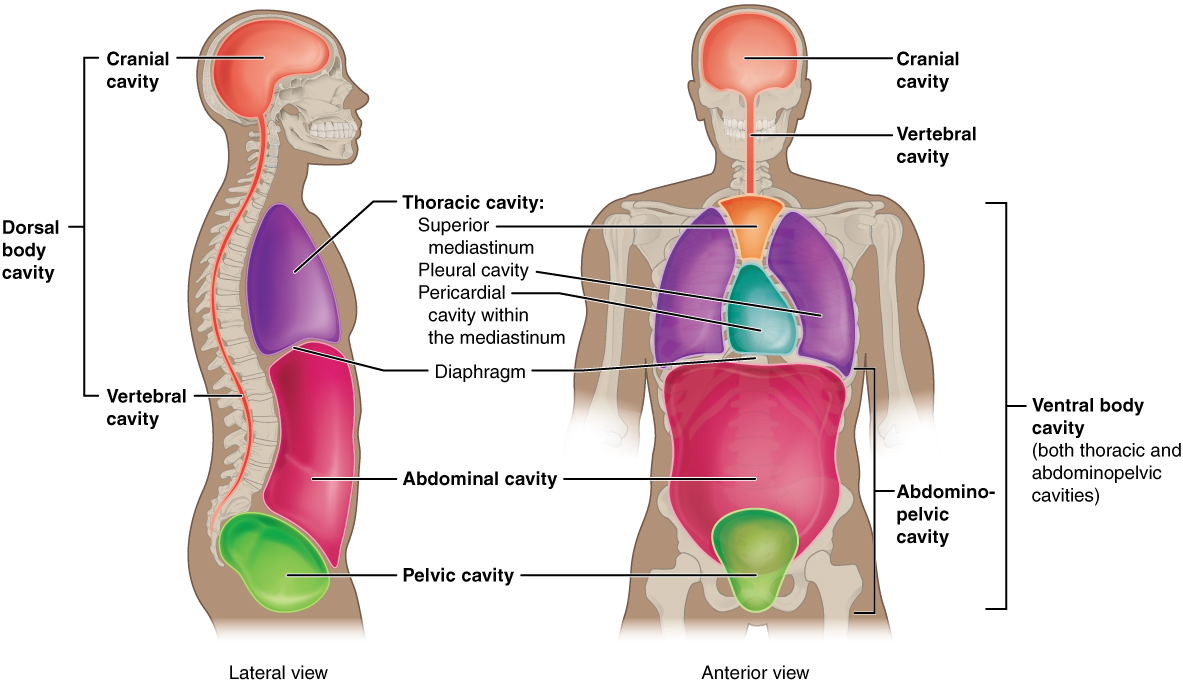
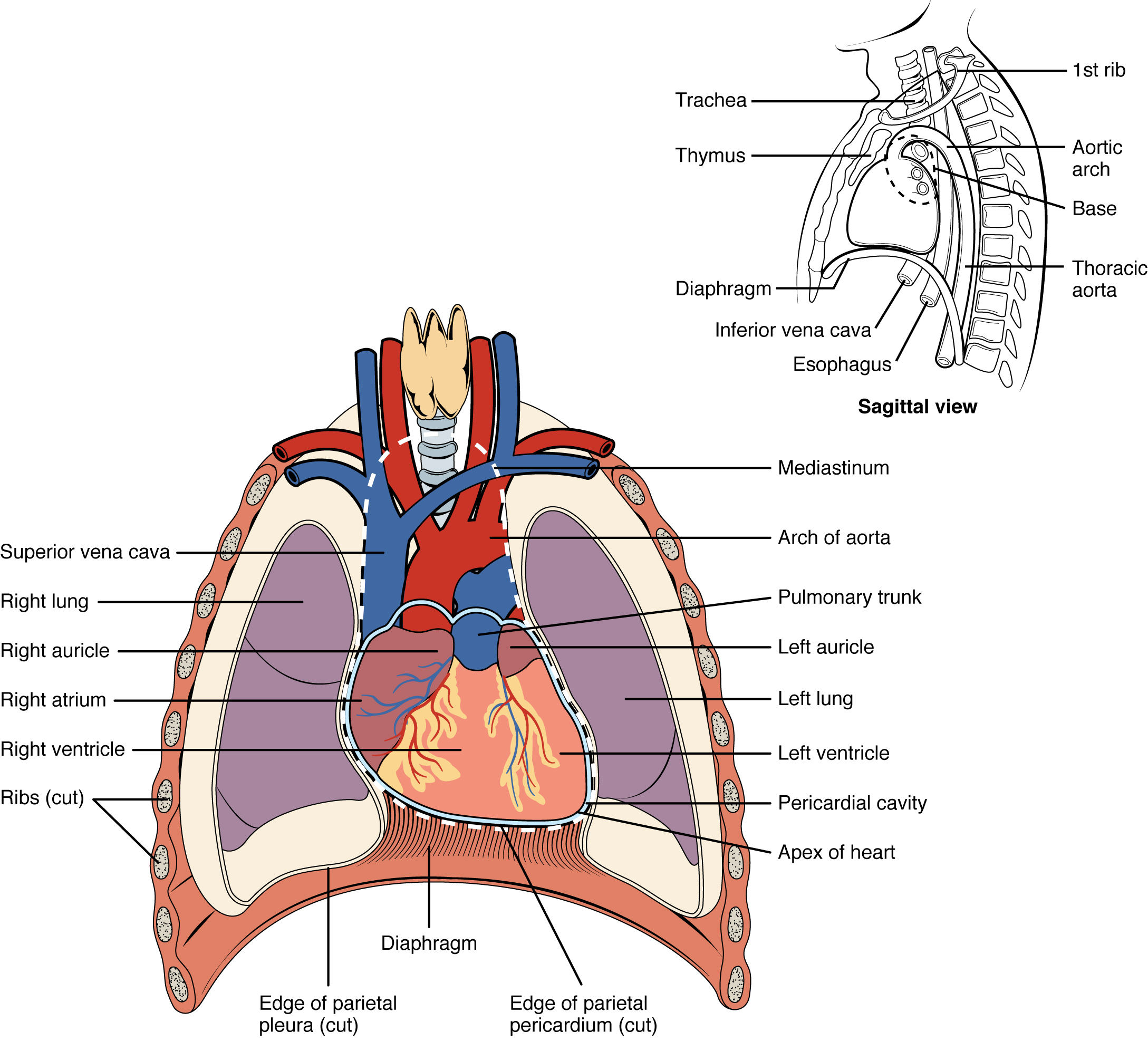
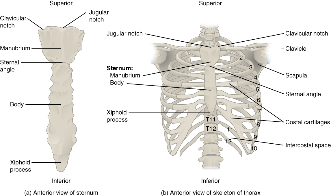
The human heart is located within the thoracic cavity, between the lungs in the space known as the mediastinum. Figure 12.1 shows the position of the heart within the thoracic cavity. Within the mediastinum, the heart is separated from the other mediastinal structures by a tough membrane known as the pericardium, or pericardial sac, and sits in its own space called the pericardial cavity. The great vessels, which carry blood to and from the heart, are attached to the superior surface of the heart, which is called the base. The base of the heart is located at the level of the third costal cartilage. The inferior tip of the heart, the apex, lies just to the left of the sternum between the junction of the fourth and fifth ribs.

Concept Check

  • On the diagram below (Figure 1), locate the mediastinum, the pericardial cavity, the base of the heart and the apex of the heart.
  • Locate the largest vein in the body superior vena cava.
Location of the heart in the thorax. Image description available.
Figure 12.1. Position of the Heart in the Thorax. The heart is located within the thoracic cavity, medially between the lungs in the mediastinum. It is about the size of a fist, is broad at the top, and tapers toward the base. From Betts, et al., 2013. Licensed under CC BY 4.0. [Image description.]

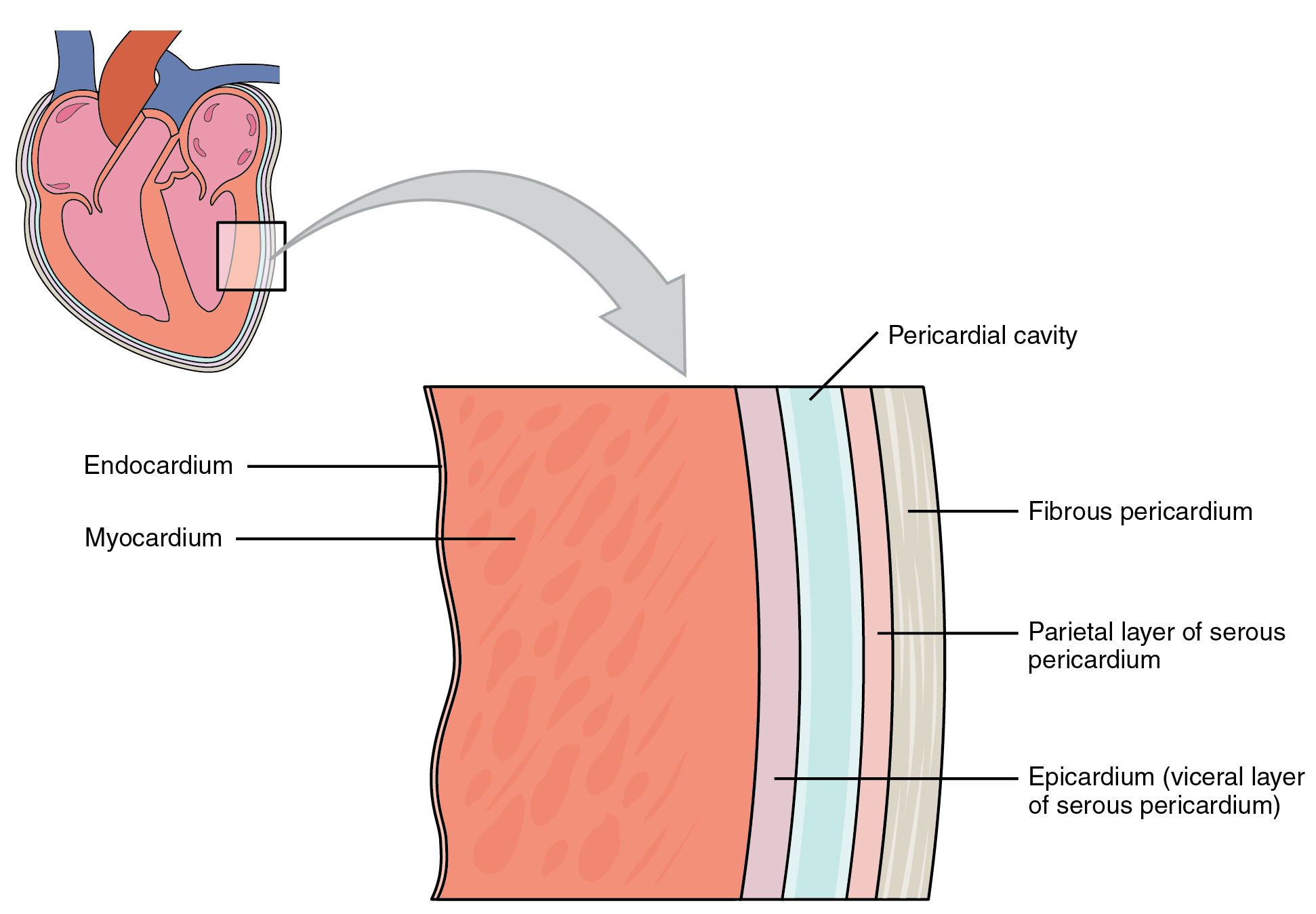
Membranes and Layers of the Heart Walls

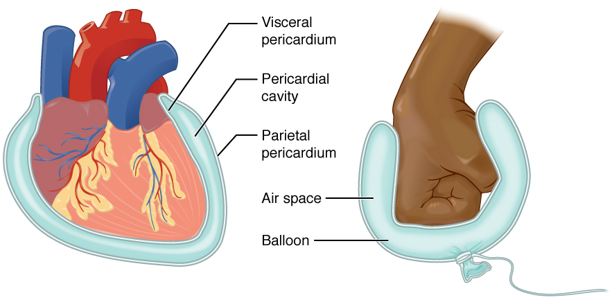
The heart and the roots of the great vessels are surrounded by a membrane known as the pericardium or pericardial sac. The pericardium consists of two distinct sub layers:

  • The sturdy outer fibrous pericardium is made of tough, dense connective tissue that protects the heart and holds it in position.
  • Separated by the pericardial cavity and containing pericardial fluid the inner serous pericardium consists of two layers:
    • the outer parietal pericardium, which is fused to the fibrous pericardium.
    • the inner visceral pericardium, or epicardium, which is fused to the heart and forms the outer layer of the heart wall.

The walls of the heart consist of three layers:

  • The outer epicardium, which is another name for the visceral pericardium mentioned above.
  • The thick, middle myocardium, which is made of muscle tissue and gives the heart its ability to contract.
  • The inner endocardium, which lines the heart chambers and is the main component of the heart valves.

Concept Check

  • Look at Figure 12.2 below, and name the layers of the heart wall and surrounding membranes, starting with the innermost layer.
  • As shown on the diagram, suggest why is the myocardium layer is thicker than the endocardium layer?


Magnified view of the heart's wall. Image description available.
Figure 12.2. Pericardial Membranes and Layers of the Heart Wall. The pericardial membrane that surrounds the heart consists of three layers and the pericardial cavity. The heart wall also consists of three layers. The pericardial membrane and the heart wall share the epicardium From Betts, et al., 2013. Licensed under CC BY 4.0. [Image description.]

Internal Structures of the Heart

The heart consists of four chambers:

  • The upper chambers are the right and left atria (singular: atrium).
  • The lower chambers are the right and left ventricles.

The interventricular septum is a muscular wall that separates the right and left ventricles. The interatrial septum separates the right and left atria.

The atrium and ventricle on each side of the heart are separated by an atrioventricular (AV) valve:

  • The right AV valve, or tricuspid valve, separates the right atrium and right ventricle.
  • The left AV valve, or bicuspid valve, separates the left ventricle and the left atrium. This valve is also called the mitral valve.

There are also two semilunar valves:

  • The pulmonary valve separates the right ventricle from the pulmonary trunk.
  • The aortic valve separates the left ventricle from the aorta (De Saix, et al., 2013).

Anatomy Labeling Activity

An interactive or media element has been excluded from this version of the text. You can view it online here:
https://ecampusontario.pressbooks.pub/medicalterminology/?p=259

Physiology of the Heart

In order for the heart to do its job of pumping blood to the lungs and to the body, nutrients and oxygen must be supplied to the cells of the heart. The heart also needs to coordinate its contractions so that all parts are working together to pump blood effectively. To understand how all of this works together to give the heart its ability to pump blood, we will examine three interdependent aspects of heart function.
  1. Circulation through the heart: Blood is pumped by the heart in order to provide oxygen and nutrients to every cell in the body.
  2. The heart as an organ (coronary blood supply): The heart is an organ, made of cells and tissues which require their own blood supply.
  3. The heart’s electrical conduction system: The heart is able to independently generate and transmit instructions to the myocardium, in order to make it contract and pump the blood.

1. Circulation Through the Heart: The Heart as a Pump

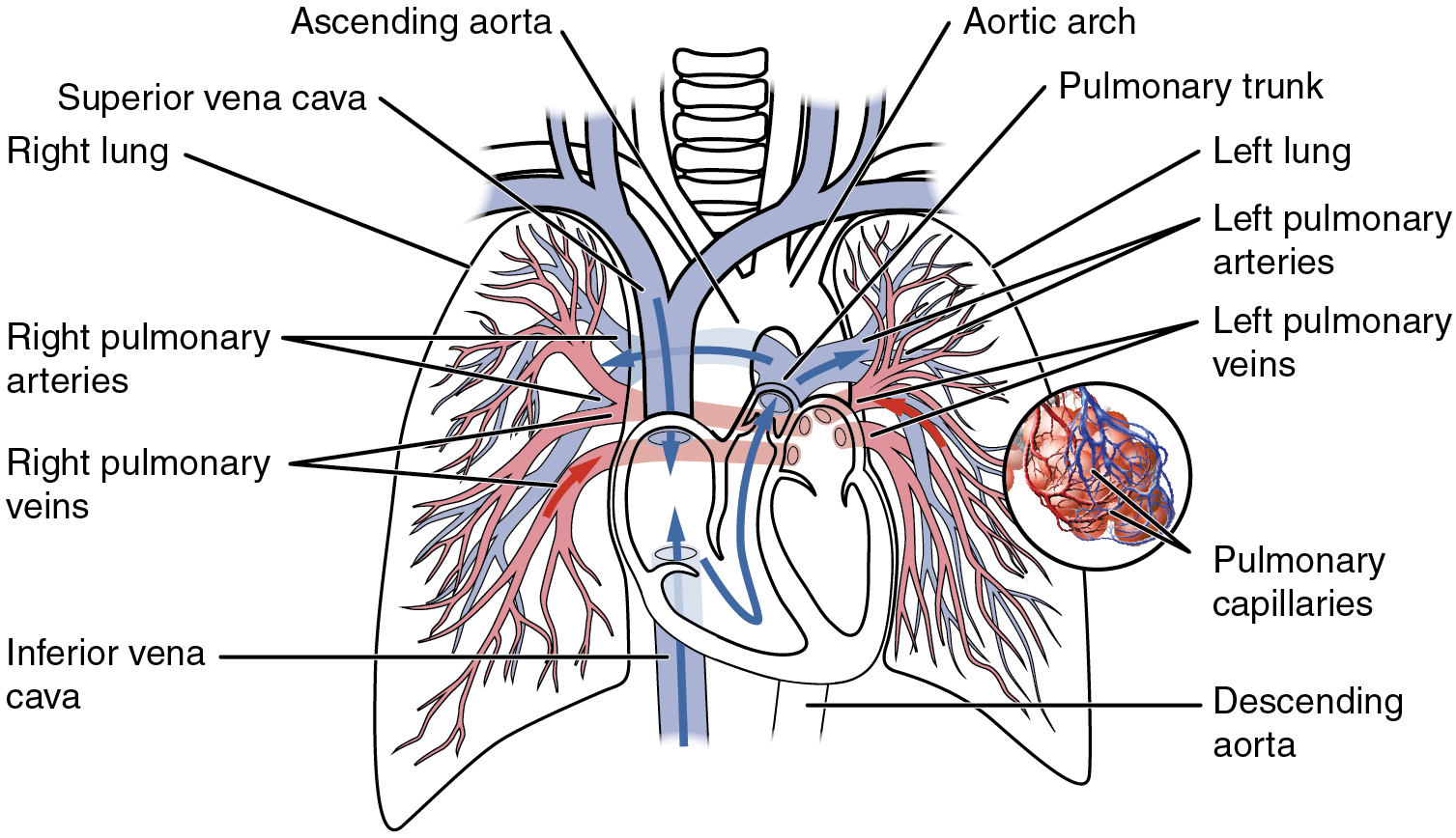
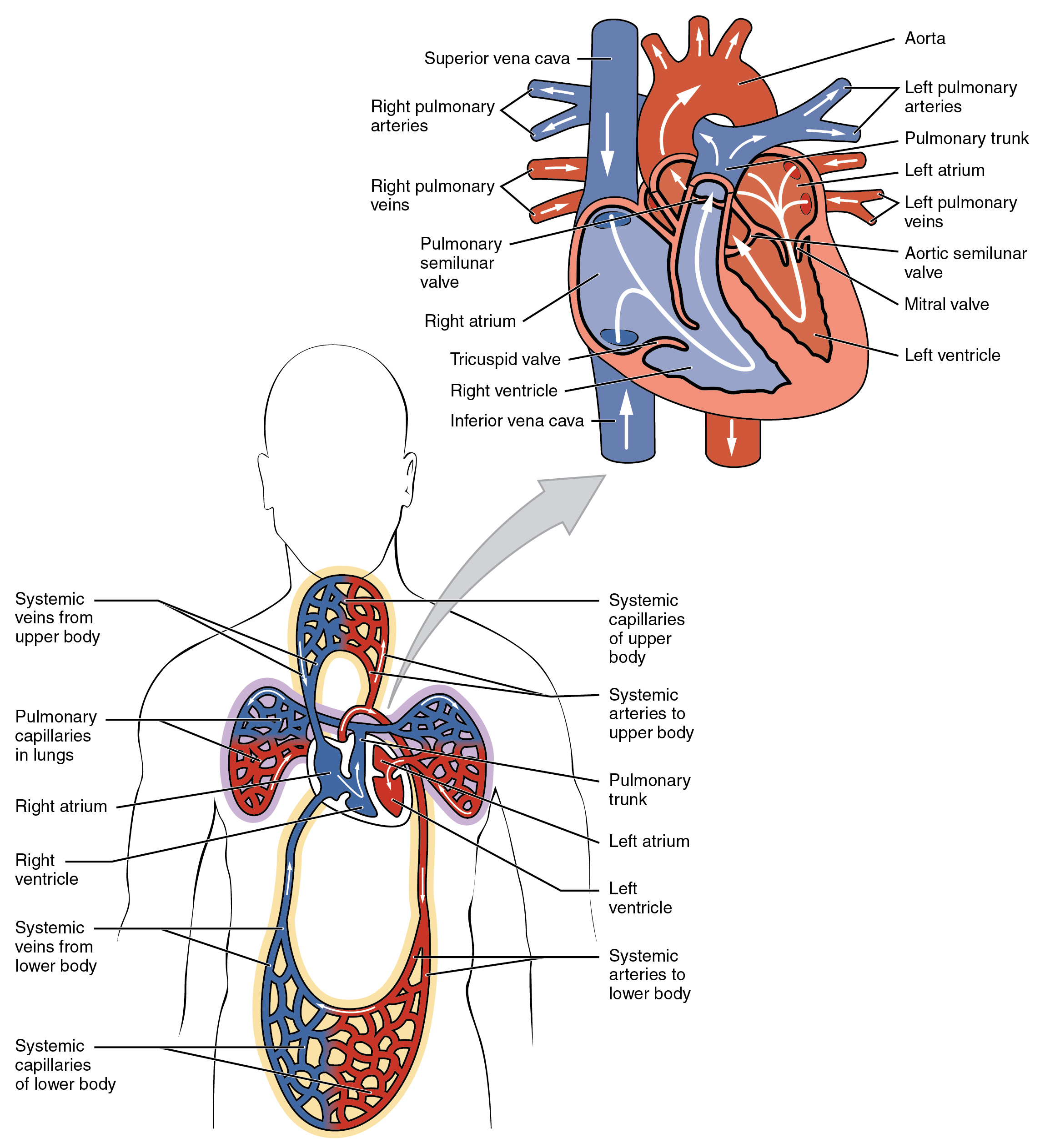
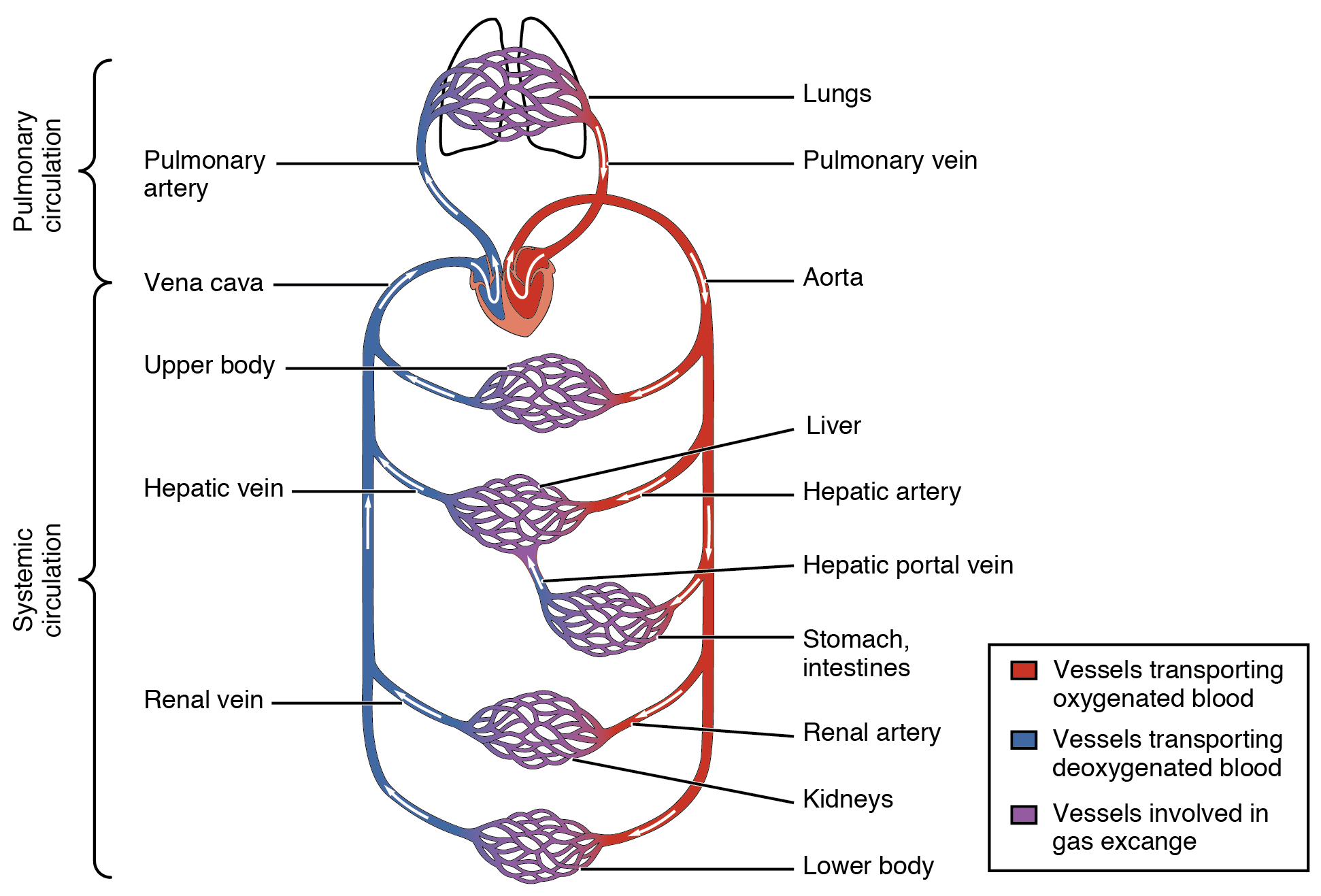
The heart pumps blood to two distinct but linked circulatory systems called the pulmonary and systemic circuits. The pulmonary circuit transports blood to and from the lungs, where it picks up oxygen and drops off carbon dioxide. The systemic circuit transports freshly oxygenated blood to virtually all of the tissues of the body and returns relatively deoxygenated blood and carbon dioxide to the heart to be sent back to the pulmonary circulation.

Did You Know?

The heart sounds heard through a stethoscope are the sounds of the four heart valves opening and closing at specific times during one cardiac cycle.
  1. Blood that is carrying carbon dioxide and waste products from the body tissues is returned to the right atrium via the superior vena cava and the inferior vena cava.
  2. From the right atrium, the deoxygenated blood moves through the tricuspid valve into the right ventricle.
  3. The right ventricle pumps deoxygenated blood through the pulmonary valve into the pulmonary trunk, which splits into the right and left pulmonary arteries, leading toward the lungs.  These arteries branch many times before reaching the pulmonary capillaries, where gas exchange occurs: carbon dioxide exits the blood and oxygen enters. The pulmonary arteries are the only arteries in the postnatal body that carry deoxygenated blood. Did you notice that they are often coloured blue on diagrams of the heart?
  4. Freshly oxygenated blood returns from the lungs to the left atrium via the pulmonary veins. These veins only postnatal veins in the body that carry highly oxygenated blood, and are often coloured red on heart images.
  5. From the left atrium, the blood moves through the mitral valve into the left ventricle.
  6. The left ventricle pumps blood through the aortic valve, into the aorta, delivering blood to all parts of the body.

Concept Check

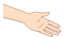
  • On Figure 12.3 below, use your finger to trace the pathway of blood flowing through the right side of the heart, naming each each of the following structures as you encounter them: Superior and inferior venae cavae, right atrium, tricuspid valve, right ventricle, pulmonary valve, right and left pulmonary arteries.
  • Suggest what would happen if the aorta experienced a blockage or constriction.
This diagram shows the network of blood vessels in the lungs.
Figure 12.3. Pulmonary Circuit Blood exiting from the right ventricle flows into the pulmonary trunk, which bifurcates into the two pulmonary arteries. These vessels branch to supply blood to the pulmonary capillaries, where gas exchange occurs within the lung alveoli. Blood returns via the pulmonary veins to the left atrium. From Betts, et al., 2013. Licensed under CC BY 4.0. [Image description.]

Pulmonary Circuit

Blood exiting from the right ventricle flows into the pulmonary trunk, which bifurcates into the two pulmonary arteries. These vessels branch to supply blood to the pulmonary capillaries, where gas exchange occurs within the lung alveoli. Blood returns via the pulmonary veins to the left atrium.

Concept Check

  • On Figure 12.4 below, use your finger to trace the pathway of blood flowing through the left side of the heart, naming each of the following structures as you encounter them: right and left pulmonary veins, left atrium, mitral valve, left ventricle, aortic valve, aorta.
Diagram of heart and circulatory system showing direction of blood flow. Image description available.
Figure 12.4. Dual System of the Human Blood Circulation. Blood flows from the right atrium to the right ventricle, where it is pumped into the pulmonary circuit. The blood in the pulmonary artery branches is low in oxygen but relatively high in carbon dioxide. Gas exchange occurs in the pulmonary capillaries (oxygen into the blood, carbon dioxide out), and blood high in oxygen and low in carbon dioxide is returned to the left atrium. From here, blood enters the left ventricle, which pumps it into the systemic circuit. Following exchange in the systemic capillaries (oxygen and nutrients out of the capillaries and carbon dioxide and wastes in), blood returns to the right atrium and the cycle is repeated. From Betts, et al., 2013. Licensed under CC BY 4.0. [Image description.]

Cardiac Cycle

The process of pumping and circulating blood is active, coordinated and rhythmic. Each heartbeat represents one cycle of the heart receiving blood and ejecting blood.

  • Diastole is the portion of the cycle in which the heart is relaxed and the atria and ventricles are filling with blood. The AV valves are open, so that blood can move from the atria to the ventricles.
  • Systole is the portion of the cycle in which the heart contracts, AV valves slam shut, and the ventricles eject blood to the lungs and to the body through the open semilunar valves. Once this phase ends, the semilunar valves close, in preparation for another filling phase.

2. The Heart as an Organ: The Coronary Blood Supply

Myocardial cells require their own blood supply to carry out their function of contracting and relaxing the heart in order to pump blood. Their own blood supply provides nutrients and oxygen and carry away carbon dioxide and waste. These functions are provided by the coronary arteries and coronary veins.

Concept Check

On the image below, locate the three main coronary arteries:

  • Anterior interventricular artery (more commonly known as the left anterior descending artery, or LAD)
  • Circumflex artery (Cx)
  • Right coronary artery (RCA)

Follow the path of each of these three arteries to try to determine which parts of the myocardium each artery (along with its many smaller branches) supplies with blood.

Anterior and posterior views of the heart and its blood vessels. Image description available.
Figure 12.5 Coronary Circulation. The anterior view of the heart shows the prominent coronary surface vessels. The posterior view of the heart shows the prominent coronary surface vessels. From Betts, et al., 2013. Licensed under CC BY 4.0. [Image description.]

3. The Heart’s Electrical Conduction System

In order for all parts of the heart to work together to beat regularly and effectively, the heart has its own electrical system, which initiates and conducts each heartbeat through the entire myocardium. Specialized groups of heart cells perform this function all on their own, without requiring messages from the central nervous system.

Watch this video:

Thumbnail for the embedded element "The Heart, Part 2 - Heart Throbs: Crash Course A&P #26"

A YouTube element has been excluded from this version of the text. You can view it online here: https://ecampusontario.pressbooks.pub/medicalterminology/?p=259

Media 12.2. The Heart, Part 2 – Heart Throbs: Crash Course A&P #26 [Online video]. Copyright 2015 by CrashCourse.

Anterior view of frontal section of the heart. Image description available.
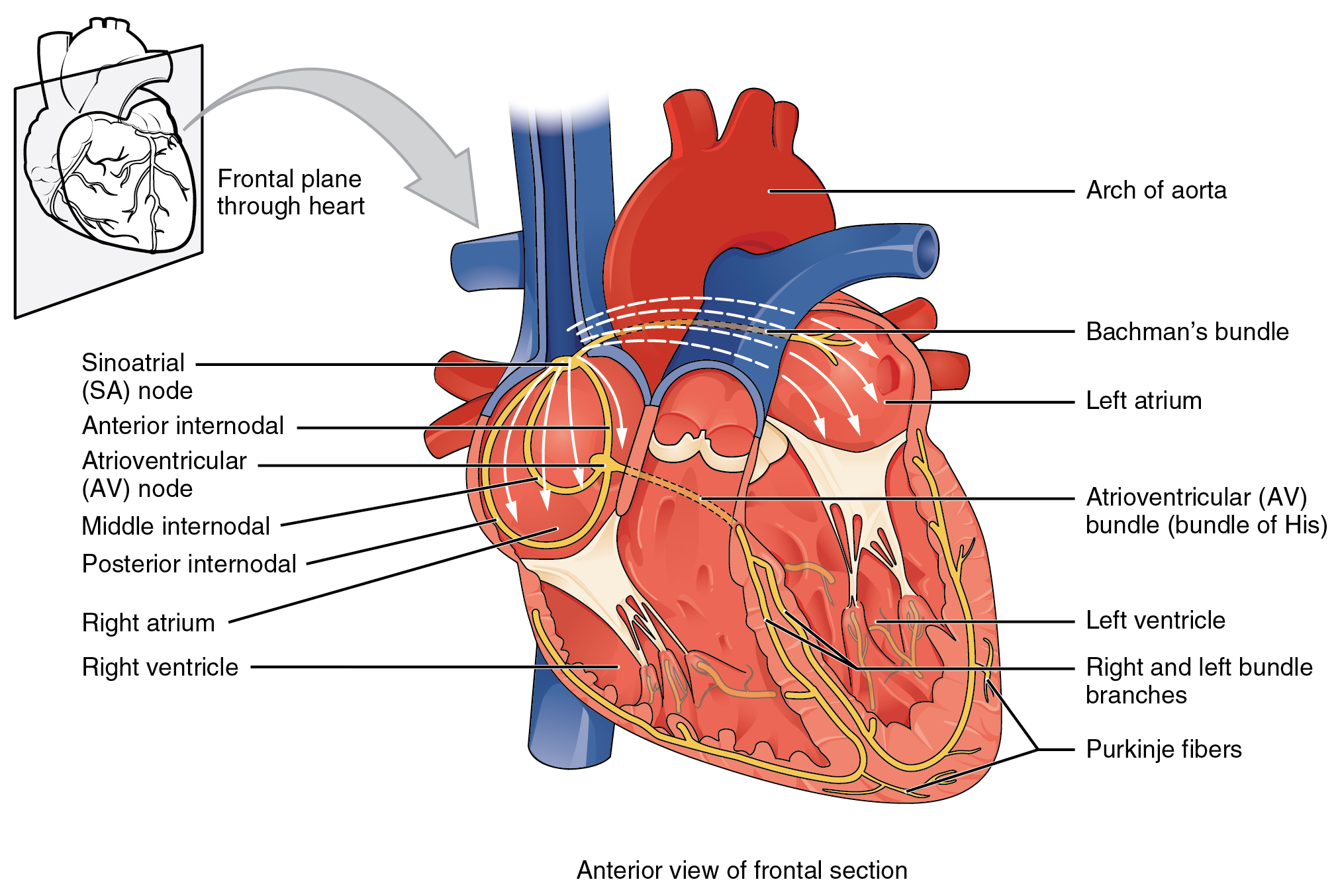
Figure 12.6. Conduction System of the Heart. Specialized conducting components of the heart include the sinoatrial node, the internodal pathways, the atrioventricular node, the atrioventricular bundle, the right and left bundle branches, and the Purkinje fibers. From Betts, et al., 2013. Licensed under CC BY 4.0. [Image description.]

Concept Check

  • On the image above, trace the electrical impulse generated by the heart’s pacemaker (the sinoatrial node, or SA node) through the rest of the conduction system, including the atrioventricular (AV) node, the atrioventricular bundle (bundle of His), the right and left bundle branches, and the Purkinje fibers.

We can detect and record the electrical activity of the heart’s conduction system using an electrocardiogram (ECG or EKG). Figure 12.7 shows the electrical impulse originating in the SA node (step 2) and travelling through the heart’s conduction system, allowing the heart to complete one cardiac cycle. Each waveform on the ECG tracing represents electricity moving through and affecting a different part of the heart. Did you notice that the AV valves close when the electrical impulse reaches the ventricles, just before systole occurs?

ECG tracing correlated to the cardiac cycle. Image description available.
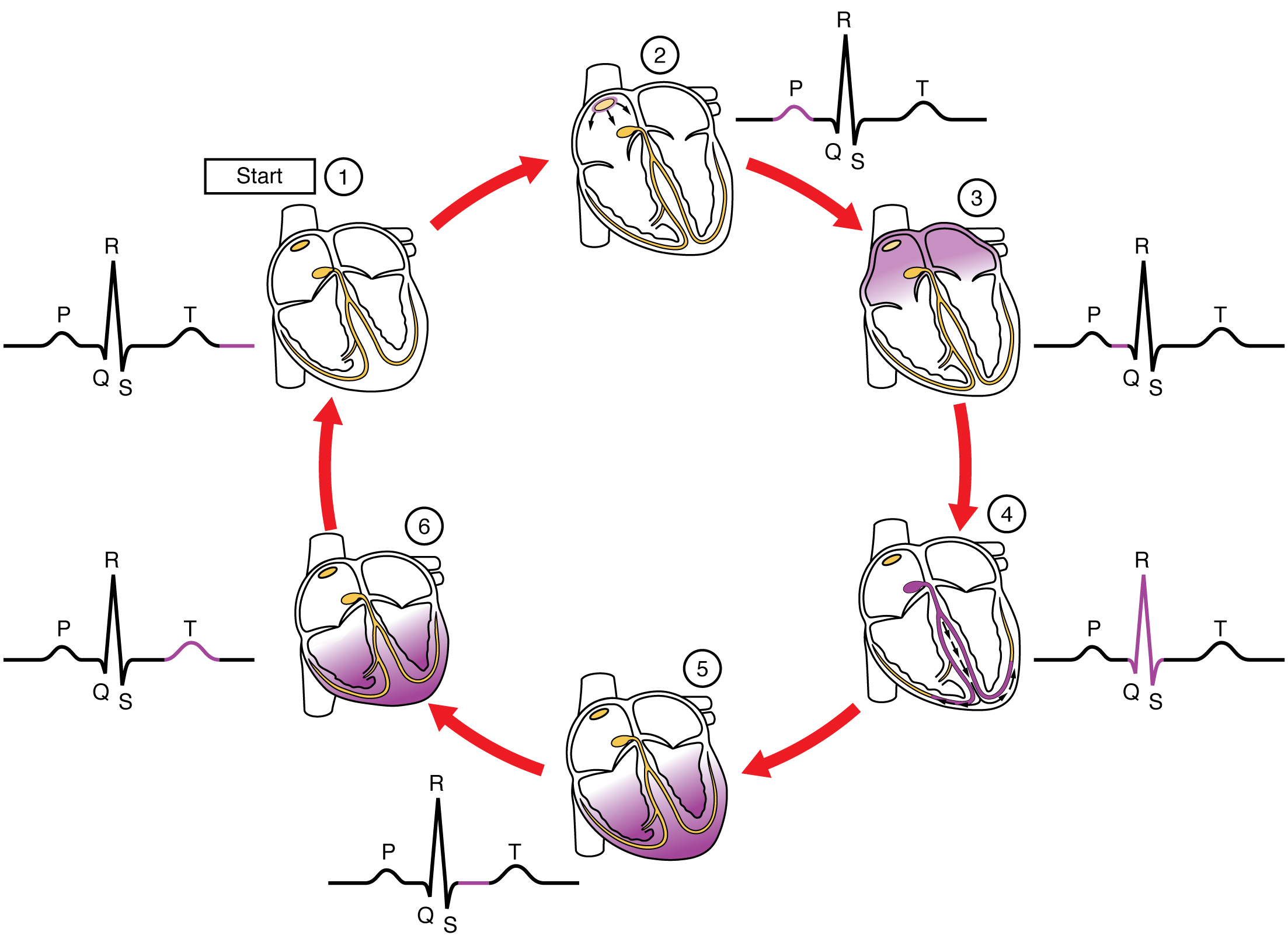
Figure 12.7. ECG Tracing Correlated to the Cardiac Cycle. This diagram correlates an ECG tracing with the electrical and mechanical events of a heart contraction. Each segment of an ECG tracing corresponds to one event in the cardiac cycle. From Betts, et al., 2013. Licensed under CC BY 4.0. [Image description.]

Heart Terms not Easily Broken into Word Parts An interactive or media element has been excluded from this version of the text. You can view it online here:
https://ecampusontario.pressbooks.pub/medicalterminology/?p=259

Heart Abbreviations

Many terms and phrases related to the cardiovascular system- heart are abbreviated.
Learn these common abbreviations by expanding the list below.

An interactive or media element has been excluded from this version of the text. You can view it online here:
https://ecampusontario.pressbooks.pub/medicalterminology/?p=259

Diseases and Disorders

Cardiomyopathy

The heart of a well-trained athlete can be considerably larger than the average person’s heart. This is because exercise results in an increase in muscle cells called hypertrophy . Hearts of athletes can pump blood more effectively at lower rates than those of non-athletes. However, when an enlarged heart is not the result of exercise, it may be due to hypertrophic cardiomyopathy. The cause of an abnormally enlarged heart muscle is unknown, but the condition is often undiagnosed and can cause sudden death in apparently otherwise healthy young people (Betts, et al., 2013).

Other types of cardiomyopathy include:

Cardiomyopathy may also be caused by myocardial infarctions, myocardial infections, pregnancy, alcohol or cocaine abuse, autoimmune and endocrine diseases. Because the myocardium is responsible for contracting and pumping blood, patients with cardiomyopathy experience impaired heart function which may lead to heart failure. (Centers for Disease Control and Prevention, 2019). To learn more about cardiomyopathy visit the CDC’s cardiomyopathy web page.

Heart Failure

Heart failure is defined as the inability of the heart to pump enough blood to meet the needs of the body. It is also called congestive heart failure (CHF). This condition causes swelling in the lower extremities and shortness of breath, due to a buildup of fluid in the lungs. It may be caused by cardiomyopathy and it may lead to hypertension and heart valve disorders (Heart & Stroke, n.d.). To learn more, visit the Heart & Stroke’s congestive heart failure web page.

Valvular Heart Disease

Concept check

Do you remember the names and locations of the 4 heart valves?

The four heart valves open and close at specific times during the cardiac cycle, in order to ensure that blood flows in only one direction through the heart. This requires that these valves open and close completely. Infections such as rheumatic disease or bacterial endocarditis can affect the heart valves and result in scar tissue formation which interferes with valve function. Other causes of heart valve disease include: congenitally malformed valves, autoimmune diseases, and other cardiovascular diseases such as aortic aneurysms and atherosclerosis (Centers for Disease Control and Prevention, 2019a).

Heart valve disease may be asymptomatic, or cause dyspnea, arrhythmias, fatigue and other symptoms. It is often detected when a heart murmur is heard through a stethoscope (Centers for Disease Control and Prevention, 2019a).

Visit the CDC’s page on valvular heart disease to learn more.
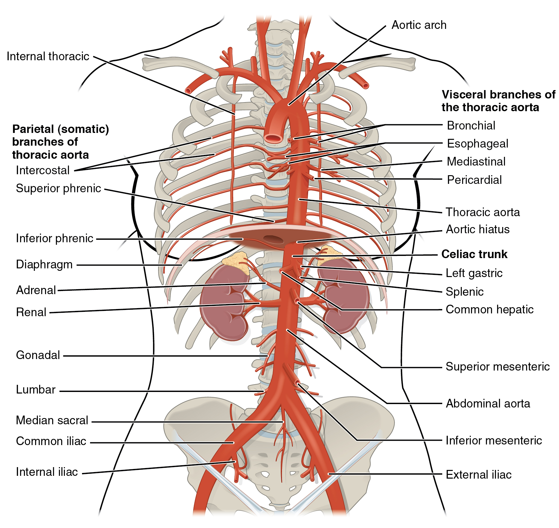
Aneurysms

An aneurysm is a defect in the wall of an artery in which the wall becomes thin and weak and starts to balloon out as blood pulses against the vessel wall. This can happen to any artery and even to the myocardial walls. Aneurysms sometimes occur in the portion of the aorta that is in the thorax (see Figure 12.8). If these aneurysms start to leak between layers of the vessel wall, the condition is known as aortic dissection. If an aortic or cardiac aneurysm bursts, there is sudden, massive internal bleeding (Centers for Disease Control and Prevention, 2019b).

Thoracic aorta with branches labelled. Image description available.
Figure 12.8. Arteries of the Thoracic and Abdominal Regions The thoracic aorta gives rise to the arteries of the visceral and parietal branches. From Betts, et al., 2013. Licensed under CC BY 4.0. [Image description.]

People who smoke, have hypertension, hypercholesterolemia, and/or atherosclerosis have an increased risk of developing aneurysms. Having a family history of aneurysms or certain genetic diseases may also increase a person’s risk of developing an aneurysm.

Aneurysms are often asymptomatic and may be detected incidentally during diagnostic tests that are being done for other reasons. They are sometimes repaired surgically and sometimes treated with medications such as antihypertensives (Centers for Disease Control and Prevention, 2019b; Tittley, n.d.). Visit the Canadian Society for Vascular Surgery’s page on thoracic aortic aneurysms to learn more.

Heart Defects

Fetal circulation is different from postnatal circulation. There are 2 extra openings in the fetal heart, the foramen ovale and the ductus arteriosus, which allow blood circulation that bypasses the immature fetal lungs. The fetal blood is reoxygenated by the mother’s lungs and transported between mother and fetus via the placenta. These two openings usually close around the time of birth (Betts, et al., 2013).

Septal defects are commonly first detected through auscultation. Unusual heart sounds may be detected because blood is not flowing and valves are not closing correctly. Medical imaging is ordered to confirm or rule out a diagnosis. In many cases, treatment may not be needed.

This diagram shows the structure of the heart with different congenital defects. The top left panel shows patent foramen ovale, the top right panel shows coarctation of the aorta, the bottom left panel shows patent ductus ateriosus and the bottom right shows tetralogy of fallot.
Figure 12.9. Congenital Heart Defects. (a) A patent foramen ovale defect is an abnormal opening in the interatrial septum, or more commonly, a failure of the foramen ovale to close. (b) Coarctation of the aorta is an abnormal narrowing of the aorta. (c) A patent ductus arteriosus is the failure of the ductus arteriosus to close. (d) Tetralogy of Fallot includes an abnormal opening in the interventricular septum. From Betts, et al., 2013. Licensed under CC BY 4.0. [Image description.]

Diseases of the Coronary Circulation

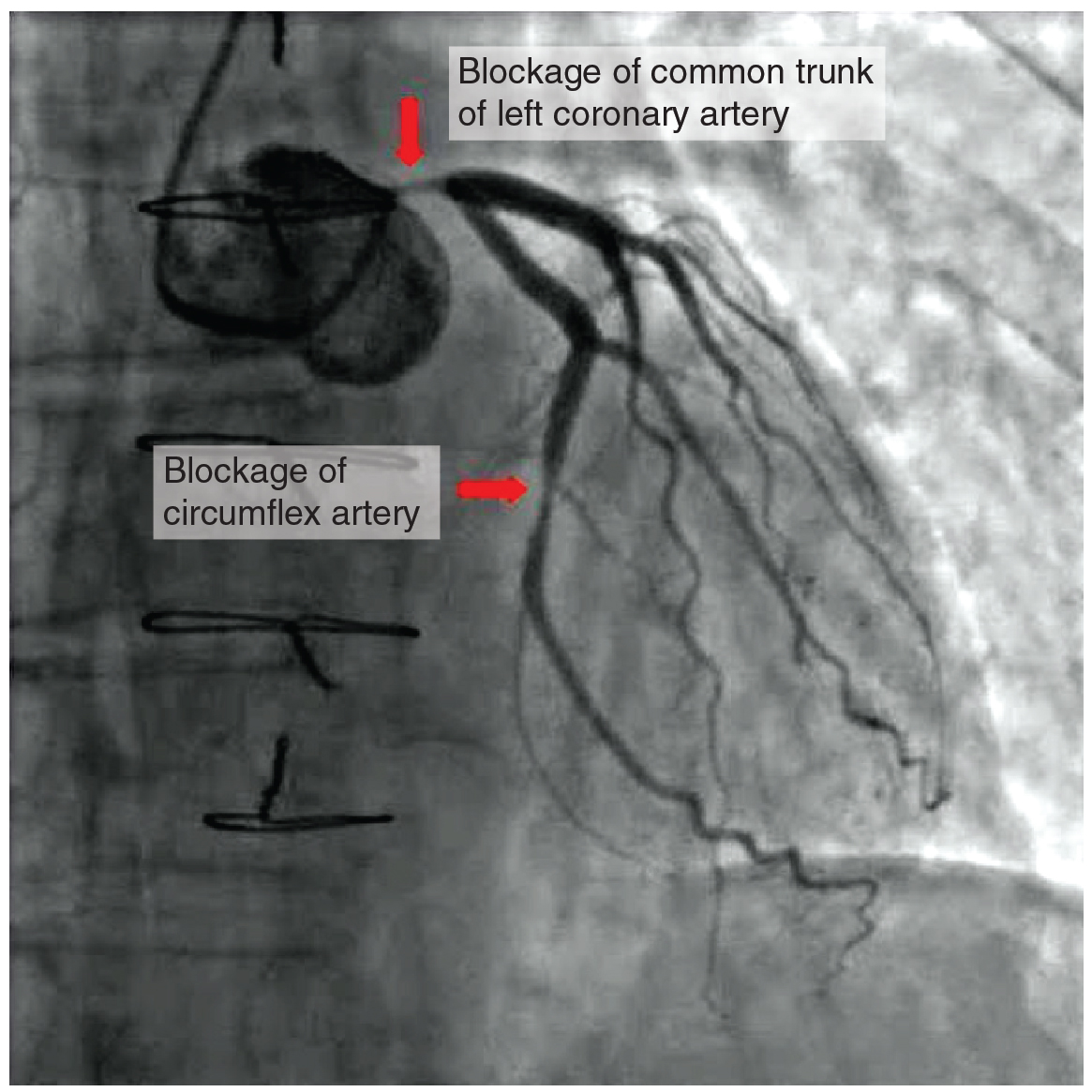
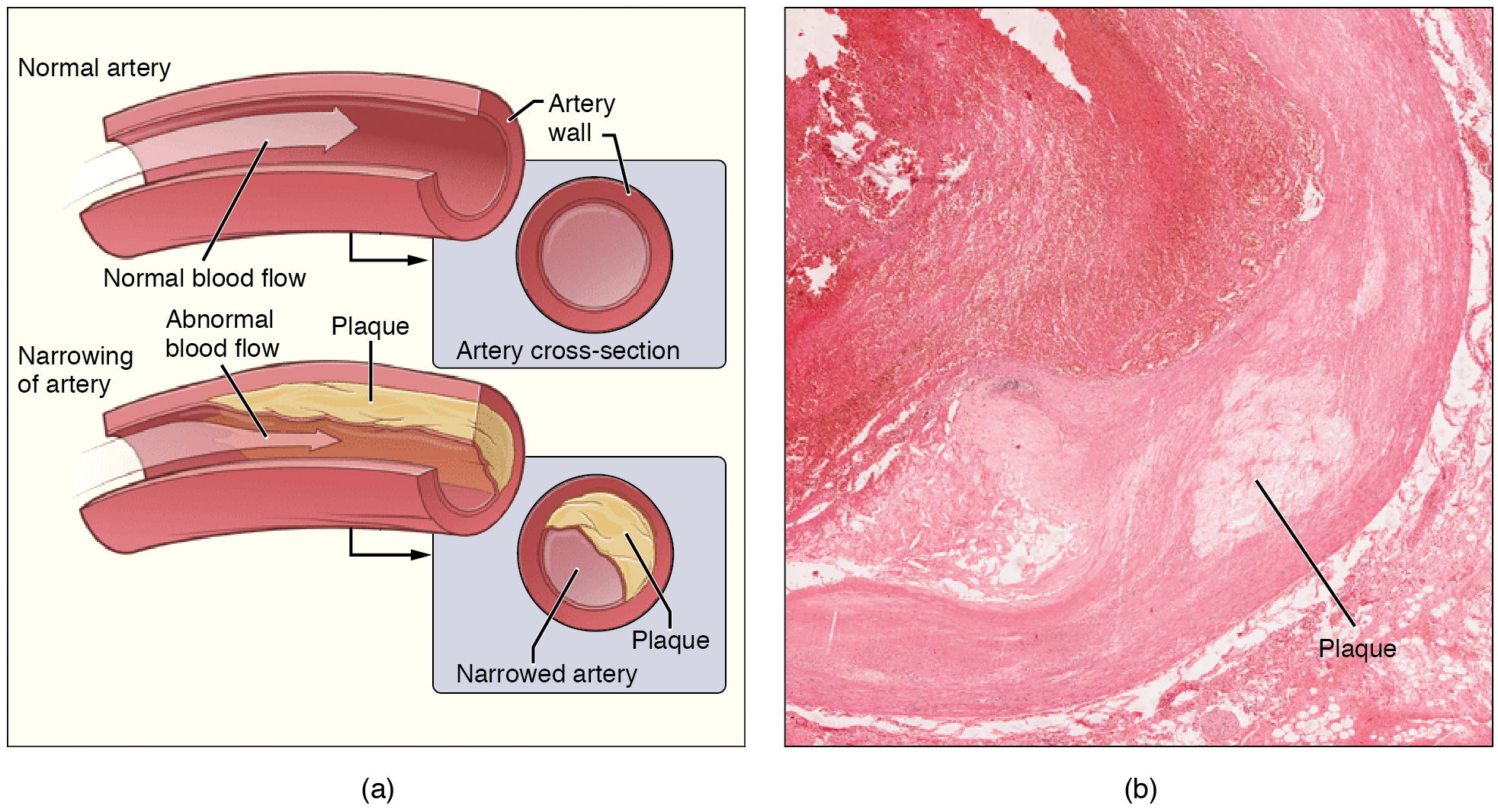
Coronary Artery Disease (CAD)

Coronary artery disease occurs when the buildup of plaque in the coronary arteries obstructs the flow of blood and decreases compliance of the vessels. This condition is called atherosclerosis. As the disease progresses and coronary blood vessels become more and more narrow, cells of the myocardium become ischemic, which causes symptoms of angina pectoris, in some patients. If untreated, coronary artery disease can lead to MI.

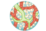
The image below shows the blockage of coronary arteries on an angiogram (Betts, et al., 2013).

This photo shows a blockage in the coronary artery and in the circumflex artery.
Figure 12.10. Angiogram of Atherosclerotic Coronary Arteries. In this coronary angiogram (X-ray), the dye makes visible two occluded coronary arteries. Such blockages can lead to decreased blood flow (ischemia) and insufficient oxygen (hypoxia) delivered to the cardiac tissues. If uncorrected, this can lead to cardiac muscle death (myocardial infarction). From Betts, et al., 2013. Licensed under CC BY 4.0.


CAD is progressive and chronic. Risk factors include smoking, family history, hypertension, obesity, diabetes, high alcohol consumption, lack of exercise, stress, and hyperlipidemia. Treatments may include medication, changes to diet and exercise, angioplasty with a balloon catheter, insertion of a stent, or coronary artery bypass graft (CABG) (Betts, et al., 2013).

Myocardial Infarction

Myocardial infarction (MI) is the medical term for a heart attack.

An MI normally results from a lack of blood flow to a region of the heart, resulting in death of the cardiac muscle cells. An MI often occurs when a coronary artery is blocked by the buildup of atherosclerotic plaque. It can also occur when a piece of an atherosclerotic plaque breaks off and travels through the coronary arterial system until it lodges in one of the smaller vessels. MIs may be triggered by excessive exercise, in which the partially occluded artery is no longer able to pump sufficient quantities of blood, or severe stress, which may induce spasm of the smooth muscle in the walls of the vessel (Betts, et al., 2013).

Did you know?

It is estimated that between 22 and 64 percent of myocardial infarctions are silent MIs.

In the case of acute MI (AMI), there is often sudden pain beneath the sternum (retrosternal pain) called angina pectoris, often radiating down the left arm in males but not in female patients. Other common symptoms include dyspnea, palpitations, nausea and vomiting, diaphoresis, anxiety, and syncope. Many of the symptoms are shared with other medical conditions, including anxiety attacks and simple indigestion, so differential diagnosis is critical (Betts, et al., 2013).

An MI can be confirmed by examining the patient’s ECG.

Other diagnostic tests include:

MIs may induce dangerous heart rhythms and even cardiac arrest. Important risk factors for MI include coronary artery disease, age, smoking, high blood levels of LDL, low levels of HDL, hypertension, diabetes mellitus, obesity, lack of physical exercise, chronic kidney disease, excessive alcohol consumption, and use of illegal drugs (Betts, et al., 2013).

Diseases of the (Electrical) Conduction System

Arrhythmia

Did you know?

Arrhythmia does not mean an absence of a heartbeat! That would be asystole, or flat line!
Arrhythmia is defined as the absence of a regular rhythm, meaning that the heart rate is either too fast, too slow or just irregular.

The heart’s natural pacemaker, the sinoatrial (SA) node initiates an electrical impulse 60-90 times per minute in a resting adult. This impulse travels through the heart’s conduction system in order to ensure a smooth, coordinated pumping action. This electrical activity can be detected and recorded through the skin using an electrocardiograph. Arrhyhmias may occur when the SA node fails to initiate an impulse, or when the conduction system fails to transmit that impulse through the heart.

In the event that the electrical activity of the heart is severely disrupted, cessation of electrical activity or fibrillation may occur. In fibrillation, the heart beats in a wild, uncontrolled manner, which prevents it from being able to pump effectively.

The most common treatment is defibrillation which uses special paddles to apply a charge to the heart from an external electrical source in an attempt to establish a normal sinus rhythm. A defibrillator effectively stops the heart so that the SA node can trigger a normal conduction cycle. External automated defibrillators (EADs) are being placed in areas frequented by large numbers of people, such as schools, restaurants, and airports. These devices contain simple and direct verbal instructions that can be followed by non-medical personnel in an attempt to save a life (Betts, et al., 2013).

Abnormal Heart Rates

Bradycardia is the condition in which resting adult heart rate drops below 60 bpm. a client exhibiting symptoms such as weakness, fatigue, dizziness, syncope, chest discomfort, palpitations or respiratory distress may indicate that the heart is not providing sufficient oxygenated blood to the tissues. If the patient is not exhibiting symptoms then bradycardia is not considered clinically significant. The term relative bradycardia may be used with a patient who has a HR in the normal range but is still suffering from these symptoms.  Most patients remain asymptomatic as long as the HR remains above 50 bpm.

Tachycardia is the condition in which the resting rate is above 100 bpm. Tachycardia is not normal in a resting patient and may be detected in pregnant women or individuals experiencing extreme stress. Some individuals may remain asymptomatic, but when present, symptoms may include dizziness, shortness of breath, rapid pulse, heart palpitations, chest pain, or syncope. Treatment depends upon the underlying cause but may include medications, implantable cardioverter defibrillators, ablation, or surgery (Betts, et al., 2013).

Heart Block

heart block refers to an interruption in the normal conduction pathway. Heart blocks are generally named after the part of the conduction system that is causing the problem. For example, bundle branch blocks occur within either the left or right atrioventricular bundle branches.

AV blocks are often described by degrees. A first-degree or partial block indicates a delay in conduction between the SA and AV nodes. A second-degree or incomplete block occurs when some impulses from the SA node reach the AV node and continue, while others do not. In the third-degree or complete block, there is no correlation between atrial activity and ventricular activity. This means that none of the impulses generated by the SA node get transmitted to the rest of the heart and the AV node must take over as the primary pacemaker, initiating contractions at 40–60 beats per minute, which is adequate to maintain consciousness.

In order to speed up the heart rate and restore full sinus rhythm, a cardiologist can implant an artificial pacemaker, which delivers electrical impulses to the heart muscle to ensure that the heart continues to contract and pump blood effectively. These artificial pacemakers are programmable by the cardiologists and can either provide stimulation temporarily upon demand or on a continuous basis. Some devices also contain built-in defibrillators (Betts, et al., 2013).

In this image the QT cycle for different heart conditions are shown. From top to bottom, the arrhythmias shown are second-degree partial block, atrial fibrillation, ventricular tachycardia, ventricular fibrillation and third degree block.
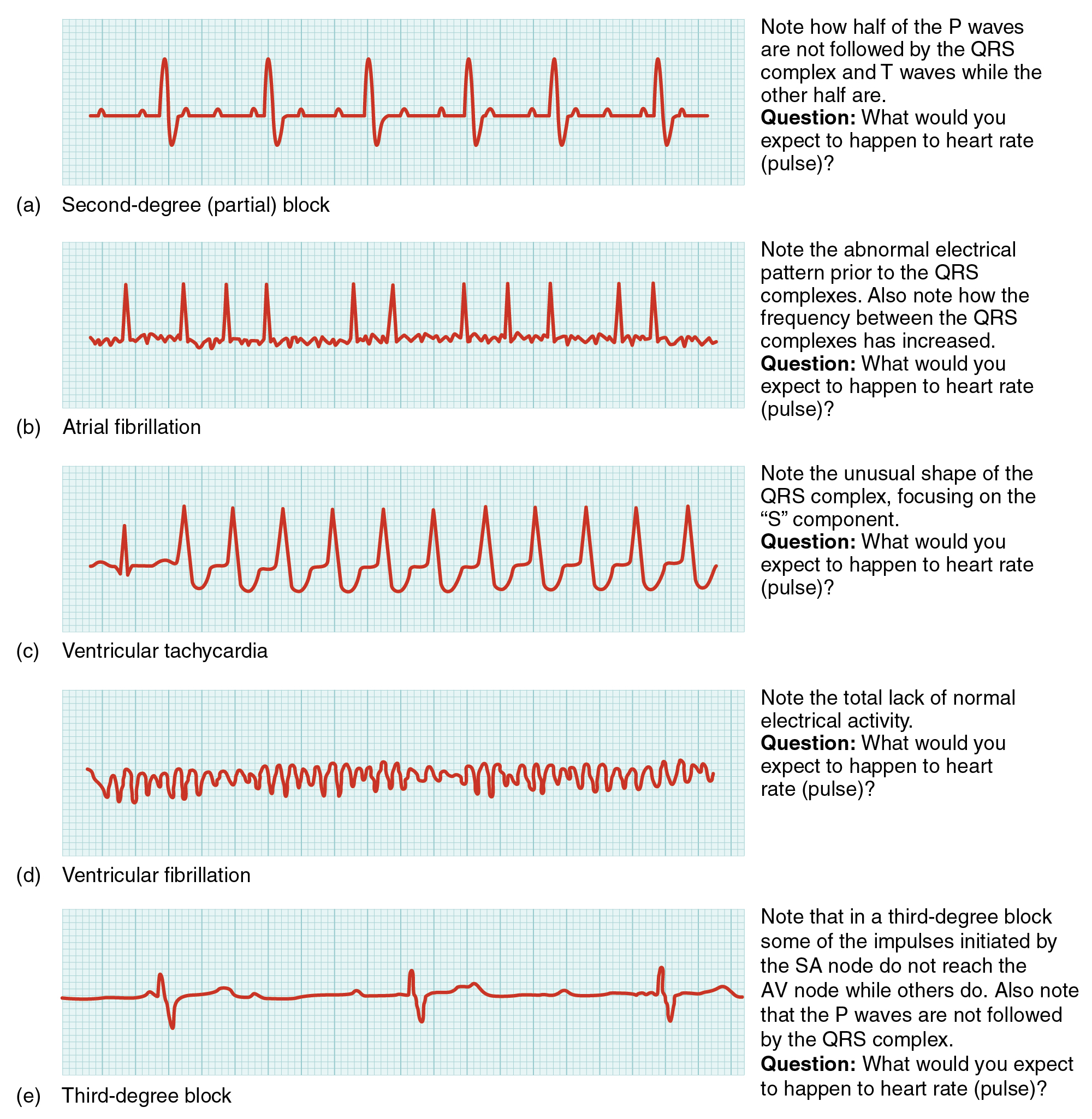
Figure 12.11. Common ECG Abnormalities. (a) In a second-degree or partial block, one-half of the P waves are not followed by the QRS complex and T waves while the other half are. (b) In atrial fibrillation, the electrical pattern is abnormal prior to the QRS complex, and the frequency between the QRS complexes has increased. (c) In ventricular tachycardia, the shape of the QRS complex is abnormal. (d) In ventricular fibrillation, there is no normal electrical activity. (e) In a third-degree block, there is no correlation between atrial activity (the P wave) and ventricular activity (the QRS complex). From Betts, et al., 2013. Licensed under CC BY 4.0. [Image description.]

Medical Terms in Context

An interactive or media element has been excluded from this version of the text. You can view it online here:
https://ecampusontario.pressbooks.pub/medicalterminology/?p=259

Medical Specialties and Procedures Related to the Heart

Cardiologists and Cardiovascular Surgeons

Cardiologists are medical doctors that specialize in diagnosing and treating heart disease non-invasively. Cardiovascular/thoracic surgeons provide surgical treatments for the heart and other thoracic organs (Canadian Medical Association, 2018). To learn more about these specialists please visit the CMA’s Canadian Specialy Profiles web page.

Cardiology Technologists

Cardiology Technologists complete a college training program and perform diagnostic tests such as electrocardiography, stress testing, Holter monitor testing, ambulatory blood pressure testing, as well as pacemaker monitoring and programming (Canadian Society of Cardiology Technologists, n.d.). Please visit the Canadian Society of Cardiology Technologists web page for more information.

Cardiovascular Perfusionists

Cardiovascular perfusionists complete a college training program and are responsible for operation of the heart-lung bypass machine during open heart surgery. They also monitor the patient’s vitals, adminstering IV fluids, and other drugs (Michener Institute of Education, n.d.). Please visit the Michener Institute’s Cardiovasular Perfusion program page for more information.

Cardiovascular System – Heart Vocabulary

5.25 liters of blood

The volume of blood ejected by the ventricle in one minute is called the cardiac output.

70 mL blood per contraction

The amount of blood ejected from the ventricle in one contraction is called the stroke volume.

Ablation

Using extreme heat or extreme cold to destroy cells in part of the heart which were causing abnormal rhythms.

Angina Pectoris

Chest pain.

Angiogram

An x-ray of the coronary blood vessels using a special catheter and an injection of dye.

Antihypertensives

Class of medications used to treat high blood pressure.

Arrhythmias

Absence of a regular heart rhythm.

Asymptomatic

Pertaining to without symptoms.

Atherosclerosis

A hardening of the arteries that involves the accumulation of plaque.

Auscultation

Listening to the heart using a stethoscope.

AV

Atrioventricular: the area of the heart where the atria and ventricles meet.

AV Valves

Atrioventricular valves: mitral (bicuspid) valve allows blood to flow from left atrium to left ventricle, tricuspid valve allows blood to flow from right atrium to right ventricle.

Bradycardia

Pertaining to a slow heart (rate).

Cardiac Troponin

The regulatory protein for muscle contraction.

Clubbing of the fingers and toes

Broadening of the nails and exaggerated curvature of the nails.

Compliance

The ability of the blood vessels to dilate and constrict as needed.

Congenital

Present at birth.

Creatine Kinase MB

An enzyme that catalyzes the conversion of creatine to phosphocreatine, consuming ATP.

CT

Computerized tomography: a special 3-dimensional x-ray, also called CAT=Computerized Axial Tomography.

Cyanosis

Abnormal condition of blue (bluish colour, lips and nail beds). Typically caused by low oxygenation.

Diabetes Mellitus

An endocrine system disorder in which the pancreas does not produce insulin or the cells of the body do not respond to insulin. This results in high levels of glucose in the blood.

Diaphoresis

Sweating.

Ductus Arteriosus

Connection between pulmonary trunk and aorta in the fetal heart.

Dyspnea

Difficult breathing.

ECG

ECG/EKG both these abbreviations mean electrocardiogram or a recording of the electrical impulses in the heart.

Echocardiography

Process of using sound to record the heart.

Electrocardiograph

Instrument used to record electrical activity within the heart.

Foramen Ovale

Opening between right and left atria, which is normal in the fetal heart.

Great Vessels

The great vessels include the superior vena cava, inferior vena cava, aorta and pulmonary trunk.

HDL

High-density lipoprotein, often referred to as ‘good’ cholesterol.

Heart Murmur

An abnormal heart sound.

Heart Rate

The number of times the heart contracts in one minute.

Hypercholesterolemia

Higher than normal levels of cholesterol in the blood.

Hyperlipidemia

Excessive fat in the blood.

Hypertension

High blood pressure.

Implantable Cardioverter Defibrillators (ICD)

An electronic implant that provides an automatic shock to convert a dangerous heart rhythm to a normal heart rhythm.

Inferior Vena Cava

One of the two largest veins in the body. It carries deoxygenated blood from the torso and legs back to the heart.

Interatrial Septum

The wall separating the right and left atria.

Interventricular Septum

The wall of myocardium that separates the right and left ventricles.

Ischemic

Ischemia is a condition in which cells receive insufficient amounts of blood and oxygen.

LDL

Low-density lipoprotein, often referred to as ‘bad’ cholesterol.

Mitral Valve

Also known as the bicuspid valve.

MRI

Magnetic Resonance Imaging: Highly detailed images produced using a strong magnet and radio waves.

Pacemaker

An electronic implant that initiates a heart beat.

Palpitations

A feeling in the chest that may be caused by an irregular heart rhythm.

Pericardial fluid

Pericardial fluid is a serous fluid which allow the 2 layers of serous pericardium to slide smoothly against each other as the heart beats.

Plaque

A fatty material including cholesterol, connective tissue, white blood cells, and some smooth muscle cells.

Polycythemia

A disorder in which too many red blood cells are produced.

Pulmonary Trunk

Very large artery referred to as a trunk, a term indicating that the vessel gives rise to several smaller arteries.

Roots of the Great Vessels

The part of each great vessel (aorta, pulmonary trunk, inferior vena cava, superior vena cava) that connects to the base of the heart.
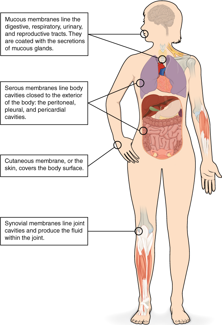
Serous

You may recall that serous membranes throughout the body are folded back on themselves, which results in a double-layered membrane separated by serous fluid. The serous membrane surrounding the lungs is called pleura. The serous membrane surrounding the abdominopelvic organs is called peritoneum.

Silent Mis

A myocardial infarction without symptoms. The patient may not know that they are having an MI.

Sinus Rhythm

This is the rhythm set by the heart’s pacemaker, the sinoatrial node and is usually approximately 60-90 beats per minute in a resting adult.

Superior Vena Cava

One of the two largest veins in the body. It carries deoxygenated blood from the head and upper extremities back to the heart.

Syncope

Fainting.

Tachycardia

Condition of a fast heart (rate).

Test Yourself

An interactive or media element has been excluded from this version of the text. You can view it online here:
https://ecampusontario.pressbooks.pub/medicalterminology/?p=259


References

Canadian Medical Association. (2018). Canadian Specialty Profiles. https://www.cma.ca/canadian-specialty-profiles

Canadian Society of Cardiology Technologists. (n.d.). Becoming a registered cardiology technologist. https://www.csct.ca/education/about-being-rct

Centers for Disease Control and Prevention. (2019). Cardiomyopathy. CDC. https://www.cdc.gov/heartdisease/cardiomyopathy.htm

Centers for Disease Control and Prevention. (2019a). Valvular heart disease. CDC. https://www.cdc.gov/heartdisease/valvular_disease.htm

Centers for Disease Control and Prevention. (2019b). Aortic aneurysm. CDC. https://www.cdc.gov/heartdisease/aortic_aneurysm.htm

[CrashCourse]. (2015, July 6). The heart, part 1 – under pressure: Crash course A&P #25 [Video]. YouTube. https://youtu.be/X9ZZ6tcxArI

[CrashCourse]. (2015, July 13). The heart, part 2 – heart throbs: Crash course A&P #26 [Video]. YouTube. https://youtu.be/FLBMwcvOaEo

Heart & Stroke. (n.d.). Heart failure. Heart and Stroke Foundation. https://www.heartandstroke.ca/heart/conditions/heart-failure

Mitchener Institute for Education. (n.d.). Cardiovascular perfusion. Michener Institute of Education at UHN. https://michener.ca/program/cardiovascular-perfusion/

Tittley, J. G. (n.d.). Thoracic aortic aneurysms (TAA). Retrieved from Canadian Society for Vascular Surgery: https://canadianvascular.ca/Thoracic-Aortic-Aneurysms-(TAA)

Image Descriptions

Figure 12.1 image description: This diagram shows the location of the heart in the thorax (sagittal and anterior views). The sagittal view labels read (from top, clockwise): first rib, aortic arch, thoracic arch, esophagus, inferior vena cava, diaphragm, thymus, trachea. The anterior view lables read (from top, clockwise): mediastinum, arch of aorta, pulmonary trunk, left auricle, left lung, left ventricle, pericardial cavity, apex of heart, edge of parietal pericardium, diaphgragm, edge of parietal pleura, ribs, right ventricle, right atrium, right auricle, right lung, superior vena cava. [Return to Figure 12.1].

Figure 12.2 image description: This image shows a magnified view of the structure of the heart wall. Labels read (from top, clockwise): pericardial cavity, fibrous pericardium, parietal layer of serous pericardium, epicardium (visceral layer of serous pericardium), myocardium, endocardium. [Return to Figure 12.2].

Figure 12.3 image description: This diagram shows the network of blood vessels in the lungs. Labels read (from top, clockwise (left-side of the body): aortic arch, pulmonary trunk, left lung, left pulmonary arteries, left pulmonary vein, pulmonary capillaries, descending aorta, (right side of body) inferior vena cava, right pulmonary veins, right pulmonary arteries, right lung, superior vena cava, ascending aorta. [Return to Figure 12.3].

Figure 12.4 image description: The top panel shows the human heart with the arteries and veins labeled (from top, clockwise): aorta, left pulmonary arteries, pulmonary trunk, left atrium, left pulmonary veins, aortic semilunar valve, mitral valve, left ventricle, inferior vena cava, right ventricle, tricuspid valve, right atrium, pulmonary semilunar valve, right pulmonary veins, right pulmonary arteries, superior vena cava. The bottom panel shows a rough map of the the human circulatory system. Labels read (from top, clockwise): systemic capillaries of upper body, systemic arteries to upper body, pulmonary trunk, left atrium, left ventricle, systemic arteries to lower body, systemic capillaries of lower body, systemic veins from lower body, right ventricle, right atrium, pulmonary capillaries in lungs, systemic veins from upper body. [Return to Figure 12.4].

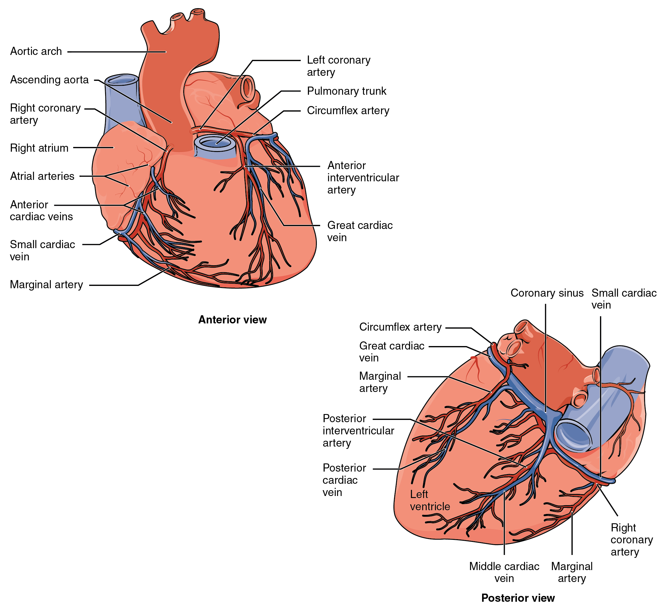
Figure 12.5 image description: The top panel of this figure shows the anterior view of the heart while the bottom panel shows the posterior view of the heart. The different blood vessels are labeled. Anterior view labels (from top of diagram, clockwise): left coronary artery, pulmonary trunk, circumflex artery, anterior interventricular artery, great cardiac vein, small cardiac vein, anterior cardiac veins, atrial arteries, right atrium, right coronary artery, ascending aorta, aortic arch. Posterior view labels (from top of diagram, clockwise): coronary sinus, small cardiac vein, right coronary artery, marginal artery, middle cardiac vein, posterior cardiac vein, posterior interventricular artery, marginal artery, great cardiac vein, circumflex artery. [Return to Figure 12.5].

Figure 12.6 image description: This image shows the anterior view of the frontal section of the heart with the major parts labeled. Labels read (from top of diagram, clockwise) arch of aorta, Bachman’s bundle, atrioventricular bundle (bundle of His), left ventricle, right and left bundle branches, Purkinje fibers, right ventricle, right atrium, posterior intermodal, middle intermodal, atrioventricular node, anterior intermodal, Sinoatrial node. [Return to Figure 12.6].

Figure 12.7 image description: This diagram shows the six different stages of heart contraction and relaxation along with the stages in the QT cycle. [Return to Figure 12.7].

Figure 12.8 image description: This diagram shows the arteries in the thoracic and abdominal cavity. Visceral branches of the thoracic aorta labels (from top): bronchial, esophageal, mediastinal, pericardial, thoracic aorta, aortic hiatus, celiac trunk, left gastric, splenic, common hepatic, superior mesenteric, abdominal aorta, inferior mesenteric, external iliac. Parietal (somatic) branches of thoracic aorta labels (from top): intercostal, superior phrenic, inferior phrenic, diaphragm, adrenal, renal, gonadal, lumbar, medial sacral, common iliac, internal iliac. [Return to Figure 12.8].

Figure 12.9 image description: This diagram shows the structure of the heart with different congenital defects. The top left panel shows patent foramen ovale (label reads foramen ovale fails to close), the top right panel shows coarctation of the aorta (label reads narrow segment of aorta), the bottom left panel shows patent ductus ateriosus (label reads Ductus arteriosus remains open) and the bottom right shows tetralogy of fallot (labels read aorta emerges from both ventricles, interventricular septal defect, enlarged right ventricle, stenosed pulmonary semilunar valve). [Return to Figure 12.9].

Figure 12.11 image description: In this image the QT cycle for different heart conditions are shown. From top to bottom, the arrhythmias shown are second-degree partial block (text reads: Note how half of the P waves are not followed by the QRS complex and T waves while the other half are. Question: what would you expect to happen to heart rate?), atrial fibrillation (text reads: Note the abnormal electric pattern prior to the QRS complexes. Also note how the frequency between the QRS complexes has increased. Question: What t would you expect to happen to heart rate?), ventricular tachycardia (text reads: Note the unusual shape of the QRS complex, focusing on the S component. Question: What would you expect to happen to heart rate?), ventricular fibrillation (text reads: Note the total lack of normal electrical activity. Question: What would you expect to happen to heart rate?), and third degree block (text reads: Note that in a third-degree block some of the impulses initiated by the SA node do not reach the AV node while others do. Also note that the P waves are not followed by the QRS complex. Question: What would you expect to happen to heart rate?). [Return to Figure 12.11].

Unless otherwise indicated, this chapter contains material adapted from Anatomy and Physiology (on OpenStax), by Betts, et al. and is used under a a CC BY 4.0 international license. Download and access this book for free at https://openstax.org/books/anatomy-and-physiology/pages/1-introduction.

13

Cardiovascular System - Blood Vessels and Blood

Learning Objectives

  • Identify the anatomy of the blood vessels and the composition of blood
  • Describe the main functions of the blood vessels and of the components of blood
  • Spell medical terms of the blood vessels and blood and use correct abbreviations
  • Identify the medical specialties associated with the blood vessels and blood
  • Explore common diseases, disorders, and procedures related to the blood vessels and blood

Blood Vessels and Blood Word Parts

Click on prefixes, combining forms, and suffixes to reveal a list of word parts to memorize for the Cardiovascular System – Blood.

An interactive or media element has been excluded from this version of the text. You can view it online here:
https://ecampusontario.pressbooks.pub/medicalterminology/?p=257

Introduction to the Blood Vessels and Blood

Our large, complex bodies need blood to deliver nutrients to and remove wastes from our trillions of cells. The heart, as discussed in the previous chapter, pumps blood throughout the body in a network of blood vessels. Together, these three components—blood, heart, and vessels—makes up the cardiovascular system.

Virtually every cell, tissue, organ, and system in the body is impacted by the circulatory system. This includes the generalized and more specialized functions of transport of materials, capillary exchange, maintaining health by transporting white blood cells and various immunoglobulins (antibodies), hemostasis, regulation of body temperature, and helping to maintain acid-base balance. Table 13.1 summarizes the important relationships between the circulatory system and the other body systems.

Table 13.1 Interaction of the Circulatory System with Other Body Systems. A table depicting the various body systems and the role of the circulatory system in each. Adapted from Betts, et al., 2013. Licensed under CC BY 4.0.
SYSTEM ROLE OF CIRCULATORY SYSTEM
Digestive
Stomach and intestinal tract.
Digestive System
Absorbs nutrients and water; delivers nutrients (except most lipids) to liver for processing by hepatic portal vein; provides nutrients essential for hematopoiesis and building hemoglobin.
Endocrine
endocrine system
Endocrine System
Delivers hormones: atrial natriuretic hormone (peptide) secreted by the heart atrial cells to help regulate blood volumes and pressures; epinephrine, ANH, angiotensin II, ADH, and thyroixine to help regulate blood pressure; estrogen to promote vascular health in women and men.
Integumentary
forearm and left hand
Integumentary System
Carries clotting factors, platelets, and white blood cells for hemostasis, fighting infection, and repairing damage; regulates temperature by controlling blood flow to the surface, where heat can be dissipated; provides some coloration of integument; acts as a blood reservoir.
Lymphatic
Cells and capillaries meant to represent the lymphatic system.
Lymphatic System
Transports various white blood cells, including those produced by lymphatic tissue, and immunoglobulins (antibodies) throughout the body to maintain health; carries excess tissue fluid not able to be reabsorbed by the vascular capillaries back to the lymphatic system for processing.
Muscular
Muscle illustration.
Muscular System
Provides nutrients and oxygen for contraction; removes lactic acid and distributes heat generated by contraction; muscular pumps aid in venous return; exercise contributes to cardiovascular health and helps to prevent atherosclerosis.
Nervous
brain and nerves
Nervous System
Produces cerebrospinal fluid (CSF) within choroid plexuses;contributes to blood-brain barrier; cardiac and vasomotor centers regulate cardiac output and blood flow through vessels via the autonomic system.
Reproductive
diagram of the penis.
Reproductive System
Aids in erection of genitalia in both sexes during sexual arousal; transports gonadotropic hormones that regulate reproductive functions.
Respiratory
the lungs.
Respiratory System
Provides blood for critical exchange of gases to carry oxygen needed for metabolic reactions and carbon dioxide generated as byproducts of these processes.
Skeletal
Hip and pelvis illustration.
Skeletal System
Provides calcium,phosphate, and other minerals critical for bone matrix; transports hormones regulating buildup and absorption of matrix including growth hormone (somatotropin), thyroid hormone, calcitronins, and parathryoid hormones; erythropoietin stimulates myeloid cell hematopoiesis; some level of protection for select vessels by bony structures.
Urinary
kidneys and bladder
Urinary System
Delivers 20% of resting circulation to kidneys for filtering, reabsorption of useful products, and secretion of excesses; regulates blood volume and pressure by regulating fluid loss in the form of urine and by releasing the enzyme renin that is essential in the renin-angiotensin-aldosterone mechanism.

Watch this video:

Thumbnail for the embedded element "Blood Vessels, Part 1 - Form and Function: Crash Course A&P #27"

A YouTube element has been excluded from this version of the text. You can view it online here: https://ecampusontario.pressbooks.pub/medicalterminology/?p=257

Media 13.1  Blood Vessels, Part 1 – Form and Function: Crash Course A&P #27 [Online video]. Copyright 2015 by CrashCourse.

Cardiovascular System – Blood Vessels and Blood Medical Terms

An interactive or media element has been excluded from this version of the text. You can view it online here:
https://ecampusontario.pressbooks.pub/medicalterminology/?p=257

Anatomy of the Blood Vessels

Blood pumped by the heart flows through a series of vessels known as arteries, arterioles, capillaries, venules, and veins before returning to the heart.

Muscle contracted and relaxed. Image description available.
Figure 13.1 Skeletal Muscle Pump. The contraction of skeletal muscles surrounding a vein compresses the blood and increases the pressure in that area. This action forces blood closer to the heart where venous pressure is lower. Note the importance of the one-way valves to assure that blood flows only in the proper direction. From Betts, et al., 2013. Licensed under CC BY 4.0[Image description.]


Concept Check

  • Select the correct bolded word: Arteries always carry blood away from/towards the heart
  • Select the correct bolded word: Veins always carry blood Away from/towards the heart.

Both arteries and veins have the same three distinct tissue layers, called tunics, for the garments first worn by ancient Romans. From the most interior layer to the outer, these tunics are the tunica intima, the tunica media, and the tunica externa (see Figure 13.3). The smooth muscle in the middle layer, the tunica media, provides the vessel with the ability to vasoconstrict and vasodilate as needed to ensure sufficient blood flow.

The top left panel of this figure shows the ultrastructure of an artery, and the top right panel shows the ultrastructure of a vein. The bottom panel shows a micrograph with the cross sections of an artery and a vein.
Figure 13.2 Structure of Blood Vessels. (a) Arteries and (b) veins share the same general features, but the walls of arteries are much thicker because of the higher pressure of the blood that flows through them. (c) A micrograph shows the relative differences in thickness. LM × 160. (Micrograph provided by the Regents of the University of Michigan Medical School © 2012). From Betts, et al., 2013. Licensed under CC BY 4.0. [Image description.]
The table below compares the features of arteries and veins.
Table 13.2. Comparison of Arteries and Veins. From Betts, et al., 2013. Licensed under CC BY 4.0.
CHARACTERISTIC ARTERIES VEINS
Direction of blood flow Conducts blood away from the heart Conducts blood toward the heart
General appearance Rounded Irregular, often collapsed
Pressure High Low
Wall thickness Thick Thin
Relative oxygen concentration Higher in systemic arteries

Lower in pulmonary arteries

Lower in systemic veins

Higher in pulmonary veins

Valves Not present Present most commonly in limbs and in veins inferior to the heart

The Major Arteries and Veins in the Human Body

Many arteries and veins share the same names, parallel one another throughout the body, and are very similar on the right and left sides of the body. For example, you will find a pair of femoral arteries and a pair of femoral veins, with one vessel on each side of the body. In contrast, some vessels closer to the midline of the body, such as the aorta, are unique and not paired.  Names of vessels may change with location. Like a street that changes name as it passes through an intersection, an artery or vein can change names as it passes an anatomical landmark. For example, the left subclavian artery becomes the axillary artery as it passes into the axillary region, and then becomes the brachial artery as it enters the upper arm. The next two diagrams illustrate the major arteries and veins in the human body.
 
The major arteries in the human body. Image description available.
Figure 13.3 Systemic Arteries. The major systemic arteries shown here deliver oxygenated blood throughout the body. From Betts, et al., 2013. Licensed under CC BY 4.0. [Image description.]
The major veins in the human body. Image description available.
Figure 13.4 Major Systemic Veins of the Body. The major systemic veins of the body are shown here in an anterior view. From Betts, et al., 2013. Licensed under CC BY 4.0. [Image description.]

Concept Check

  • Without looking back at the images of the main arteries and veins of the body, can you name and locate 3 arteries and 3 veins in your body?

Physiology of the Blood Vessels

Arteries and veins transport blood in two distinct circuits: the systemic circuit and the pulmonary circuit. Systemic arteries provide blood rich in oxygen to the body’s tissues. The blood returned to the heart through systemic veins has less oxygen, since much of the oxygen carried by the arteries has been delivered to the cells. In contrast, in the pulmonary circuit, arteries carry blood low in oxygen exclusively to the lungs for gas exchange. Pulmonary veins then return freshly oxygenated blood from the lungs to the heart to be pumped back out into systemic circulation.

Oxgenated and deoxygenated blood flow through the major organs. Image description available.
Figure 13.5 Cardiovascular Circulation. The pulmonary circuit moves blood from the right side of the heart to the lungs and back to the heart. The systemic circuit moves blood from the left side of the heart to the head and body and returns it to the right side of the heart to repeat the cycle. The arrows indicate the direction of blood flow, and the colors show the relative levels of oxygen concentration. From Betts, et al., 2013. Licensed under CC BY 4.0. [Image description.]

Blood Pressure

Blood pressure is the force exerted by blood upon the walls of the blood vessels or the chambers of the heart. Blood pressure may be measured in capillaries and veins, as well as the vessels of the pulmonary circulation; however, the general term ‘blood pressure’ refers to the pressure of blood flowing in the arteries of the systemic circulation. Blood pressure is one of the critical parameters measured on virtually every patient in every healthcare setting.
The technique used today was developed more than 100 years ago by a pioneering Russian physician, Dr. Nikolai Korotkoff. Turbulent blood flow through the vessels can be heard as a soft ticking while measuring blood pressure; these sounds are known as Korotkoff sounds. Blood pressure is measured in mm Hg and is usually obtained from the brachial artery using a sphygmomanometer  and a stethoscope. Blood pressure is recorded as systolic pressure over diastolic pressure.

Did You Know?

120/80 mm Hg is a normal, healthy blood pressure. 60-100 beats per minute is a normal, resting, adult pulse.

Five variables influence blood flow and blood pressure:

  • Cardiac output
  • Vessel Compliance
  • Volume of the blood
  • Viscosity of the blood
  • Blood vessel length and diameter

Pulse

Each time the heart ejects blood forcefully into the circulation, the arteries must expand and then recoil to accommodate the surge of blood moving through them. This expansion and recoiling of the arterial wall is called the pulse and allows us to measure heart rate. Pulse can be palpated manually by placing the tips of the fingers across an artery that runs close to the body surface, such as the radial artery or the common carotid artery. These sites and other pulse sites are shown in the figure below.

Both the rate and the strength of the pulse are important clinically. A high or irregular pulse rate can be caused by physical activity or other temporary factors, but it may also indicate a heart condition. The pulse strength indicates the strength of ventricular contraction and cardiac output. If the pulse is strong, then systolic pressure is high. If it is weak, systolic pressure has fallen, and medical intervention may be warranted.

Pulse points in a woman’s body. Image description available.
Figure 13.6 Pulse Sites. The pulse is most readily measured at the radial artery, but can be measured at any of the pulse points shown. From Betts, et al., 2013. Licensed under CC BY 4.0. [Image description.]

The Composition (Anatomy) of Blood and the Functions of the Components

Blood is a connective tissue made up of cellular elements and an extracellular matrix. The cellular elements are referred to as the formed elements and include red blood cells (RBCs)white blood cells (WBCs), and platelets. The extracellular matrix, called plasma, makes blood unique among connective tissues because it is fluid. This fluid, which is mostly water, perpetually suspends the formed elements and enables them to circulate throughout the body within the cardiovascular system.

Did You Know?

Blood constitutes approximately 8% of adult body weight.

In the laboratory, blood samples are often centrifuged in order to separate the components of blood from one another (see the figure below). Erythrocytes are the heaviest elements in blood and settle at the very bottom of the tube. Above the erythrocyte layer we see the buffy coat, a pale, thin layer of leukocytes and thrombocytes, which together make up less than 1% of the sample of whole blood. Above the buffy coat is the blood plasma, normally a pale, straw-colored fluid, which constitutes the remainder of the sample.

In normal blood, about 45 percent of a sample is erythrocytes, which is referred to as the hematocrit. The hematocrit of any one sample can vary significantly, however, about 36–50 percent, according to gender and other factors. Not counting the buffy coat, which makes up less than 1% of the blood, we can estimate the mean plasma percentage to be the percent of blood that is not erythrocytes: approximately 55%.

This figure shows three test tubes with a red and yellow liquid in them. The left panel shows normal blood, the center panel shows anemic blood and the right panel shows polycythemic blood.
Figure 13.7 Composition of Blood. The cellular elements of blood include a vast number of erythrocytes and comparatively fewer leukocytes and platelets. Plasma is the fluid in which the formed elements are suspended. A sample of blood spun in a centrifuge reveals that plasma is the lightest component. It floats at the top of the tube separated from the heaviest elements, the erythrocytes, by a buffy coat of leukocytes and platelets. Hematocrit is the percentage of the total sample that is comprised of erythrocytes. Depressed and elevated hematocrit levels are shown for comparison. From Betts, et al., 2013. Licensed under CC BY 4.0. [Image description.]
The table below provides a useful summary of the components of blood and their functions.
Table 13.3 Major Blood Components. This table displays the components of blood and their associated functions. Adapted from Betts, et al., 2013. Licensed under CC BY 4.0.
COMPONENT AND % OF BLOOD SUBCOMPONENT AND % OF COMPONENT TYPE AND % (WHERE APPROPRIATE) SITE OF PRODUCTION MAJOR FUNCTION(S)
Plasma 46 – 63 percent Water 92 percent Fluid Absorbed by intestinal tract or produced by metabolism Transport medium
Plasma proteins Albumin 54 – 60 percent Liver Maintain osmotic concentration, transport lipid molecules
Globulins 35 – 38 percent Alpha globulins – liver Transport, maintain osmotic concentration
Beta globulins – liver Transport, maintain osmotic concentration
Gamma globulins (immunoglobulins) – plasma cells Immune responses
Fibrinogen 4 – 7 percent Liver  Blood clotting in hemostasis
Regulatory proteins < 1 percent Hormones and enzymes Various sources Regulate various body functions
Other solutes 1 percent Nutrients, gases, and wastes Absorbed by intestinal tract, exchanged in respiratory system, or produced by cells Numerous and varied
Formed elements 37 – 54 percent Erythrocytes 99 percent Erythrocytes Red bone marrow Transport gases, primarily oxygen and some carbon dioxide
Leukocytes
< 1 percent
Platelets
< 1 percent
Granular Leukocytes:
neutrophils
eosinophils
basophils
Red bone marrow Nonspecific immunity
Agranular leukocytes:
lymphocytes
monocytes
Lymphocytes:
bone marrow and lyphatic tissue
Lymphocytes:
specific immunity
Monocytes: redbone marrow Monocytes:
nonspecific immunity
Platelets
< 1 percent
n/a Megakaryocytes:
Red Bone Marrow
Hemostasis

Concept Check

Use the table above to answer these questions:

  • What substance makes up most of the plasma?
  • What are some general functions of plasma and its components?
  • What is the function of erythrocytes?
  • What is the overall function of leukocytes? (Hint: which word appears in all 3 chart cells that list leukocyte functions?)
  • What is the function of platelets?

Blood Plasma

Like other fluids in the body, plasma is composed primarily of water. In fact, it is about 92% water. Dissolved or suspended within this water is a mixture of substances, most of which are proteins. The major components of plasma and their functions are summarized in the table above.

Formed Elements (Erythrocytes, Leukocytes, Thrombocytes)

The table below summarizes the main facts about the formed elements in blood.

Table 13.4 Summary of Formed Elements in Blood. Adapted from Betts, et al., 2013. Licensed under CC BY 4.0.
FORMED ELEMENT MAJOR SUBTYPES NUMBER PRESENT PER MICROLITER (µL) AND MEAN (RANGE) APPEARANCE IN A STANDARD BLOOD SMEAR SUMMARY OF FUNCTIONS COMMENTS

Erythrocytes (red blood cells)

Red Blood Cell
Red Blood Cell
n/a 5.2 million ( 4.4-5.0 million) Flattened biconcave disk; no nucleus; pale red colour Transport oxygen and some carbon dioxide between tissues and lungs Lifespan of approximately 120 days
Leukocytes (white blood cells)  n/a 7000 (5000 – 10,000) Obvious dark-staining nucleus All function in body defenses Exit capillaries and move into tissues; lifespan of usually a few hours or days
Leukocytes (white blood cells) Types


Granulocytes including neutrophils, eosinophils, and basophils 4360 (1800-9950) Abundant granules in cytoplasm; nucleus normal lobed Nonspecific (innate) resistance to disease Classified according to membrane-bound granules in cytoplasm

Neutrophils

Neutrophil
Neutrophil Cell
4150 (1800-7300) Nuclear lobes increase with age; pale lilac granules Phagocytic; particularly effective against bacteria. Release cytotoxic chemicals from granules Most common leukocyte; lifespan of minutes to days

Eosinophils

Eosinophils
Eosinophils Cell
165 (0-700) Nucleus generally two-lobed; bright red-orange granules Phagocytic cells; particularly effective with antigen-antibody complexes. Release antihistamines. Increase in allergies and parasitic infections Lifespan of minutes to days

Basophils

Basophils
Basophil Cell
44 (0-150) Nucleus generally two-lobed but difficult to see due to presence of heavy, dense, dark purple granules Promotes inflammation Least common leukocyte; lifespan unknown
Agranulocytes including lymphocytes and monocytes 2640 (1700-4950) Lack abundant granules in cytoplasm; have a simple-shaped nucleus that may be indented Body defenses Group consists of two major cell types from different lineages

Lymphocytes

Lymphocytes Image
Lymphocytes Cell
2185 (1500-4000) Spherical cells with a single often large nucleus occupying much of the cell’s volume; stains purple; see in large (natural killer cells) and small (B and T cells) variants Primarily specific (adaptive) immunity; T cells directly attack other cells (cellular immunity). B cells release antibodies (humoral immunity); natural killer cells are similar to T cells but nonspecific Initial cells originate in bone marrow, but secondary production occurs in lymphatic tissue; several distinct subtypes; memory cells form after exposure to a pathogen and rapidly increase responses to subsequent exposure; lifespan of many years

Monocytes

Monocytes Image
Monocytes Cell
455 (200-950) Largest leukocyte with an indented or horseshoe-shaped nucleus Very effective phagocytic cells engulfing pathogens or worn out cells; also serve as antigen-presenting cells (APCs) for other components of the immune system Produced in red bone marrow; referred to as macrophages after leaving circulation

Platelets

Platelets Image
Platelete Cells
n/a 350,000 (150,000 – 500,000) Cellular fragments surrounded by a plasma membrane and containing granules; purple stain Hemostasis plus release growth factors for repair and healing of tissue Formed from megakaryocytes that remain in the red bone marrow and shed platelets into circulation

Hemopoiesis/Hematopoiesis

The lifespan of the formed elements is very brief. Although one type of leukocyte (memory cells) can survive for years, most erythrocytes, leukocytes, and platelets normally live only a few hours to a few weeks. Thus, the body must form new blood cells and platelets quickly and continuously, a process known as hemopoiesis.

In children, hemopoiesis can occur in the medullary cavity of long bones; in adults, the process is largely restricted to the cranial and pelvic bones, the vertebrae, the sternum, and the proximal epiphyses of the femur and humerus. Throughout adulthood, the liver and spleen maintain their ability to generate the formed elements. This process is referred to as extramedullary hemopoiesis. When a disease such as bone cancer destroys the bone marrow, causing hemopoiesis to fail, extramedullary hemopoiesis may be initiated .

All formed elements arise from stem cells of the red bone marrow, called hemopoietic stem cell, or hemocytoblast. Hemopoiesis begins when the hemopoietic stem cell is exposed to appropriate chemical stimuli collectively called hemopoietic growth factors, which prompt it to divide and differentiate. One daughter cell remains a hemopoietic stem cell, allowing hemopoiesis to continue. The other daughter cell becomes either of two types of more specialized stem cells. Follow the chart below from top to bottom to learn how stem cells become mature formed elements of blood.
This flowchart shows the pathways in which a multipotent hemotopoietic stem cell differentiates into the different cell types found in blood. Image description available.
Figure 13.8 Hematopoietic System of Bone Marrow. Hemopoiesis is the proliferation and differentiation of the formed elements of blood. From Betts, et al., 2013. Licensed under CC BY 4.0. [Image description.]

Erythrocytes

The most abundant formed elements in blood, erythrocytes are basically sacs packed with an oxygen-carrying compound called hemoglobin. Production of erythrocytes in the red bone marrow occurs at the staggering rate of more than 2 million cells per second. For this production to occur, raw materials including iron, copper, zinc B-vitamins, glucose, lipids, and amino acids must be present in adequate amounts. Erythrocytes live only 120 days on average, and thus must be continually replaced. Worn-out erythrocytes are phagocytized by macrophages and their hemoglobin is broken down. The breakdown products are recycled or removed as wastes.

This photograph shows a few red blood cells.
Figure 13.9 Shape of Red Blood Cells. Erythrocytes are biconcave discs with very shallow centers. This shape optimizes the ratio of surface area to volume, facilitating gas exchange. It also enables them to fold up as they move through narrow blood vessels. From Betts, et al., 2013. Licensed under CC BY 4.0.

Leukocytes

Leukocytes protect the body against invading microorganisms and body cells with mutated DNA, and they clean up debris, thus they are a major component of the body’s defenses against disease. Figure 13.10 shows the different types of leukocytes.
This figure shows micrographs of the different types of leukocytes. From left to right, the order of leukocytes shown are: basophil, eosinophil, neutrophil, monocyte, and lymphocyte.
Figure 13.10 Leukocytes. (Micrographs provided by the Regents of University of Michigan Medical School © 2012). From Betts, et al., 2013. Licensed under CC BY 4.0.

Concept Check

  • What is hemoglobin?
  • Can you name the 5 types of leukocytes?


Leukocytes routinely leave the bloodstream to perform their defensive functions in the body’s tissues, where they are often given distinct names, such as macrophage or microglia, depending on their function. As shown in Figure 1 below, they leave the capillaries—the smallest blood vessels—or other small vessels through a process known as emigration or diapedesis in which they squeeze through adjacent cells in a blood vessel wall.

Once they have exited the capillaries, some leukocytes will take up fixed positions in lymphatic tissue, bone marrow, the spleen, the thymus, or other organs. Others will move about through the tissue spaces, sometimes wandering freely, and sometimes moving toward the direction in which they are drawn by chemical signals, a mechanism known as positive chemotaxis.

Leukocytes responding to chemical signals from injured cells. Image description available.
Figure 13.11 Emigration. Leukocytes exit the blood vessel and then move through the connective tissue of the dermis toward the site of a wound. Some leukocytes, such as the eosinophil and neutrophil, are characterized as granular leukocytes. They release chemicals from their granules that destroy pathogens; they are also capable of phagocytosis. The monocyte differentiates into a [pb_glossary id="969"]macrophage[/pb_glossary] that then [pb_glossary id="1618"]phagocytizes[/pb_glossary] the pathogens. From Betts, et al., 2013. Licensed under CC BY 4.0. [Image description.]
Lymphocytes

Lymphocytes are one of the types of leukocytes and will be discussed in more detail here, since they tie into the next chapter which discussed the body’s defenses The three major groups of lymphocytes include natural killer cells, B cells, and T cells.

    • Natural killer (NK) cells are capable of recognizing cells that do not express “self” proteins on their plasma membrane or that contain foreign or abnormal markers. These “nonself” cells include cancer cells, cells infected with a virus, and other cells with atypical surface proteins.
    • B lymphocytes (B cells) and T lymphocytes (T cells), play prominent roles in defending the body against specific pathogens (disease-causing microorganisms) and are involved in specific immunity. B cells undergo a maturation process in the bone marrow, whereas T cells undergo maturation in the thymus. This site of the maturation process gives rise to the name B and T cells.
      • Plasma cells, a type of B cell, produce the antibodies or immunoglobulins that bind to specific foreign or abnormal components of plasma membranes.
      • T cells provide immunity by physically attacking foreign or diseased cells.
      • Memory cells are a variety of both B and T cells that form after exposure to a pathogen and mount rapid responses upon subsequent exposures. Unlike other leukocytes, memory cells live for many years.

Platelets

After entering the circulation, approximately one-third of the newly-formed platelets migrate to the spleen for storage for later release in response to any rupture in a blood vessel. They then become activated to perform their primary function, which is to limit blood loss. Platelets remain only about 10 days, then are phagocytized by macrophages.

Platelets are key players in hemostasis, the process by which the body seals a ruptured blood vessel and prevents further loss of blood. Although rupture of larger vessels usually requires medical intervention, hemostasis is quite effective in dealing with small, simple wounds. There are three steps to the process: vascular spasm, the formation of a platelet plug, and coagulation (blood clotting). Failure of any of these steps will result in hemorrhageThe figure below summarizes the steps of hemostatis.


Process of clotting blood. Image description available.
Figure 13.12 Hemostasis. (a) An injury to a blood vessel initiates the process of hemostasis. Blood clotting involves three steps. First, vascular spasm constricts the flow of blood. Next, a platelet plug forms to temporarily seal small openings in the vessel. Coagulation then enables the repair of the vessel wall once the leakage of blood has stopped. (b) The synthesis of fibrin in blood clots involves either an intrinsic pathway or an extrinsic pathway, both of which lead to a common pathway. (credit a: Kevin MacKenzie). From Betts, et al., 2013. Licensed under CC BY 4.0. [Image description.]

Fibrinolysis is the process in which a clot is degraded in a healing vessel. An anticoagulant is any substance that opposes coagulation. Several circulating plasma anticoagulants play a role in limiting the coagulation process to the region of injury and restoring a normal, clot-free condition of blood.

Concept Check

  • Can you explain what happens in each step of hemostasis?
  • Describe an anticoagulant.

Physiology of Blood

Although carrying oxygen and nutrients to cells and removing wastes from cells is the main function of blood, it is important to realize that blood also serves in defense, distribution of heat, and maintenance of homeostasis.

Transportation

  • Nutrients from the foods you eat are absorbed in the digestive tract. Most of these travel in the bloodstream directly to the liver, where they are processed and released back into the bloodstream for delivery to body cells.
  • Oxygen from the air you breathe diffuses into the blood, which moves from the lungs to the heart, which then pumps it out to the rest of the body.
  • Endocrine glands scattered throughout the body release their products, called hormones, into the bloodstream, which carries them to distant target cells.
  • Blood also picks up cellular wastes and byproducts, and transports them to various organs for removal. For instance, blood moves carbon dioxide to the lungs for exhalation from the body, and various waste products are transported to the kidneys and liver for excretion from the body in the form of urine or bile.

Defense

  • Leukocytes protect the organism from disease-causing bacteria, cells with mutated DNA that could multiply to become cancerous, or body cells infected with viruses.
  • When damage to the vessels results in bleeding, blood platelets and certain proteins dissolved in the plasma, interact to block the ruptured areas of the blood vessels involved. This protects the body from further blood loss.

Homeostasis

  • If you were exercising on a warm day, your rising core body temperature would trigger several homeostatic mechanisms, including increased transport of blood from your core to your body periphery, which is typically cooler. As blood passes through the vessels of the skin, heat would be dissipated to the environment, and the blood returning to your body core would be cooler. In contrast, on a cold day, blood is diverted away from the skin to maintain a warmer body core. In extreme cases, this may result in frostbite.
  • Blood helps to regulate the water content of body cells.
  • Blood also helps to maintain the chemical balance of the body. Proteins and other compounds in blood act as buffers, which thereby help to regulate the pH of body tissues. The pH of blood ranges from 7.35 to 7.45.

Concept Check

These three terms all sound similar. Can you explain them by breaking down the word parts?

  • Hemostasis
  • Homeostasis
  • Hemopoiesis


Blood Types

In order to understand blood types, it is important to understand several terms that relate to the body’s immune functions (discussed in detail in the next chapter)

  • Antigens are substances that the body does not recognize as belonging to itself (“self”) and that therefore trigger a defensive response from the leukocytes of the immune system. Many people have antigens on the surfaces of their red blood cells. More than 50 antigens have been identified on erythrocyte membranes, but the most significant in terms of their potential harm to patients are classified in two groups: the ABO blood group and the Rh blood group.
  • Antibodies are proteins which are produced by plasma cells in response to a “non-self” antigen being present in the body. Antibodies attach to the antigens on the plasma membranes of the erythrocytes in a blood transfusion and cause them to adhere to one another.
  • Agglutination refers to the resulting clumps of red blood cells that are formed in such an antigen-antibody reaction. These clumps can block small blood vessels, thereby cutting of the supply of oxygen and nutrients to the tissues.
  • Hemolysis, or the breakdown of the erythrocyte’s cell membrane, takes place as the clumps of red cells start to degrade. The resulting release of the cell’s contents, mainly hemoglobin, into the bloodstream can cause kidney failure.

ABO Blood Group

ABO blood types are genetically determined. Each type is determined by the presence or absence of certain antigens on the individual’s red blood cell membrane, as well as the presence or absence of certain antibodies. Normally the body must be exposed to a foreign antigen before an antibody can be produced. This is not the case for the ABO blood group, in which some blood types come preloaded with their own set of antibodies against another type. The table below shows the ABO blood group as well as the universal donor and recipient in relation to blood transfusions.

This table shows the different blood types, the antibodies in plasma, the antigens in the red blood cell, and the blood compatible blood types in an emergency. Table described in text below figure..
Figure 13.13 ABO Blood Groups. From Betts, et al., 2013. Licensed under CC BY 4.0.
  • Blood Type A
    • People whose erythrocytes have A antigens on their erythrocyte membrane surface.
    • People who have type A blood, without any prior exposure to incompatible blood, have preformed anti-B antibodies circulating in their blood. These antibodies will cause a serious immune reaction if they encounter blood that has B antigens.
  • Blood Type B
    • People whose erythrocytes have B antigens.
    • People with type B blood has preformed anti-A antibodies.
  • Blood Type AB
    • People can also have both A and B antigens on their erythrocytes, in which case they are blood type AB.
    • Individuals with type AB blood, do not have preformed antibodies to either A or B antigens.
  • Blood Type O
    • People with neither A nor B antigens are designated blood type O.
    • People with type O blood have both anti-A and anti-B antibodies circulating in their blood plasma.

Rh Blood Group

The Rh blood group is classified according to the presence or absence of a second erythrocyte antigen identified as Rh. Those who have the Rh D antigen present on their erythrocytes are described as Rh positive (Rh+) and those who lack it are Rh negative (Rh). Note that the Rh group is distinct from the ABO group, so any individual, no matter their ABO blood type, may have or lack this Rh antigen. When identifying a patient’s blood type, the Rh group is designated by adding the word positive or negative to the ABO type. For example, A positive (A+) means ABO group A blood with the Rh antigen present, and AB negative (AB) means ABO group AB blood without the Rh antigen.

Hemolytic Disease of the Newborn (HDN)

Antibodies to the Rh antigen are produced only in Rh individuals after exposure to the antigen. This process, called sensitization, occurs following a transfusion with Rh-incompatible blood or, more commonly, with the birth of an Rh+ baby to an Rh mother.

  • In a first pregnancy problems are rare, since the baby’s Rh+ cells rarely cross the placenta. However, during or immediately after birth, the Rh mother can be exposed to the baby’s Rh+ cells (Figure below). Research has shown that this occurs in about 13−14 percent of such pregnancies. After exposure, the mother’s immune system begins to generate anti-Rh antibodies.
  • In a second pregnancy if a mother should conceive a Rh+ baby, the Rh antibodies she has produced can cross the placenta into the fetal bloodstream and destroy the fetal RBCs. This condition, known as hemolytic disease of the newborn (HDN) or erythroblastosis fetalis. This may cause anemia in mild cases, but the agglutination and hemolysis can be so severe that without treatment the fetus may die in the womb or shortly after birth.
    • A drug known as RhoGAM, short for Rh immune globulin, can temporarily prevent the development of Rh antibodies in the Rh mother, thereby averting this potentially serious disease for the fetus. RhoGAM antibodies destroy any fetal Rh+ erythrocytes that may cross the placental barrier. RhoGAM is normally administered to Rh mothers during weeks 26−28 of pregnancy and within 72 hours following birth.
This figure shows an umbilical artery and vein passing through the placenta on the top left. The top right panel shows the first exposure to Rh+ antibodies in the mother. The bottom right panel shows the response when the second exposure in the form of another fetus takes place. Textboxes detail the steps in each process.
Figure 13.14 Erythroblastosis Fetalis. The first exposure of an Rh− mother to Rh+ erythrocytes during pregnancy induces sensitization. Anti-Rh antibodies begin to circulate in the mother’s bloodstream. A second exposure occurs with a subsequent pregnancy with an Rh+ fetus in the uterus. Maternal anti-Rh antibodies may cross the placenta and enter the fetal bloodstream, causing agglutination and hemolysis of fetal erythrocytes. From Betts, et al., 2013. Licensed under CC BY 4.0. [Image description.]

Blood Transfusions

Figure 13.15 is an example of a commercially produced “bedside” card which enables quick typing of both a recipient’s and donor’s blood before transfusion. The card contains three reaction sites or wells. One is coated with an anti-A antibody, one with an anti-B antibody, and one with an anti-D antibody (tests for the presence of Rh factor D). Mixing a drop of blood and saline into each well enables the blood to interact with a preparation of type-specific antibodies, also called anti-seras. Agglutination of RBCs in a given site indicates a positive identification of the blood antigens, in this case A and Rh antigens for blood type A+. To avoid serious and potentially fatal immune reactions, the donor’s and recipient’s blood types must match
This figure shows three different red blood cells with different blood types.
Figure 13.15. Cross Matching Blood Types. From Betts, et al., 2013. Licensed under CC BY 4.0.

To avoid transfusion reactions, it is best to transfuse only matching blood types; that is, a type B+ recipient should ideally receive blood only from a type B+ donor and so on. That said, in emergency situations, when acute hemorrhage threatens the patient’s life, there may not be time for cross matching to identify blood type. In these cases, blood from a universal donor may be transfused.

Blood Vessel Medical Terms Not Easily Broken into Word Parts

An interactive or media element has been excluded from this version of the text. You can view it online here:
https://ecampusontario.pressbooks.pub/medicalterminology/?p=257

Common Diseases and Disorders of Blood Vessels and/or Blood

Arteriosclerosis

Arteriosclerosis is normally defined as the more generalized loss of compliance, “hardening of the arteries,” whereas atherosclerosis is a more specific term for the build-up of plaque in the walls of the vessel and is a specific type of arteriosclerosis.

When arteriosclerosis causes vessel compliance to be reduced, pressure and resistance within the vessel increase. This is a leading cause of hypertension and coronary heart disease, as it causes the heart to work harder to overcome this resistance. Any artery in the body can be affected by these pathological conditions, and individuals who have pathologies like coronary artery disease, may also be at risk for other vascular injuries, like strokes or peripheral arterial disease.

Atherosclerosis is a type of arteriosclerosis in which plaques form when circulating triglycerides, cholesterol and other substances seep between the damaged endothelial lining cells and become trapped within the artery wall, resulting in narrowed arteries and impaired blood flow (see Figure 13.16) (Betts, et al., 2013).

Normal and narrowing arteries. Image description available.
Figure 13.16 Atherosclerosis. (a) Atherosclerosis can result from plaques formed by the buildup of fatty, calcified deposits in an artery. (b) Plaques can also take other forms, as shown in this micrograph of a coronary artery that has a buildup of connective tissue within the artery wall. LM × 40. (Micrograph provided by the Regents of University of Michigan Medical School © 2012). From Betts, et al., 2013. Licensed under CC BY 4.0. [Image description.]

Sometimes a plaque can rupture, causing microscopic tears in the artery wall that allow blood to leak into the tissue on the other side. When this happens, platelets rush to the site to clot the blood. This clot can further obstruct the artery and—if it occurs in a coronary or cerebral artery—cause a sudden heart attack or stroke. Alternatively, plaque can break off and travel through the bloodstream as an embolus until it blocks a more distant, smaller artery.

Peripheral arterial disease (PAD, also called peripheral vascular disease, PVD), occurs when atherosclerosis affects arteries in the legs. A major risk factor for both arteriosclerosis and atherosclerosis is advanced age, as the conditions tend to progress over time. There is also a distinct genetic component, and pre-existing hypertension and/or diabetes also greatly increase the risk. However, obesity, poor nutrition, lack of physical activity, and tobacco use all are major risk factors.

Treatment of atherosclerosis includes lifestyle changes, such as weight loss, smoking cessation, regular exercise, and adoption of a diet low in sodium and saturated fats. Medications to reduce cholesterol and blood pressure may be prescribed. For blocked coronary arteries, angioplasty or coronary artery bypass graft (CABG) surgery may be warranted. In an carotid endarterectomy, plaque is surgically removed from the walls of a the carotid artery, which is the main source of oxygenated blood for the brain (Betts, et al., 2013).

Edema and Varicose Veins

Despite the presence of valves and the contributions of other anatomical and physiological adaptations that assist in moving blood through veins, over the course of a day, some blood will inevitably pool, especially in the lower limbs, due to the pull of gravity. Any blood that accumulates in a vein will increase the pressure within it, which can then be reflected back into the smaller veins, venules, and eventually even the capillaries. This increased pressure in the capillaries will push of fluids out of the capillaries and into the interstitial fluid, causing a condition called edema.

Most people experience a daily accumulation of tissue fluid, especially if they spend much of their work life on their feet (like most health professionals). However, clinical edema goes beyond normal swelling and requires medical treatment. Edema has many potential causes, including hypertension and heart failure, severe protein deficiency, renal failure, and many others. In order to treat edema, which is a sign rather than a discrete disorder, the underlying cause must be diagnosed and alleviated.

This photo shows a person’s leg with varicose veins
Figure 13.17 Varicose Veins. Varicose veins are commonly found in the lower limbs. (credit: Thomas Kriese). From Betts, et al., 2013. Licensed under CC BY 4.0.

Edema may be accompanied by varicose veins, especially in the superficial veins of the legs (see Figure 13.17). This disorder arises when defective valves allow blood to accumulate within the veins, causing them to distend, twist, and become visible on the surface of the skin. Varicose veins may occur in both sexes, but are more common in women and are often related to pregnancy. More than simple cosmetic blemishes, varicose veins are often painful and sometimes itchy or throbbing. Without treatment, they tend to grow worse over time. The use of support hose, as well as elevating the feet and legs whenever possible, may be helpful in alleviating this condition (Betts, et al., 2013).


Hypertension

Hypertension is defined as chronic and persistent blood pressure measurements of 140/90 mm Hg or above. Pressures between 120/80 and 140/90 mm Hg are defined as prehypertension. Hypertension is typically a silent disorder and patients may fail to recognize the seriousness of their condition and fail to follow their treatment plan, putting them at risk for a heart attack or stroke. Hypertension may also lead to an aneurysm, peripheral arterial disease, chronic kidney disease, or heart failure (Betts, et al., 2013).

Hemorrhage

Minor blood loss is managed by hemostasis and repair. Hemorrhage is a loss of blood that cannot be controlled by hemostatic mechanisms. Initially, the body responds to hemorrhage by initiating mechanisms aimed at increasing blood pressure and maintaining blood flow. Ultimately, however, blood volume will need to be restored, either through physiological processes or through medical intervention. If blood loss is less than 20 percent of total blood volume, fast-acting homeostatic mechanisms causing increased cardiac output and vasoconstriction, would usually return blood pressure to normal and redirect the remaining blood to the tissues. Blood volume will then need to be restored via slower-acting homeostatic mechanisms, to increase body fluids and erythrocyte production (Betts, et al., 2013).

Circulatory Shock

The loss of too much blood may lead to circulatory shock, a life-threatening condition in which the circulatory system is unable to maintain blood flow to adequately supply sufficient oxygen and other nutrients to the tissues to maintain cellular metabolism. It should not be confused with emotional or psychological shock. Typically, the patient in circulatory shock will demonstrate an increased heart rate but decreased blood pressure. Urine output will fall dramatically, and the patient may appear confused or lose consciousness. Unfortunately, shock is an example of a positive-feedback loop that, if uncorrected, may lead to the death of the patient (Betts, et al., 2013).

There are several recognized forms of shock:

  • Hypovolemic shock in adults is typically caused by hemorrhage, although in children it may be caused by fluid losses related to severe vomiting or diarrhea.
  • Cardiogenic shock results from the inability of the heart to maintain cardiac output. Most often, it results from a myocardial infarction (heart attack), but it may also be caused by arrhythmias, valve disorders, cardiomyopathies, cardiac failure, or simply insufficient flow of blood through the cardiac vessels.
  • Vascular shock occurs when arterioles lose their normal muscular tone and dilate dramatically. It may arise from a variety of causes, and treatments almost always involve fluid replacement and medications, called inotropic or pressor agents, which restore tone to the muscles of the vessels.
  • Anaphylactic shock is a severe allergic response that causes the widespread release of histamines, triggering vasodilation throughout the body.
  • Obstructive shock, as the name would suggest, occurs when a significant portion of the vascular system is blocked. It is not always recognized as a distinct condition and may be grouped with cardiogenic shock, including pulmonary embolism and cardiac tamponade. Treatments depend upon the underlying cause and, in addition to administering fluids intravenously, often include the administration of anticoagulants, removal of fluid from the pericardial cavity, or air from the thoracic cavity, and surgery as required. The most common cause is a pulmonary embolism. Other causes include stenosis of the aortic valve; cardiac tamponade; and a pneumothorax (Betts, et al., 2013).

Blood Disorders

Erythrocyte Disorders

Changes in the levels of RBCs can have significant effects on the body’s ability to effectively deliver oxygen to the tissues (Betts, et al., 2013).

Did You Know?

Did you know?
‘O2 sat’ or ‘percent sat’ is the percent saturation; that is, the percentage of hemoglobin sites occupied by oxygen in a patient’s blood.

Anemia

The size, shape, and number of erythrocytes, and the number of hemoglobin molecules can have a major impact on a person’s health. When the number of RBCs or hemoglobin is deficient, the general condition is called anemia. There are more than 400 types of anemia.

Anemia can be broken down into three major groups: those caused by blood loss, those caused by faulty or decreased RBC production, and those caused by excessive destruction of RBCs. In addition to these causes, various disease processes also can lead to anemias. These include chronic kidney diseases often associated with a decreased production of EPO, hypothyroidism, some forms of cancer, lupus, and rheumatoid arthritis(Betts, et al., 2013).

Blood Loss Anemias:

Causes:

  • Bleeding from wounds or other lesions, including ulcers, hemorrhoids, inflammation of the stomach (gastritis), and some cancers of the gastrointestinal tract
    • The excessive use of aspirin or other nonsteroidal anti-inflammatory drugs such as ibuprofen can trigger ulceration and gastritis
  • Excessive menstruation and loss of blood during childbirth.

Anemias Caused by Faulty or Decreased RBC Production:

  • Sickle cell anemia
This photograph shows red blood cells of a person suffering from sickle cell anemia. Instead of being discoid shaped like healthy blood cells, sickle red blood cells are shaped like a sickle.
Figure 13.18 Sickle Cells. (credit: Janice Haney Carr). From Betts, et al., 2013. Licensed under CC BY 4.0. [Image description.]
    • A genetic disorder involving the production of an abnormal type of hemoglobin which delivers less oxygen to tissues and causes erythrocytes to assume a sickle (or crescent) shape.
  • Iron deficiency anemia
    • The most common type of anemia and results when the amount of available iron is insufficient to allow production of sufficient heme.
  • Vitamin deficiency anemia (Generally insufficient vitamin B12 and folate).
  • Megaloblastic anemia involves a deficiency of vitamin B12 and/or folate, often due to inadequate dietary intake.
  • Pernicious anemia is caused by poor absorption of vitamin B12 and is often seen in patients with Crohn disease, surgical removal of the intestines or stomach (common in some weight loss surgeries), intestinal parasites, and AIDS[/pb_glossary.
  • Aplastic anemia is the condition in which myeloid stem cells are defective or replaced by cancer cells, resulting in insufficient quantities of RBCs being produced. This condition by be inherited, or it may be triggered by radiation, medication, chemotherapy, or infection.
  • Thalassemia is an inherited condition typically occurring in individuals from the Middle East, the Mediterranean, African, and Southeast Asia, in which maturation of the RBCs does not proceed normally. The most severe form is called Cooley’s anemia (Betts, et al., 2013).

Polycythemia

Polycythemia is an elevated RBC count and is detected in a patient’s elevated [pb_glossary id="1613"]hematocrit. It can occur transiently in a person who is dehydrated; when water intake is inadequate or water losses are excessive, the plasma volume falls. As a result, the hematocrit rises. A mild form of polycythemia is chronic but normal in people living at high altitudes. Some elite athletes train at high elevations specifically to induce this phenomenon. Finally, a type of bone marrow disease called polycythemia vera causes an excessive production of immature erythrocytes. Polycythemia vera can dangerously elevate the viscosity of blood, raising blood pressure and making it more difficult for the heart to pump blood throughout the body. It is a relatively rare disease that occurs more often in men than women, and is more likely to be present in elderly patients those over 60 years of age (Betts, et al., 2013).

Platelet Disorders/Clotting Disorders

Thrombocytosis

Thrombocytosis is a condition in which there are too many platelets. This may trigger thrombosis, a potentially fatal disorder. A thrombus (plural = thrombi) is an aggregation of platelets, erythrocytes, and even WBCs typically trapped within a mass of fibrin strands. While the formation of a clot is a normal step in hemostasis, thrombi can form within an intact or only slightly damaged blood vessel, adhering to the vessel wall and decreasing or obstructing the flow of blood. (Betts, et al., 2013).

Thrombophilia

Thrombophilia, also called hypercoagulation, is a condition in which there is a tendency to form thrombosis. This may be an inherited disorder or may be caused by other conditions including lupus, immune reactions to heparin, polycythemia vera, thrombocytosis, sickle cell disease, pregnancy, and even obesity.

When a portion of a thrombus breaks free from the vessel wall and enters the circulation, it is referred to as an embolus. An embolus that is carried through the bloodstream can be large enough to block a vessel critical to a major organ. When it becomes trapped, an embolus is called an embolism. In the heart, brain, or lungs, an embolism may accordingly cause a heart attack, a stroke, or a pulmonary embolism (Betts, et al., 2013).

Thrombocytopenia

Thrombocytopenia is a condition in which there is an insufficient number of platelets, possibly leading to ineffective blood clotting and excessive bleeding (Betts, et al., 2013).

Hemophilia

Hemophilia is a group of related genetic disorders in which certain plasma clotting factors are lacking or inadequate or nonfunctional. Patients with hemophilia bleed from even minor internal and external wounds, and leak blood into joint spaces after exercise and into urine and stool. Regular infusions of clotting factors isolated from healthy donors can help prevent bleeding in hemophiliac patients. At some point, genetic therapy will become a viable option (Betts, et al., 2013).

Leukocyte Disorders

Leukopenia

Leukopenia is a condition in which too few leukocytes are produced. If this condition is pronounced, the individual may be unable to ward off disease (Betts, et al., 2013).

Leukocytosis

Leukocytosis is excessive leukocyte proliferation. Although leukocyte counts are high, the cells themselves are often nonfunctional, leaving the individual at increased risk for disease (Betts, et al., 2013).

Leukemia

Leukemia is a cancer involving an abundance of leukocytes. It may involve only one specific type of leukocyte from either the myeloid line (myelocytic leukemia) or the lymphoid line (lymphocytic leukemia). In chronic leukemia, mature leukocytes accumulate and fail to die. In acute leukemia, there is an overproduction of young, immature leukocytes. In both conditions the cells do not function properly (Betts, et al., 2013).

Lymphoma

Lymphoma is a form of cancer in which masses of malignant T and/or B lymphocytes collect in lymph nodes, the spleen, the liver, and other tissues. As in leukemia, the malignant leukocytes do not function properly, and the patient is vulnerable to infection. Some forms of lymphoma tend to progress slowly and respond well to treatment. Others tend to progress quickly and require aggressive treatment, without which they are rapidly fatal (Betts, et al., 2013).

Other Conditions Related to Abnormal Leukocyte Counts

Table 13.5. Conditions Related to Abnormal White Blood Cell Counts. From Betts, et al., 2013. Licensed under CC BY 4.0.
CELL TYPE CONDITIONS RELATED TO HIGH COUNTS CONDITIONS RELATED TO LOW COUNTS
Neutrophil Infection, inflammation, burns, unusual stress Drug toxicity, other disorders
Eosinophil Allergies, parasitic worm infestations, some autoimmune diseases Drug toxicity, stress
Basophil Allergies, parasitic infections, hypothyroidism Pregnancy, stress, hyperthyroidism
Lymphocyte Viral infections, some cancers chronic illness, immunosuppression (due to HIV or steroid therapy)
Monocyte Viral or fungal infections, tuberculosis, some forms of leukemia, other chronic diseases Bone marrow suppression

Bone Marrow Biopsy/Bone Marrow Transplant

Sometimes, a healthcare provider will order a bone marrow biopsy, a diagnostic test of a sample of red bone marrow, or a bone marrow transplant, a treatment in which a donor’s healthy bone marrow—and its stem cells—replaces the faulty bone marrow of a patient. These tests and procedures are often used to assist in the diagnosis and treatment of various severe forms of anemia, such as thalassemia major and sickle cell anemia, as well as some types of cancer, specifically leukemia.

In the past, bone marrow sampling or transplant was very painful, as the procedure involved inserting a large-bore needle into the region near the iliac crest of the pelvic bones. Now, direct sampling of bone marrow can often be avoided as stem cells can be isolated in just a few hours from a sample of a patient’s blood. The isolated stem cells are then grown in culture using the appropriate hemopoietic growth factors, and analyzed or sometimes frozen for later use.

For an individual requiring a transplant, a matching donor is essential to prevent the immune system from destroying the donor cells—a phenomenon known as tissue rejection. To treat patients with bone marrow transplants, it is first necessary to destroy the patient’s own diseased marrow through radiation and/or chemotherapy. Donor bone marrow stem cells are then infused into the recipient's bloodstream, so that they can establish themselves in the recipient’s bone marrow (Betts, et al., 2013).

Common Cardiovascular System - Blood, Abbreviations

Many terms and phrases related to the cardiovascular system - blood are abbreviated. Learn these common abbreviations by expanding the list below.

An interactive or media element has been excluded from this version of the text. You can view it online here:
https://ecampusontario.pressbooks.pub/medicalterminology/?p=257

Medical Terms in Context

An interactive or media element has been excluded from this version of the text. You can view it online here:
https://ecampusontario.pressbooks.pub/medicalterminology/?p=257

Medical Specialties and Procedures Related to the Blood Vessels and Blood

Vascular Surgeons

Vascular surgery is a specialty in which the physician treats diseases of the blood and lymphatic vessels. This includes repair and replacement of diseased or damaged vessels, removal of plaque from vessels, minimally invasive procedures including the insertion of venous catheters, and traditional surgery (Betts, et al., 2013; Canadian Society for Vascular Surgery, n.d.). For more information, please visit Canadian Society for Vascular Surgery website.

Hematologists

Hematologists are specialist physicians that diagnose and treat blood disorders. These physicians must be well-versed in a wide array of laboratory procedures, basic medical disciplines, and clinical medicine (Canadian Medical Association, 2018). To learn more about hematologists, visit the Canadian Medical Association's specialty profile on hematology (PDF file).

Diagnostic Vascular Technologist

Also known as Canadian Registered Vascular Sonographers (CRVS®), these specialists are technologists that image the vascular system (Sonography Canada, 2020). To learn more, visit the Sonography Canada Credentials web page.

Phlebotomist

Phlebotomists are professionals trained to draw blood (phleb- = “a blood vessel”; -tomy = “to cut”). When more than a few drops of blood are required, phlebotomists perform a venipuncture, typically of a surface vein in the arm. They perform a capillary stick on a finger, an earlobe, or the heel of an infant when only a small quantity of blood is required. An arterial stick is collected from an artery and used to analyze blood gases. After collection, the blood may be analyzed by medical laboratories or perhaps used for transfusions, donations, or research (Betts, et al., 2013).

Medical Laboratory Technologist/Assistant

Medical or clinical laboratories employ a variety of individuals in technical positions. Training is provided through a variety of institutions and certification is through the Canadian Society for Medical Laboratory Science. Two specialized positions are:

  • Medical laboratory technologists (MLT) perform complex analyses of tissue, blood and other body fluids.
  • Medical laboratory assistants (MLA) spend the majority of their time processing samples, and in some cases, collecting them (Canadian Society for Medical Laboratory Science, n.d.)

Cardiovascular System-Blood Vocabulary

ABG

Arterial blood gas. This test measures blood pH, oxygen and CO2 levels in a sample of arterial blood, usually taken from the wrist.

AIDS

Acquired immunodeficiency syndrome, caused by infection with the HIV virus.

Aneurysm

Weakening of the wall of a blood vessel, causing it to thin and balloon out, and possibly eventually burst, resulting in internal bleeding.

Angioplasty

A balloon-tip catheter is fed through a blood vessel up to the site of the narrowing, the balloon is inflated to re-open the artery. A stent is sometimes placed at the site to reinforce the arterial wall and to prevent re-occlusion.

Anti-B Antibodies

Proteins that will mount an immune response against B antigens.

Antibodies

Also called immunoglobulins, proteins produced by B lymphocytes in response to a non-self antigen.

Antigens

A substance that provokes an immune response. This happens because the immune system sees the antigen as foreign, or 'non-self" (does not belong in that body).

Arteries

Blood vessels that transport blood away from the heart.

Arterioles

A very small artery that leads to a capillary.

Arteriosclerosis

Hardening of arteries.

Atherosclerosis

A hardening of the arteries that involves the accumulation of plaque.

Brachial Artery

Large artery in the upper arm near the biceps muscle.

Capillary

A microscopic channel that supplies blood to the tissues through perfusion.

Cardiac Output

Cardiac output is the measurement of blood flow from the heart through the ventricles, and is usually measured in liters per minute. Any factor that causes cardiac output to increase, by elevating heart rate or stroke volume or both, will elevate blood pressure and promote blood flow.

Cardiac Tamponade

The pericardial sac surrounding the heart has filled with blood or other fluid and the resulting pressure is preventing the heart from beating effectively.

Cardiogenic

Originating from the heart.

Carotid Artery

A large artery in the neck.

Celiac Disease

Inflammation of the intestines caused by exposure to gluten.

Centrifuged

A centrifuge is a common piece of laboratory equipment used to spin test tubes at a high speed in order to separate components in a liquid by weight.

Chemoreceptors

Cells that sense changes in chemical levels.

Chemotaxis

Movement in response to chemicals; a phenomenon in which injured or infected cells and nearby leukocytes emit the equivalent of a chemical “911” call, attracting more leukocytes to the site.

Compliance

The ability of any compartment to expand to accommodate increased content. The greater the compliance of an artery, the more effectively it is able to expand to accommodate surges in blood flow without increased resistance or blood pressure.

Coronary Artery Bypass Graft (CABG)

In a coronary bypass procedure, a non-vital superficial vessel from another part of the body (often the great saphenous vein) or a synthetic vessel is inserted to create a path around the blocked area of a coronary artery.

Coronary Heart Disease

Also called coronary artery disease (CAD); the blood vessels that supply blood to the myocardium become hardened and narrowed, impairing the delivery of oxygen to the heart muscle.

Crohn Disease

A type of inflammatory bowel disease.

Diapedesis

dia- = “through”; -pedan = “to leap”

Diastolic Pressure

The diastolic pressure is the lower value (usually about 80 mm Hg) and represents the arterial pressure of blood during ventricular relaxation, or diastole.

Edema

Swelling.

Embolus

A freely moving piece of a substance (plaque or blood clot) that travels through the circulation until it blocks a smaller blood vessel, cutting of the supply of oxygen to the tissue.

Endothelium

The lining of the lumen of a blood vessel.

Epiphyses

The ends of long bones, singular is epiphysis.

EPO

Erythropoietin is a hormone produced by the kidneys that plays an important role in the homeostasis of red blood cells levels in the body.

Erythrocytes

Red blood cells.

Extramedullary Hemopoiesis

Hemopoiesis outside the medullary cavity of adult bones.

Heart Rate

The number of times the heart contracts in one minute.

Hematocrit

A lab test which measures the percentage red blood cells in a sample of whole blood. It represents how much of the person's blood is made up of red blood cells, by volume.

Hemolysis

Breaking apart of the erythrocyte cell membrane, allowing its contents to leak out.

Hemopoiesis

Also called hematopoiesis; from the Greek root haima- = “blood”; -poiesis = “production”.

Hemopoietic Growth Factors

Chemical messengers which promote the proliferation and differentiation of formed elements and include erythropoietin, thrombopoietin, colony-stimulating factors, and interleukins.

Hemorrhage

Excessive or uncontrolled bleeding from the blood vessels.

Hemostasis

The process by which the body seals a ruptured blood vessel to prevent further blood loss.

Homeostasis

Biological process that results in stable equilibrium.

Hypertension

High blood pressure.

Hypothyroidism

Underactive thyroid gland, insufficient production of thyroid hormones (T3 and T4).

Hypovolemic

hypo=below, lower than normal, volemic=pertaining to volume (in this case, the volume of blood in the body).

Hypoxemia

Low blood oxygen levels.

Hypoxia

Literally: 'lower than normal amount of oxygen to tissues'. Hypoxia means that a tissue is not getting enough oxygen to survive and cell death is likely.

Ischemia

Insufficient blood and oxygen to cells of an organ. These cells are starving for oxygen, but they are still alive.

Leukocytes

White blood cells.

Lupus

An autoimmune disease in which the body mounts an immune response against its own tissues, causing chronic inflammation and tissue damage.

Macrophages

A type of leukocyte (usually a monocyte) that has the ability to ingest and destroy other cells or pathogens.

Medulla Oblongata

A part of the brain stem responsible for control of heart rate and breathing.

Perfusion

The delivery of blood to an area/tissue/organ.

Peripheral Arterial Disease

The obstruction of vessels in peripheral regions of the body.

pH

A measure of how acidic or alkaline a substance is, as determined by the number of free hydrogen ions in the substance.

Phagocytized

Also phagocytosed, this is the process by which certain cells are able to 'eat' other cells or substances by engulfing them

Placenta

The organ of gas and nutrient exchange between the baby and the mother.

Plaque

A fatty material including cholesterol, connective tissue, white blood cells, and some smooth muscle cells.

Plasma Cells

A type of B lymphocyte that produces antibodies which bind to specific foreign or abnormal antigens, in order to destroy them.

Pneumothorax

An excessive amount of air is present in the thoracic cavity, outside of the lungs, putting pressure on the lungs and interfering with venous return, pulmonary function, and delivery of oxygen to the tissues.

Polycythemia Vera

A type of bone marrow disease that causes an excessive production of immature erythrocytes.

Pulmonary Embolism

A piece of a blood clot or other substance has broken free from its original location and traveled through the bloodstream to lodge in a smaller vessel in the lungs. This causes an obstruction in that vessel and hypoxia to the tissues supplied by that vessel.

Rheumatoid Arthritis

An autoimmune disorder in which the body mounts an immune response against its own joint tissues, causing inflammation and damage to the joints.

Sickle Cell Disease

Also called sickle cell anemia: A genetic disorder involving the production of an abnormal type of hemoglobin which delivers less oxygen to tissues and causes erythrocytes to assume a sickle (or crescent) shape.

Silent Disorder

A disease or disorder that often lacks signs or symptoms.

Sphygmomanometer

A blood pressure cuff attached to a measuring device, or gauge.

Systolic Pressure

The systolic pressure is the higher value (typically around 120 mm Hg) and reflects the arterial pressure resulting from the ejection of blood during ventricular contraction, or systole.

Thalassemia

An inherited condition typically occurring in individuals from the Middle East, the Mediterranean, African, and Southeast Asia, in which maturation of the RBCs does not proceed normally. The most severe form is called Cooley’s anemia.

Thrombocytes

Also called platelets, these are cell fragments that aid in blood clotting.

Thrombocytosis

A condition in which there are too many platelets.

Thrombosis

Formation of unwanted blood clots.

Tissue Rejection

Also called organ rejection. The recipient's immune system recognizes the transplanted tissue, the graft, as non-self and mounts an immune response against it, ultimately destroying it.

Vasoconstrict

The smooth muscle layer in the blood vessel wall contracts, causing the vessel diameter to narrow. This increases blood pressure in the vessel.

Vasodilate

The smooth muscle layer in the wall of the blood vessel relaxes, allowing the vessel to widen. This decreases blood pressure in the vessel.

Vein

Blood vessels that carry blood back to the heart.

Venules

Extremely small veins.

Vessel Compliance

The ability of any compartment to expand to accommodate increased content. The greater the compliance of an artery, the more effectively it is able to expand to accommodate surges in blood flow without increased resistance or blood pressure.

Viscosity

The thickness of fluids that affects their ability to flow.


Test Yourself

An interactive or media element has been excluded from this version of the text. You can view it online here:
https://ecampusontario.pressbooks.pub/medicalterminology/?p=257

References

Canadian Medical Association. (2018, August). Hematology profile. CMA Specialty Profiles. https://www.cma.ca/sites/default/files/2019-01/hematology-e.pdf

Canadian Society for Medical Laboratory Science. (n.d.). Who are lab professionals. https://www.csmls.org/Medical-Laboratory-Professionals/About/Who-are-Lab-Professionals.aspx

Canadian Society for Vascular Surgery. (2020). Patients: What is vascular surgery? https://canadianvascular.ca/Patients

[CrashCourse]. (2015, July 20). Blood vessels, part 1 - form and function: Crash course A&P #27 [Video]. YouTube. https://youtu.be/v43ej5lCeBo

Sonography Canada. (2020). Credentials. https://sonographycanada.ca/certification/credentials


Image Descriptions

Figure 13.1 image description: The left panel shows the structure of a skeletal muscle vein pump when the muscle is relaxed, and the right panel shows the structure of a skeletal muscle vein pump when the muscle is contracted.[Return to Figure 13.1].

Figure 13.2 image description: The top left panel of this figure shows the ultrastructure of an artery (labels read from top: tunica externa, tunica media, tunica intima, smooth muscle, internal esastic membrane, vasa vasorum, external elastic membrane, nervi vasorum, endothelium, elastic fiber), and the top right panel shows the ultrastructure of a vein (labels read from top: tunica exerna, tunica media, tunica intima, vasa vasorum, smooth muscle, endothelium). The bottom panel shows a micrograph with the cross sections of an artery and a vein. [Return to Figure 13.2].

Figure 13.3 image description: The major arteries in the human body. Labels read (from top, clockwise) right common carotid, left common carotid, axillary, pulmonary trunk, descending aorta, diaphragm, renal, superior mesenteric, gonadal, inferior mesenteric, common iliac, internal iliac, deep femoral, femoral, descending genicular, doraslis pedis, plantar arch, fibular, anterior tibial, posterial tibial, popliteal, palmer arches, exernal iliac, ulnar, radial, brachial, celiac trunk, ascending aorta, aortic arch, brachiocephalic trunk, right subclavian, vertebral. [Return to Figure 13.3].

Figure 13.4 image description: The major veins in the human body. Labels read (from top, clockwise) internal jugular, brachiocephalic, superior vena cava, intercostal, inferior vena cava, gonadal, lumbar, right and left common iliac, external iliac, internal iliac, deep femoral, femoral, posterior tibial, anterior tibial, dorsal venous arch, plantar venous arch, fibular, small saphenous, popliteal, great saphenous, digital, palmar venous arches, ulnar, median antebrachial, medial cubital, hepatic, basilic, brachial, cephalic, axillary, subclavian, externa jugular. [Return to Figure 13.4].

Figure 13.5 image description: This diagram shows how oxygenated and deoxygenated blood flow through the major organs in the body. Pulmonary circulation involves the lungs, pulmonary artery and vein, vena cava, and aorta. Systemic circulation involves the upper body, hepatic vein, renal vein, aorta, liver, hepatic artery, hepatic portal vein, stomach, intestines, renal artery, kidneys, and lower body. [Return to Figure 13.5].

Figure 13.6 image description: The pulse points as shown on a woman's body. Labels read (from top) temporal artery, facial artery, common carotid artery, brachial artery, radial artery, femoral artery, popliteal artery, posterior tibial artery, dorsalis pedis artery. [Return to Figure 13.6].

Figure 13.7 image description: This figure shows three test tubes with a red and yellow liquid in them. The left panel shows normal blood, the center panel shows anemic blood and the right panel shows polycythemic blood. Labels indicate plasma (water, proteins, nutrients, hormones etc.), buffy coat (white blood cells, platelets), and hematocrit (red blood cells). [Return to Figure 13.7].

Figure 13.8 image description: This flowchart shows the pathways in which a multipotent hemotopoietic stem cell differentiates into the different cell types found in blood. From the top (multipotent hematopoietic stem cell can divide and some cells remain stem cells, while the remaining cell goes down one of two paths depending on the chemical signals received: myleoid stem cell or lymphoid stem cell. A myeloid stem cell then can become either a megakaryoblast (which then turns into a magakaryocyte, then becomes platelets), or it can become a proerythroblas (which then becomes a reticulocyte, then becoming an erythrocite), or it can become a myeloblast (which then becomes either a basophil, neutrophil, eosinophil), or it can become a monoblast (which then it becomes a monocyte). If the cell becomes a lymphoid stem cell, it then becomes a lymphoblas, which then becomes either a natural killer cell or a small lymphocyte ( either T or B lymphocyte). [Return to Figure 13.8].

Figure 13.11 image description: This figure shows how leukocytes respond to chemical signals from injured cells. The top panel shows chemical signals sent out by the injured cells (text labels read: 1) Leukocytes in the blood respond to chemical attractants released by pathogens and chemical signals from nearby injured cells). The middle panel shows leukocytes migrating to the injured cells (text labels read: 2)the leukocytes squeeze between the capillary wall as they follow the chemical signlas to where they are most concentrated (positive chemotaxis)). The bottom panel shows macrophages phagocytosing the pathogens (text label reads: 3) Within the damaged tissue, monocytes differentiate into macrophages that pgagocytize the pathogens.The eosinophils and neutrophils release chemicals that break apart pathogens. They are also capable of phagocytosis.). [Return to Figure 13.11].

Figure 13.12 image description: This figure details the steps in the clotting of blood. Each step is shown along with a detailed text box describing the steps on the left. On the right, a signaling pathway shows the different chemical signals involved in the clotting process. The steps described: 1. Injury: a blood vessel is severed. Blood and blood components (e.g. erythrocytes, white blood cells, etc.) are leaking out of the breaks. 2. Vascular spasm: the smooth muscle in the vessel wall contracts near the injury point reducing blood loss. 3. Platelet plug formation: platelets are activated by chemicals released from the injury site and by contact with underlying collagen. The platelets become spiked and stick to each other and the wound site. Initial platelets are activated by chemicals released from the injured cells and by contact with broken collagen. Bound platelets release chemicals that activate and attract other platelets. platelets move toward source of chemical signals and bind. Platelet plug grows in size. 4. Coagulation. In coagulation, fibrinogen is converted to fibrin (see part b), which forms a mesh that traps more platelets and erythrocytes, producing a clot. Part B Fibryn synthesis cascade: Intrinsic pathway (damaged vessel wall), Extrinsic pathway (trauma to extravascular cells), final common pathway (cross-linked fibrin clot). [Return to Figure 13.12].

Figure 13.14 image description: This figure shows an umbilical artery and vein passing through the placenta on the top left. The top right panel shows the first exposure to Rh+ antibodies in the mother. The bottom right panel shows the response when the second exposure in the form of another fetus takes place. Textboxes detail the steps in each process: First exposure birth of first Rh+ infant: 1. During birth, Rh+ fetal erythrocytes leak into maternal blood after breakage of the embryonic chorion, which normally isolates the fetal and maternal blood. 2) Maternal B cells are activated by the Rh antigen and produce large amounts of anti-Rh antibodies. Second exposure: Rh+ fetus: 3) Rh antibody titer in mother's blood is elevated after first exposure. 4) Rh antibodies are small enough to cross the embryonic chorion and attach the fetal erythrocites. [Return to Figure 13.14].

Figure 13.16 image description: The left panel (a) shows the cross-section of a normal and a narrowed artery. A normal artery has no plaque along the artery walls which means there is normal blood flow. In a narrow artery, plague forms on the arterial walls causing abnormal blood flow. The right panel (b) shows a micrograph of an artery with plaque in it. [Return to Figure 13.16].

Figure 13.18 image description: This photograph shows red blood cells of a person suffering from sickle cell anemia. Instead of being discoid shaped like healthy blood cells, sickle red blood cells are shaped like a sickle. [Return to Figure 13.18].

Unless otherwise indicated, this chapter contains material adapted from Anatomy and Physiology (on OpenStax), by Betts, et al. and is used under a a CC BY 4.0 international license. Download and access this book for free at https://openstax.org/books/anatomy-and-physiology/pages/1-introduction.

14

Lymphatic and Immune Systems

Learning Objectives

  • Identify the anatomy of the lymphatic and immune systems
  • Describe the main functions of lymphatic and immune systems
  • Spell lymphatic and immune systems medical terms and use correct abbreviations
  • Identify the medical specialties associated with lymphatic and immune systems
  • Explore common diseases, disorders and procedures related to lymphatic and immune systems

Word Parts for the Lymphatic and Immune Systems

Click on prefixes, combining forms, and suffixes to reveal a list of word parts to memorize for the Lymphatic and Immune Systems.

An interactive or media element has been excluded from this version of the text. You can view it online here:
https://ecampusontario.pressbooks.pub/medicalterminology/?p=1563

Introduction to the Lymphatic and Immune Systems

The lymphatic system is a series of vessels, ducts, and trunks that remove interstitial fluid from the tissues and return it the blood. The lymphatic vessels are also used to transport dietary lipids and cells of the immune system. Cells of the immune system, lymphocytes, all come from the hematopoietic system of the bone marrow. Primary lymphoid organs, the bone marrow and thymus gland, are the locations where lymphocytes proliferate and mature. Secondary lymphoid organs are the site in which mature lymphocytes congregate to mount immune responses. Many immune system cells use the lymphatic and circulatory systems for transport throughout the body to search for and then protect against pathogens.

This chapter begins by describing the anatomy and physiology of the lymphatic system, whose immune functions lead us into a discussion of the body’s multifaceted defenses, which together make up the immune system. Since the lymphatic system shares organs with a number of other body systems, the pathology discussed near the end of this chapter mainly focuses on disorders of the immune system.

Watch this video:

Thumbnail for the embedded element "Lymphatic System: Crash Course A&P #44"

A YouTube element has been excluded from this version of the text. You can view it online here: https://ecampusontario.pressbooks.pub/medicalterminology/?p=1563

Media 14.1 Lymphatic System: Crash Course A&P #44 [Online video]. Copyright 2015 by CrashCourse.


Lymphatic and Immune Systems Medical Terms

An interactive or media element has been excluded from this version of the text. You can view it online here:
https://ecampusontario.pressbooks.pub/medicalterminology/?p=1563

Anatomy and Physiology of the Lymphatic System

The lymphatic vessels begin as open-ended capillaries, which feed into larger and larger lymphatic vessels, and eventually empty into the bloodstream. Along the way, the lymph travels through the lymph nodes, which are commonly found near the groin, armpits, neck, chest, and abdomen. Humans have about 500–600 lymph nodes throughout the body (see Figure 14.1). Several organs and tissues that participate in immunity are also part of the lymphatic system.

Lymphatic system in the human body. Image description available.
Figure 14.1 Anatomy of the Lymphatic System. Lymphatic vessels in the arms and legs convey lymph to the larger lymphatic vessels in the torso. From Betts, et al., 2013. Licensed under CC BY 4.0. [Image description.]

Did you know?

Lymphatic vessels and blood vessels are similar in structure and function. Lymph is not actively pumped by the heart, but is forced through the vessels by the movements of the body muscles. (Betts, et al., 2013).

Lymphatic Capillaries

An important function of the lymphatic system is to return the fluid (lymph) to the blood. Lymph may be thought of as recycled blood plasma. Blood pressure causes leakage of fluid from the blood capillaries, resulting in the accumulation of fluid in the interstitial space. In humans, 20 liters of plasma is released into the interstitial space of the tissues each day due to capillary leakage. The blood vessels reabsorb 17 liters of this interstitial fluid, leaving three litres in the tissues for the lymphatic system to transport back to the circulation. If the lymphatic system is damaged in some way, such as by being blocked by cancer cells or destroyed by injury, interstitial fluid accumulates in the tissue spaces, causing a condition called lymphedema.

Lymphatic capillaries, also called the terminal lymphatics, are vessels where interstitial fluid enters the lymphatic system to become lymph. Located in almost every tissue in the body, these vessels are interlaced among the arterioles and venules of the circulatory system in the soft connective tissues of the body. See Figure 14.2. Exceptions are the central nervous system, bone marrow, bones, teeth, and the cornea of the eye, which do not contain lymph vessels.

lymph capillaries in the tissue spaces. Image description available.
Figure 14.2 Lymphatic Capillaries. Lymphatic capillaries are interlaced with the arterioles and venules of the cardiovascular system. Collagen fibers anchor a lymphatic capillary in the tissue (inset). Interstitial fluid slips through spaces between the overlapping endothelial cells that compose the lymphatic capillary.From Betts, et al., 2013. Licensed under CC BY 4.0. [Image description.]


Larger Lymphatic Vessels, Trunks, and Ducts

The lymphatic capillaries empty into larger lymphatic vessels, which are similar to veins in terms of their three-tunic structure and the presence of valves. These one-way valves are located fairly close to one another, and each one causes a bulge in the lymphatic vessel, giving the vessels a beaded appearance (see Figure 14.2).

In general, superficial lymphatics, follow the same routes as veins, whereas deep lymphatic vessels of the viscera generally follow the paths of arteries. The superficial and deep lymphatics eventually merge to form larger lymphatic structures known as lymphatic trunks. On the right side of the body, the right sides of the head, thorax, and right upper limb trunks drain lymph fluid into the right subclavian vein via the right lymphatic duct (see Figure 14.3). On the left side of the body, the trunks from the remaining portions of the body drain into the larger thoracic duct, which drains into the left subclavian vein. The thoracic duct itself begins just beneath the diaphragm in the cisterna chyli.

Figure 14.3 Major Trunks and Ducts of the Lymphatic System. The thoracic duct drains a much larger portion of the body than does the right lymphatic duct. From Betts, et al., 2013. Licensed under CC BY 4.0. [Image description.]

Primary Lymphoid Organs

The primary lymphoid organs are the bone marrow and thymus gland. The lymphoid organs are where lymphocytes mature, proliferate, and are selected, which enables them to attack pathogens without harming the cells of the body.

  • Bone Marrow
    • Recall that all blood cells, including lymphocytes, are formed in the red bone marrow.  The B cell undergoes nearly all of its development in the red bone marrow, whereas the immature T cell, called a thymocyte, leaves the bone marrow and matures largely in the thymus gland.
  • Thymus
    • The thymus gland, where T cells mature, is a bilobed organ found in the space between the sternum and the aorta of the heart (see Figure 14.4). Connective tissue holds the lobes closely together but also separates them and forms a capsule.
    • The loss of immune function with age is called immunosenescence. One major cause of age-related immune deficiencies is thymic involution.
      • The shrinking of the thymus gland begins at birth at a rate of about three percent tissue loss per year.  This shrinking continues until 35–45 years of age then the rate declines to about one percent loss per year for the rest of one’s life. At that pace, the total loss of thymic epithelial tissue and thymocytes would occur at about 120 years of age. So, in theory, 120 years could be the maximum life span.
Illustration of a woman showing the location of the thymus. Image description available.
Figure 14.4 Location, Structure, and Histology of the Thymus. The thymus lies above the heart. The trabeculae and lobules, including the darkly staining cortex and the lighter staining medulla of each lobule, are clearly visible in the light micrograph of the thymus of a newborn. LM × 100. (Micrograph provided by the Regents of the University of Michigan Medical School © 2012). From Betts, et al., 2013. Licensed under CC BY 4.0. [Image description.]

Concept Check

  • Do you remember what the suffix “-oid” means?
  • Can you explain the term lymphoid?

Did You Know?

The thymus gland produces a hormone called thymosin and is therefore also considered to be part of the endocrine system.


 Secondary Lymphoid Organs

Lymphocytes develop and mature in the primary lymphoid organs, but they mount immune responses from the secondary lymphoid organs, which include the lymph nodes, spleen, and lymphoid nodules. A naïve lymphocyte is one that has left the primary organ, where it learned to function immunologically, and entered a secondary lymphoid organ where it waits to encounter an antigen against which it will mount a response (see Figure 14.5).

Nnaïve T cell become activated T cells or memory cells. Image description available.
Figure 14.5 Clonal Selection and Expansion of T Lymphocytes. Stem cells differentiate into T cells with specific receptors, called clones. The clones with receptors specific for antigens on the pathogen are selected for and expanded. From Betts, et al., 2013. Licensed under CC BY 4.0. [Image description.]

Lymph Nodes

Lymph nodes function to remove debris and pathogens from the lymph, and are thus sometimes referred to as the “filters of the lymph” (see Figure 14.6). Any bacteria that infect the interstitial fluid are taken up by the lymphatic capillaries and transported to a regional lymph node. Dendritic cells and macrophages within this organ internalize and kill many of the pathogens that pass through, thereby removing them from the body. The lymph node is also the site of adaptive immune responses mediated by T cells, B cells, and accessory cells of the adaptive immune system.

Structure of a lymph node. Image description available.
Figure 14.6 Structure and Histology of a Lymph Node. Lymph nodes are masses of lymphatic tissue located along the larger lymph vessels. The micrograph of the lymph nodes shows a germinal center, which consists of rapidly dividing B cells surrounded by a layer of T cells and other accessory cells. LM × 128. (Micrograph provided by the Regents of the University of Michigan Medical School © 2012). From Betts, et al., 2013. Licensed under CC BY 4.0. [Image description.]

Did You Know?

You can live without your spleen. Do you remember the term for “surgical removal of the spleen”?

Spleen

The spleen is a vascular organ that is somewhat fragile due to the absence of a capsule. It is about 12 cm long and is attached to the lateral border of the stomach. The spleen is sometimes called the “filter of the blood” because of its extensive vascularization and the presence of macrophages and dendritic cells that remove microbes and other materials from the blood, including dying red blood cells. The spleen also functions as the location of immune responses to blood-borne pathogens.

The spleen. Image description available.
Figure 14.7 Spleen. (a) The spleen is attached to the stomach. (b) A micrograph of spleen tissue shows the germinal center. The marginal zone is the region between the red pulp and white pulp, which sequesters particulate antigens from the circulation and presents these antigens to lymphocytes in the white pulp. EM × 660. (Micrograph provided by the Regents of the University of Michigan Medical School © 2012). From Betts, et al., 2013. Licensed under CC BY 4.0. [Image description.]

Lymphoid Nodules

The other lymphoid tissues, the lymphoid nodules, consist of a dense cluster of lymphocytes without a surrounding fibrous capsule. These nodules are located in the respiratory and digestive tracts, areas routinely exposed to environmental pathogens.

Tonsils are lymphoid nodules located along the inner surface of the pharynx and are important in developing immunity to oral pathogens (see Figure 14.8). The tonsil located at the back of the throat, the pharyngeal tonsil, is sometimes referred to as the adenoid when swollen. Such swelling is an indication of an active immune response to infection. Tonsils have deep grooves called crypts, which accumulate all sorts of materials taken into the body through eating and breathing and actually “encourage” pathogens to penetrate deep into the tonsillar tissues where they are eliminated. A major function of tonsils is to help children’s bodies recognize, destroy, and develop immunity to common environmental pathogens so that they will be protected in their later lives. Tonsils are often removed in children who have recurring throat infections since swollen palatine tonsils can interfere with breathing and/or swallowing.

Locations of the tonsils. Image description available.
Figure 14.8. Locations and Histology of the Tonsils. (a) The pharyngeal tonsil is located on the roof of the posterior superior wall of the nasopharynx. The palatine tonsils lay on each side of the pharynx. (b) A micrograph shows the palatine tonsil tissue. LM × 40. (Micrograph provided by the Regents of the University of Michigan Medical School © 2012). From Betts, et al., 2013. Licensed under CC BY 4.0. [Image description.]

Concept Check

Tonsils are named after their locations.

  • Look at the figure above and determine which anatomical structure is closely associated with each set of tonsils and was therefore used to name the tonsils, for example, the lingual tonsils are named after the tongue (lingula).
  • Can you tell which structures were used to name the palatine tonsils and the pharyngeal tonsils?

Bronchus-associated lymphoid tissue (BALT) consists of lymphoid follicular structures with an overlying epithelial layer found along the bifurcations of the bronchi, and between bronchi and arteries. These tissues, in addition to the tonsils, are effective against inhaled pathogens.

Mucosa-associated lymphoid tissue (MALT) consists of an aggregate of lymphoid follicles directly associated with mucous membrane. MALT makes up dome-shaped structures found underlying the mucosa of the gastrointestinal tract, breast tissue, lungs, and eyes. Peyer’s patches, a type of MALT in the small intestine, are especially important for immune responses against ingested substances (see Figure `14.9). Peyer’s patches contain specialized cells that sample material from the intestinal lumen and transport it to nearby follicles so that adaptive immune responses to potential pathogens can be mounted.

Mucousa associated lymphoid tissue nodule. Image description available.
Figure 14.9 Mucosa-associated Lymphoid Tissue (MALT) Nodule. LM × 40. (Micrograph provided by the Regents of the University of Michigan Medical School © 2012). From Betts, et al., 2013. Licensed under CC BY 4.0. [Image description.]

The Organization of the Immune System

The immune system is a collection of barriers, cells, and soluble proteins that interact and communicate with each other in extraordinarily complex ways. The modern model of immune function is organized into a three phases immune response (based on the timing of their effects). Ideally, this response will rid the body of a pathogen entirely (see Figure 14.10).

Think of a primary infection as a race between the pathogen and the immune system:

  1. The pathogen bypasses Barrier defenses and starts to multiply in the host’s body.
  2. During the first 4 to 5 days, the innate immune response will partially control, but not stop the pathogen growth.
  3. The slower but more specific and effective adaptive immune response gears up and becomes progressively stronger, it will begin to clear the pathogen from the body. This clearance is referred to as seroconversion. It should be noted that seroconversion does not necessarily mean a patient is getting well.
This figure shows a lateral view of a human face in the top left. A magnified callout shows the germinal center of the palatine tonsil. Another magnified view shows how the innate immune system works. This process is described in greater detail in the text below the figure.

Figure 14.10 Cooperation between Innate and Adaptive Immune Responses. The innate immune system enhances adaptive immune responses so they can be more effective. From Betts, et al., 2013. Licensed under CC BY 4.0.

Phase 1: Barrier Defenses

Barrier defenses are part of the body’s most basic innate defense mechanisms. They are not a response to infections, but rather are continuously working to protect against pathogens by preventing them from entering the body, destroying them after they enter, or flushing them out before they can establish themselves.

Barrier defenses examples:

  • Skin:
    • Keratinized cells of the surface are too dry for bacteria to grow are continuously sloughed off, along with pathogens that are on their surfaces.
  • Skin (sweat glands, sebaceous glands):
    • Lower pH than pathogens prefer, may contain substances that are toxic to pathogens, washing action.
  • Oral Cavity (salivary glands):
    • Lysozyme is an enxyme that destroys bacteria.
  • Stomach:
    • LowpH which is fatal to many pathogens.
  • Mucosal:
    • Traps both microbes and debris, and facilitates their removal.
  • Normal flora (nonpathogenic bacteria):
    • Prevents pathogens from growing on mucosal surfaces.

Phase 2: Innate Immune Response

Innate immune responses are critical to the early control of infections. Whereas barrier defenses are the body’s first line of physical defense against pathogens, innate immune responses are the first line of physiological defense. Innate responses occur rapidly, but with less specificity and effectiveness than the adaptive immune response. Within the first few days of an infection, a series of antibacterial proteins are induced, each with activities against certain bacteria. Additionally, interferons are induced that protect cells from viruses in their vicinity. Finally, the innate immune response does not stop when the adaptive immune response is developed. In fact, both can cooperate and one can influence the other in their responses against pathogens.

Innate immune responses (and early induced responses) are in many cases ineffective at completely controlling pathogen growth but they slow pathogen growth and allow time for the adaptive immune response to strengthen and either control or eliminate the pathogen. The innate immune system also sends signals to the cells of the adaptive immune system, guiding them in how to attack the pathogen.

Watch this video:

Thumbnail for the embedded element "Immune System, Part 1: Crash Course A&P #45"

A YouTube element has been excluded from this version of the text. You can view it online here: https://ecampusontario.pressbooks.pub/medicalterminology/?p=1563

Media 14.2 Immune System, Part 1: Crash Course A&P #45 [Online video]. Copyright 2015 by CrashCourse.

Cells of the Innate Immune Response

Phagocytes: Macrophages and Neutrophils

A phagocyte is a cell that is able to surround and engulf a particle or cell, a process called phagocytosis. The phagocytes of the immune system engulf other particles or cells, either to clean an area of debris, old cells, or to kill pathogenic organisms such as bacteria. Macrophages, neutrophils, and dendritic cells are the major phagocytes of the immune system and are the body’s fast acting, front line immunological defense against organisms that have breached barrier defenses and have entered the body.

Macrophages not only participate in innate immune responses but have also evolved to cooperate with lymphocytes as part of the adaptive immune response. Macrophages exist in many tissues of the body, either freely roaming through connective tissues or fixed to reticular fibers within specific tissues such as lymph nodes. When pathogens breach the body’s barrier defenses, macrophages are the first line of defense.

A neutrophil is a phagocytic cell that is attracted via chemotaxis from the bloodstream to infected tissues. contains cytoplasmic granules, which in turn contain a variety of vasoactive mediators such as histamine. Whereas macrophages act like sentries, always on guard against infection, neutrophils can be thought of as military reinforcements that are called into a battle to hasten the destruction of the enemy.

A monocyte is a circulating precursor cell that differentiates into either a macrophage or dendritic cell, which can be rapidly attracted to areas of infection by signal molecules of inflammation.

Natural Killer Cells

NK cells are a type of lymphocyte that have the ability to induce apoptosis in cells infected with pathogens such as intracellular bacteria and viruses. If apoptosis is induced before the virus has the ability to synthesize and assemble all its components, no infectious virus will be released from the cell, thus preventing further infection.

Soluble Mediators of the Innate Immune Response
 The previous discussions have alluded to chemical signals that can induce cells to change various physiological characteristics, such as the expression of a particular receptor. These soluble factors are secreted during innate or early induced responses, and later during adaptive immune responses.

Concept Check

Do you know the difference between these terms?

  • Intercellular
  • Intracellular
  • Interstitial

Cytokines and Chemokines

A cytokine is signaling molecule that allows cells to communicate with each other over short distances. Cytokines are secreted into the intercellular space, and the action of the cytokine induces the receiving cell to change its physiology. A chemokine is a soluble chemical mediator similar to cytokines except that its function is to attract cells (chemotaxis) from longer distances.

Early Induced Proteins

Early induced proteins are those that are not constitutively present in the body, but are made as they are needed early during the innate immune response. Interferons are an example of early induced proteins. Cells infected with viruses secrete interferons that travel to adjacent cells and induce them to make antiviral proteins. Thus, even though the initial cell is sacrificed, the surrounding cells are protected.

Inflammatory Response

The hallmark of the innate immune response is inflammation. Stub a toe, cut a finger, or do any activity that causes tissue damage and inflammation will result, with its four characteristics: heat, redness, pain, and swelling (“loss of function” is sometimes mentioned as a fifth characteristic). It is important to note that inflammation does not have to be initiated by an infection, but can also be caused by tissue injuries. The release of damaged cellular contents into the site of injury is enough to stimulate the response, even in the absence of breaks in physical barriers that would allow pathogens to enter (by hitting your thumb with a hammer, for example). The inflammatory reaction brings in phagocytic cells to the damaged area to clear cellular debris and encourages the entry of clotting factors to set the stage for wound repair. Inflammation also facilitates the transport of antigen to lymph nodes by dendritic cells for the development of the adaptive immune response.

Inflammatory response. Stages of this response are detailed in the text below the figure.
Figure 14.11 Inflammatory Response. From Betts, et al., 2013. Licensed under CC BY 4.0.

The above image summarizes the following events in the inflammatory response:

    • The released contents of injured cells stimulate the release of substances from mast cells including histamine, leukotrienes, and prostaglandins.
    • Histamine increases blood flow to the area by vasodilation, resulting in heat and redness. Histamine also increases the permeability of local capillaries, causing plasma to leak out and form interstitial fluid, resulting in swelling
    • Leukotrienes attract neutrophils from the blood by chemotaxis.
      When local infections are severe, neutrophils are attracted to the sites of infections in large numbers, and as they phagocytose the pathogens and subsequently die, their accumulated cellular remains are visible as pus at the infection site.
    • Prostaglandins cause vasodilation by relaxing vascular smooth muscle and are a major cause of the pain associated with inflammation. Nonsteroidal anti-inflammatory drugs such as aspirin and ibuprofen relieve pain by inhibiting prostaglandin production.

Concept Check

  • Do you remember the suffix used to describe ‘inflammation’?
  • Describe what causes the pain associated with inflammation.

Acute inflammation is a short-term innate immune response to an insult to the body. If the cause of the inflammation is not resolved, however, it can lead to chronic inflammation, which is associated with major tissue destruction and fibrosis.

Phase 3: Adaptive Immune Response

Watch this video:

Thumbnail for the embedded element "Immune System, Part 2: Crash Course A&P #46"

A YouTube element has been excluded from this version of the text. You can view it online here: https://ecampusontario.pressbooks.pub/medicalterminology/?p=1563

Media 14.3 Immune System, Part 2: Crash Course A&P #46 [Online video]. Copyright 2015 by CrashCourse.

Benefits of the Adaptive Immune Response

  • Specificity
    • The ability to specifically recognize and mount a response against almost any pathogen.
    • Antigens, are recognized by receptors on the surface of B and T lymphocytes.
  • Immunological Memory
    • The first exposure to a pathogen is called a primary adaptive response.
    • Symptoms of a first infection, called primary disease, are always relatively severe because it takes time for an initial adaptive immune response to a pathogen to become effective.
    • Upon re-exposure to the same pathogen, a secondary adaptive immune response is generated, which is stronger and faster that the primary response, often eliminating the pathogen before it can cause damage or even symptoms.
    • This secondary response is the basis of immunological memory, which gives us immunity.
Graph depicting levels of antibodies over time. Image description available.
Figure 14.12 Primary and Secondary Antibody Responses. Antigen A is given once to generate a primary response and later to generate a secondary response. When a different antigen is given for the first time, a new primary response is made. From Betts, et al., 2013. Licensed under CC BY 4.0. [Image description.]
  • Self Recognition
    • The ability to distinguish between self-antigens, those that are normally present in the body, and foreign antigens, those that might be on a potential pathogen.
    • As T and B cells mature, there are mechanisms in place that prevent them from recognizing self-antigen, preventing a damaging immune response against the body. When these mechanisms fail, their breakdown leads to autoimmune diseases.

Lymphocytes: B Cells, T Cells, Plasma Cells

As stated above, lymphocytes are the primary cells of adaptive immune responses. These cells were introduced in the previous chapter and are summarized in the following table:

Table 14.1 Cells of the Adaptive Immune Response. From Betts, et al., 2013. Licensed under CC BY 4.0.
CELL TYPE DESCRIPTION AND DETAILS
Plasma Cell B cell (lymphocyte) that has been activated through exposure to an antigen and produces antibodies against that antigen (see the figure below)
There are 5 classes of antibodies (IgM, IgG, IgE, IgA, IgD), each functioning in different ways:

IgM promotes chemotaxis, opsonization, and cell lysis, making it a very effective antibody against bacteria at early stages of a primary antibody response

IgG is the one that crosses the placenta to protect the developing fetus from disease and exits the blood to the interstitial fluid to fight extracellular pathogens

IgA is the only antibody to leave the interior of the body to protect body surfaces. IgA is also of importance to newborns, because this antibody is present in mother’s breast milk (colostrum), which serves to protect the infant

IgE is associated with allergies and anaphylaxis

T Cell Different T cell types have the ability to either secrete soluble factors that communicate with other cells of the adaptive immune response or destroy cells infected with intracellular pathogen
    • Cytotoxic T Cell (Tc) kill target cells by inducing apoptosis using the same mechanism as NK cells: killing a virally infected cell before the virus can complete its replication cycle results in the production of no infectious particles
    • Helper T Cell (Th) release cytokines, which help to develop and regulate other immune system cells
    • Suppressor T Cell (also called regulatory T cell) control T Cell response, in order to prevent too many T cells from being formed during an immune repsonse
Memory Cell B cells and T cells formed during primary exposure to a pathogen (see the figure below)
Remain in the body for a long time after an infection and are able to mount a fast and effective immune response to a pathogen if it is encountered a second time, preventing the pathogen from causing disease
Primary and secondary response B cell immune responses. Image description available.
Figure 14.13 Clonal Selection of B Cells. During a primary B cell immune response, both antibody-secreting plasma cells and memory B cells are produced. These memory cells lead to the differentiation of more plasma cells and memory B cells during secondary responses. From Betts, et al., 2013. Licensed under CC BY 4.0. [Image description.]

Active Versus Passive Immunity

Immunity to pathogens, and the ability to control pathogen growth so that damage to the tissues of the body is limited, can be acquired by:

  1. The active development of an immune response in the infected individual.
    or
  2. The passive transfer of immune components from an immune individual to a non-immune one.

The downside to this passive immunity is the lack of the development of immunological memory. Once the antibodies are transferred, they are effective for only a limited time before they degrade.

Table 14.2 Active Versus Passive Immunity. From Betts, et al., 2013. Licensed under CC BY 4.0.
IMMUNITY NATURAL ARTIFICIAL
Active: resistance to pathogens acquired during an adaptive immune response Result of memory cells formed during the adaptive immune response to a pathogen Vaccine response. Through vaccination, one avoids the disease that results from the first exposure to the pathogen, yet reaps the benefits of protection from immunological memory.  Vaccination was one of the major medical advances of the twentieth century and led to the eradication of smallpox and the control of many infectious diseases, including polio, measles, and whooping cough
Passive: transfer of antibodies from an immune person to a nonimmune person Trans-placental antibodies from mother to fetus and maternal antibodies in breast milk protect newborn from infections Immunoglobulin injections taken from animals previously exposed to a specific pathogen; a fast-acting method of temporarily protecting an individual who was possibly exposed to a pathogen

Evasion of the Immune System by Pathogens

The immune system and pathogens are in a slow, evolutionary race to see who stays on top. Early childhood is a time when the body develops much of its immunological memory that protects it from diseases in adulthood. Pathogens have shown the ability, however, to evade the body’s immune responses, as described below.

    • Protective adaptations: It is important to keep in mind that although the immune system has evolved to be able to control many pathogens, pathogens themselves have evolved ways to evade the immune response. An example is in Mycobactrium tuberculosis, which has evolved a complex cell wall that is resistant to the digestive enzymes of the macrophages that ingest them, and thus persists in the host, causing the chronic disease tuberculosis.
    • Multiple strains: Bacteria sometimes evade immune responses because they exist in multiple strains, each having different surface antigens and requiring individual adaptive immune responses. One example is a small group of strains of S. aureus, called methicillin-resistant Staphylococcus aureus (MRSA), which has become resistant to multiple antibiotics..
    • Antigen mutation: Because viruses’ surface molecules mutate continuously, viruses like influenza change enough each year that the flu vaccine for one year may not protect against the flu common to the next. New vaccine formulations must be derived for each flu season.
    • Genetic recombination: An example is the influenza virus, which contains gene segments that can recombine when two different viruses infect the same cell. Recombination between human and pig influenza viruses led to the 2010 H1N1 swine flu outbreak.
    • Immunosuppression: Pathogens, especially viruses, can produce immunosuppressive molecules that impair immune function.

Tissue Transplantation

With the use of tissue typing and anti-rejection drugs, transplantation of organs and the control of the anti-transplant immune response have made huge strides in the past 50 years. 

Immunosuppressive drugs such as cyclosporine A have made transplants more successful, but tissue matching is still key. Family members, since they share a similar genetic background, are much more likely to share MHC molecules than unrelated individuals do.

One disease of transplantation occurs with bone marrow transplants, which are used to treat various diseases, including SCID and leukemia. Because the bone marrow cells being transplanted contain lymphocytes capable of mounting an immune response, and because the recipient’s immune response has been destroyed before receiving the transplant, the donor cells may attack the recipient tissues, causing graft-versus-host disease. Symptoms of this disease, which usually include a rash and damage to the liver and mucosa, are variable, and attempts have been made to moderate the disease by first removing mature T cells from the donor bone marrow before transplanting it.

Immune Responses Against Cancer

It is clear that with some cancers, like Kaposi’s sarcoma (see Figure 14.14), for example, that a healthy immune system does a good job at controlling them. This disease, which is caused by the human herpes virus, is almost never observed in individuals with strong immune systems. Other examples of cancers caused by viruses include liver cancer caused by the hepatitis B virus and cervical cancer caused by the human papilloma virus. As these last two viruses have vaccines available for them, getting vaccinated can help prevent these two types of cancer by stimulating the immune response.

This photograph shows lesions on the surface of skin which are characteristic of Karposi's sarcoma.
Figure 14.14 Karposi’s Sarcoma Lesions. (credit: National Cancer Institute). From Betts, et al., 2013. Licensed under CC BY 4.0.

On the other hand, as cancer cells are often able to divide and mutate rapidly, they may escape the immune response, just as certain pathogens such as HIV do.

There are three stages in the immune response to many cancers:

    1. Elimination occurs when the immune response first develops toward tumor-specific antigens specific to the cancer and actively kills most cancer cells.
    2. Equilibrium is the period that follows, during which the remaining cancer cells are held in check.
    3. Escape of the immune response, and resulting disease, occurs because many cancers mutate and no longer express any specific antigens for the immune system to respond to.

This fact has led to extensive research in trying to develop ways to enhance the early immune response to completely eliminate the early cancer and thus prevent a later escape. One method that has shown some success is the use of cancer vaccines. These differ from other vaccines in that they are directed against the cells of one’s own body. Treated cancer cells are injected into cancer patients to enhance their anti-cancer immune response and thereby prolong survival. The immune system has the capability to detect these cancer cells and proliferate faster than the cancer cells do, thus overwhelming the cancer in a similar way as they do for viruses. Cancer vaccines are being developed for malignant melanoma and renal (kidney) cell carcinoma.

Immune Responses and Stress

In order to protect the entire body from infection, the immune system is required to interact with other organ systems, sometimes in complex ways. For example, hormones such as cortisol (naturally produced by the adrenal cortex) and prednisone (synthetic) are well known for their abilities to suppress T cell immune mechanisms, hence, their prominent use in medicine as long-term, anti-inflammatory drugs.

One well-established interaction of the immune, nervous, and endocrine systems is the effect of stress on immune health. In the human vertebrate evolutionary past, stress was associated with the fight-or-flight response, largely mediated by the central nervous system and the adrenal medulla. This stress was necessary for survival since fighting or fleeing usually resolved the problem in one way or another. It has been found that short-term stress diverts the body’s resources towards enhancing innate immune responses. This has the ability to act fast and would seem to help the body prepare better for possible infections associated with the trauma that may result from a fight-or-flight exchange.

On the other hand, there are no physical actions to resolve most modern day stresses, including short-term stressors like taking examinations and long-term stressors such as being unemployed or losing a spouse. The effect of stress can be felt by nearly every organ system, and the immune system is no exception (see Table 14.3). Chronic stress, unlike short-term stress, may inhibit immune responses even in otherwise healthy adults. The suppression of both innate and adaptive immune responses is clearly associated with increases in some diseases.

Table 14.3 Effects of Stress on Body Systems. From Betts, et al., 2013. Licensed under CC BY 4.0.
SYSTEM STRESS-RELATED ILLNESS
Integumentary system Acne, skin rashes, irritation
Nervous system Headaches, depression, anxiety, irritability, loss of appetite, lack of motivation, reduced mental performance
Muscular and skeletal systems Muscle and joint pain, neck and shoulder pain
Circulatory system Increased heart rate, hypertension, increased probability of heart attacks
Digestive system Indigestion, heartburn, stomach pain, nausea, diarrhea, constipation, weight gain or loss
Immune system Depressed ability to fight infections
Male reproductive system Lowered sperm production, impotence, reduced sexual desire
Female reproductive system Irregular menstrual cycle, reduced sexual desire

Anatomy Labeling Activity

An interactive or media element has been excluded from this version of the text. You can view it online here:
https://ecampusontario.pressbooks.pub/medicalterminology/?p=1563

Medical Terms not Easily Broken into Word Parts

An interactive or media element has been excluded from this version of the text. You can view it online here:
https://ecampusontario.pressbooks.pub/medicalterminology/?p=1563

Lymphatic and Immune System Abbreviations

An interactive or media element has been excluded from this version of the text. You can view it online here:
https://ecampusontario.pressbooks.pub/medicalterminology/?p=1563

Diseases and Disorders of the Lymphatic and Immune Systems

The immune response can be under-reactive or over-reactive, leading to a state of disease. The factors that maintain immunological homeostasis are complex and incompletely understood.

Underactive Immune System: Immunodeficiencies

Suppressed immunity can result from inherited genetic defects or by acquiring viruses (Betts, et al., 2013).

Inherited Immunodeficiencies/SCID

While many inherited immunodeficiencies exist, the most serious is severe combined immunodeficiency disease (SCID). This complex disease is caused by many different genetic defects which result in impaired B cell and T cell arms of the adaptive immune response. Children with this disease usually die of opportunistic infections within their first year of life unless they receive a bone marrow transplant. Such a procedure had not yet been perfected for David Vetter, the “boy in the bubble,” who was treated for SCID by having to live in a sterile plastic cocoon for the 12 years before his death from infection in 1984. One of the features that make bone marrow transplants work as well as they do is the proliferative capability of hematopoietic stem cells of the bone marrow. Only a small amount of bone marrow from a healthy donor is given intravenously to the recipient. It finds its own way to the bone where it populates it, eventually reconstituting the patient’s immune system, which is usually destroyed beforehand by treatment with radiation or chemotherapeutic drugs (Betts, et al., 2013).

New treatments for SCID using gene therapy, inserting nondefective genes into cells taken from the patient and giving them back, have the advantage of not needing the tissue match required for standard transplants. Although not a standard treatment, this approach holds promise, especially for those in whom standard bone marrow transplantation has failed (Betts, et al., 2013).

Acquired Immunodeficiency/HIV and AIDS

 Although many viruses cause suppression of the immune system, only HIV wipes it out completely. HIV is transmitted through semen, vaginal fluids, and blood, and can be caught by risky sexual behaviors and the sharing of needles by intravenous drug users. There are sometimes, but not always, flu-like symptoms in the first 1 to 2 weeks after infection. The presence of anti-HIV antibodies indicates a positive HIV test. Because seroconversion takes different lengths of time in different individuals, multiple HIV tests are given months apart to confirm or eliminate the possibility of infection.

After seroconversion, the amount of virus circulating in the blood drops and stays at a low level for several years. During this time, the levels of CD4 T cells decline steadily, until at some point, the immune response is so weak that opportunistic disease and eventually death result.

Treatment for the disease consists of drugs that target virally encoded proteins that are necessary for viral replication but are absent from normal human cells. By targeting the virus itself and sparing the cells, this approach has been successful in significantly prolonging the lives of HIV-positive individuals (Betts, et al., 2013).

Overactive Immune System: Hypersensitivities and Autoimmune Diseases

Hypersensitivities

Over-reactive immune responses include the hypersensitivities: allergies and inflammatory responses to nonpathogenic environmental substances (Betts, et al., 2013). The table below compares different hypersensitivities.
Table 14.4 Table Summarizing Types of Hypersensitivities. From Betts, et al., 2013. Licensed under CC BY 4.0.
TYPE OF HYPERSENSITIVITY DETAILS AND EXPLANATION
Type I
    • Allergies and allergic asthma
    • Major symptoms of inhaled allergens are the nasal edema and runny nose caused by the increased vascular permeability and increased blood flow of nasal blood vessels
    • ‘Immediate Hypersensitivity’: usually rapid and occur within just a few minutes
    • Mild allergies are usually treated with antihistamines
    • Severe allergies that may cause anaphylactic shock, which can be fatal within 20 to 30 minutes if untreated; epinephrine raises blood pressure and relaxes bronchial smooth muscle and is routinely used to counteract the effects of anaphylactic shock
Type II
    • Occurs during mismatched blood transfusions and blood compatibility diseases such as erythroblastosis fetalis
Type III
    • Occurs with diseases such as systemic lupus erythematosus
Type IV
    • ‘Delayed hypersensitivity’-takes 24-72 hours to develop
    • A standard cellular immune response in which the first exposure to an antigen is called sensitization, such that on re-exposure, an immune response results
    • The classical test for delayed hypersensitivity is the tuberculin test for tuberculosis, where bacterial proteins from M. tuberculosis are injected into the skin. A couple of days later, a positive test, as indicated by an induration, means that the patient has been exposed to the bacteria and exhibits a cellular immune response to it
    • Another type of delayed hypersensitivity is contact sensitivity, where substances such as the metal nickel cause a red and swollen area upon contact with the skin in an individual who was previously sensitized to the metal.

Autoimmune Responses

The worst cases of the immune system over-reacting are autoimmune diseases in which the immune systems begin to attack cells of the patient’s own body, causing chronic inflammation and significant damage. The trigger for these diseases is often unknown, although environmental and genetic factors are likely involved. Treatments are usually based on resolving the symptoms using immunosuppressive and anti-inflammatory drugs. Figure 14.15 below provides two examples of autoimmune diseases: rheumatoid arthritis (RA) and systemic lupus erythematosus (SLE) (Betts, et al., 2013).

Rheumatoid arthritis xray and lupus symptoms.
Figure 14.15 Autoimmune Disorders: Rheumatoid Arthritis and Lupus. (a) Extensive damage to the right hand of a rheumatoid arthritis sufferer is shown in the x-ray. (b) The diagram shows a variety of possible symptoms of systemic lupus erythematosus.  From Betts, et al., 2013. Licensed under CC BY 4.0.. [Image description.]

Overall, there are more than 80 different autoimmune diseases, which are a significant health problem in the elderly. Table 14.5 below lists several of the most common autoimmune diseases, the antigens that are targeted (autoantigen or “self” antigen), and the resulting tissue damage (Betts, et al., 2013).

Table 14.5 Autoimmune Diseases. From Betts, et al., 2013. Licensed under CC BY 4.0.
DISEASE AUTOANTIGEN SYMPTOMS
Celiac disease Tissue transglutaminase Damage to small intestine
Diabetes mellitus type I Beta cells of pancreas Low insulin production; inability to regulate serum glucose
Graves’ disease Thyroid-stimulating hormone receptor (antibody blocks receptor) Hyperthyroidism
Hashimoto’s thyroiditis Thyroid-stimulating hormone receptor (antibody mimics hormone and stimulates receptor) Hypothyroidism
Lupus erythematosus Nuclear DNA and proteins Damage of many body systems
Myasthenia gravis Acetylcholine receptor in neuromuscular junctions Debilitating muscle weakness
Rheumatoid arthritis Joint capsule antigens Chronic inflammation of joints

Lymphoma

Lymphoma was briefly discussed in the previous chapter.

Medical Terms in Context

An interactive or media element has been excluded from this version of the text. You can view it online here:
https://ecampusontario.pressbooks.pub/medicalterminology/?p=1563

An interactive or media element has been excluded from this version of the text. You can view it online here:
https://ecampusontario.pressbooks.pub/medicalterminology/?p=1563

An interactive or media element has been excluded from this version of the text. You can view it online here:
https://ecampusontario.pressbooks.pub/medicalterminology/?p=1563

Medical Specialties and Procedures Related to the Lymphatic and Immune Systems

Clinical Immunology/Allergy is a medical specialty that diagnoses and treats diseases of the immune system (Canadian Medical Association, 2018). For more information, please visit the Canadian Medical Association Specialty Profiles Clinical Immunology page (PDF File).

Skin testing (for allergies) is done by a clinical immunologist/allergist to identify allergens in Type I hypersensitivity. In skin testing, allergen extracts are injected into the epidermis, and a positive result of the wheal and flare response usually occurs within 30 minutes. The soft center is due to fluid leaking from the blood vessels and the redness is caused by the increased blood flow to the area that results from the dilation of local blood vessels at the site (Betts, et al., 2013).

Lymphatic System and Immune System Vocabulary

Active immunity

Immunity developed from an individual’s own immune system.

Acute inflammation

Inflammation occurring for a limited time period; rapidly developing.

Adaptive immune response

Relatively slow but very specific and effective immune response controlled by lymphocytes.

Afferent lymphatic vessels

Lead into a lymph node.

Allergens

Antigens that evoke type 1 hypersensitivity (allergy) responses.

Anaphylactic Shock

Also called anaphylaxis. An inhaled, ingested or injected (bee sting) allergen causes a significant drop in blood pressure along with contractions of smooth muscles of the airways.

Antibody

Antigen-specific protein secreted by plasma cells, immunoglobulin.

Antigen

Molecule recognized by the receptors of b and t lymphocytes.

Apoptosis

Programmed cell death.

B cells

Lymphocytes that act by differentiating into an antibody-secreting plasma cell.

Barrier defenses

Antipathogen defenses deriving from a barrier that physically prevents pathogens from entering the body to establish an infection.

Bone marrow

Tissue found inside bones, the site of all blood cell differentiation and maturation of b lymphocytes.

Bronchus-associated lymphoid tissue (balt)

Lymphoid nodule associated with the respiratory tract.

CD4 T Cells

CD4 is the receptor that HIV uses to get inside T cells and reproduce. CD4+ helper T cells play an important role in T cell immune responses and antibody responses.

Chemokine

Soluble, long-range, cell-to-cell communication molecule.

Chemotaxis

Movement in response to chemicals; a phenomenon in which injured or infected cells and nearby leukocytes emit the equivalent of a chemical “911” call, attracting more leukocytes to the site.

Chronic inflammation

Inflammation occurring for long periods of time.

Chyle

Lipid-rich lymph inside the lymphatic capillaries of the small intestine.

Cisterna chyli

Bag-like vessel that forms the beginning of the thoracic duct.

Complement

Enzymatic cascade of constitutive blood proteins that have antipathogen effects, including the direct killing of bacteria.

Crypts

Histologically, tonsils do not contain a complete capsule, and the epithelial layer invaginates deeply into the interior of the tonsil to form tonsillar crypts.

Cytokine

Soluble, short-range, cell-to-cell communication molecule.

Deep Lymphatic Vessels

Lymphatic vessels of the organs.

Efferent lymphatic vessels

Lead out of a lymph node.

Erythroblastosis fetalis

Disease of rh factor-positive newborns in rh-negative mothers with multiple rh-positive children; resulting from the action of maternal antibodies against fetal blood.

Genetic mutation that affects both t cell and b cell arms of the immune response.

Genetic Recombination

The combining of gene segments from two different pathogens.

Graft-versus-host disease

In bone marrow transplants, occurs when the transplanted cells mount an immune response against the recipient.

Histamine

Vasoactive mediator in granules of mast cells and is the primary cause of allergies and anaphylactic shock.

HIV

Human Immunodeficiency Virus. An infectious disease usually transmitted via blood or sexual fluids. It attacks the immune system and can lead to AIDS.

Hypersensitivities

Reacting to something that would not normally evoke a reaction.

Immune system

Series of barriers, cells, and soluble mediators that combine to response to infections of the body with pathogenic organisms.

Immunity

After an infection, memory cells remain in the body for a long time and can very quickly mount an immune response against the same pathogen if it tries to re-infect. This protects us from getting diseases from the same pathogen over again.

Immunological memory

Ability of the adaptive immune response to mount a stronger and faster immune response upon re-exposure to a pathogen.

Induration

A firm, raised reddened patch of skin.

Inflammation

Basic innate immune response characterized by heat, redness, pain, and swelling.

Innate immune response

Rapid but relatively nonspecific immune response.

Intercellular

Between cells.

Interferons

Early induced proteins made in virally infected cells that cause nearby cells to make antiviral proteins.

Interstitial Fluid

Fluid that has leaked out of blood capillaries into the tissue spaces.

Interstitial

Between cells of the tissues, often used interchangeably with ‘intercellular’.

Interstitial Space

Spaces between individual cells in the tissues.

Intracellular

Inside the cell membrane or within the cell.

Leukemia

A cancer involving an abundance of leukocytes. It may involve only one specific type of leukocyte from either the myeloid line (myelocytic leukemia) or the lymphoid line (lymphocytic leukemia). In chronic leukemia, mature leukocytes accumulate and fail to die. In acute leukemia, there is an overproduction of young, immature leukocytes. In both conditions the cells do not function properly.

Lymph

Fluid contained within the lymphatic system.

Lymph node

One of the bean-shaped organs found associated with the lymphatic vessels.

Lymphatic capillaries

Smallest of the lymphatic vessels and the origin of lymph flow.

Lymphatic system

Network of lymphatic vessels, lymph nodes, and ducts that carries lymph from the tissues and back to the bloodstream.

Lymphatic trunks

Large lymphatics that collect lymph from smaller lymphatic vessels and empties into the blood via lymphatic ducts.

Lymphocytes

White blood cells characterized by a large nucleus and small rim of cytoplasm.

Lymphoid nodules

Unencapsulated patches of lymphoid tissue found throughout the body.

Lymphoma

A form of cancer in which masses of malignant T and/or B lymphocytes collect in lymph nodes, the spleen, the liver, and other tissues. These leukocytes do not function properly, and the patient is vulnerable to infection.

Macrophage

Ameboid phagocyte found in several tissues throughout the body.

Mast cell

Cell found in the skin and the lining of body cells that contains cytoplasmic granules with vasoactive mediators such as histamine.

Memory t cells

Long-lived immune cell reserved for future exposure to an pathogen.

MHC

Major Histocompatibility Complex molecules, also called Human Leukocyte Antigen (HLA) are protein structures found on the outside of cells that help the immune system recognize non-self antigens.

Monocyte

Precursor to macrophages and dendritic cells seen in the blood.

Mucosa-associated lymphoid tissue (malt)

Lymphoid nodule associated with the mucosa.

Mucosal

Mucous membranes line body cavities that open to the outside world, including the respiratory tract, gastrointestinal tract, urinary tract and reproductive tracts.

Naïve lymphocyte

Mature b or t cell that has not yet encountered antigen for the first time.

Natural killer cell (nk)

Cytotoxic lymphocyte of innate immune response.

Neutrophil

Phagocytic white blood cell recruited from the bloodstream to the site of infection via the bloodstream.

Opsonization

An antibody or an antimicrobial protein binds to a pathogen, thereby marking it as a target for phagocytes.

Passive immunity

Transfer of immunity to a pathogen to an individual that lacks immunity to this pathogen usually by the injection of antibodies.

Pathogens

Disease causing agents.

Phagocytosis

Movement of material from the outside to the inside of the cells via vesicles made from invaginations of the plasma membrane.

Plasma cell

Differentiated b cell that is actively secreting antibody.

Primary adaptive response

Immune system’s response to the first exposure to a pathogen.

Primary lymphoid organ

Site where lymphocytes mature and proliferate, red bone marrow and thymus gland.

Right lymphatic duct

Drains lymph fluid from the upper right side of body into the right subclavian vein.

S. aureus

Staphylococcus aureus is a bacterium that is commonly found in minor skin infections, as well as in the nose of some healthy people.

Secondary adaptive response

Immune response observed upon re-exposure to a pathogen, which is stronger and faster than a primary response.

Secondary lymphoid organs

Sites where lymphocytes mount adaptive immune responses, examples include lymph nodes and spleen.

Seroconversion

The reciprocal relationship between virus levels in the blood and antibody levels. As the antibody levels rise, the virus levels decline, and this is a sign that the immune response is being at least partially effective (partially, because in many diseases, seroconversion does not necessarily mean a patient is getting well).

Severe combined immunodeficiency disease (scid)

Genetic mutation that affects both t cell and b cell arms of the immune response.

Spleen

Secondary lymphoid organ that filters pathogens from the blood (white pulp) and removes degenerating or damaged blood cells (red pulp).

Superficial Lymphatics

Lymphatic vessels of the subcutaneous tissues of the skin.

Systemic Lupus Erythematosus

SLE is an autoimmune disease in which the immune system recognizes its own cell antigens as being “non-self” and mounts an immune response against them. As a result, many body tissues and vital organs become chronically inflamed and damaged.

T cell

Lymphocyte that acts by secreting molecules that regulate the immune system or by causing the destruction of foreign cells, viruses, and cancer cells.

Thoracic duct

Large duct that drains lymph from the lower limbs, left thorax, left upper limb, and the left side of the head.

Thymocytes

Lymphocytes that develop into T-cells in the thymus gland.

Thymus

Primary lymphoid organ, where t lymphocytes proliferate and mature.

Tonsils

Lymphoid nodules associated with the nasopharynx.

Tissue typing

The determination of MHC molecules in the tissue to be transplanted to better match the donor to the recipient.

Vaccine

A killed or weakened pathogen or its components that, when administered to a healthy individual, leads to the development of immunological memory (a weakened primary immune response) without causing much in the way of symptoms.

Vasodilation

The smooth muscle layer in the wall of the blood vessel relaxes, allowing the vessel to widen. This decreases blood pressure in the vessel.

Wheal and flare response

A soft, pale swelling at the site surrounded by a red zone.

Test Yourself

An interactive or media element has been excluded from this version of the text. You can view it online here:
https://ecampusontario.pressbooks.pub/medicalterminology/?p=1563

References

Canadian Medical Association. (2018, August). Clinical immunology/allergy profile. Canadian Medical Association Specialty Profiles. https://www.cma.ca/sites/default/files/2019-01/immunology-allergy-e.pdf.

[CrashCourse]. (2015, November 30). Lymphatic system: Crash course A&P #44 [Video]. YouTube. https://youtu.be/I7orwMgTQ5I

[CrashCourse]. (2015, December 8). Immune system, part 1: Crash course A&P #45 [Video]. YouTube. https://youtu.be/GIJK3dwCWCw

[CrashCourse]. (2015, December 14). Immune system, part 2: Crash course A&P #46 [Video]. YouTube. https://youtu.be/2DFN4IBZ3rI

Image Descriptions

Figure 14.1 image description: The left panel shows a female human body, and the entire lymphatic system is shown Labels read (clockwise from top): thymus, lymph nodes, thymus, spleen, lymph vessel, bone marrow, right lymphatic duct, entering vein, tonsil, adenoid. The right panel shows magnified images of the thymus and the lymph node. Labels read (clockwise from top): tissue cell, interstitial fluid, lymphatic capillary, blood capillary, lymphatic vessel. Label of lymph node reads masses of lymphocytes and macrophages. [Return to Figure 14.1].

Figure 14.2 image description: This image shows the lymph capillaries in the tissue spaces. Labels read (clockwise, from top): lymph capillary, tissue cells, venule, lymphatic vessel, tissue fluid, arteriole). It also shows a magnified image shows the interstitial fluid and the lymph vessels. Labels read (clockwise, from top): collagen fiber, interstitial fluid, lymph, lymph vessel endothelial cells, backflow prevention valve, endothelial flaps. [Return to Figure 14.2].

Figure 14.3 image description: This figure shows the lymphatic trunks and the duct system in the human body. Labels read (clockwise from top) thoracic duct, cisterna chyli of thoracic duct, drained by thoracic duct, drained by right lymphatic duct. Callouts to the left and right show the magnified views of the left and right jugular vein respectively. Labels read (right lymphatic duct): right internal jugular vein, right subclavian vein, right lymphatic duct; (left jugular vein): left internal jugular vein, thoracic duct drains into subclavian vein, left subclavian vein.[Return to Figure 14.3].

Figure 14.4 image description: The left panel of this figure shows the head and chest of a woman and the location of the thymus is marked. Labels read (clockwise, from top) lymph nodes, spleen, heart, thymus, right lymphatic duct entering vein, tonsil, adenoid. The top right panel shows a micrograph of the thymus. Labels read (from left to right): medulla, cortex, trabeculae, fibrous capsule. The bottom right panel shows a magnified view of the structure of the thymus. Labels read (clockwise, from top): thymocytes, trabecula, fibrous capsule, cortex, medulla (layers), medullary epithelial cell, blood vessel, macrophage, dendritic cell, cortical epithelial cell.[Return to Figure 14.4].

Figure 14.5 image description:This flowchart shows the process in which a naïve T cell become activated T cells in the left part of the pathway and memory cells in the right part of the pathway. A naive T cell becomes an activated T cell when an antigen-presenting cell is introduced. The antigen is extracted from a pathogen and then either activated T cells are cloned and destroy the infected cells in the body, and/or memory T cells are produced and are activated if this antigen is encountered again. [Return to Figure 14.5].

Figure 14.6 image description: The left panel of this figure shows a micrograph of the cross section of a lymph node. Labels indicate the connective tissue capsule, cortex, and subcapsular sinus. The right panel shows the structure of a lymph node. Labels indicate (from top, clockwise) the efferent lymphatic vessels, connective tissue capsule, subcapsular sinus, cortex, afferent lymphatic vessels, trabecula, germinal centers. [Return to Figure 14.6].

Figure 14.7 image description: The top left panel shows the location of the spleen in the human body. The top center panel shows a close up view of the location of the spleen. Labels read (clockwise, from top): hilum, spleen, diaphragm, splenic vein, splenic artery. The top right panel shows the blood vessels and spleen tissue. Labels read (from left to right, top then bottom) red pulp, trabecula (bottom) white pulp, arteriole, venule. The bottom panel shows a histological micrograph Labels read (clockwise, from top): trabecula, marginal zone, central artery or arteriole, germinal center, venuous sinus, red pulp, arterial capillaries. [Return to Figure 14.7].

Figure 14.8 image description: The top panel of this image shows the locations of the tonsils. Labels read (clockwise from top):palatine tonsil, palatine bone, tongue, mandible, hyoid, trachea, esophagus. Callout shows the location of the pharyngeal tonsil. Labels read (from top): brain, sphenoidal sinus, sphenoid bone, pharyngeal tonsil, nasopharynx. Another callout details the location of the palatine tonsil. Labels read (from top): palatine tonsil, lingual tonsil, epiglottis. Another callout shows a photograph of the back of the throat where the tonsils are located. Labels read (from top) hard palate, soft palate, uvula, palatine tonsils (swollen due to infection) and tongue. The bottom panel shows the histological micrograph of the tonsils. Labels read (from top): crpyt, stratified squamous epithelium, germinal centers. [Return to Figure 14.8].

Figure 14.9 image description: This figure shows a micrograph of a mucosa associated lymphoid tissue (MAST) nodule. Labels indicate the mucosa and Peyer’s patches (which appear to be dark purple). [Return to Figure 14.9].

Figure 14.12 image description: This graph shows the antibody concentration as a function of time in primary and secondary response. Initial exposure indicates a low concentration of antibody, which then elevates over time during the primary immune response. It decreases a little during secondary exposure, but then spikes during the secondary immune response. [Return to Figure 14.12].

Figure 14.13 image description: This flow chart shows how the clonal selection of B cells takes place. The left panel shows the primary response and the right panel shows the secondary response. During a primary B cell immune response, both antibody-secreting plasma cells and memory B cells are produced. These memory cells lead to the differentiation of more plasma cells and memory B cells during secondary responses. [Return to Figure 14.13].

Figure 14.15 image description: The left panel of this figure shows an x-ray image of a person’s hand with rheumatoid arthritis, and the right panel of this figure shows a woman’s body with labels showing the different responses in the body when the patient suffers from lupus. Labels (from top, clockwise) read: psychological: fatigue, loss of appetite, face butterfly rash, pleura inflammation, pericardium inflammation, fingers and toes poor circulation, joints arthritis, muscles aches, mouth and nose ulcers, systemic: low-grade fever photosensitivity.[Return to Figure 14.51].

Unless otherwise indicated, this chapter contains material adapted from Anatomy and Physiology (on OpenStax), by Betts, et al. and is used under a a CC BY 4.0 international license. Download and access this book for free at https://openstax.org/books/anatomy-and-physiology/pages/1-introduction.

15

Digestive System

Learning Objectives

  • Identify the anatomy of the digestive system
  • Describe the main functions of the digestive system
  • Spell the medical terms of the digestive system and use correct abbreviations
  • Identify the medical specialties associated with the digestive system
  • Explore common diseases, disorders, and procedures related to the digestive system

Digestive System Word Parts

Click on prefixes, combining forms, and suffixes to reveal a list of word parts to memorize for the Digestive System.

An interactive or media element has been excluded from this version of the text. You can view it online here:
https://ecampusontario.pressbooks.pub/medicalterminology/?p=261

Introduction to the Digestive System

The digestive system is continually at work, yet people seldom appreciate the complex tasks it performs in a choreographed biologic symphony. Consider what happens when you eat an apple. Of course, you enjoy the apple’s taste as you chew it, but in the hours that follow, unless something goes amiss and you get a stomachache, you don’t notice that your digestive system is working. You may be taking a walk or studying or sleeping, having forgotten all about the apple, but your stomach and intestines are busy digesting it and absorbing its vitamins and other nutrients. By the time any waste material is excreted, the body has appropriated all it can use from the apple. In short, whether you pay attention or not, the organs of the digestive system perform their specific functions, allowing you to use the food you eat to keep you going.

This chapter examines the structure and functions of these organs, and explores the mechanics and chemistry of the digestive processes. The function of the digestive system is to break down the foods you eat, release their nutrients, and absorb those nutrients into the body. Although the small intestine is the workhorse of the system, where the majority of digestion occurs, and where most of the released nutrients are absorbed into the blood or lymph, each of the digestive system organs makes a vital contribution to this process (see Figure 15.1).

The digestive system. Image description available.
Figure 15.1 Components of the Digestive System. All digestive organs play integral roles in the life-sustaining process of digestion. From Betts, et al., 2013. Licensed under CC BY 4.0. [Image description.]

Watch this video:

Thumbnail for the embedded element "Digestive System, Part 1: Crash Course A&P #33"

A YouTube element has been excluded from this version of the text. You can view it online here: https://ecampusontario.pressbooks.pub/medicalterminology/?p=261

Media 15.1 Digestive System, Part 1: Crash Course A&P #33. Copyright 2015 by CrashCourse.

Digestive System Medical Terms

Now that you have memorized the word parts see if you can break down the following Digestive terms and define them.

An interactive or media element has been excluded from this version of the text. You can view it online here:
https://ecampusontario.pressbooks.pub/medicalterminology/?p=261

Practice with this activity:

An interactive or media element has been excluded from this version of the text. You can view it online here:
https://ecampusontario.pressbooks.pub/medicalterminology/?p=261

Anatomy (Structures) of the Digestive System

The Mouth

The cheeks, tongue, and palate frame the mouth, which is

Did You Know?

You can eat upside down. Food doesn’t need gravity to reach your stomach. Peristalsis, a wave-like muscle movement, pushes food along.

also called the oral cavity (or buccal cavity). The structures of the mouth are illustrated in Figure 15.2.

At the entrance to the mouth are the lips, or labia (singular = labium). Their outer covering is skin, which transitions to a mucous membrane in the mouth proper. Lips are very vascular with a thin layer of keratin; hence, the reason they are red.

The pocket-like part of the mouth that is framed on the inside by the gums and teeth, and on the outside by the cheeks and lips is called the oral vestibule. Moving farther into the mouth, the opening between the oral cavity and throat (oropharynx) is called the fauces (like the kitchen “faucet”). The main open area of the mouth, or oral cavity proper, runs from the gums and teeth to the fauces.

When you are chewing, you do not find it difficult to breathe simultaneously. The next time you have food in your mouth, notice how the arched shape of the roof of your mouth allows you to handle both digestion and respiration at the same time. This arch is called the palate. The anterior region of the palate serves as a wall (or septum) between the oral and nasal cavities as well as a rigid shelf against which the tongue can push food. It is created by the maxillary and palatine bones of the skull and, given its bony structure, is known as the hard palate. If you run your tongue along the roof of your mouth, you’ll notice that the hard palate ends in the posterior oral cavity, and the tissue becomes fleshier. This part of the palate, known as the soft palate, is composed mainly of skeletal muscle. You can therefore manipulate, subconsciously, the soft palate—for instance, to yawn, swallow, or sing (see Figure 15.2).

Anterior view of the structures of the mouth. Image description available.
Figure 15.2 Mouth. The mouth includes the lips, tongue, palate, gums, and teeth. From Betts, et al., 2013. Licensed under CC BY 4.0. [Image description.]

A fleshy bead of tissue called the uvula drops down from the center of the posterior edge of the soft palate. Although some have suggested that the uvula is a vestigial organ, it serves an important purpose. When you swallow, the soft palate and uvula move upward, helping to keep foods and liquid from entering the nasal cavity. Unfortunately, it can also contribute to the sound produced by snoring. Two muscular folds extend downward from the soft palate, on either side of the uvula. Toward the front, the palatoglossal arch lies next to the base of the tongue; behind it, the palatopharyngeal arch forms the superior and lateral margins of the fauces. Between these two arches are the palatine tonsils, clusters of lymphoid tissue that protect the pharynx. The lingual tonsils are located at the base of the tongue.

Tongue

Perhaps you have heard it said that the tongue is the strongest muscle in the body. Those who stake this claim cite its strength proportionate to its size. Although it is difficult to quantify the relative strength of different muscles, it remains indisputable that the tongue is a workhorse, facilitating ingestion, mechanical digestion, chemical digestion (lingual lipase), sensation (of taste, texture, and temperature of food), swallowing, and vocalization.

The tongue is attached to the mandible, the styloid processes of the temporal bones, and the hyoid bone. The hyoid is unique in that it only distantly/indirectly articulates with other bones. The tongue is positioned over the floor of the oral cavity. A medial septum extends the entire length of the tongue, dividing it into symmetrical halves.

The top and sides of the tongue are studded with papillae, extensions of lamina propria of the mucosa, which are covered in stratified squamous epithelium (see Figure 15.3).

Structures of the tongue and lingual papillae. Image description available.
Figure 15.3 Tongue. This superior view of the tongue shows the locations and types of lingual papillae. From Betts, et al., 2013. Licensed under CC BY 4.0. [Image description.]

Salivary Glands

Many small salivary glands are housed within the mucous membranes of the mouth and tongue. These minor exocrine glands are constantly secreting saliva, either directly into the oral cavity or indirectly through ducts, even while you sleep. In fact, an average of 1 to 1.5 liters of saliva is secreted each day. Usually just enough saliva is present to moisten the mouth and teeth. Secretion increases when you eat, because saliva is essential to moisten food and initiate the chemical breakdown of carbohydrates. Small amounts of saliva are also secreted by the labial glands in the lips. In addition, the buccal glands in the cheeks, palatal glands in the palate, and lingual glands in the tongue help ensure that all areas of the mouth are supplied with adequate saliva.

Concept Check

  • Describe how the anatomy of the mouth permits breathing and chewing at the same time
  • Explain the role saliva performs in the digestive system

Pharynx

The pharynx (throat) is involved in both digestion and respiration. It receives food and air from the mouth, and air from the nasal cavities. When food enters the pharynx, involuntary muscle contractions close off the air passageways. A short tube of skeletal muscle lined with a mucous membrane, the pharynx runs from the posterior oral and nasal cavities to the opening of the esophagus and larynx. It has three subdivisions. The most superior, the nasopharynx, is involved only in breathing and speech. The other two subdivisions, the oropharynx and the laryngopharynx, are used for both breathing and digestion. The oropharynx begins inferior to the nasopharynx and is continuous below with the laryngopharynx. The inferior border of the laryngopharynx connects to the esophagus, whereas the anterior portion connects to the larynx, allowing air to flow into the bronchial tree.

Esophagus

The esophagus is a muscular tube that connects the pharynx to the stomach. It is approximately 25.4 cm (10 in) in length, located posterior to the trachea, and remains in a collapsed form when not engaged in swallowing. As you can see in Figure 15.4, the esophagus runs a mainly straight route through the mediastinum of the thorax. To enter the abdomen, the esophagus penetrates the diaphragm through an opening called the esophageal hiatus.

The esophagus. Image description available.
Figure 15.4 Esophagus. The upper esophageal sphincter controls the movement of food from the pharynx to the esophagus. The lower esophageal sphincter controls the movement of food from the esophagus to the stomach. From Betts, et al., 2013. Licensed under CC BY 4.0. [Image description.]

Passage of Food Through the Esophagus

The upper esophageal sphincter, which is continuous with the inferior pharyngeal constrictor, controls the movement of food from the pharynx into the esophagus. The upper two-thirds of the esophagus consists of both smooth and skeletal muscle fibers, with the latter fading out in the bottom third of the esophagus. Rhythmic waves of peristalsis, which begin in the upper esophagus, propel the bolus of food toward the stomach. Meanwhile, secretions from the esophageal mucosa lubricate the esophagus and food. Food passes from the esophagus into the stomach at the lower esophageal sphincter (also called the gastroesophageal or cardiac sphincter). Recall that sphincters are muscles that surround tubes and serve as valves, closing the tube when the sphincters contract and opening it when they relax.

Stomach

There are four main regions in the stomach: the cardia, fundus, body, and pylorus (see Figure 15.5). The cardia (or cardiac region) is the point where the esophagus connects to the stomach and through which food passes into the stomach. Located inferior to the diaphragm, above and to the left of the cardia, is the dome-shaped fundus. Below the fundus is the body, the main part of the stomach. The funnel-shaped pylorus connects the stomach to the duodenum. The wider end of the funnel, the pyloric antrum, connects to the body of the stomach. The narrower end is called the pyloric canal, which connects to the duodenum. The smooth muscle pyloric sphincter is located at this latter point of connection and controls stomach emptying. In the absence of food, the stomach deflates inward, and its mucosa and submucosa fall into a large fold called a ruga.

Cross-section of the stomach. Image description available.
Figure 15.5 Stomach. The stomach has four major regions: the cardia, fundus, body, and pylorus. The addition of an inner oblique smooth muscle layer gives the muscularis the ability to vigorously churn and mix food. From Betts, et al., 2013. Licensed under CC BY 4.0. [Image description.]

The convex lateral surface of the stomach is called the greater curvature; the concave medial border is the lesser curvature. The stomach is held in place by the lesser omentum, which extends from the liver to the lesser curvature, and the greater omentum, which runs from the greater curvature to the posterior abdominal wall.

Small Intestines

Did You Know?

Your body absorbs 90 per cent of our nutrients through the small intestine, into your blood.

Chyme released from the stomach enters the small intestine, which is the primary digestive organ in the body. Not only is this where most digestion occurs, it is also where practically all absorption occurs. The longest part of the alimentary canal, the small intestine is about 3.05 meters (10 feet) long in a living person (but about twice as long in a cadaver due to the loss of muscle tone). Since this makes it about five times longer than the large intestine, you might wonder why it is called “small.” In fact, its name derives from its relatively smaller diameter of only about 2.54 cm (1 in), compared with 7.62 cm (3 in) for the large intestine. As we’ll see shortly, in addition to its length, the folds and projections of the lining of the small intestine work to give it an enormous surface area, which is approximately 200 m2, more than 100 times the surface area of your skin. This large surface area is necessary for complex processes of digestion and absorption that occur within it.

The coiled tube of the small intestine is subdivided into three regions. From proximal (at the stomach) to distal, these are the duodenum, jejunum, and ileum (see Figure 15.6).

The small intestine. Image description available.
Figure 15.6 Small Intestine. The three regions of the small intestine are the duodenum, jejunum, and ileum. From Betts, et al., 2013. Licensed under CC BY 4.0. [Image description.]

Large Intestines

The large intestine is the terminal part of the alimentary canal. The primary function of this organ is to finish absorption of nutrients and water, synthesize certain vitamins, form feces, and eliminate feces from the body.

The large intestine runs from the appendix to the anus. It frames the small intestine on three sides. Despite its being about one-half as long as the small intestine, it is called large because it is more than twice the diameter of the small intestine, about 3 inches.

The large intestine is subdivided into four main regions: the cecum, the colon, the rectum, and the anus. The ileocecal valve, located at the opening between the ileum and the large intestine, controls the flow of chyme from the small intestine to the large intestine.
The first part of the large intestine is the cecum, a sac-like structure that is suspended inferior to the ileocecal valve. It is about 6 cm (2.4 in) long, receives the contents of the ileum, and continues the absorption of water and salts. The appendix (or vermiform appendix) is a winding tube that attaches to the cecum. Although the 7.6-cm (3-in) long appendix contains lymphoid tissue, suggesting an immunologic function, this organ is generally considered vestigial. However, at least one recent report assumes the survival advantage conferred by the appendix: In diarrheal illness, the appendix may serve as a bacterial reservoir to repopulate the enteric bacteria for those surviving the initial phases of the illness. Moreover, its twisted anatomy provides a haven for the accumulation and multiplication of enteric bacteria. The mesoappendix, the mesentery of the appendix, tethers it to the mesentery of the ileum.

Cecum

The first part of the large intestine is the cecum, a sac-like structure that is suspended inferior to the ileocecal valve. It is about 6 cm (2.4 in) long, receives the contents of the ileum, and continues the absorption of water and salts. The appendix (or vermiform appendix) is a winding tube that attaches to the cecum. Although the 7.6-cm (3-in) long appendix contains lymphoid tissue, suggesting an immunologic function, this organ is generally considered vestigial. However, at least one recent report assumes a survival advantage conferred by the appendix: In diarrheal illness, the appendix may serve as a bacterial reservoir to repopulate the enteric bacteria for those surviving the initial phases of the illness. Moreover, its twisted anatomy provides a haven for the accumulation and multiplication of enteric bacteria. The mesoappendix, the mesentery of the appendix, tethers it to the mesentery of the ileum.

Colon

The cecum blends seamlessly with the colon. Upon entering the colon, the food residue first travels up the ascending colon on the right side of the abdomen. At the inferior surface of the liver, the colon bends to form the right colic flexure (hepatic flexure) and becomes the transverse colon. The region defined as hindgut begins with the last third of the transverse colon and continues on. Food residue passing through the transverse colon travels across to the left side of the abdomen, where the colon angles sharply immediately inferior to the spleen, at the left colic flexure (splenic flexure). From there, food residue passes through the descending colon, which runs down the left side of the posterior abdominal wall. After entering the pelvis inferiorly, it becomes the s-shaped sigmoid colon, which extends medially to the midline (see Figure 15.7). The ascending and descending colon, and the rectum (discussed next) are located in the retroperitoneum. The transverse and sigmoid colon are tethered to the posterior abdominal wall by the mesocolon.

The large intestine. Image description available.
Figure 15.7 Large Intestine. The large intestine includes the cecum, colon, and rectum. From Betts, et al., 2013. Licensed under CC BY 4.0. [Image description.]

Accessory Organs of Digestion

Chemical digestion in the small intestine relies on the activities of three accessory digestive organs: the liver, pancreas, and gallbladder (see Figure 15.8). The digestive role of the liver is to produce bile and export it to the duodenum. The gallbladder primarily stores, concentrates, and releases bile. The pancreas produces pancreatic juice, which contains digestive enzymes and bicarbonate ions, and delivers it to the duodenum.

Concept Check

On the Figure 6 diagram locate the following anatomical organs and consider how these organs support the digestive process

  • Liver
  • Pancreas
  • Gallbladder
Accessory organs of the digestive system. Image description available.
Figure 15.8 Accessory Organs. The liver, pancreas, and gallbladder are considered accessory digestive organs, but their roles in the digestive system are vital. From Betts, et al., 2013. Licensed under CC BY 4.0. [Image description.]

Liver

The liver is the largest gland in the body, weighing about three pounds in an adult. It is also one of the most important organs. In addition to being an accessory digestive organ, it plays a number of roles in metabolism and regulation. The liver lies inferior to the diaphragm in the right upper quadrant of the abdominal cavity and receives protection from the surrounding ribs.

The liver is divided into two primary lobes: a large right lobe and a much smaller left lobe. In the right lobe, some anatomists also identify an inferior quadrate lobe and a posterior caudate lobe, which are defined by internal features. The liver is connected to the abdominal wall and diaphragm by five peritoneal folds referred to as ligaments.

The porta hepatis (“gate to the liver”) is where the hepatic artery and hepatic portal vein enter the liver. These two vessels, along with the common hepatic duct, run behind the lateral border of the lesser omentum on the way to their destinations. The hepatic portal vein delivers partially deoxygenated blood containing nutrients absorbed from the small intestine and actually supplies more oxygen to the liver than do the much smaller hepatic arteries. In addition to nutrients, drugs and toxins are also absorbed. After processing the bloodborne nutrients and toxins, the liver releases nutrients needed by other cells back into the blood, which drains into the central vein and then through the hepatic vein to the inferior vena cava. With this hepatic portal circulation, all blood from the alimentary canal passes through the liver. This largely explains why the liver is the most common site for the metastasis of cancers that originate in the alimentary canal.

Bile produced by the liver is a mixture secreted by the liver to accomplish the emulsification of lipids in the small intestine.
 

Bilirubin, the main bile pigment, is a waste product produced when the spleen removes old or damaged red blood cells from the circulation. These breakdown products, including proteins, iron, and toxic bilirubin, are transported to the liver via the splenic vein of the hepatic portal system. In the liver, proteins and iron are recycled, whereas bilirubin is excreted in the bile. It accounts for the green color of bile. Bilirubin is eventually transformed by intestinal bacteria into stercobilin, a brown pigment that gives your stool its characteristic color! In some disease states, bile does not enter the intestine, resulting in white (‘acholic’) stool with a high fat content, since virtually no fats are broken down or absorbed.

Between meals, bile is produced but conserved. The valve-like hepatopancreatic ampulla closes, allowing bile to divert to the gallbladder, where it is concentrated and stored until the next meal.

Pancreas

The soft, oblong, glandular pancreas lies transversely in the retroperitoneum behind the stomach. Its head is nestled into the “c-shaped” curvature of the duodenum with the body extending to the left about 15.2 cm (6 in) and ending as a tapering tail in the hilum of the spleen. It is a curious mix of exocrine (secreting digestive enzymes) and endocrine (releasing hormones into the blood) functions (Figure 15.9).

This figure shows the pancreas and its major parts. A magnified view of a small region of the pancreas shows the pancreatic islet cells, the acinar cells and the pancreatic duct.
Figure 15.9 Exocrine and Endocrine Pancreas. The pancreas has a head, a body, and a tail. It delivers pancreatic juice to the duodenum through the pancreatic duct.From Betts, et al., 2013. Licensed under CC BY 4.0. [Image description.]

The exocrine part of the pancreas arises as little grape-like cell clusters, each called an acinus (plural = acini), located at the terminal ends of pancreatic ducts. These acinar cells secrete enzyme-rich pancreatic juice into tiny merging ducts that form two dominant ducts. The larger duct fuses with the common bile duct (carrying bile from the liver and gallbladder) just before entering the duodenum via a common opening (the hepatopancreatic ampulla). The smooth muscle sphincter of the hepatopancreatic ampulla controls the release of pancreatic juice and bile into the small intestine. The second and smaller pancreatic duct, the accessory duct (duct of Santorini), runs from the pancreas directly into the duodenum, approximately 1 inch above the hepatopancreatic ampulla. When present, it is a persistent remnant of pancreatic development.

Scattered through the sea of exocrine acini are small islands of endocrine cells, the islets of Langerhans. These vital cells produce the hormones pancreatic polypeptide, insulin, glucagon, and somatostatin.

Gallbladder

The gallbladder is 8–10 cm (~3–4 in) long and is nested in a shallow area on the posterior aspect of the right lobe of the liver. This muscular sac stores, concentrates, and, when stimulated, propels the bile into the duodenum via the common bile duct. It is divided into three regions. The fundus is the widest portion and tapers medially into the body, which in turn narrows to become the neck. The neck angles slightly superiorly as it approaches the hepatic duct. The cystic duct is 1–2 cm (less than 1 in) long and turns inferiorly as it bridges the neck and hepatic duct.

The simple columnar epithelium of the gallbladder mucosa is organized in rugae, similar to those of the stomach. There is no submucosa in the gallbladder wall. The wall’s middle, muscular coat is made of smooth muscle fibers. When these fibers contract, the gallbladder’s contents are ejected through the cystic duct and into the bile duct (Figure 15.10). Visceral peritoneum reflected from the liver capsule holds the gallbladder against the liver and forms the outer coat of the gallbladder. The gallbladder’s mucosa absorbs water and ions from bile, concentrating it by up to 10-fold (Betts, et al., 2013).

Concept check

  • Locate the cystic duct on the diagram shown.
  • Consider what complications could arise if this duct was blocked or obstructed.
Gallbladder. Image description available.
Figure 15.10 Gallbladder. The gallbladder stores and concentrates bile, and releases it into the two-way cystic duct when it is needed by the small intestine. From Betts, et al., 2013. Licensed under CC BY 4.0. [Image description.]

Watch this video:

Watch How the Body Works : The Architecture of the Liver (video) to see the structure of the liver and how this structure supports the functions of the liver, including the processing of nutrients, toxins, and wastes. At rest, about 1500 mL of blood per minute flow through the liver. What percentage of this blood flow comes from the hepatic portal system? (Betts, et al., 2013).

Anatomy Labeling Activity

An interactive or media element has been excluded from this version of the text. You can view it online here:
https://ecampusontario.pressbooks.pub/medicalterminology/?p=261

Physiology (Function) of the Digestive System

The main functions of the digestive system are:

Digestive Processes

The processes of digestion include six activities: ingestion, propulsion, mechanical or physical digestion, chemical digestion, absorption, and defecation.

The first of these processes, ingestion, refers to the entry of food into the alimentary canal through the mouth. There, the food is chewed and mixed with saliva, which contains enzymes that begin breaking down the carbohydrates in the food plus some lipid digestion via lingual lipase. Chewing increases the surface area of the food and allows an appropriately sized bolus to be produced.

Food leaves the mouth when the tongue and pharyngeal muscles propel it into the esophagus. This act of swallowing, the last voluntary act until defecation, is an example of propulsion, which refers to the movement of food through the digestive tract. It includes both the voluntary process of swallowing and the involuntary process of peristalsis. Peristalsis consists of sequential, alternating waves of contraction and relaxation of alimentary wall smooth muscles, which act to propel food along (see Figure 15.11). These waves also play a role in mixing food with digestive juices. Peristalsis is so powerful that foods and liquids you swallow enter your stomach even if you are standing on your head.

Peristalic movement of food. Image description available.
Figure 15.11. Peristalsis. Peristalsis moves food through the digestive tract with alternating waves of muscle contraction and relaxation. From Betts, et al., 2013. Licensed under CC BY 4.0. [Image description.]

Digestion includes both mechanical and chemical processes. Mechanical digestion is a purely physical process that does not change the chemical nature of the food. Instead, it makes the food smaller to increase both surface area and mobility. It includes mastication, or chewing, as well as tongue movements that help break food into smaller bits and mix food with saliva. Although there may be a tendency to think that mechanical digestion is limited to the first steps of the digestive process, it occurs after the food leaves the mouth, as well. The mechanical churning of food in the stomach serves to further break it apart and expose more of its surface area to digestive juices, creating an acidic “soup” called chyme. Segmentation, which occurs mainly in the small intestine, consists of localized contractions of circular muscle of the muscularis layer of the alimentary canal. These contractions isolate small sections of the intestine, moving their contents back and forth while continuously subdividing, breaking up, and mixing the contents. By moving food back and forth in the intestinal lumen, segmentation mixes food with digestive juices and facilitates absorption.

In chemical digestion, starting in the mouth, digestive secretions break down complex food molecules into their chemical building blocks (for example, proteins into separate amino acids). These secretions vary in composition, but typically contain water, various enzymes, acids, and salts. The process is completed in the small intestine.

Food that has been broken down is of no value to the body unless it enters the bloodstream and its nutrients are put to work. This occurs through the process of absorption, which takes place primarily within the small intestine. There, most nutrients are absorbed from the lumen of the alimentary canal into the bloodstream through the epithelial cells that make up the mucosa. Lipids are absorbed into lacteals and are transported via the lymphatic vessels to the bloodstream.

In defecation, the final step in digestion, undigested materials are removed from the body as feces.

Digestive System: From Appetite Suppression to Constipation

Age-related changes in the digestive system begin in the mouth and can affect virtually every aspect of the digestive system. Taste buds become less sensitive, so food isn’t as appetizing as it once was. A slice of pizza is a challenge, not a treat, when you have lost teeth, your gums are diseased, and your salivary glands aren’t producing enough saliva. Swallowing can be difficult, and ingested food moves slowly through the alimentary canal because of reduced strength and tone of muscular tissue. Neurosensory feedback is also dampened, slowing the transmission of messages that stimulate the release of enzymes and hormones.

Pathologies that affect the digestive organs—such as hiatal hernia, gastritis, and peptic ulcer disease—can occur at greater frequencies as you age. Problems in the small intestine may include duodenal ulcers, maldigestion, and malabsorption. Problems in the large intestine include hemorrhoids, diverticular disease, and constipation. Conditions that affect the function of accessory organs—and their abilities to deliver pancreatic enzymes and bile to the small intestine—include jaundice, acute pancreatitis, cirrhosis, and gallstones.

In some cases, a single organ is in charge of a digestive process. For example, ingestion occurs only in the mouth and defecation only in the anus. However, most digestive processes involve the interaction of several organs and occur gradually as food moves through the alimentary canal (see Figure 15.12).

Digestive processes. Image description available.
Figure 15.12. Digestive Processes. The digestive processes are ingestion, propulsion, mechanical digestion, chemical digestion, absorption, and defecation. From Betts, et al., 2013. Licensed under CC BY 4.0. [Image description.]

Some chemical digestion occurs in the mouth. Some absorption can occur in the mouth and stomach, for example, alcohol and aspirin.

Regulatory Mechanisms

Neural and endocrine regulatory mechanisms work to maintain the optimal conditions in the lumen needed for digestion and absorption. These regulatory mechanisms, which stimulate digestive activity through mechanical and chemical activity, are controlled both extrinsically and intrinsically.

Watch this video:

Thumbnail for the embedded element "Digestive System, Part 3: Crash Course A&P #35"

A YouTube element has been excluded from this version of the text. You can view it online here: https://ecampusontario.pressbooks.pub/medicalterminology/?p=261

Media 15.2 Digestive System, Part 3: Crash Course A&P #35. Copyright 2015 by CrashCourse.

Medical Terms not Easily Broken into Word Parts

An interactive or media element has been excluded from this version of the text. You can view it online here:
https://ecampusontario.pressbooks.pub/medicalterminology/?p=261

Common Digestive Abbreviations

An interactive or media element has been excluded from this version of the text. You can view it online here:
https://ecampusontario.pressbooks.pub/medicalterminology/?p=261

Diseases and Disorders of the Digestive System

Gastroesophageal Reflux Disease

This condition is largely caused by gastric acid flowing upwards from the stomach into the esophagus. Those suffering from the condition will often feel a burning sensation radiating near the top of the stomach. (Mayo Clinic Staff, 2020). To learn more about GERD visit the Mayo Clinic’s Gastroesophageal Reflux Disease (GERD) page.

Cholecystitis

This condition is known as inflammation of the gall bladder. Gall stone development can block the gall bladder’s release of bile leading to an inflammatory response. Surgical removal (cholecystectomy) or laser stone crushing known as lithotripsy are often the treatment options (“Cholecystitis”, 2019). To learn more about cholecystitis visit the Radiology Info’s cholecystitis web page.

Cirrhosis

Cirrhosis is condition whereby the liver scars. Advanced cirrhosis is life threatening. It generally can not be reversed. It is caused by different forms of liver disease and chronic alcoholism. (Mayo Clinic Staff, 2018).

Cirrhosis often has no signs or symptoms until liver damage is extensive and may include:

To learn more about Cirrhosis visit the Mayo Clinic’s Cirrohsis web page .

Esophageal Cancer

This is cancer of the esophagus. The cancer can occur anywhere along the esophageal tube, and can be caused by factors including tobacco use, alcohol, and chronic acid reflux (Canadian Digestive Health Foundation, 2020). To learn more about esophageal cancer, visit the CDHF’s Esophageal Cancer web page.

Hepatitis A, B and C

Inflammation of the liver is referred to as hepatitis. This condition can by caused by several factors such as viruses, alcohol consumption, toxins, and drug interactions. In some cases it can also be caused by an autoimmune response in the body.  There are five types of viral hepatitis, A, B, C, D, and E (Booth, 2018). To learn more, visit Healthline’s article on Hepatitis.

Celiac Sprue (Celiac Disease)

Individuals who posses celiac disease have an immune sensitivity reaction occurring in the small intestines when they consume gluten. Typically people with this condition are genetically pre-disposed to the condition. Damage to the small intestine will occur if continued consumption of gluten occurs. Individuals once diagnosed eat a gluten free diet as a best approach for management of the condition. (Celiac Disease Foundation, n.d.). To learn more, visit the Celiac Disease Foundation’s What is Celiac Disease? article .

Crohn’s Disease and Ulcerative Colitis

Crohn’s disease and ulcerative colitis are chronic inflammatory bowel diseases (IBD) whereby a section or segments of the digestive tract experience inflammation. Crohn’s disease can occur anywhere along the digestive tract from the mouth to the anus, although it is most often found in the small intestines. This often leads to malabsorption of nutrients from food. Ulcerative colitis is localized inflammation and ulcers in the colon (Crohn’s and Colitis Canada, 2019). To learn more, visit Crohn’s and Colitis Canada’s page about Crohn’s and Colitis diseases.

Colon Cancer

Cancer formation in the colon portion of the digestive tract. It is typically found in older adults. Colon cancer is often diagnosed through a colonoscopy. (Canadian Digestive Health Foundation, 2020a). To learn more, visit the CDHF’s page on colon cancer.

Hernia

A hernia occurs when an organ or fatty tissue squeezes through a weak spot in a surrounding muscle or connective tissue. A hiatal hernia is found in the upper stomach region.

Irritable Bowel Syndrome

Irritable bowel syndrome (IBS) is a common disorder affecting the large intestines. IBS often involves abdominal pain as sensitive nerve tissue within the colon react to movement of food and waste through the digestive tract. Along with the abdominal pain individuals often experience gas and bloating. Diet and lifestyle modifications often help in the management of the condition. (Canadian Digestive Health Foundation, 2020b). To learn more about irritable bowel syndrome, visit the CDHF’s web page on IBS.

Polyps

A polyp is a small growth of tissue protruding outward from the intestinal wall. Some cancers in the intestines start off as a polyp. Typically, they are found in people over the age of 50.  Polyps start as a small collection of cells found within the colon. Most are harmless but can transition over time into a cancerous growth (Mayo Clinic Staff, 2019). To learn more about polyps review the Mayo Clinic’s patient information page on polyps.

Medical Terms in Context

An interactive or media element has been excluded from this version of the text. You can view it online here:
https://ecampusontario.pressbooks.pub/medicalterminology/?p=261

An interactive or media element has been excluded from this version of the text. You can view it online here:
https://ecampusontario.pressbooks.pub/medicalterminology/?p=261

Medical Specialties and Procedures Related to the Digestive System

Gastroenterology

This specialty is focused on the diagnosis and treatment of conditions afflicting the digestive system. Gastroenterology is a branch of internal medicine. A physician who specializes in this area is known as a gastroenterologist. (Canadian Medical Association, 2018). To learn more about gastroenterology visit the Canadian Medical Association’s Gastroenterology profile page (PDF File).

Procedures

Upper and Lower Gastrointestinal Series

This is a diagnostic a procedure involving the introduction of a contrast medium known as barium. Barium can be introduced by ingesting or by enema. After induction of the barium, x-rays can be taken of the upper and lower gastrointestinal system structures (Johns Hopkins Medicine, 2020).  To learn more, visit Johns Hopkins Medicine’s web page on barium x-rays.

Fecal Occult Blood Test

This is a test for hidden blood in a fecal sample. A patient is provided with a card to place a small segment of fecal output on. The sample is viewed under a microscope to look for blood. Blood detection can be an indicator of an abnormal growth occurring in the intestines (Johns Hopkins Medicine, 2020a).

Stool Culture

This procedure involves the collection of a small sample of feces which is analyzed for abnormal bacterial growth through a culture check (Johns Hopkins Medicine, 2020a).

Esophagogastroduodenoscopy

An EGD (upper endoscopy) is a procedure by which a physician examines the upper gastrointestinal tract (esophagus, stomach, duodenum) using a special instrument called an endoscope. The physician examines the tissues and is able to take a biopsy, if needed. (Johns Hopkins Medicine, 2020a.)

Digestive System Vocabulary

Ampulla

A sac-like enlargement of a canal or duct.

Bicarbonate

A by-product of the body’s metabolism.

Carbohydrates

The sugars, starches and fibers found in fruits, grains, vegetables and milk products.

Convex

Curved outwards.

Distal

Away from the center of the body or from the point of attachment.

Emulsification

The process of breaking down the fat into smaller blood cells which makes it easy for enzymes to function and digest food.

Exocrine

To secrete externally, directly or through a duct.

Fundus

A part of a hollow organ.

Hiatal

Location where the diaphragm has a small opening (hiatus) through which the esophagus passes before connecting.

Hilum

A concave region where blood vessels, lymphatic vessels, and nerves also enter the lungs.

Labia

Lips of the mouth.

Lacteals

The lymphatic vessels of the small intestine which absorb digested fats.

Lingual Tonsils

A collection of lymphatic tissue located in the lamina propria of the root of the tongue.

Lymphoid

Resembling lymph or lymphatic tissues.

Malabsorption

A disorder that occurs when people are unable to absorb nutrients from their diets.

Maldigestion

Poor breakdown of food.

Nasal Cavity

The inside of your nose.

Neurosensory

Relating to afferent nerves.

Omentum

Fatty tissue that stretches over the abdomen, plays a role in immune response and the growth of certain cancers.

Palatine Tonsils

A pair of soft tissue masses located at the rear of the throat (pharynx).

Proximal

Situated nearer to the center of the body or the point of attachment.

Pyloric Sphincter

A band of smooth muscle at the junction between the pylorus of the stomach and the duodenum of the small intestine.

Quadrate

A square or rectangular shape.

Stratified Squamous Epithelium

Cells arranged in layers upon a basal membrane.

Test Yourself

An interactive or media element has been excluded from this version of the text. You can view it online here:
https://ecampusontario.pressbooks.pub/medicalterminology/?p=261

References

Booth, S. (2018, August 13). Everything you need to know about hepatitis A, B, C, D, E. Healthline. https://www.healthline.com/health-news/everything-you-need-to-know-about-hepatitis#1

Canadian Medical Association. (2018, August). Gastroenterology profile. CMA Specialty Profiles. https://www.cma.ca/sites/default/files/2019-01/gastronenterology-e.pdf

Canadian Digestive Health Foundation. (2020). Esophageal cancer. https://cdhf.ca/digestive-disorders/esopagheal-cancer/what-is-esophageal-cancer/

Canadian Digestive Health Foundation. (2020a). Colon cancer. https://cdhf.ca/digestive-disorders/colon-cancer/

Canadian Digestive Health Foundation. (2020b). Irritable bowel syndrome. https://cdhf.ca/digestive-disorders/irritable-bowel-syndrome-ibs/

Celiac Disease Foundation. (n.d.) What is celiac disease?. Available from: https://celiac.org

“Cholecystitis”. (2019, January 15). Radiologyinfo.org. https://www.radiologyinfo.org/en/info.cfm?pg=cholecystitis

Crohns and Colitis Canada. (n.d.). What are Chron’s and colitis? https://crohnsandcolitis.ca/About-Crohn-s-Colitis/What-are-Crohns-and-Colitis

Johns Hopkins Medicine. (2020). Barium x-rays (upper and lower GI). https://www.hopkinsmedicine.org/health/conditions-and-diseases/barium-xrays-upper-and-lower-gi

Johns Hopkins Medicine. (2020a). Digestive diagnostic procedures. https://www.hopkinsmedicine.org/health/treatment-tests-and-therapies/digestive-diagnostic-procedures

Mayo Clinic Staff. (2020, May 22). Gastroesophageal reflux disease. Mayo Clinic. https://www.mayoclinic.org/diseases-conditions/gerd/symptoms-causes/syc-20361940

Mayo Clinic Staff. (2018, December 7). Cirrhosis. Mayo Clinic. Available from: https://www.mayoclinic.org/diseases-conditions/cirrhosis/symptoms-causes/syc-20351487

Mayo Clinic Staff. (2019, October 19). Colon polyps. Mayo Clinic. https://www.mayoclinic.org/diseases-conditions/colon-polyps/symptoms-causes/syc-20352875

Image Descriptions

Figure 15.1 image description: This diagram shows the digestive system of a human being, with the major organs labeled. Labels read (clockwise, from top): salivary glands: parotid gland, sublingual gland, submandibular gland; pharynx, stomach, spleen, pancreas, large intestine: transverse colon, ascending colon, descending colon, cecum, sigmoid colon, appendix, rectum, anal canal, anus; small intestine: duodenum, jejunum, iliem, gall bladder, liver, esophagus, tongue, mouth. [Return to Figure 15.1].

Figure 15.2 image description: This diagram shows an anterior view of the structure of the mouth. The teeth, lips, tongue, gums and many other parts are labeled. Labels read (clockwise from top): superior lip, superior labial frenulum, gingivae, palatoglossal arch, fauces, palatopharyngeal arch, palatine tonsil, tongue, lingual frenulum, opening duct of submandibular gland, gingivae, inferior labial frenulum, inferior lip, oral vestibule, incisors, cuspid, premolars, molars, cheek, uvula, soft palate, hard palate. [Return to Figure 15.2].

Figure 15.3 image description: This diagram shows the structures of the tongue and lingual papillae. Labels read (from top): epiglottis, palatopharyngeal arch, palatine tonsil, lingual tonsil, palatoglossal arch, terminal sulcus, foliate papillae, circumvallate papilla, dorsum of tongue, fungiform papilla, filiform papilla. [Return to Figure 15.3].

Figure 15.4 image description: This diagram shows the esophagus, going from the mouth to the stomach. The upper and the lower esophageal sphincter are labeled. Labels read (from top): upper esophageal sphincter, trachea, esophagus, lower esophageal sphincter, stomach. [Return to Figure 15.4].

Figure 15.5 image description: This image shows a cross-section of the stomach, and the major parts: the cardia, fundus, body and pylorus are labeled. Labels read (from top of stomach): esophagus, muscular externa (longitudinal layer, circular layer, oblique layer), cardia, fundus, serosa, lesser and greater curvatures, lumen, rugae of mucosa, pyloric antrum, pyloric canal, pyloric sphincter valve at pylorus, duodenum. [Return to Figure 15.5].

Figure 15.6 image description: This diagram shows the small intestine. The different parts of the small intestine are labeled. Labels read (from top of small intestine): duodenum, jejunum, ilieum, large intestine, rectum. [Return to Figure 15.6].

Figure 15.7 image description: This image shows the large intestine; the major parts of the large intestine are labeled. Labels read (from start of large intestinal tract): vermiform complex, cecum, ileum, ascending colon, transverse colon, right colic hepatic flexure, left colic splenic flexure, descending colon, sigmoid colon, rectum, anal canal. [Return to Figure 15.7].

Figure 15.8 image description: This diagram shows the accessory organs of the digestive system. The liver, spleen, pancreas, gallbladder and their major parts are shown. Labels read: liver (right lobe, quadrate lobe, left lobe, caudate lobe), spleen, pancreas, pancreatic duct, gall bladder right hepatic duct, cystic duct, common hepatic duct, common bile duct, left hepatic duct. [Return to Figure 15.8].

Figure 15.9 image description: This figure shows the pancreas and its major parts Labels read (from left to right): common bile duct, head of pancreas, pancreatic duct, lobules, tail of pancreas. A magnified view of a small region of the pancreas shows the pancreatic islet cells, the acinar cells, exocrine cells, and the pancreatic duct. [Return to Figure 15.9].

Figure 15.10 image description: This figure shows the gallbladder and its major parts are labeled. Labels read (starting in gallbladder): body, fundus, neck, cystic duct, common hepatic duct, common bile duct, left and right hepatic ducts, liver. [Return to Figure 15.10].

Figure 15.11 image description: This image shows the peristaltic movement of food. In the left image, the food bolus is towards the top of the esophagus and arrows pointing downward show the direction of movement of the peristaltic wave. In the center image, the food bolus and the wave movement are closer to the center of the esophagus and in the right image, the bolus and the wave are close to the bottom end of the esophagus. [Return to Figure 15.11].

Figure 15.12 image description: This image shows the different processes involved in digestion. The image shows how food travels from the mouth through the major organs. Associated textboxes list the various digestive processes: Absorption (nutrients and water to blood vessels and lymph vessels (small intestine), water to blood vessels (large intestine)), propulsion (swallowing (oropharynx), peristalsis (esophagus, stomach, small intestine, large intestine), chemical digestion, mechanical digestion (chewing (mouth), churning (stomach), segmentation (small intestine)). Parts of the digestive tract are labelled: ingestion of food, pharynx, esophagus, stomach, small intestine, large intestine, feces, anus, defecation. [Return to Figure 15.12].

Unless otherwise indicated, this chapter contains material adapted from Anatomy and Physiology (on OpenStax), by Betts, et al. and is used under a a CC BY 4.0 international license. Download and access this book for free at https://openstax.org/books/anatomy-and-physiology/pages/1-introduction.

16

Skeletal System

Learning Objectives

  • Identify the anatomy of the skeletal system
  • Describe the main functions of the skeletal system
  • Spell the medical terms of the skeletal system and use correct abbreviations
  • Explore common diseases, disorders, and procedures related to the skeletal system
  • Identify the medical specialties associated with the skeletal system

Skeletal System Word Parts

Click on prefixes, combining forms, and suffixes to reveal a list of word parts to memorize for the Musculoskeletal System.

An interactive or media element has been excluded from this version of the text. You can view it online here:
https://ecampusontario.pressbooks.pub/medicalterminology/?p=265

Introduction to the Skeletal System

The skeletal system forms the framework of the body. It is the body system composed of bones, cartilage and ligaments.  Each bone serves a particular function and varies in size, shape and strength. Bones are weight-bearing structures in your body and can therefore change in thickness as you gain or lose weight. The skeletal system performs the following critical functions for the human body:

Watch this video:

Thumbnail for the embedded element "The Skeletal System: Crash Course A&P #19"

A YouTube element has been excluded from this version of the text. You can view it online here: https://ecampusontario.pressbooks.pub/medicalterminology/?p=265

Media 16.1. The Skeletal System: Crash Course A&P #19 [Online video]. Copyright 2015 by CrashCourse.

Skeletal System Medical Terms

An interactive or media element has been excluded from this version of the text. You can view it online here:
https://ecampusontario.pressbooks.pub/medicalterminology/?p=265

Anatomy (Structures) of the Skeletal System

The skeletal system includes all of the bones, cartilages, and ligaments of the body that support and give shape to the body and body structures. The skeleton consists of the bones of the body. For adults, there are 206 bones in the skeleton. Younger individuals have higher numbers of bones because some bones fuse together during childhood and adolescence to form an adult bone. The primary functions of the skeleton are to provide a rigid, internal structure that can support the weight of the body against the force of gravity, and to provide a structure upon which muscles can act to produce movements of the body.

In addition to providing for support and movements of the body, the skeleton has protective and storage functions. It protects the internal organs, including the brain, spinal cord, heart, lungs, and pelvic organs. The bones of the skeleton serve as the primary storage site for important minerals such as calcium and phosphate. The bone marrow found within bones stores fat and houses the blood-cell producing tissue of the body.

The skeleton is subdivided into two major divisions: the axial and appendicular.

The Axial Skeleton

The axial skeleton forms the vertical, central axis of the body and includes all bones of the head, neck, chest, and back (see Figure 16.1). It serves to protect the brain, spinal cord, heart, and lungs. It also serves as the attachment site for muscles that move the head, neck, and back, and for muscles that act across the shoulder and hip joints to move their corresponding limbs.

The axial skeleton of the adult consists of 80 bones including the skull, the vertebral column, and the thoracic cage. The skull is formed by 22 bones. Also associated with the head are an additional seven bones, including the hyoid bone and the ear ossicles (three small bones found in each middle ear). The vertebral column consists of 24 bones, each called a vertebra, plus the sacrum and coccyx. The thoracic cage includes the 12 pairs of ribs, and the sternum, the flattened bone of the anterior chest.

anterior and posterior views of the skeleton. Image description available.
Figure 16.1 Axial and Appendicular Skeleton. The axial skeleton supports the head, neck, back, and chest and thus forms the vertical axis of the body. It consists of the skull, vertebral column (including the sacrum and coccyx), and the thoracic cage, formed by the ribs and sternum. The appendicular skeleton is made up of all bones of the upper and lower limbs. From Betts, et al., 2013. Licensed under CC BY 4.0. [Image description.]

Did You Know?

The axial skeleton has 80 bones and includes bones of the skull (and face), vertebral column, and thoracic cage.

The cranium or skull supports the face and protects the brain. It is subdivided into the bones of the skull and the bones of the face.

Bones of the Skull

  • Frontal – forms the forehead
  • Parietal – the upper lateral sides of the cranium
  • Occipital – the posterior skull and base of the cranial cavity
  • Temporal – the lower lateral sides of the cranium
  • Sphenoid -the ‘keystone’ bone that forms part of the base of skull and eye sockets
  • Ethmoid – forms part of the nose and orbit and base of cranium
  • Auditory ossicles – the small bones of the middle ear
  • External auditory meatus – the external opening of ear and temporal bone

Bones of the Face

Bones of the Vertebral Column

The vertebral column is also known as the spinal column or spine (see Figure 16.2). It consists of a sequence of vertebrae (singular = vertebra), each of which is separated and united by an intervertebral disc. Together, the vertebrae and intervertebral discs form the vertebral column. It is a flexible column that supports the head, neck, and body and allows for their movements. It also protects the spinal cord, which passes down the back through openings in the vertebrae.

Anterior and lateral views of vertebral column. Image description available.
Figure 16.2 Vertebral Column. The adult vertebral column consists of 24 vertebrae, plus the sacrum and coccyx. The vertebrae are divided into three regions: cervical C1–C7 vertebrae, thoracic T1–T12 vertebrae, and lumbar L1–L5 vertebrae. The vertebral column is curved, with two primary curvatures (thoracic and sacrococcygeal curves) and two secondary curvatures (cervical and lumbar curves). From Betts, et al., 2013. Licensed under CC BY 4.0. [Image description.]

Types of Vertebrae

Bones of the Thoracic Cavity

The thoracic cage (rib cage) forms the thorax (chest) portion of the body. It consists of the 12 pairs of ribs with their costal cartilages and the sternum (see Figure 16.3). The ribs are anchored posteriorly to the 12 thoracic vertebrae (T1–T12). The thoracic cage protects the heart and lungs.

Anterior views of sternum and skeleton of thorax. Image description available.
Figure 16.3 Thoracic Cage. The thoracic cage is formed by the (a) sternum and (b) 12 pairs of ribs with their costal cartilages. The ribs are anchored posteriorly to the 12 thoracic vertebrae. The sternum consists of the manubrium, body, and xiphoid process. The ribs are classified as true ribs (1–7) and false ribs (8–12). The last two pairs of false ribs are also known as floating ribs (11–12). From Betts, et al., 2013. Licensed under CC BY 4.0. [Image description.]

Ribs

There are 12 sets of ribs and can be divided as such:

Sternum

The sternum, also known as the breast bone, is divided into 3 parts:

Concept Check

Answer the following questions:

  • What is the medical term for the upper jaw bone and for the lower jaw bone?
  • What medical term is used for the bones of the inner ear?
  • How many bones make up the cervical region of the vertebral column?

The Appendicular Skeleton

The appendicular skeletonincludes all bones of the upper and lower limbs, plus the bones that attach each limb to the axial skeleton. There are 126 bones in the appendicular skeleton of an adult.   

Did You Know?

The appendicular skeleton has 126 bones. It is divided into the bones of the upper limbs and lower limb s that attach each limb to skeleton. (Betts, et al., 2013)

Bones of the Pectoral Girdle

  • Scapula – the shoulder blades
  • Clavicle – the collar bones. It connects the sternum to the scapula
  • Acromion – the extension that forms the bony point of the shoulder

Bones of the Upper Limbs

The bones of the upper limbs include the bones of the arms, wrists, and hands.

Bones of the Arm
  • Humerus – the bone in upper arm
  • Radius – the bone that runs thumb-side of the forearm
  • Ulna – the bone that runs on the side of the little finger of the forearm
Bones of the lower arm. Image description available.
Figure 16.4 Ulna and Radius. The ulna is located on the medial side of the forearm, and the radius is on the lateral side. These bones are attached to each other by an interosseous membrane. From Betts, et al., 2013. Licensed under CC BY 4.0. [Image description.]
Bones of the Wrist and Hand
  • Carpals – the wrist bones
  • Metacarpals – the bones in the palm of hand
  • Phalanges – the finger and toe bones

Each phalanx has three bones: the distal, medial, and proximal. The exception is the thumb and big toe which has two bones: distal and proximal. See Fig 16.5 below. There are 30 bones in each upper limb. Can you count them on your limb?

Bones of the hands with labels. Image description available.
Figure 16.5 Bones of the Hands. From Betts, et al., 2013. Licensed under CC BY 4.0. [Image description.]

Bones of the Pelvic Region

The bones of the pelvic region protect the reproductive, urinary, and excretory organs.

  • Pelvic girdle – the hip or coxal bone.  It is formed by the fusion of three bones during adolescence
  • Illium – the largest part of the hip bone
  • Ischium – the lower portion of pelvic girdle
  • Pubis – the anterior portion of pelvic girdle
  • Pelvis – consists of four bones: the left and right hip bones as well as the sacrum and coccyx
  • Acetabulum – the large socket in the pelvic bones that holds the head of the femur

The shape of the pelvic girdle is different for males than females. In the male, it is a funnel shape. In the female it is shaped like a basin to accommodate for the fetus during pregnancy.

Did you know?

The femur is the longest and strongest bone of the body, and accounts for approximately one-quarter of a person’s total height (Betts, et al., 2013).

Bones of the Lower Limbs

The bones of the lower limb include bones of the leg and the feet.

Bones of the Leg
Bones of the lower legs. Image description available.
Figure 16.6 Tibia and Fibula. The tibia is the larger, weight-bearing bone located on the medial side of the leg. The fibula is the slender bone of the lateral side of the leg and does not bear weight. From Betts, et al., 2013. Licensed under CC BY 4.0. [Image description.]
Bones of the Ankles and Feet


Bones of the foot. Image description available.
Figure 16.7 Bones of the Foot. The bones of the foot are divided into three groups. The posterior foot is formed by the seven tarsal bones. The mid-foot has the five metatarsal bones. The toes contain the phalanges. From Betts, et al., 2013. Licensed under CC BY 4.0. [Image description.]

Concept Check

Answer the following questions:

  • Is the humerus the same as the funny bone?
  • What is the medical term for the kneecap?

Anatomy Labeling Activity

An interactive or media element has been excluded from this version of the text. You can view it online here:
https://ecampusontario.pressbooks.pub/medicalterminology/?p=265

Physiology (Function) of the Skeletal System

The bones of the skeletal system is comprised of an inner spongy tissue referred to as bone marrow. There are two types of bone marrow, red and yellow. The red bone marrow produces the red blood cells and it does so by a process called hematopoiesis. The yellow bone marrow contains adipose tissues which can be a source of energy. The bones of the skeletal system also store minerals such as calcium and phosphate. These minerals are important for the physiological processes in the body and are released into the bloodstream when levels are low in the body.

Joints

Watch this video:

Thumbnail for the embedded element "Joints: Crash Course A&P #20"

A YouTube element has been excluded from this version of the text. You can view it online here: https://ecampusontario.pressbooks.pub/medicalterminology/?p=265

Media 16.2. Joints: Crash Course A&P #20 [Online video]. Copyright 2015 by CrashCourse.

Most bones connect to at least one other bone in the body. The area where bones meet bones or where bones meet cartilage are called articulations.  Joints can be classified based on their ability to move. At movable joints, the articulating surfaces of the adjacent bones can move smoothly against each other. However, other joints may be connected to each other by connective tissue or cartilage. These joints are designed for stability and provide for little or no movement. Importantly, joint stability and movement are related to each other. This means that stable joints allow for little or no mobility between the adjacent bones. Conversely, joints that provide the most movement between bones are the least stable.

Based on the function of joints, there are 3 types of joints:

Did You Know?

The left and right hip bones are connected by an amphiarthrosis joint.

  • Synarthrosis joints which allow no movement
    •  example: joints of the skull
  • Amphiarthrosis joints which allow some movement
    •  example: joints of the pubic symphysis
  • Diarthrosis joints which allow for free movement
    •  example: joints of the knee

Structures associated with joints are:

  • Cartilage –  the elastic connective tissue that is found at the ends of bones, nose tip, etc.
  • Synovial membrane – the lining or covering of synovial joints
  • Synovial fluid – the lubricating fluid found between synovial joints
  • Ligaments – the tough, elastic connective tissue that connects bone to bone
  • Tendons – the fibrous connective tissue that attaches muscle to bone
  • Bursa – the closed, fluid-filled sacs that works as a cushion
  • Meniscus – C_shaped cartilage that act as shock absorbers between bones

Body Movements

Synovial joints are movable joints and provide most of the body movements.  Body movement occurs when the bones, joints and muscles work together.

Movements of the body (part 1 of 2). Image description available.
Figure 16.8 Movements of the Body, Part 1. Synovial joints give the body many ways in which to move. (a) and (b) Flexion and extension motions are in the sagittal (anterior and posterior) plane of motion. These movements take place at the shoulder, hip, elbow, knee, wrist, metacarpophalangeal, metatarsophalangeal, and interphalangeal joints. (c) and (d) Anterior bending of the head or vertebral column is flexion, while any posterior-going movement is extension. (e) Abduction and adduction are motions of the limbs, hand, fingers, or toes in the coronal (medial and lateral) plane of movement. Moving the limb or hand laterally away from the body, or spreading the fingers or toes, is abduction. Adduction brings the limb or hand toward or across the midline of the body, or brings the fingers or toes together. Circumduction is the movement of the limb, hand, or fingers in a circular pattern, using the sequential combination of flexion, adduction, extension, and abduction motions. Adduction/abduction and circumduction take place at the shoulder, hip, wrist, metacarpophalangeal, and metatarsophalangeal joints. (f) Turning of the head side to side or twisting of the body is rotation. Medial and lateral rotation of the upper limb at the shoulder or lower limb at the hip involves turning the anterior surface of the limb toward the midline of the body (medial or internal rotation) or away from the midline (lateral or external rotation). From Betts, et al., 2013. Licensed under CC BY 4.0. [Image description.]

Flexion and Extension

Flexion and extension are movements that take place within the sagittal plane and involve anterior or posterior movements of the body or limbs. For the vertebral column, flexion (anterior flexion) is an anterior (forward) bending of the neck or body, while extension involves a posterior-directed motion, such as straightening from a flexed position or bending backward. Lateral flexion is the bending of the neck or body toward the right or left side. These movements of the vertebral column involve both the joints as well as the associated intervertebral disc.

In the limbs, flexion decreases the angle between the bones (bending of the joint), while extension increases the angle and straightens the joint (see Figure 16.8(a-d)). You will discover in the muscular system chapter that the associated muscles to these movements are flexor and extensor.

Abduction and Adduction

Abduction and adduction motions occur within the coronal plane and involve medial-lateral motions of the limbs, fingers, toes, or thumb.  For example, abduction is raising the arm at the shoulder joint, moving it laterally away from the body, while adduction brings the arm down to the side of the body (see Figure 16.8(e)). In the muscular system chapter you will discover that the associated muscles to these movements are abductor and adductor.

Circumduction

Circumduction is the movement of a body region in a circular manner, in which one end of the body region being moved stays relatively stationary while the other end describes a circle. It involves the sequential combination of flexion, adduction, extension, and abduction at a joint (see Figure 16.8(e)).

Rotation

Rotation can occur within the vertebral column, at a pivot joint, or at a ball-and-socket joint. Rotation of the neck or body is the twisting movement produced by the summation of the small rotational movements available between adjacent vertebrae. At a pivot joint, one bone rotates in relation to another bone.

Rotation can also occur at the ball-and-socket joints of the shoulder and hip. Here, the humerus and femur rotate around their long axis, which moves the anterior surface of the arm or thigh either toward or away from the midline of the body (see see Figure 16.8(f)).

Movements of the body (part 2 of 2). Image described in figure caption.
Figure 16.9 Movements of the Body, Part 2. (g) Supination of the forearm turns the hand to the palm forward position in which the radius and ulna are parallel, while forearm pronation turns the hand to the palm backward position in which the radius crosses over the ulna to form an “X.” (h) Dorsiflexion of the foot at the ankle joint moves the top of the foot toward the leg, while plantar flexion lifts the heel and points the toes. (i) Eversion of the foot moves the bottom (sole) of the foot away from the midline of the body, while foot inversion faces the sole toward the midline. (j) Protraction of the mandible pushes the chin forward, and retraction pulls the chin back. (k) Depression of the mandible opens the mouth, while elevation closes it. (l) Opposition of the thumb brings the tip of the thumb into contact with the tip of the fingers of the same hand and reposition brings the thumb back next to the index finger. From Betts, et al., 2013. Licensed under CC BY 4.0.

Supination and Pronation

Supination and pronation are movements of the forearm. In the anatomical position, the upper limb is held next to the body with the palm facing forward. This is the supinated position of the forearm. In this position, the radius and ulna are parallel to each other. When the palm of the hand faces backward, the forearm is in the pronated position, and the radius and ulna form an X-shape.

Pronation is the movement that allows the palm of the hand to face backward while in supination the palm of the hand faces forward. It helps to remember that supination is the motion you use when scooping up soup with a spoon (see Figure 16.9(g)).

Dorsiflexion and Plantar Flexion

Dorsiflexion and plantar flexion are movements at the ankle joint, which is a hinge joint. Lifting the front of the foot, so that the top of the foot moves (upward) toward the anterior leg is dorsiflexion, while lifting the heel of the foot from the ground or pointing the toes downward is plantar flexion. These are the only movements available at the ankle joint (see Figure 16.9(h)).

Inversion and Eversion

Inversion and eversion are complex movements that involve the multiple plane joints among the tarsal bones of the posterior foot (intertarsal joints) and thus are not motions that take place at the ankle joint. Inversion is the turning of the foot to angle the bottom of the foot toward the midline, while eversion turns the bottom of the foot away from the midline. The foot has a greater range of inversion than eversion motion. These are important motions that help to stabilize the foot when walking or running on an uneven surface and aid in the quick side-to-side changes in direction used during active sports such as basketball, racquetball, or soccer (see Figure 16.9(i)).

Protraction and Retraction

Protraction and retraction are anterior-posterior movements of the scapula or mandible. Protraction of the scapula occurs when the shoulder is moved forward, as when pushing against something or throwing a ball. Retraction is the opposite motion, with the scapula being pulled posteriorly and medially, toward the vertebral column. For the mandible, protraction occurs when the lower jaw is pushed forward, to stick out the chin, while retraction pulls the lower jaw backward (see Figure 16.9(j)).

Depression and Elevation

Depression and elevation are downward and upward movements of the scapula or mandible. The upward movement of the scapula and shoulder is elevation, while a downward movement is depression. These movements are used to shrug your shoulders. Similarly, elevation of the mandible is the upward movement of the lower jaw used to close the mouth or bite on something, and depression is the downward movement that produces opening of the mouth (see Figure 16.9(k)).

Concept Check

  • Discuss the joints involved and movements required for you to cross your arms together in front of your chest.
  • Differentiate between pronation and supination.

Musculoskeletal System Movement Terms

An interactive or media element has been excluded from this version of the text. You can view it online here:
https://ecampusontario.pressbooks.pub/medicalterminology/?p=265

Medical Terms in Context

An interactive or media element has been excluded from this version of the text. You can view it online here:
https://ecampusontario.pressbooks.pub/medicalterminology/?p=265

An interactive or media element has been excluded from this version of the text. You can view it online here:
https://ecampusontario.pressbooks.pub/medicalterminology/?p=265

Common Diseases and Disorders

Osteoporosis

Health Canada (2018) describes osteoporosis as bone loss that causes bones to become weak and thin over time. This weakness can lead to fractures from simple movements and occur often in the wrist, shoulder, spine, and hip. To learn more about osteoporosis please visit the Osteoporosis Health Canada website.

Arthritis

Arthritis  often presents as edema , arthralgia, and ankylosis (Centers for Disease Control and Prevention, 2019). Common types of arthritis are osteoarthritis (OA), rheumatoid arthritis (RA), fibromyalgia, Gout and lupus. To learn more about arthritis visit the CDC’s Arthritis Basics web page.

Osteoarthritis

Osteoarthritis is the most common form of arthritis and according to the Arthritis Society affects Canadians more than the combination of all other types of arthritis. The breakdown of cartilage and bone occurs over time when joints are exposed to heavy workloads either through occupation, obesity and/or prior injury to a joint. Common symptoms are pain, stiffness and aching that worsens over time. While there is no cure, symptoms can be managed through exercise, medications and in severe cases joint replacements (Arthritis Society, 2020).

Rheumatoid arthritis

The CDC describes rheumatoid arthritis (RA) as an autoimmune and inflammatory disease. Autoimmune diseases are disorders in which the immune system over reacts and begins to attack itself.  In the case of RA inflammation to the joint tissues of the hands, wrists and knees is painful and debilitating. Treatments may include immunosuppressive drugs and anti-inflammatory drugs (Betts, et al., 2013). RA can also affect other tissues throughout the body and cause problems in organs such as the lungs, heart, and eyes. RA can affect children and in this case it is referred to as juvenile rheumatoid arthritis (Centers for Disease Control and Prevention, 2020a).

Gout 

According to the Arthritis Society, gout is an inflammatory arthritis caused when the immune system attacks the crystals that form when uric acid builds up in a joint. Gout has periods of exacerbation and remission and is commonly treated through lifestyle changes and medication. While any joint can be effected it is common in the lower extremities and most often in the big toe (Choy & MacMullan, 2017). To learn more about the causes and treatments please visit the Arthritis Society’s web page about gout.

Myasthenia Gravis

The National Institute of Neurological Disorders and Strokes describes myasthenia gravis as a "chronic autoimmune neuromuscular disorder that causes weakness in the skeletal muscle” (Office of Communications and Public Liaison, 2020). To learn more,  visit the National Institute of Neurological Disorders and Stroke’s Myasthenia Gravis fact sheet.

Fibromyalgia

Fibromyalgia is a challenging disease to diagnose since symptoms manifest differently and are similar to other diseases. Symptoms may include chronic fatigue, gastrointestinal problems, headaches and increased pain sensitivity. Historically, fibromyalgia was often misdiagnosed or dismissed as not real. According to The Canadian Women’s Health Network, there is agreement on the definition and treatment for fibromyalgia but it is recommended to find a specialist who is familiar with fibromyalgia (Canadian Women’s Health Network, 2012). To learn more about the diagnosis and treatment for fibromyalgia please visit the Canadian Women’s Health Network’s fibromyalgia web page. 

Osteomyelitis

Osteomyelitis is a bone infection caused when staphylococcus bacteria travels through the blood stream from an infection in one part of the body to the bone. Staphylococcus bacteria is found on the skin and it can transfer to the bone through a wound and/or surgical contamination. The risk increases as people age or if their immune system is compromised (Mayo Clinic Staff, 2018). To learn more about the causes, symptoms and treatments for osteomyelitis please visit the Mayo Clinic’s osteomyelitis web page. 

Disorders of the Curvature of the Spine

Developmental anomalies, pathological changes, or obesity can enhance the normal vertebral column curves, resulting in the development of abnormal or excessive curvatures (see Figure 16.10). Disorders associated with the curvature of the spine include:

Abnormal curvature of the spinal column. Image description available.
Figure 16.10 Abnormal Curvatures of the Vertebral Column. (a) Scoliosis is an abnormal lateral bending of the vertebral column. (b) An excessive curvature of the upper thoracic vertebral column is called kyphosis. (c) Lordosis is an excessive curvature in the lumbar region of the vertebral column. From Betts, et al., 2013. Licensed under CC BY 4.0. [Image description.]
Changes to spine in osteoporosis. Image description available.
Figure 16.11. Osteoporosis. Osteoporosis is an age-related disorder that causes the gradual loss of bone density and strength. When the thoracic vertebrae are affected, there can be a gradual collapse of the vertebrae. This results in kyphosis, an excessive curvature of the thoracic region. From Betts, et al., 2013. Licensed under CC BY 4.0. [Image description.]

Fractures

A fracture is a broken bone. It will heal whether or not a physician resets it in its anatomical position. If the bone is not reset correctly, the healing process will keep the bone in its deformed position. Crepitation or crepitus  is the creaking or popping sound that is heard when fractured bones move against each other Fractures are classified by their complexity, location, and other features (see Figure 16.12).  Some fractures may be described using more than one term because it may have the features of more than one type (e.g., an open transverse fracture) (Betts, et al., 2013; Canadian Orthopaedics Foundation, n.d.).

Types of fractures include:

Types of fractures in the leg. Image description available.
Figure 16.12. Types of Fractures. Compare healthy bone with different types of fractures: (a) closed fracture, (b) open fracture, (c) transverse fracture, (d) spiral fracture, (e) comminuted fracture, (f) impacted fracture, (g) greenstick fracture, and (h) oblique fracture. From Betts, et al., 2013. Licensed under CC BY 4.0. [Image description.]


Bone Cancer

There are three types of primary bone cancers: osteosarcoma, Ewing Sarcomas and chondrosarcoma. These are considered primary cancers because they originate in the bones. Osteosarcoma and Ewing Sarcomas are cancers found in children, teenagers, and young adults. Ewing Sarcomas is considered to be the more aggressive of the two cancers since it tends to metastasize quickly. Osteosarcoma is the most common type of bone cancer and it begins in the tissues of growing bones. Chondrosarcoma is a slow-growing bone cancer that affects adults and rarely metastasizes (Government of Canada, 2013). To learn more, visit the Public Health Agency of Canada’s web page on bone cancer.

Diagnostic Procedures

Common diagnostic procedures related specifically to the skeletal system include x-rays, bone mineral density testing, and arthroscopy.

Medical Specialties Related to the Skeletal System

Orthopedic Surgeon

Orthopedic Surgeons are medical doctors who complete an additional 5-years of specialized training in the prevention, diagnosis, treatment and surgery of disorders and diseases related to the musculoskeletal systems (Canadian Medical Association, 2018). For more details please visit the Canadian Medical Association’s page on Orthopedic Surgery (PDF file).

Rheumatologist

Rheumatologists are medical doctors who have additional training as internists with a sub-specialty in rheumatology. Many rheumatology disorders have an underlying autoimmune disorders. Subsequently, rheumatologists are interested in autoimmune disorders and their impact on multiple body systems including the musculoskeletal systems (Canadian Medical Association, 2018a). For more details please follow the link to the Canadian Medical Association’s page on Rheumatology (PDF file).

Doctor of Chiropractic (DC)/Chiropractor

A Doctor of Chiropractic (DC) is regulated and licensed by each province in Canada. Chiropractors have seven years of University Education, a supervised internship, and national examinations. Chiropractors are trained in the prevention, assessment and treatment of the spine, muscular system and nervous system. Chiropractors focus on spinal adjustments, nutrition, and preventing injury without the use of pharmaceuticals or surgical procedures (Canadian Chiropractic Association, 2020; 2020a).. To learn more visit the Canadian Chiropractic Association website.

Physiotherapist

A physiotherapist in Canada has a Master’s degree in physiotherapy and has successfully completed a national Physiotherapy Competency Examination (PCE). Physiotherapists use an evidenced-based approach when assessing and designing treatment plans for their clients. Treatments may include exercises, massage, joint manipulation, and occupational retraining (Canadian Physiotherapy Association, 2020). To learn more please visit the Canadian Physiotherapy Association website.

Skeletal System Vocabulary

Amphiarthrosis

Joints with some movement.

Ankylosis

Abnormal condition of stiffness.

Appendicular Skeleton

Consists of all the bones in the upper and lower limbs.

Arthralgia

Painful joint(s).

Arthritis

Inflammation of the joints.

Articulations

Also known as joints. It is where bones meet bones or bones meet bones.

Autoimmune Diseases/Disorders

Autoimmune diseases are disorders in which the immune system over reacts and begins to attack itself.

Axial Skeleton

Forms the vertical, central axis of the body and includes all bones of the head, neck, chest, and back.

Chronic

A condition that lasts a long time with periods of remission and exacerbation.

Diarthrosis

Freely moveable joints.

Edema

Swelling.

Hematopoiesis

The process in which the body produces blood.

Osteoarthritis

Inflammation of bones and joints.

Osteoporosis

Abnormal condition of bones that are porous.

Synarthrosis

Joints with no movements.

Test Yourself

An interactive or media element has been excluded from this version of the text. You can view it online here:
https://ecampusontario.pressbooks.pub/medicalterminology/?p=265

References

Arthritis Society. (2020). Osteoarthritis. https://arthritis.ca/about-arthritis/arthritis-types-(a-z)/types/osteoarthritis

Canadian Chiropractic Association. (2020). Regulation and education. https://www.chiropractic.ca/about-chiropractic/canadian-chiropractors/regulation-and-education/

Canadian Chiropractic Association. (2020a). How our experts can help. https://www.chiropractic.ca/about-chiropractic/how-our-experts-can-help/

Canadian Medical Association. (2018, August). Orthopedic surgery profile. CMA Specialty Profiles. https://www.cma.ca/sites/default/files/2019-01/orthopedic-surgery-e.pdf

Canadian Medical Association. (2018a, August). Rheumatology profile. CMA Specialty Profiles. https://www.cma.ca/sites/default/files/2019-01/rheumatology-e.pdf

Canadian Orthopaedic Foundation. (n.d.). Long bone fractures. https://whenithurtstomove.org/about-orthopaedics/conditions-and-ailments/long-bone-fractures/

Canadian Physiotherapy Association. (2020). Physiotherapy in Canada. https://physiotherapy.ca/physiotherapy-canada

Canadian Women’s Health Network. (2012). Fibromyalgia. http://www.cwhn.ca/node/40784

Centers for Disease Control and Prevention. (2019). Arthritis basics. CDC Arthritis Program. https://www.cdc.gov/arthritis/basics/index.html

Centers for Disease Control and Prevention. (2020). Rheumatoid arthritis. CDC Arthritis Program. https://www.cdc.gov/arthritis/basics/rheumatoid-arthritis.html

Choy, G., & MacMullan, P. (2017). Gout. Arthritis Society. https://arthritis.ca/about-arthritis/arthritis-types-(a-z)/types/gout

Government of Canada. (2013). Bone cancer. Public Health Agency of Canada. https://www.canada.ca/en/public-health/services/chronic-diseases/cancer/bone-cancer.html

Government of Canada. (2018). Osteoporosis. Public Health Agency of Canada. https://www.canada.ca/en/public-health/services/chronic-diseases/osteoporosis.html

Mayo Clinic Staff. (2018). Osteomyelitis. Mayo Clinic Patient Care and Health Information. https://www.mayoclinic.org/diseases-conditions/osteomyelitis/symptoms-causes/syc-20375913

Mayo Clinic Staff. (2018a). Arthroscopy. Mayo Clinic Patient Care and Health Information. https://www.mayoclinic.org/tests-procedures/arthroscopy/about/pac-20392974

Office of Communications and Public Liaison. (2020). Myasthenia gravis fact sheet. National Institute of Neurological Disorders and Stroke. https://www.ninds.nih.gov/Disorders/Patient-Caregiver-Education/Fact-Sheets/Myasthenia-Gravis-Fact-Sheet

Ontario Association of Radiologists. (2020). X-ray. https://oarinfo.ca/patient-info/radiology-medical-services/x-ray

National Cancer Institute. Dual x-ray absorptiometry. National Institutes of Health. https://www.cancer.gov/publications/dictionaries/cancer-terms/def/dual-x-ray-absorptiometry

Image Descriptions

Figure 16.1 image description: This diagram shows the human skeleton and identifies the major bones. The left panel shows the anterior view (from the front) and the right panel shows the posterior view (from the back). Labels read (from the top of skull): skull (cranial portion, facial portion), pectoral shoulder girdle, clavicle, scapula, thoracic cage (sternum, ribs), upper limb (humerus, ulna, radius, carpals, meta carpals, phalanges), vertebral column, pelvic girdle (hip bones), lower limb (femur, patella, tibia, fibula, tarsals, metatarsals, phalanges). [Return to Figure 16.1].

Figure 16.2 image description: This image shows the structure of the vertebral column. The left panel shows the front view of the vertebral column. Labels and the right panel shows the side view of the vertebral column. labels read (from top): 7 cervical vertebrae (C1-C7) form cervical curve, 12 thoracic vertebrae (T1-T12) form thoracic curve, intervertebral disc, 5 lumbar vertebrae (L1-L5) form lumbar curve, Fused vertebrae of sacrum and coccyx form sacrococcygeal curve, sacrum, coccyx. [Return to Figure 16.2].

Figure 16.3 image description: This figure shows the skeletal structure of the rib cage. The left panel shows the anterior view of the sternum. Labels read (from top): clavicular notch, jugular notch, manubrium, sternal angle, body, xiphoid process. The right panel shows the anterior panel of the sternum including the entire rib cage. Labels read (from top): jugular notch, clavicular notch, clavicle, sternum (manubrium, body, xyphoid process), scapula, sternal angle, costal cartilages, intercostal space. Ribs are numbered 1-12 from the top. [Return to Figure 16.3].

Figure 16.4 image description: This diagram labels the bones of the lower arm (excluding the hands). Labels read (from top): olecranon process, head of radius, radial notch of the ulna, trochlear notch, coronoid process, radial tuberosity, proximal radioulnar joint, neck of radius, radius, interosseous membrane, ulna, ulnar notch of the radius, head of the ulna, distal radioulnar joint, styloid process of ulna, styloid process of radius. [Return to Figure 16.4].

Figure 16.5 image description: This diagram shows an anterior and posterior view of the hands with corresponding labels. Anterior view labels read (from top): middle finger, ring finger, index finger, little finger, thumb, phalanges (distal, proximal), metacarpals, carpals, ulna, radius. Posterior view lables read (frop top): Phalanges (distal, middle, proximal), head shaft and base of proximal phalange, head shaft and base of metatarsal, metatarsals 1-5, carpals, ulna, radius. [Return to Figure 16.5].

Figure 16.6 image description: This image shows the structure of the tibia and the fibula. The left panel shows the anterior view. Labels read (from top): lateral condyle, medial condyle, tibial tuberosity, anterior border, interosseous membrane, fibula, tibia, medial malleolus, lateral malleolus, articular surface.The right panel shows the posterior view. Labels read (from top): articular surface of medial and lateral condyles, medial condyle, head of fibula, soleal line, interosseous membrane, tibla, fibula, medial malleolus, lateral malleolus, articular surface. [Return to Figure 16.6].

Figure 16.7 image description: This figure shows the bones of the foot. The left panel shows the superior view. Labels read (from toes): distal, proximal phalanges, distal phalage, middle phalange, proximal phalange, medial cuneiform, intermediate and lateral cuneiforms, navicular, cuboid, talus, trochlea of talus, calcaneus. The top right panel shows the medial view. Labels read (from left to right starting at toe): first metatarsal, medial cuneiform, intermediate cuneiform, navicular, talus, calcaneus, facet for medial malleolus, sustentaculum tali (talar shelf), calcaneal tuberosity. The bottom right panel shows the lateral view. Labels read (from left at the heel, to right): calcaneus, talus, facelt for lateral malleolus, cuboid, navicular, intermediate and lateral cuneiforms, fifth metatarsal. [Return to Figure 16.7].

Figure 16.8 image description: This multi-part image shows different types of movements that are possible by different joints in the body. Labels read (from top, left): a and b angular movements: flexion and extension at the shoulders and knees, c) angular movements: flexion and extension of the neck (arrows pointing left and right to indicate movement). Labels (from bottom, left) read: d) angular movements: flexion and extension of the vertical column, e) angular movements abduction, adduction, and cicumduction of the upper limb at the shoulder, f) rotation of the head, neck, and lower limb. [Return to Figure 16.8].

Figure 16.10 image description: This image shows the changes to the abnormal curves of the vertebral columns in different diseases. The left panel shows the change in the curve of the vertebral column in scoliosis, the middle panel shows the change in the curve of the vertebral column in kyphosis, and the right panel shows the change in the curve of the vertebral column in lordosis. [Return to Figure 16.10].

Figure 16.11 image description: This figure shows the changes to the spine in osteoporosis. The left panel shows the structure of normal vertebrae and the right panel shows the curved vertebrae in osteoporosis. [Return to Figure 16.11].

Figure 16.12 image description: In this illustration, each type of fracture is shown on the right femur from an anterior view. In the closed fracture, the femur is broken in the middle of the shaft with the upper and lower halves of the bone completely separated. However, the two halves of the bones are still aligned in that the broken edges are still facing each other. In an open fracture, the femur is broken in the middle of the shaft with the upper and lower halves of the bone completely separated. Unlike the closed fracture, in the open fracture, the two bone halves are misaligned. The lower half is turned laterally and it has protruded through the skin of the thigh. The broken ends no longer line up with each other. In a transverse fracture, the bone has a crack entirely through its width, however, the broken ends are not separated. The crack is perpendicular to the long axis of the bone. Arrows indicate that this is usually caused by compression of the bone in a superior-inferior direction. A spiral fracture travels diagonally through the diameter of the bone. In a comminuted fracture, the bone has several connecting cracks at its middle. It is possible that the bone could splinter into several small pieces at the site of the comminuted fracture. In an impacted fracture, the crack zig zags throughout the width of the bone like a lightning bolt. An arrow indicates that these are usually caused by an impact that pushes the femur up into the body. A greenstick fracture is a small crack that does not extend through the entire width of the bone. The oblique fracture shown here is travelling diagonally through the shaft of the femur at about a thirty degree angle. [Return to Figure 16.12].

Unless otherwise indicated, this chapter contains material adapted from Anatomy and Physiology (on OpenStax), by Betts, et al. and is used under a a CC BY 4.0 international license. Download and access this book for free at https://openstax.org/books/anatomy-and-physiology/pages/1-introduction.

17

Muscular System

Learning Objectives

  • Identify the anatomy of the muscular system
  • Describe the main functions of the muscular system
  • Spell the medical terms of the muscular system and use correct abbreviations
  • Explore common diseases, disorders, and procedures related to the muscular system
  • Identify the medical specialties associated with the muscular system

Muscular System Word Parts

Click on prefixes, combining forms, and suffixes to reveal a list of word parts to memorize for the Muscular System.

An interactive or media element has been excluded from this version of the text. You can view it online here:
https://ecampusontario.pressbooks.pub/medicalterminology/?p=2208

Introduction to the Muscular System

When most people think of muscles, they think of the muscles that are visible just under the skin, particularly of the limbs. These are skeletal muscles, so-named because most of them move the skeleton. But there are two additional types of muscles: the smooth muscle and the cardiac muscle. The body has over 600 muscles which contribute significantly to the body’s weight.

Watch this video:

Thumbnail for the embedded element "Muscles, Part 2 - Organismal Level: Crash Course A&P #22"

A YouTube element has been excluded from this version of the text. You can view it online here: https://ecampusontario.pressbooks.pub/medicalterminology/?p=2208

Media 17.1 Muscles, Part 2 – Organismal Level: Crash Course A&P #22 [Online video]. Copyright 2015 by CrashCourse.

Muscular System Medical Terms

An interactive or media element has been excluded from this version of the text. You can view it online here:
https://ecampusontario.pressbooks.pub/medicalterminology/?p=2208

Anatomy (Structures) of the Muscular System

Muscle is one of the four primary tissue types of the body, and it is made up of specialized cells called fibers. The body contains three types of muscle tissue: skeletal muscle, cardiac muscle, and smooth muscle  (see Figure 17.1). All three muscle tissues have some properties in common; they all exhibit a quality called excitability as their plasma membranes can change their electrical states (from polarized to depolarized) and send an electrical wave called an action potential along the entire length of the membrane. Fascia is fibrous connective tissue that encloses muscles.

This figure show the micrographs of skeletal muscle, smooth muscle, and cardiac muscle cells.
Figure 17.1 The Three Types of Muscle Tissue. The body contains three types of muscle tissue: (a) skeletal muscle, (b) smooth muscle, and (c) cardiac muscle.  (Micrographs provided by the Regents of University of Michigan Medical School © 2012). From Betts, et al., 2013. Licensed under CC BY 4.0.

Three Types of Muscle Tissues

  • Skeletal – closely associated with the skeletal system. Also known as striated muscles and are responsible for voluntary muscle movement – such as swallowing, etc.
  • Smooth – mainly associated with the walls of the internal organs. Also known as visceral muscles and are responsible for involuntary muscle movement – such as breathing, etc.
  • Cardiac – heart muscle or myocardium.  Its appearance is similar to a skeletal muscle and is responsible for the pumping of blood. It gives the heart beat.

Did You Know?

The gluteus maximus is the largest muscle and the heart is the hardest working muscle.

Skeletal Muscle

Skeletal muscles act not only to produce movement but also to stop movement, such as resisting gravity to maintain posture. Small, constant adjustments of the skeletal muscles are needed to hold a body upright or balanced in any position. Muscles also prevent excess movement of the bones and joints, maintaining skeletal stability and preventing skeletal structure damage or deformation.

Skeletal muscles are located throughout the body at the openings of internal tracts to control the movement of various substances. These muscles allow functions, such as swallowing, urination, and defecation, to be under voluntary control. Skeletal muscles also protect internal organs (particularly abdominal and pelvic organs) by acting as an external barrier or shield to external trauma and by supporting the weight of the organs.

Skeletal muscles contribute to the maintenance of homeostasis in the body by generating heat.  This heat is very noticeable during exercise, when sustained muscle movement causes body temperature to rise, and in cases of extreme cold, when shivering produces random skeletal muscle contractions to generate heat.

Smooth Muscle

Smooth muscle, so named because the cells do not have striations, is present in the walls of hollow organs like the urinary bladder, uterus, stomach, intestines, and in the walls of passageways, such as the arteries and veins of the circulatory system, and the tracts of the respiratory, urinary, and reproductive systems. Smooth muscle is also present in the eyes, where it functions to change the size of the iris and alter the shape of the lens; and in the skin where it causes hair to stand erect in response to cold temperature or fear.

Cardiac Muscle

Cardiac muscle tissue is only found in the heart. Highly coordinated contractions of cardiac muscle pump blood into the vessels of the circulatory system. Similar to skeletal muscle, cardiac muscle is striated and organized into sarcomeres, possessing the same banding organization as skeletal muscle (see Figure 17.1). Cardiac muscle fibers cells also are extensively branched and are connected to one another at their ends by intercalated discs. An intercalated disc allows the cardiac muscle cells to contract in a wave-like pattern so that the heart can work as a pump.


Concept Check

  • Compare and contrast the 3 types of muscles tissues.
  • Where in the body do you find each of the muscle types?


Physiology (Function) of the Muscular System

The main function of the muscular system is to assist with movement. Muscles work as antagonistic pairs. As one muscle contracts, the other muscle relaxes. This contraction pulls on

Did You Know?

The tongue is made up of many muscles and muscles work in pairs.

the bones and assists with movement.  Contraction is the shortening of the muscle fibers while relaxation lengthens the fibers. This sequence of relaxation and contraction is influenced by the nervous system. 

Muscles also work to keep the posture of the body. This is done through muscle contraction where the trunk is kept straight either when sitting or standing.

Naming of Muscles

There are many nomenclatures for naming muscles. Some of these include:

Muscular system. Image description available.
Figure 17.2. Overview of the Muscular System. On the anterior and posterior views of the muscular system above, superficial muscles (those at the surface) are shown on the right side of the body while deep muscles (those underneath the superficial muscles) are shown on the left half of the body. For the legs, superficial muscles are shown in the anterior view while the posterior view shows both superficial and deep muscles. From Betts, et al., 2013. Licensed under CC BY 4.0. [Image description.]
Table 17.1. Understanding a Muscle Name from the Latin. Adapted from Betts, et al., 2013. Licensed under CC BY 4.0.
EXAMPLE WORD LATIN ROOT 1 LATIN ROOT 2 MEANING TRANSLATION
abductor digiti minimi abductor ab = away from duct = to move a muscle that moves away from A muscle that moves the little finger or toe away
digiti digitus = digit n/a refers to a finger or toe
minimi minimus = mini, tiny n/a little
adductor digiti minimi adductor ad = to, toward duct = to move a muscle that moves towards A muscle that moves the little finger or toe toward
digiti digitus = digit n/a refers to a finger or toe
minimi minimus = mini, tiny n/a little
 Muscular System Medical Abbreviations

An interactive or media element has been excluded from this version of the text. You can view it online here:
https://ecampusontario.pressbooks.pub/medicalterminology/?p=2208

Common Diseases and Disorders

Duchenne Muscular Dystrophy

Duchenne Muscular Dystrophy (DMD) is caused by the inability of the body to make dystrophin (a muscle protein). This causes the muscles to become weak as the person ages. This disease primarily effects boys and signs and symptoms typically present before the age of five. Signs and symptoms may include frequent falls and trouble keeping up with peers. Since all muscles are affected, the person will eventually require a wheelchair and assistance with breathing (Muscular Dystrophy Canada, 2020). To learn more please visit Muscular Dystrophy Canada’s neuromuscular disorders web page.

Cerebral Palsy

Cerebral Palsy (CP) is caused by an interruption to the normal development of a person’s brain leading to weakness with muscles. Depending on the area of the brain that is affected, signs and symptoms will vary in the type and severity between individuals. Balance and coordination are often challenging due the inability to control muscles (Centers for Disease Control and Prevention, 2019; Ontario Federation for Cerebral Palsy, 2018). To learn more about Cerebral palsy please visit the Centers for Disease Control and Prevention.

Carpal Tunnel Syndrome

Carpal tunnel syndrome may present with pain, numbness or weakness to the hand(s) caused by pressure on the median nerve. Some causes for this pressure are work related such as keyboarding with improper body mechanics, illness such as arthritis, and even pregnancy (Healthwise Staff, 2018). To learn more, visit Health Link BC’s Carpal Tunnel web page.

Paralysis

Paralysis is the loss of strength and control of the muscles in parts of the body. Paralysis can be localized where it affects specific areas such as the face, feet, vocal chords, etc., or it can be generalized where it affects a larger area of the body. There are various types of generalized paralysis, including:
  • Paresis – a partial paralysis wherein there is still some control of the muscles
  • Paraplegia – paralysis that affects both legs and lower part of the body.
  • Quadriplegia – affects both arms, both legs and sometimes from the neck down
  • Hemiplegia – affects one side of the body. For example, the arm and leg on the same side of the body (Cleveland Clinic, 2017)

To learn more about paralysis, please visit the Cleveland Clinic’s Paralysis information web page.

Sprain and Strain

A sprain is an injury to a joint whereby a ligament is stretched or torn.

A strain is an injury to a muscle whereby a tendon is stretched or torn.

Diagnostic Procedures

Electromyography (EMG) is a procedure that assesses the function of nerve cells that control muscles. Electrodes, either attached to the skin or inserted into the muscle, allow for the recording of electrical impulses. EMG can indicate functional problems with the peripheral nerves, muscles, or with the signals between the nerves and the muscles. This is just one test in a series of tests that assist in the diagnosis of neuromuscular disorders (Mayo Clinic Staff, 2019; Body Restoration, 2020). To learn more, please visit the Mayo Clinic’s Electromyography web page.

Magnetic Resonance Imaging (MRI) is a test that uses radio frequency waves and a magnetic field to produce clear images that aid in the diagnosis of a wide range of conditions (London Health Sciences Centre, 2020). Leung (2017) notes that there has been increased clinical use in using MRI for the treatment and monitoring of muscular disorders due to the high-quality MRI images that distinguish skeletal muscles from fat (para. 4).

Range of Motion Testing is a diagnostic procedures used to determine the amount of movement around a specific joint.

Medical Terminology in Context

An interactive or media element has been excluded from this version of the text. You can view it online here:
https://ecampusontario.pressbooks.pub/medicalterminology/?p=2208

Medical Specialties Related to Muscular System

Orthopedic Surgeon

Orthopedic Surgeons are medical doctors who complete an additional 5-years of specialized training in the prevention, diagnosis, treatment and surgery of disorders and diseases related to the musculoskeletal systems (Canadian Medical Association, 2018). For more details please visit the Canadian Medical Association’s page on Orthopedic Surgery (PDF file).

Neurologist

Neurologists are medical doctors who complete an additional 5 years of specialized training in the prevention, diagnosis, and treatment of disorders and conditions related to the brain, spinal cord, nerves and muscles (Canadian Medical Association, 2018a). For more details visit the Canadian Medical Association’s page on Neurology profile (PDF File).

Kinesiologist

Kinesiologists are regulated health-care professionals with a four-year degree in kinesiology or related discipline. In Ontario, a kinesiologist must be registered and in good standing with the College of Kinesiologists of Ontario. Kinesiologists work in a variety of settings that assist people with pain management, injury prevention, and health promotion through biomechanics (College of Kinesiologists of Ontario, n.d.). To learn more, visit the College of Kinesiologists of Ontario’s website.

Muscular System Vocabulary

Antagonistic

In opposition to each other.

Cardiac muscle

The heart muscle also known as the myocardium. Its appearance is similar to skeletal muscle. It pumps blood and gives the heart beat.

Electromyography (EMG)

Measures muscle response or electrical activity in response to a nerve’s stimulation of the muscle.

Fibromyalgia

Pain in the fibrous tissues of muscles.

Hemostasis

Biological process that results in stable equilibrium.

Hemiplegia

Paralysis that effects one side of the body.

Magnetic Resonance Imaging (MRI)

Radio frequency waves and a strong magnetic field provide clear and detailed pictures of internal organs and tissues.

Myasthenia Gravis

Grave or serious muscle weakness.

Paraplegia

Paralysis that affects both legs and lower part of the body.

Paresis

Partial paralysis wherein there is still some control of the muscles.

Quadriplegia

Affects both arms, both legs and sometimes from the neck down.

Skeletal muscle 

Also known as striated muscles. Skeletal muscles are responsible for voluntary muscle movement.

Smooth muscle

Also known as visceral muscles. Smooth muscle is mainly associated with the walls of internal organs. Smooth muscles are responsible for involuntary muscle movement.

Sprain

Injury to a joint whereby a ligament is stretched or torn.

Strain

Injury to a muscle whereby a tendon is stretched or torn.

Test Yourself

An interactive or media element has been excluded from this version of the text. You can view it online here:
https://ecampusontario.pressbooks.pub/medicalterminology/?p=2208

References

Body Restoration. (2020). Electromyography (EMG) test: Diagnosing nerve and muscle injuries. https://bodyrestoration.ca/electromyography-test-edmonton-2/

Canadian Medical Associatoin. (2018, August). Orthopedic surgery profile. CMA Specialty Profiles. https://www.cma.ca/sites/default/files/2019-01/orthopedic-surgery-e.pdf

Canadian Medical Association. (2018a, August). Neurology profile. CMA Specialty Profiles. https://www.cma.ca/sites/default/files/2019-01/neurology-e.pdf

Centers for Disease Control and Prevention. (2019, April 30). What is cerebral palsy? CDC. https://www.cdc.gov/ncbddd/cp/facts.html

College of Kinesiologists of Ontario. (n.d.). About kinesiology. https://www.coko.ca/patients-and-clients/about-kinesiology/

[CrashCourse]. (2015, July 15). Muscles, part 2 – organismal level: Crash course A&P #22 [Video]. YouTube. https://youtu.be/I80Xx7pA9hQ

Healthwise Staff. (2018, September 20). Carpal tunnel syndrome: Topic overview. HealthLink BC. https://www.healthlinkbc.ca/health-topics/hw213308

Leung, J. (2016, November 25). Magnetic resonance imaging patterns of muscle involvement in genetic muscle diseases: a systematic review. Journal of Neurology, 264(7), 1320-1333. https://dx.doi.org/10.1007%2Fs00415-016-8350-6

Mayo Clinic Staff. (2019, May 21). Electromyography (EMG). Mayo Clinic Patient Care and Information. https://www.mayoclinic.org/tests-procedures/emg/about/pac-20393913

Muscular Dystrophy Canada. (2020). About Neuromuscular disorders: Duchenne muscular dystrophpy. https://muscle.ca/discover-md/types-of-neuromuscular-disorders/

Ontario Federation for Cerebral Palsy. (2018). About cerebral palsy. OFCP. https://www.ofcp.ca/about-cerebral-palsy

Image Descriptions

Figure 17.2 image description: The top panel shows the anterior view of the human body with the major muscles labeled. Labels read (from top, head): occipitofrontalis (frontal belly), sternocleidomastoid, trapezius, deltoid, pectorailis minor, serratus anterior, pectoralis major, arm muscles: biceps brachii, brachialis, brachioradialis, pronator teres, flexor carpi radialis, abdomnial: rectus abdominis, abdominal external oblique, lower body: tensor fasciae latae, illiopsoas, penctineus, adductor longus, sartorius, gracilis, rectus femoris, vastus lateralis, vasus medialis, biularis longus, tibialis anterior. The bottom panel shows the posterior view of the human body with the major muscles labeled. Labels read (from top, head, left side): epicranial aponeurosis, occipitofrontalis, splenius capitis, levator scapulae, rhombus, trapezius, supraspinatus, teras minor, infraspinatus, teres major, triceps brachii, seratus posterior inferior, external oblique, lower body: gluteus medius, gluteus maximus, semimebranosus, peroneus longus, tibialis posterior, (right side, from top) trapezius, deltpid, latissimus dorsi, arm: brachioradialis, extersor carpi radialis, extensor digitorum, extensor carpi ulnaris, flexor carpi ulnaris, lower body: gluteus minimus, gemellus muscles, biceps femoris, semitendinosus, gracilis, gastrocnemius, soleus. [Return to Figure 17.2].

Unless otherwise indicated, this chapter contains material adapted from Anatomy and Physiology (on OpenStax), by Betts, et al. and is used under a a CC BY 4.0 international license. Download and access this book for free at https://openstax.org/books/anatomy-and-physiology/pages/1-introduction.

18

Sensory Systems

Learning Objectives

    • Identify the anatomy of the sensory systems
    • Describe the main functions of the sensory systems
    • Spell the medical terms of the sensory systems and use correct abbreviations
    • Identify the medical specialties associated with the sensory systems
    • Explore common diseases, disorders, and procedures related to the sensory systems

Sensory System Word Parts

Click on prefixes, combining forms, and suffixes to reveal a list of word parts to memorize for the Sensory System.

An interactive or media element has been excluded from this version of the text. You can view it online here:
https://ecampusontario.pressbooks.pub/medicalterminology/?p=263

Introduction to the Sensory Systems

Ask anyone what the senses are, and they are likely to list the five major senses as taste, smell, touch, hearing, and sight. However, these are not all of the senses. The most obvious omission from this list is balance. Touch can be further subdivided into pressure, vibration, stretch, and hair-follicle position, on the basis of the type of mechanoreceptors that perceive these touch sensations. Other overlooked senses include temperature perception by thermoreceptors and pain perception by nociceptors.

Within the realm of physiology, senses can be classified as either general or special. A general sense is one that is distributed throughout the body and has receptor cells within the structures of other organs. Mechanoreceptors in the skin, muscles, or the walls of blood vessels are examples of this type. General senses often contribute to the sense of touch, as described above, or to proprioception and kinesthesia , or to a visceral sense, which is most important to autonomic functions. A special sense is one that has a specific organ devoted to it, namely the eye, inner ear, tongue, or nose.

Gustation (Taste) and Olfaction (Smell)

Watch this video:

Thumbnail for the embedded element "Taste & Smell: Crash Course A&P #16"

A YouTube element has been excluded from this version of the text. You can view it online here: https://ecampusontario.pressbooks.pub/medicalterminology/?p=263

Media 18.1 Taste & Smell: Crash Course A&P #16 [Online video]. Copyright 2015 from CrashCourse.

Sensory System Medical Terms

An interactive or media element has been excluded from this version of the text. You can view it online here:
https://ecampusontario.pressbooks.pub/medicalterminology/?p=263

Gustation (Taste)

Gustation is the special sense associated with the tongue. The surface of the tongue, along with the rest of the oral cavity, is lined by a stratified squamous epithelium. Raised bumps called papillae contain the structures for gustatory transduction. There are four types of papillae, based on their appearance:

Within the structure of the papillae are taste buds that contain specialized gustatory receptor cells for the transduction of taste stimuli. These receptor cells are sensitive to the chemicals contained within foods that are ingested, and they release neurotransmitters based on the amount of the chemical in the food. Neurotransmitters from the gustatory cells can activate sensory neurons in the facial, glossopharyngeal, and vagus cranial nerves.

Only a few recognized submodalities exist within the sense of taste, or gustation. Until recently, only four tastes were recognized: sweet, salty, sour, and bitter. Research at the turn of the 20th century led to recognition of the fifth taste, umami, during the mid-1980s. Very recent research has suggested that there may also be a sixth taste for fats, or lipids.

Olfaction (Smell)

Did You Know?

The human body can detect over 10,000 odours.

Like taste, olfaction, is also responsive to chemical stimuli. The olfactory receptor neurons are located in a small region within the superior nasal cavity. The nasal epithelium, including the olfactory cells, can be harmed by airborne toxic chemicals.
Scent receptor messages travel to the cerebrum, specifically to the primary olfactory cortex that is located in the inferior and medial areas of the temporal lobe and additionally to the hypothalamus, where smells become associated with long-term memory and emotional response.


Concept Check

  • Which parts of the brain are active with recording and associating scents with memories and emotions?
  • Recall and list the four types of papillae (taste buds) found on the tongue.

Audition (Hearing), Equilibrium (Balance), and Somatosensation (Touch)

Watch this video:

Thumbnail for the embedded element "Hearing & Balance: Crash Course A&P #17"

A YouTube element has been excluded from this version of the text. You can view it online here: https://ecampusontario.pressbooks.pub/medicalterminology/?p=263

Media 18.2 Hearing & Balance: Crash Course A&P #17 [Online video]. Copyright 2015 by CrashCourse.

Audition (Hearing)

Hearing, or audition, is the transduction of sound waves into a neural signal that is made possible by the structures of the ear (see Figure 18.1).

This image shows the structure of the ear with the major parts labeled.
Figure 18.1 Structures of the Ear. The external ear contains the auricle, ear canal, and tympanic membrane. The middle ear contains the ossicles and is connected to the pharynx by the Eustachian tube. The inner ear contains the cochlea and vestibule, which are responsible for audition and equilibrium, respectively. From Betts, et al., 2013. Licensed under CC BY 4.0. [Image description.]
  • The external ear consists of the auricle sometimes referred to as the pinna, ear canal, and tympanic membrane.
    •  The C-shaped curves of the auricle direct sound waves toward the auditory canal. The canal enters the skull through the external auditory meatus of the temporal bone. At the end of the auditory canal is the tympanic membrane, which vibrates after it is struck by sound waves.
  • The middle ear consists of the ossicles, oval window, and tympanic membrane.
    • The three ossicles are the malleus, incus, and stapes, which are Latin names that roughly translate to hammer, anvil, and stirrup. The malleus is attached to the tympanic membrane and articulates with the incus. The incus, in turn, articulates with the stapes. The stapes is then attached to the inner ear, where the sound waves will be transduced into a neural signal. Vibrations of the ossicles travel through the oval window, moving fluid  in a wave-like motion. The frequency of the fluid waves match the frequencies of the sound waves. The middle ear is connected to the pharynx through the Eustachian tube, which helps equilibrate air pressure across the tympanic membrane. The tube is normally closed but will pop open when the muscles of the pharynx contract during swallowing or yawning.
  • The inner ear is often described as a bony labyrinth, as it is composed of a series of canals embedded within the temporal bone.
    • It consists of the cochlea that is responsible for hearing and the vestibule that is responsible for balance. The neural signals from these two regions are relayed to the brain stem through separate fiber bundles. However, these two distinct bundles travel together from the inner ear to the brain stem as the vestibulocochlear nerve. Sound is transduced into neural signals within the cochlear region of the inner ear, which contains the sensory neurons of the spiral ganglia. These ganglia are located within the spiral-shaped cochlea of the inner ear. The cochlea is attached to the stapes through the oval window.

The image below is a cross-sectional view of the cochlea that shows the scala vestibuli and scala tympani run along both sides of the cochlear duct (see Figure 18.2). The cochlear duct contains several organs of Corti, which tranduce the wave motion of the two scala into neural signals. The organs of Corti lie on top of the basilar membrane, which is the side of the cochlear duct located between the organs of Corti and the scala tympani. As the fluid waves move through the scala vestibuli and scala tympani, the basilar membrane moves at a specific spot, depending on the frequency of the waves. Higher frequency waves move the region of the basilar membrane that is close to the base of the cochlea. Lower frequency waves move the region of the basilar membrane that is near the tip of the cochlea.

Cross-section of the cochlea. Image description available.
Figure 18.2 Cross Section of the Cochlea. The three major spaces within the cochlea are highlighted. The scala tympani and scala vestibuli lie on either side of the cochlear duct. The organ of Corti, containing the mechanoreceptor hair cells, is adjacent to the scala tympani, where it sits atop the basilar membrane. From Betts, et al., 2013. Licensed under CC BY 4.0. [Image description.]

The cochlea encodes auditory stimuli for frequencies between 20 and 20,000 Hz, which is the range of sound that human ears can detect. The unit of Hertz measures the frequency of sound waves in terms of cycles produced per second. Frequencies as low as 20 Hz are detected by hair cells at the apex, or tip, of the cochlea. Frequencies in the higher ranges of 20 KHz are encoded by hair cells at the base of the cochlea, close to the round and oval windows. Most auditory stimuli contain a mixture of sounds at a variety of frequencies and intensities (represented by the amplitude of the sound wave). The hair cells along the length of the cochlear duct, which are each sensitive to a particular frequency, allow the cochlea to separate auditory stimuli by frequency, just as a prism separates visible light into its component colours.

Did You Know?

Sound travels at speed of 1,130 feet per second.

Equilibrium (Balance)

Along with audition, the inner ear is responsible for encoding information about equilibrium.  The cells that sense head position, head movement, and body motion are located within the vestibule of the inner ear. Head position is sensed by otolith organs, whereas head movement is sensed by the semicircular canals (see Figure 18.3). The neural signals generated in the vestibular ganglion are transmitted through the vestibulocochlear nerve to the brain stem and cerebellum.
The left panel of this image shows a person’s head in a still position. Underneath this, the ampullary nerve is shown. The right panel shows a person rotating his head, and the below that, the direction of movement of the cupula is shown.
Figure 18.3 Rotational Coding by Semicircular Canals. Rotational movement of the head is encoded by the hair cells in the base of the semicircular canals. As one of the canals moves in an arc with the head, the internal fluid moves in the opposite direction, causing the cupula and stereocilia to bend. The movement of two canals within a plane results in information about the direction in which the head is moving, and activation of all six canals can give a very precise indication of head movement in three dimensions. From Betts, et al., 2013. Licensed under CC BY 4.0. [Image description.]

Somatosensation (Touch)

Did You Know?

With the aging process humans lose sensory receptors cells including cells which detect pain and temperature changes.

Somatosensation is considered a general sense, as opposed to the special senses discussed in this section. Somatosensation is the group of sensory modalities that are associated with touch, proprioception, and interoception. These modalities include pressure, vibration, light touch, tickle, itch, temperature, pain, proprioception, and kinesthesia. This means that its receptors are not associated with a specialized organ, but are instead spread throughout the body in a variety of organs. Many of the somatosensory receptors are located in the skin, but receptors are also found in muscles, tendons, joint capsules, ligaments, and in the walls of visceral organs.

The two types of somatosensory signals that are transduced by free nerve endings are pain and temperature. Temperature receptors are stimulated when local temperatures differ from body temperature. Some thermoreceptors are sensitive to just cold and others to just heat. Nociception is the sensation of potentially damaging stimuli. Mechanical, chemical, or thermal stimuli beyond a set threshold will elicit painful sensations. Stressed or damaged tissues release chemicals that activate receptor proteins in the nociceptors.

​For example, the sensation of heat associated with spicy foods involves capsaicin, the active molecule in hot peppers. Capsaicin molecules bind to a transmembrane ion channel in nociceptors that is sensitive to temperatures above 37°C. The dynamics of capsaicin binding with this transmembrane ion channel is unusual in that the molecule remains bound for a long time. Because of this, it will decrease the ability of other stimuli to elicit pain sensations through the activated nociceptor. For this reason, capsaicin can be used as a topical analgesic, such as in products such as Icy Hot™.

Concept Check

  • What structure exists within the ear to assist with maintaining equilibrium?
  • What are the medical terms used to describe the sense of taste and touch?

Ear Anatomy Labeling Activity An interactive or media element has been excluded from this version of the text. You can view it online here:
https://ecampusontario.pressbooks.pub/medicalterminology/?p=263

Vision (Sight)

Watch this video:

Thumbnail for the embedded element "Vision: Crash Course A&P #18"

A YouTube element has been excluded from this version of the text. You can view it online here: https://ecampusontario.pressbooks.pub/medicalterminology/?p=263

Media 18.3 Vision: Crash Course A&P #18 [Online video]. Copyright 2015 by CrashCourse.

Vision is the special sense of sight that is based on the transduction of light stimuli received through the eyes. The eyes are located within either orbit in the skull. The bony orbits surround the eyeballs, protecting them and anchoring the soft tissues of the eye (see Figure 18.4). The eyelids, with lashes at their leading edges, help to protect the eye from abrasions by blocking particles that may land on the surface of the eye.

The inner surface of each lid is a thin membrane known as the palpebral conjunctiva. The conjunctiva extends over the sclera, connecting the eyelids to the eyeball. Tears are produced by the lacrimal gland, located beneath the lateral edges of the nose. Tears produced by this gland flow through the lacrimal duct to the medial corner of the eye, where the tears flow over the conjunctiva, washing away foreign particles.

Lateral view of the eye. Image description available.
Figure 18.4 The Eye in the Orbit. The eye is located within the orbit and surrounded by soft tissues that protect and support its function. The orbit is surrounded by cranial bones of the skull. From Betts, et al., 2013. Licensed under CC BY 4.0. [Image description.]

Movement of the eye within the orbit is accomplished by the contraction of six extraocular muscles that originate from the bones of the orbit and insert into the surface of the eyeball. Four of the muscles are arranged at the cardinal points around the eye and are named for those locations. They are the:

When each of these muscles contract, the eye moves toward the contracting muscle. For example, when the superior rectus contracts, the eye rotates to look up.

The eye itself is a hollow sphere composed of three layers of tissue.

The eye is also divided into two cavities:

The retina is composed of several layers and contains specialized cells for the initial processing of visual stimuli. The photoreceptors (rods and cones) change their membrane potential when stimulated by light energy. The change in membrane potential alters the amount of neurotransmitter that the photoreceptor cells release onto bipolar cells in the outer synaptic layer. It is the bipolar cell in the retina that connects a photoreceptor to a retinal ganglion cell (RGC) in the inner synaptic layer. There, amacrine cells additionally contribute to retinal processing before an action potential is produced by the RGC. The axons of RGCs, which lie at the innermost layer of the retina, collect at the optic disc and leave the eye at the optic nerve (see Figure 18.5). Because these axons pass through the retina, there are no photoreceptors at the very back of the eye, where the optic nerve begins. This creates a “blind spot” in the retina, and a corresponding blind spot in our visual field.

Lateral and medial view of eye ball. Image description available.
Figure 18.5. Structure of the Eye. The sphere of the eye can be divided into anterior and posterior chambers. The wall of the eye is composed of three layers: the fibrous tunic, vascular tunic, and neural tunic. Within the neural tunic is the retina, with three layers of cells and two synaptic layers in between. The center of the retina has a small indentation known as the fovea. From Betts, et al., 2013. Licensed under CC BY 4.0. [Image description.]

Photoreceptors in the retina (rods and cones) are located behind the axons, RGCs, bipolar cells, and retinal blood vessels. A significant amount of light is absorbed by these structures before the light reaches the photoreceptor cells. At the exact center of the retina is a small area known as the fovea. At the fovea, the retina lacks the supporting cells and blood vessels, and only contains photoreceptors. Therefore, visual acuity, is greatest at the fovea. This is because the fovea is where the least amount of incoming light is absorbed by other retinal structures (see Figure 18.5). As one moves in either direction from this central point of the retina, visual acuity drops significantly.

Example: Visual Acuity (VA)  between the fovea and peripheral retina.

The difference in visual acuity between the fovea and peripheral retina is easily evidenced by looking directly at a word in the middle of this paragraph. The visual stimulus in the middle of the field of view falls on the fovea and is in the sharpest focus. Without moving your eyes off that word, notice that words at the beginning or end of the paragraph are not in focus. The images in your peripheral vision are focused by the peripheral retina, and have vague, blurry edges and words that are not as clearly identified. As a result, a large part of the neural function of the eyes is concerned with moving the eyes and head so that important visual stimuli are centered on the fovea.

There are three types of cone opsins, that are sensitive to different wavelengths of light and provide us with colour vision. By comparing the activity of the three different cones, the brain can extract colour information from visual stimuli (see Figure 18.6). For example, a bright blue light that has a wavelength of approximately 450 nm would activate the “red” cones minimally, the “green” cones marginally, and the “blue” cones predominantly. The relative activation of the three different cones is calculated by the brain, which perceives the colour as blue. However, cones cannot react to low-intensity light, and rods do not sense the colour of light. Therefore, our low-light vision is, in essence, in grayscale. In other words, in a dark room, everything appears as a shade of gray. If you think that you can see colours in the dark, it is most likely because your brain knows what colour something is and is relying on that memory.

Colour sensitivity of photopigments. Chart description available.
Figure 18.6 Comparison of Colour Sensitivity of Photopigments. Comparing the peak sensitivity and absorbance spectra of the four photopigments suggests that they are most sensitive to particular wavelengths. From Betts, et al., 2013. Licensed under CC BY 4.0. [Image description.]

Sensory System Medical Terms

An interactive or media element has been excluded from this version of the text. You can view it online here:
https://ecampusontario.pressbooks.pub/medicalterminology/?p=263

Anatomy Labeling Activity

An interactive or media element has been excluded from this version of the text. You can view it online here:
https://ecampusontario.pressbooks.pub/medicalterminology/?p=263

An interactive or media element has been excluded from this version of the text. You can view it online here:
https://ecampusontario.pressbooks.pub/medicalterminology/?p=263

Sensory System Terms not Easily Broken into Word Parts

An interactive or media element has been excluded from this version of the text. You can view it online here:
https://ecampusontario.pressbooks.pub/medicalterminology/?p=263

Common Abbreviations for the Sensory System

An interactive or media element has been excluded from this version of the text. You can view it online here:
https://ecampusontario.pressbooks.pub/medicalterminology/?p=263

Diseases and Disorders of the Sensory Systems

Olfactory Diseases and Disorders

Anosmia

Blunt force trauma to the face, such as that common in many car accidents, can lead to the loss of the olfactory nerve, and subsequently, loss of the sense of smell. This condition is known as anosmia. When the frontal lobe of the brain moves relative to the ethmoid bone, the olfactory tract axons may be sheared apart. Professional fighters often experience anosmia because of repeated trauma to face and head. In addition, certain pharmaceuticals, such as antibiotics, can cause anosmia by killing all the olfactory neurons at once. If no axons are in place within the olfactory nerve, then the axons from newly formed olfactory neurons have no guide to lead them to their connections within the olfactory bulb. There are temporary causes of anosmia, as well, such as those caused by inflammatory responses related to respiratory infections or allergies. 

Loss of the sense of smell can result in food tasting bland. A person with an impaired sense of smell may require additional spice and seasoning levels for food to be tasted. Anosmia may also be related to some presentations of mild depression, because the loss of enjoyment of food may lead to a general sense of despair. The ability of olfactory neurons to replace themselves decreases with age, leading to age-related anosmia. This explains why some elderly people salt their food more than younger people do. However, this increased sodium intake can increase blood volume and blood pressure, increasing the risk of cardiovascular diseases in the elderly (Betts, et al., 2013).

Ears, Nose, and Throat Diseases and Disorders

Otitis Media

Otitis Media is known as inflammation of the middle ear canal that involves the eardrum. It is commonly seen in younger children due to bacterial and viral infections. Symptoms include possible fever, cough and cold symptoms, hearing loss, irritability and otalgia. Treatment involves symptomatic control as well as antibiotic (Amoxicillin) use if necessary (Government of Canada, 2013). To learn more about otitis media review this website by the Government of Canada.

Otitis Externa

Otitis Externa is inflammation of the external ear canal and is known as swimmer’s ear because it is associated with its exposure to water. Its clinical presentation and management are the same as otitis media (Piercefield, et al., 2011). To learn more about otitis externa review this website from the Centers for Disease Control and Prevention.

Conductive Hearing Loss

Hearing loss occurs when something disrupts sound through the mid and outer ear, such as physical damage to the ear drum (perforation). Hearing loss can be managed with pharmacotherapy, surgery or a combination of the two (Centers for Disease Control and Prevention, 2020).

Sensorineural Hearing Loss

This hearing loss results from damage to the neural structures. Tumors as well as exposure to loud noises (acute/chronic) can lead to this type of hearing loss (Centers for Disease Control and Prevention, 2020). To learn more about sensorineural hearing loss review the Centers for Disease Control and Prevention’s web page about sensorineural hearing loss.

Tinnitus

Tinnitus is a condition of ringing in the ears. It is due to inflammation of the middle ear. It is the first indicator of nerve damage, particularly in sensorineural hearing loss (Veterans Review and Appeal Board, 2011). For more information review this Discussion Paper on Hearing Loss from the Veterans Review and Appeal Board.

Otosclerosis

This is hardening of the ear due to new bone formation of the inner ear ossicles. Its etiology is of idiopathic or hereditary origin. Clinical features are consistent with that of conductive hearing loss. Further management is required by an ENT surgeon (Veterans Affairs Canada, 2019). To learn more visit the Veterans Affairs Canada web page on Otosclerosis.

Rhinitis

Is inflammation of the nasal cavity mucosal lining which can lead to congestion and rhinorrhea (runny nose). The causes are due to allergic reactions as well as viruses. Treatment regimens include symptomatic management, saline sprays and oral antihistamines (Naclerio, Bachert, & Baraniuk, 2010). To learn more about rhinitis and other nasal conditions please view Naclerio, Bachert, & Baraniuk’s article Pathophysiology of Nasal Congestion.

Dacryostenosis

Also known as nasolacrimal obstruction. Is an obstruction of the nasolacrimal duct. It prevents tears from draining from the eyes into the ducts and thus individuals have excessive tearing. Its etiology is congenital and is the result of the duct not forming properly. This condition is managed via observation as it resolves overtime (usually after 1 year) (Government of Canada, 2013). To learn more, visit the Government of Canada’s web page about dacryostenosis.

Eye Diseases and Disorders

Blindness

The term “blindness” may cover a broad spectrum of visual disability such as the inability to see because of injury, disease, or a congenital condition. The parameters for legal blindness are visual field is 20 degrees or narrower and/or visual acuity is 20/200 or less in both eyes even after correction. Many of the conditions described below lead to visual disability, low vision, and legal blindness (Canadian National Institute for the Blind, n.d). To learn more about blindness visit this website by the Canadian National Institute for the Blind.

Cataract

A cataract is a clouding of the normally clear lens of your eye. For people who have cataracts, it’s like seeing through cloudy lenses or windows. The lens of the eye loses its flexibility due to the aging process leading in some cases to senile cataracts. Infants sometime are born with congenital cataracts. Treatment usually involves surgery to remove the clouding of the lens (Mayo Clinic Staff, 2018). To learn more, visit the Mayo Clinic’s web page about cataracts.

Conjunctivitis

This is a condition involving inflammation of the conjunctiva. Its common causes are due to allergens, chemicals, foreign objects, as well as bacterial and viral pathogens. The cause of conjunctivitis determines if it is transmissible from one individual to another. Pink eye caused by the adenovirus for example, is highly contagious compared to pollen which is not. Management involves treating the underlying cause of the conjunctivitis (Centers for Disease Control and Prevention, 2019). To learn more about conjunctivitis please visit the Centers for Disease Control and Prevention’s web page about conjunctivitis.

Diabetic Retinopathy

This is a disease of the retina caused by diabetes mellitus. The retinal veins dilate leading to swelling as fluid leaks from blood vessels into the retina. It is estimated that 20% of newly diagnosed diabetics suffer from diabetic retinopathy (American Optometric Association, 2020). To learn more visit the American Optometric Association ‘s website on diabetic retinopathy.

Glaucoma

This disease is part of a group of eye diseases which lead to progressive degeneration of the optic nerve. This, in turn, can lead to loss of nerve tissue that results in gradual irreversible vision loss and potential blindness if not detected and treated early. The most common form of glaucoma is primary open angle glaucoma. This form is associated with elevated pressure caused by a backup of fluid in the eye (Canadian Association of Optometrists, n.d.). To learn more visit the Canadian Association of Optometrists’ web page about glaucoma.

Macular Degeneration/ Age-related Macular Degeneration (AMD)

Progressive damage of a portion of the retina known as the macula. Severe central vision is lost with peripheral vision retained. This is the leading cause of blindness in people over the age of 55 (Canadian Association of Optometrists, n.d.a). To learn more visit the Canadian Association of Optometrists’ web page on AMD.

Nystagmus

This is condition whereby involuntary repetitive eye movements that make it impossible to fixate on a single object. The condition is often referred to dancing eyes (Dubow, 2020). To learn more information about nystagmus view this website from All About Vision.

Retinal Detachment

According to the Eye Physicians & Surgeons of Ontario, this condition occurs when the retina gets pulled away or separated from its normal position. Flashing lights, floaters and what appears to be a grey curtain are all symptoms of a retinal tear which can lead to a retinal detachment. An ophthalmologist is a retinal specialist who can repair the retinal detachment. If left untreated a retinal detachment could lead to blindness (as cited in Canadian Opthamological Society, n.d.). To learn more visit the Canadian Ophthalmological Society’s page on Retinal Tear and Detachment.

Strabismus

This is a condition where the affected eye rotates due to mismatched eye coordination. Each eye is focused differently as described below:

  • Estropia: the convergence of one or both eyes medially.
  • Extropia: the deviation of one eye laterally.
  • Hypertropia: the deviation of one eye superiorly.
  • Hypotropia: the deviation of one eye inferiorly.

If not managed, the brain may reject input from one eye resulting in vision loss of the respective eye (amblyopia). Amblyopia is a condition also known as lazy eye which is caused when there is an imbalance of stimuli from the brain to the eyes (one eye receives more than the other). It usually occurs in childhood and requires early intervention to rectify this condition (Government of Canada, 2013). To learn more about strabismus go to Pediatric and Adolescent Care- Chapter 8 – Eyes by the Government of Canada.


Medical Terms in Context

An interactive or media element has been excluded from this version of the text. You can view it online here:
https://ecampusontario.pressbooks.pub/medicalterminology/?p=263

An interactive or media element has been excluded from this version of the text. You can view it online here:
https://ecampusontario.pressbooks.pub/medicalterminology/?p=263

Medical Specialties and Procedures Related to the Sensory Systems

Several medical specialties support the sensory systems. An optometrist is an eye specialist that examines and evaluates for ocular pathology and an optometrist prescribes corrective lenses.  An ophthalmologist evaluates and manage eye pathology as well as perform surgery.  An otorhinolaryngologist (ENT) is a physician that specializes in ears, nose and throat treatment and conditions. An audiologist evaluates and manages individuals with hearing loss.

Sensory System Vocabulary

Acoustic Neuroma

A benign tumour in the internal auditory canal.

Blepharitis

Inflammation of eyelids.

Epistaxis

Nosebleed.

Glossopharyngeal

Pertaining to tongue and throat.

Hyperopia

Farsightedness. Near objects look blurred but distant objects are more clearly visible.

Kinesthesia

Body movement.

Mechanoreceptors

A sensory neuron that responds to mechanical pressure.

Myopia

Nearsightedness. Near objects are clear and seen but far objects are not.

Nociceptors

Sensory neurons that respond to pain.

Ophthalmia Neonatorum

Conjunctivitis in newborns (severe).

Pharyngitis

Inflammation of the pharynx.

Pharyngotonsillitis

Inflammation of the pharynx and tonsils.

Proprioception

Body movement.

Rhinitis

Inflammation of the nasal cavity which can lead to rhinorrhea.

Rhinorrhea

Runny nose.

Sinusitis

Inflammation of the sinuses.

Stye

Acute infection of eyelash hair follicle.

Thermoreceptors

Specialized neurons that respond to changes in temperature.

Tonsillitis

Inflammation of the tonsils.

Tympanic Membrane

Ear drum.

Visceral

Pertaining to internal organs.

Visual Acuity

Sharpness of vision.

Test Yourself

An interactive or media element has been excluded from this version of the text. You can view it online here:
https://ecampusontario.pressbooks.pub/medicalterminology/?p=263

References

American Optometric Association. (2020). Diabetic retinopathy. https://www.aoa.org/patients-and-public/eye-and-vision-problems/glossary-of-eye-and-vision-conditions/diabetic-retinopathy

Canadian National Institute for the Blind. (n.d.). What is blindness? https://cnib.ca/en/sight-loss-info/blindness/what-blindness?region=on

CrashCourse. (2015, May 11). Vision: Crash course A&P #18 [Online video]. YouTube. https://www.youtube.com/watch?v=o0DYP-u1rNM

CrashCourse. (2015, APril 27). Taste & smell: Crash course A&P #16 [Online video]. YouTube. https://www.youtube.com/watch?v=mFm3yA1nslE

CrashCourse. (2015, May 4). Hearing & balance: Crash course A&P #17 [Online video]. YouTube. https://www.youtube.com/watch?v=Ie2j7GpC4JU

Centers for Disease Control and Prevention. (2019). Conjunctivitis (pink eye). CDC. https://www.cdc.gov/conjunctivitis/about/causes.html

Centers for Disease Control and Prevention. (2020). Types of hearing loss. CDC. https://www.cdc.gov/ncbddd/hearingloss/types.html

Dubow, B. (2020). Nystagmus: Involuntary eye movements. All About Vision. https://www.allaboutvision.com/conditions/nystagmus.htm

Government of Canada. (2013). Pediatric and adolescent care- chapter 9 – ears, nose, throat, and mouth. Indigenous Services Canada. https://www.canada.ca/en/indigenous-services-canada/services/first-nations-inuit-health/health-care-services/nursing/clinical-practice-guidelines-nurses-primary-care/pediatric-adolescent-care/chapter-9-ears-nose-throat-mouth.html#a24

Government of Canada. (2013a). Pediatric and adolescent care – chapter 8 – eyes. Indigenous Services Canada. https://www.canada.ca/en/indigenous-services-canada/services/first-nations-inuit-health/health-care-services/nursing/clinical-practice-guidelines-nurses-primary-care/pediatric-adolescent-care/chapter-8-eyes.html#a8.3.8

Mayo Clinic Staff. (2018). Cataracts overview. Mayo Clinic Patient Care & Health Information. https://www.mayoclinic.org/diseases-conditions/cataracts/symptoms-causes/syc-20353790

Naclerio, R.M., Bachert, C., & Baraniuk, J.N. (2010). Pathophysiology of nasal congestion. International Journal of General Medicine, 3, 47-57. https://dx.doi.org/10.2147%2Fijgm.s8088

Ontario Association of Optometrists. (n.d.). AMD (age-related macular degeneration). https://opto.ca/health-library/amd%20and%20low%20vision

Ontario Association of Optometrists. (n.d.a). Glaucomoa. https://opto.ca/health-library/about-glaucoma

Piercefield, E.W., Collier, S.A., Hlavsa, M.C., & Beach, M.J. (2011). Estimated burden of acute otitis externa – United States, 2003-2007. CDC Morbidity and Mortaliity Weekly Report. https://www.cdc.gov/mmwr/preview/mmwrhtml/mm6019a2.htm?s_cid=mm6019a2_w

Veterans Affairs Canada. (2019). Otosclerosis. https://www.veterans.gc.ca/eng/health-support/physical-health-and-wellness/compensation-illness-injury/disability-benefits/benefits-determined/entitlement-eligibility-guidelines/otoscl

Veterans Review and Appeal Board. (2011, December). Discussion paper on hearing loss. http://vrab-tacra.gc.ca/Publications/Discussion-Paper-Document-de-travail-3-eng.cfm

Image Descriptions

Figure 18.1 image description: This image shows the structure of the ear with the major parts labeled. The ear is divided up into 3 parts from left to right: external ear, middle ear, and inner ear. Labels for each part read: external ear (auricle, ear canal), middle ear (tympanic membrane, malleus, incus, tympanic cavity), inner ear (stapes, vestibule, vestibular nerve, cochlear nerve, cochlea, round window, eustachian tube). [Return to Figure 18.1].

Figure 18.2 image description: This diagram shows the structure of the cochlea in the inner ear. Labels read (from top, counter clockwise): bony cochlear wall, scala vestibuli, cochlear duct, tectorial membrane, basilar membrane, scala tympani, spiral ganglion, cochlear branch of N VIII, organ of Corti. [Return to Figure 18.2].

Figure 18.3 image description: The left panel of this image shows a person’s head in a still position. Underneath this, the ampullary nerve is shown. Labels read: cupula, ampulla, ampullary nerve). The right panel shows a person rotating his head, and the below that, the direction of movement of the cupula is shown. Label reads: as the head rotates, cupula bends in opposite direction of the rotation. [Return to Figure 18.3].

Figure 18.4 image description: This diagram shows the lateral view of the eye. The major parts are labeled. Labels read (from top): eye brow, orbicularis oculi muscle, levator palpebrae superioris muscle, palpebral conjuctiva, eyelashes, cornea, cojunctiva. [Return to Figure 18.4].

Figure 18.5 image description: This diagram shows a lateral and medial view of the eye ball. The major parts are labelled. Labels read (from top, clockwise): posterior cavity (vitreous chamber, scleral venous sinus (canal of Schlemm), suspensory ligaments, lens, cornea, iris, pupil); anterior cavity (contains aqueous humor, posterior chamber, anterior chamber, suspensory ligaments); Ciliary body (ciliary process and muscle), medial rectus muscle, optic disc (blind spot), central retinal artery and vein, foveal centralis, retina, choroid, sclera, lateral rectus muscle. [Return to Figure 18.5].

Figure 18.6 image description: This graph shows the normalized absorbance versus wavelength for different cell types in the eye. The Y-axis is normalized absorbance, and the X axis is wavelength (nm) with the colours violet, blue, cyan, green, yellow, and red across the bottom. The lines in the graph indicate blue cones which peak at 420 nm, rods which peak at 498 nm, green cones which peak at 534 nm, and red cones which peak at 564 nm. Blue cones line is labelled as short, green cones as medium, and red cones as long. [Return to Figure 18.6].

Unless otherwise indicated, this chapter contains material adapted from Anatomy and Physiology (on OpenStax), by Betts, et al. and is used under a a CC BY 4.0 international license. Download and access this book for free at https://openstax.org/books/anatomy-and-physiology/pages/1-introduction.


19

Nervous System

Learning Objectives

  • Identify the anatomy of the nervous system
  • Describe the main functions of the nervous system
  • Spell the medical terms of the nervous system and use correct abbreviations
  • Identify the medical specialties associated with the nervous system
  • Explore common diseases, disorders, and procedures related to the nervous system

Nervous System Word Parts

Click on prefixes, combining forms, and suffixes to reveal a list of word parts to memorize for the Nervous System.

An interactive or media element has been excluded from this version of the text. You can view it online here:
https://ecampusontario.pressbooks.pub/medicalterminology/?p=267

Introduction to the Nervous System

The picture you have in your mind of the nervous system probably includes the brain, the nervous tissue contained within the cranium, and the spinal cord, the extension of nervous tissue within the vertebral column. That suggests it is made of two organs—and you may not even think of the spinal cord as an organ—but the nervous system is a very complex structure. Within the brain, many different and separate regions are responsible for many different and separate functions. It is as if the nervous system is composed of many organs that all look similar and can only be differentiated using tools such as the microscope or electrophysiology .

Watch this video:

Thumbnail for the embedded element "The Nervous System, Part 1: Crash Course A&P #8"

A YouTube element has been excluded from this version of the text. You can view it online here: https://ecampusontario.pressbooks.pub/medicalterminology/?p=267

Media 19.1 The Nervous System, Part 1: Crash Course A&P #8 [Online video]. Copyright 2015 by CrashCourse.

Nervous System Medical Terms

An interactive or media element has been excluded from this version of the text. You can view it online here:
https://ecampusontario.pressbooks.pub/medicalterminology/?p=267

Anatomy (Structures) of the Nervous System

The Central and Peripheral Nervous Systems

The nervous system can be divided into two major regions: the central and peripheral nervous systems. The central nervous system (CNS) is the brain and spinal cord, and the peripheral nervous system (PNS) is everything else (see Figure 19.1). The brain is contained within the cranial cavity of the skull, and the spinal cord is contained within the vertebral cavity of the vertebral column. It is a bit of an oversimplification to say that the CNS is what is inside these two cavities and the peripheral nervous system is outside of them, but that is one way to start to think about it. In actuality, there are some elements of the peripheral nervous system that are within the cranial or vertebral cavities. The peripheral nervous system is so named because it is on the periphery—meaning beyond the brain and spinal cord. Depending on different aspects of the nervous system, the dividing line between central and peripheral is not necessarily universal.
Central and peripheral nervous system. Image description available.
Figure 19.1 Central and Peripheral Nervous System. The structures of the PNS are referred to as ganglia and nerves, which can be seen as distinct structures. The equivalent structures in the CNS are not obvious from this overall perspective and are best examined in prepared tissue under the microscope. From Betts, et al., 2013. Licensed under CC BY 4.0. [Image description.]

Nervous tissue, present in both the CNS and PNS, contains two basic types of cells: neurons and glial cells. Neurons are the primary type of cell that most anyone associates with the nervous system. They are responsible for the computation and communication that the nervous system provides. They are electrically active and release chemical signals to target cells. Glial cells, or glia, are known to

Did You Know?

The brain has over 100 billion neurons.

play a supporting role for nervous tissue. Ongoing research pursues an expanded role that glial cells might play in signalling, but neurons are still considered the basis of this function. Neurons are important, but without glial support they would not be able to perform their function. A glial cell is one of a variety of cells that provide a framework of tissue that supports the neurons and their activities. The neuron is the more functionally important of the two, in terms of the communicative function of the nervous system. To describe the functional divisions of the nervous system, it is important to understand the structure of a neuron.

Neurons are cells and therefore have a soma, or cell body, but they also have extensions of the cell; each extension is generally referred to as a process. There is one important process that every neuron has called an axon, which is the fiber that connects a neuron with its target. Another type of process that branches off from the soma is the dendrite. Dendrites are responsible for receiving most of the input from other neurons.

Looking at nervous tissue, there are regions that predominantly contain cell bodies and regions that are largely composed of just axons. These two regions within nervous system structures are often referred to as gray matter (the regions with many cell bodies and dendrites) or white matter (the regions with many axons). Figure 19.2 demonstrates the appearance of these regions in the brain and spinal cord. The colours ascribed to these regions are what would be seen in “fresh,” or unstained, nervous tissue. Gray matter is not necessarily gray. It can be pinkish because of blood content, or even slightly tan, depending on how long the tissue has been preserved. White matter is white because axons are insulated by a lipid-rich substance called myelin. Lipids can appear as white (“fatty”) material, much like the fat on a raw piece of chicken or beef. Actually, gray matter may have that colour ascribed to it because next to the white matter, it is just darker—hence, gray.

The distinction between gray matter and white matter is most often applied to central nervous tissue, which has large regions that can be seen with the unaided eye. When looking at peripheral structures, often a microscope is used and the tissue is stained with artificial colours. That is not to say that central nervous tissue cannot be stained and viewed under a microscope, but unstained tissue is most likely from the CNS—for example, a frontal section of the brain or cross section of the spinal cord.

Gray matter and white matter of the brain. Image description available.
Figure 19.2 Gray Matter and White Matter. A brain removed during an autopsy, with a partial section removed, shows white matter surrounded by gray matter. Gray matter makes up the outer cortex of the brain. (credit: modification of work by “Suseno”/Wikimedia Commons). From Betts, et al., 2013. Licensed under CC BY 4.0. [Image description.]

The Adult Brain

The adult brain is separated into four major regions: the cerebrum, the diencephalon, the brain stem, and the cerebellum. The cerebrum is the largest portion and contains the cerebral cortex and subcortical nuclei. It is divided into two halves by the longitudinal fissure.

The Cerebrum

The iconic gray mantle of the human brain, which appears to make up most of the mass of the brain, is the cerebrum (see Figure 19.3). The wrinkled portion is the cerebral cortex, and the rest of the structure is beneath that outer covering. There is a large separation between the two sides of the cerebrum called the longitudinal fissure. It separates the cerebrum into two distinct halves, a right and left cerebral hemisphere. Deep within the cerebrum, the white matter of the corpus callosum provides the major pathway for communication between the two hemispheres of the cerebral cortex.

Lateral and anterior view of the cerebrum. Image description available.
Figure 19.3 The Cerebrum. The cerebrum is a large component of the CNS in humans, and the most obvious aspect of it is the folded surface called the cerebral cortex. From Betts, et al., 2013. Licensed under CC BY 4.0. [Image description.]

Did You Know?

The brain is about 75% water and is the fattest organ in the body.

Many of the higher neurological functions, such as memory, emotion, and consciousness, are the result of cerebral function. The complexity of the cerebrum is different across vertebrate species. The cerebrum of the most primitive vertebrates is not much more than the connection for the sense of smell. In mammals, the cerebrum comprises the outer gray matter that is the cortex (from the Latin word meaning “bark of a tree”) and several deep nuclei that belong to three important functional groups. The basal nuclei are responsible for cognitive processing, the most important function being that associated with planning movements. The basal forebrain contains nuclei that are important in learning and memory. The limbic cortex is the region of the cerebral cortex that part of the limbic system, a collection of structures involved in emotion, memory, and behavior.

Cerebral Cortex

The cerebrum is covered by a continuous layer of gray matter that wraps around either side of the forebrain—the cerebral cortex. This thin, extensive region of wrinkled gray matter is responsible for the higher functions of the nervous system. A gyrus (plural = gyri) is the ridge of one of those wrinkles, and a sulcus (plural = sulci) is the groove between two gyri. The pattern of these folds of tissue indicates specific regions of the cerebral cortex.

The head is limited by the size of the birth canal, and the brain must fit inside the cranial cavity of the skull. Extensive folding in the cerebral cortex enables more gray matter to fit into this limited space. If the gray matter of the cortex were peeled off of the cerebrum and laid out flat, its surface area would be roughly equal to one square meter.

The folding of the cortex maximizes the amount of gray matter in the cranial cavity. During embryonic development, as the telencephalon expands within the skull, the brain goes through a regular course of growth that results in everyone’s brain having a similar pattern of folds. The surface of the brain can be mapped on the basis of the locations of large gyri and sulci. Using these landmarks, the cortex can be separated into four major regions, or lobes (see Figure 19.4). The lateral sulcus that separates the temporal lobe from the other regions is one such landmark. Superior to the lateral sulcus are the parietal lobe and frontal lobe, which are separated from each other by the central sulcus. The posterior region of the cortex is the occipital lobe, which has no obvious anatomical border between it and the parietal or temporal lobes on the lateral surface of the brain. From the medial surface, an obvious landmark separating the parietal and occipital lobes is called the parieto-occipital sulcus. The fact that there is no obvious anatomical border between these lobes is consistent with the functions of these regions being interrelated.

Lateral view of the cerebral cortex. Image description available.
Figure 19.4 Lobes of the Cerebral Cortex. The cerebral cortex is divided into four lobes. Extensive folding increases the surface area available for cerebral functions. From Betts, et al., 2013. Licensed under CC BY 4.0. [Image description.]

Concept Check

  • Identify the two major divisions of the nervous system.
  • Describe the cerebral cortex.
  • What are the halves of the cerebrum know as?

The Diencephalon

The diencephalon is deep beneath the cerebrum and constitutes the walls of the third ventricle. The diencephalon can be described as any region of the brain with “thalamus” in its name. The two major regions of the diencephalon are the thalamus itself and the hypothalamus (see Figure 19.5). There are other structures, such as the epithalamus, which contains the pineal gland, or the subthalamus, which includes the subthalamic nucleus that is part of the basal nuclei.

Thalamus

The thalamus is a collection of nuclei that relay information between the cerebral cortex and the periphery, spinal cord, or brain stem. All sensory information, except for the sense of smell, passes through the thalamus before processing by the cortex. For example, the portion of the thalamus that receives visual information will influence what visual stimuli are important, or what receives attention.

The cerebrum also sends information down to the thalamus, which usually communicates motor commands. This involves interactions with the cerebellum and other nuclei in the brain stem. The cerebrum interacts with the basal nuclei, which involves connections with the thalamus. The primary output of the basal nuclei is to the thalamus, which relays that output to the cerebral cortex. The cortex also sends information to the thalamus that will then influence the effects of the basal nuclei.

Hypothalamus

Inferior and slightly anterior to the thalamus is the hypothalamus, the other major region of the diencephalon. The hypothalamus is a collection of nuclei that are largely involved in regulating homeostasis. The hypothalamus is the executive region in charge of the autonomic nervous system and the endocrine system through its regulation of the anterior pituitary gland. Other parts of the hypothalamus are involved in memory and emotion as part of the limbic system.

Location of the thalamus, hypothalamus, and pituitary gland in the brain. Image description available.
Figure 19.5 The Diencephalon. The diencephalon is composed primarily of the thalamus and hypothalamus, which together define the walls of the third ventricle. The thalami are two elongated, ovoid structures on either side of the midline that make contact in the middle. The hypothalamus is inferior and anterior to the thalamus, culminating in a sharp angle to which the pituitary gland is attached. From Betts, et al., 2013. Licensed under CC BY 4.0. [Image description.]

Brain Stem

The midbrain and hindbrain (composed of the pons and the medulla) are collectively referred to as the brain stem (see Figure 19.6). The structure emerges from the ventral surface of the forebrain as a tapering cone that connects the brain to the spinal cord. Attached to the brain stem, but considered a separate region of the adult brain, is the cerebellum. The midbrain coordinates sensory representations of the visual, auditory, and somatosensory perceptual spaces. The pons is the main connection with the cerebellum. The pons and the medulla regulate several crucial functions, including the cardiovascular and respiratory systems and rates.

The cranial nerves connect through the brain stem and provide the brain with the sensory input and motor output associated with the head and neck, including most of the special senses. The major ascending and descending pathways between the spinal cord and brain, specifically the cerebrum, pass through the brain stem.

This figure shows the location of the midbrain, pons and the medulla in the brain.
Figure 19.6 The Brain Stem. The brain stem comprises three regions: the midbrain, the pons, and the medulla.From Betts, et al., 2013. Licensed under CC BY 4.0. [Image description.]

Midbrain

One of the original regions of the embryonic brain, the midbrain is a small region between the thalamus and pons. It is separated into the tectum and tegmentum, from the Latin words for roof and floor, respectively. The cerebral aqueduct passes through the center of the midbrain, such that these regions are the roof and floor of that canal.

Pons

The word pons comes from the Latin word for bridge. It is visible on the anterior surface of the brain stem as the thick bundle of white matter attached to the cerebellum. The pons is the main connection between the cerebellum and the brain stem. The bridge-like white matter is only the anterior surface of the pons; the gray matter beneath that is a continuation of the tegmentum from the midbrain. Gray matter in the tegmentum region of the pons contains neurons receiving descending input from the forebrain that is sent to the cerebellum.

Medulla

The medulla is the region known as the myelencephalon in the embryonic brain. The initial portion of the name, “myel,” refers to the significant white matter found in this region—especially on its exterior, which is continuous with the white matter of the spinal cord. The tegmentum of the midbrain and pons continues into the medulla because this gray matter is responsible for processing cranial nerve information. A diffuse region of gray matter throughout the brain stem, known as the reticular formation, is related to sleep and wakefulness, such as general brain activity and attention.

The Cerebellum

The cerebellum, as the name suggests, is the “little brain.” It is covered in gyri and sulci like the cerebrum, and looks like a miniature version of that part of the brain (see Figure 19.7). The cerebellum is largely responsible for comparing information from the cerebrum with sensory feedback from the periphery through the spinal cord. It accounts for approximately 10 percent of the mass of the brain.

Location of the cerebellum in the brain. Image description available.
Figure 19.7 The Cerebellum. The cerebellum is situated on the posterior surface of the brain stem. Descending input from the cerebellum enters through the large white matter structure of the pons. Ascending input from the periphery and spinal cord enters through the fibers of the inferior olive. Output goes to the midbrain, which sends a descending signal to the spinal cord. From Betts, et al., 2013. Licensed under CC BY 4.0. [Image description.]

Concept Check

  • What is the primary processing purpose of the medulla?
  • Identify the structure in the brain responsible for sensory feedback through the spinal cord. Suggest what may happen if this function failed.

The Spinal Cord

The description of the CNS is concentrated on the structures of the brain, but the spinal cord is another major organ of the system. Whereas the brain develops out of expansions of the neural tube into primary and then secondary vesicles, the spinal cord maintains the tube structure and is only specialized into certain regions. As the spinal cord continues to develop in the newborn, anatomical

Did You Know?

The bundle of nerve fibers making up the spinal cord is no thicker than the human thumb.

features mark its surface. The anterior midline is marked by the anterior median fissure, and the posterior midline is marked by the posterior median sulcus. Axons enter the posterior side through the dorsal (posterior) nerve root, which marks the posterolateral sulcus on either side. The axons emerging from the anterior side do so through the ventral (anterior) nerve root. Note that it is common to see the terms dorsal (dorsal = “back”) and ventral (ventral = “belly”) used interchangeably with posterior and anterior, particularly in reference to nerves and the structures of the spinal cord. You should learn to be comfortable with both.

On the whole, the posterior regions are responsible for sensory functions and the anterior regions are associated with motor functions. This comes from the initial development of the spinal cord, which is divided into the basal plate and the alar plate. The basal plate is closest to the ventral midline of the neural tube, which will become the anterior face of the spinal cord and gives rise to motor neurons. The alar plate is on the dorsal side of the neural tube and gives rise to neurons that will receive sensory input from the periphery.

The length of the spinal cord is divided into regions that correspond to the regions of the vertebral column. The name of a spinal cord region corresponds to the level at which spinal nerves pass through the intervertebral foramina. Immediately adjacent to the brain stem is the following divisions of the spinal cord:

  • cervical region
  • thoracic region
  • lumbar region
  • sacral region

The spinal cord is not the full length of the vertebral column because the spinal cord does not grow significantly longer after the first or second year, but the skeleton continues to grow. The nerves that emerge from the spinal cord pass through the intervertebral formina at the respective levels. As the vertebral column grows, these nerves grow with it and result in a long bundle of nerves that resembles a horse’s tail and is named the cauda equina. The sacral spinal cord is at the level of the upper lumbar vertebral bones. The spinal nerves extend from their various levels to the proper level of the vertebral column.

Neurons

Neurons are the cells considered to be the basis of nervous tissue. They are responsible for the electrical signals that communicate information about sensations, and that produce movements in response to those stimuli, along with inducing thought processes within the brain. An important part of the function of neurons is in their structure, or shape. The three-dimensional shape of these cells makes the immense numbers of connections within the nervous system possible.

Parts of a Neuron

As you learned in the first section, the main part of a neuron is the cell body, which is also known as the soma (soma = “body”). The cell body contains the nucleus and most of the major organelles. But what makes neurons special is that they have many extensions of their cell membranes, which are generally referred to as processes. Neurons are usually described as having one, and only one, axon—a fiber that emerges from the cell body and projects to target cells. That single axon can branch repeatedly to communicate with many target cells. It is the axon that propagates the nerve impulse, which is communicated to one or more cells. The other processes of the neuron are dendrites, which receive information from other neurons at specialized areas of contact called synapses. The dendrites are usually highly branched processes, providing locations for other neurons to communicate with the cell body. Information flows through a neuron from the dendrites, across the cell body, and down the axon. This gives the neuron a polarity—meaning that information flows in this one direction. Figure 19.8 shows the relationship of these parts to one another.

Anatomy of a neuron. Image description available.
Figure 19.8 Parts of a Neuron. The major parts of the neuron are labeled on a multipolar neuron from the CNS. From Betts, et al., 2013. Licensed under CC BY 4.0. [Image description.]

Where the axon emerges from the cell body, there is a special region referred to as the axon hillock. This is a tapering of the cell body toward the axon fiber. Within the axon hillock, the cytoplasm changes to a solution of limited components called axoplasm. Because the axon hillock represents the beginning of the axon, it is also referred to as the initial segment.

Many axons are wrapped by an insulating substance called myelin, which is actually made from glial cells. Myelin acts as insulation much like the plastic or rubber that is used to insulate electrical wires. A key difference between myelin and the insulation on a wire is that there are gaps in the myelin covering of an axon. Each gap is called a node of Ranvier and is important to the way that electrical signals travel down the axon. The length of the axon between each gap, which is wrapped in myelin, is referred to as an axon segment. At the end of the axon is the axon terminal, where there are usually several branches extending toward the target cell, each of which ends in an enlargement called a synaptic end bulb. These bulbs are what make the connection with the target cell at the synapse.

Types of Neurons

There are many neurons in the nervous system—a number in the trillions. And there are many different types of neurons. They can be classified by many different criteria. The first way to classify them is by the number of processes attached to the cell body. Using the standard model of neurons, one of these processes is the axon, and the rest are dendrites. Because information flows through the neuron from dendrites or cell bodies toward the axon, these names are based on the neuron’s polarity (see Figure 19.9).

Possible shapes of neurons. Image description available.
Figure 19.9 Neuron Classification by Shape. Unipolar cells have one process that includes both the axon and dendrite. Bipolar cells have two processes, the axon and a dendrite. Multipolar cells have more than two processes, the axon and two or more dendrites. From Betts, et al., 2013. Licensed under CC BY 4.0. [Image description.]

Unipolar cells have only one process emerging from the cell. True unipolar cells are only found in invertebrate animals, so the unipolar cells in humans are more appropriately called “pseudo-unipolar” cells. Invertebrate unipolar cells do not have dendrites.

Bipolar cells have two processes, which extend from each end of the cell body, opposite to each other. One is the axon and one the dendrite. Bipolar cells are not very common. They are found mainly in the olfactory epithelium (where smell stimuli are sensed), and as part of the retina.

Multipolar neurons are all of the neurons that are not unipolar or bipolar. They have one axon and two or more dendrites (usually many more). With the exception of the unipolar sensory ganglion cells, and the two specific bipolar cells mentioned above, all other neurons are multipolar.

Neurons can also be classified on the basis of where they are found, who found them, what they do, or even what chemicals they use to communicate with each other. Some neurons referred to in this section on the nervous system are named on the basis of those sorts of classifications (see Figure 19.10). For example, a multipolar neuron that has a very important role to play in a part of the brain called the cerebellum is known as a Purkinje (commonly pronounced per-KIN-gee) cell. It is named after the anatomist who discovered it (Jan Evangilista Purkinje, 1787–1869).

Other neuron classifications. Image description available.
Figure 19.10 Other Neuron Classifications. Three examples of neurons that are classified on the basis of other criteria. (a) The pyramidal cell is a multipolar cell with a cell body that is shaped something like a pyramid. (b) The Purkinje cell in the cerebellum was named after the scientist who originally described it. (c) Olfactory neurons are named for the functional group with which they belong. From Betts, et al., 2013. Licensed under CC BY 4.0. [Image description.]

Glial Cells

Glial cells, or neuroglia or simply glia, are the other type of cell found in nervous tissue. They are considered to be supporting cells, and many functions are directed at helping neurons complete their function for communication. The name glia comes from the Greek word that means “glue,” and was coined by the German pathologist Rudolph Virchow, who wrote in 1856: “This connective substance, which is in the brain, the spinal cord, and the special sense nerves, is a kind of glue (neuroglia) in which the nervous elements are planted.” Today, research into nervous tissue has shown that there are many deeper roles that these cells play. And research may find much more about them in the future.

There are six types of glial cells. Four of them are found in the CNS and two are found in the PNS. Table 19.1 outlines some common characteristics and functions.

Table 19.1: Glial Cell Types by Location and Basic Function. From Betts, et al., 2013. Licensed under CC BY 4.0.
CNS GLIA PNS GLIA BASIC FUNCTION
Astrocyte Satellite cell Support
Oligodendrocyte Schwann cell Insulation, myelination
Microglia Immune surveillance and phagocytosis
Ependymal cell Creating CSF

Glial Cells of the CNS

One cell providing support to neurons of the CNS is the astrocyte, so named because it appears to be star-shaped under the microscope (astro- = “star”). Astrocytes have many processes extending from their main cell body (not axons or dendrites like neurons, just cell extensions). Those processes extend to interact with neurons, blood vessels, or the connective tissue covering the CNS that is called the pia mater (see Figure 19.11). Generally, they are supporting cells for the neurons in the central nervous system. Some ways in which they support neurons in the central nervous system are by maintaining the concentration of chemicals in the extracellular space, removing excess signaling molecules, reacting to tissue damage, and contributing to the blood-brain barrier (BBB). The blood-brain barrier is a physiological barrier that keeps many substances that circulate in the rest of the body from getting into the central nervous system, restricting what can cross from circulating blood into the CNS. Nutrient molecules, such as glucose or amino acids, can pass through the BBB, but other molecules cannot. This actually causes problems with drug delivery to the CNS. Pharmaceutical companies are challenged to design drugs that can cross the BBB as well as have an effect on the nervous system.

Glial cells of the CNS. Image description available.
Figure 19.11 Glial Cells of the CNS. The CNS has astrocytes, oligodendrocytes, microglia, and ependymal cells that support the neurons of the CNS in several ways. From Betts, et al., 2013. Licensed under CC BY 4.0. [Image description.]

Like a few other parts of the body, the brain has a privileged blood supply. Very little can pass through by diffusion. Most substances that cross the wall of a blood vessel into the CNS must do so through an active transport process. Because of this, only specific types of molecules can enter the CNS. Glucose—the primary energy source—is allowed, as are amino acids. Water and some other small particles, like gases and ions, can enter. But most everything else cannot, including white blood cells, which are one of the body’s main lines of defense. While this barrier protects the CNS from exposure to toxic or pathogenic substances, it also keeps out the cells that could protect the brain and spinal cord from disease and damage. The BBB also makes it harder for pharmaceuticals to be developed that can affect the nervous system. Aside from finding efficacious substances, the means of delivery is also crucial.

Oligodendrocyte, sometimes called just “oligo,” which is the glial cell type that insulates axons in the CNS. The name means “cell of a few branches” (oligo- = “few”; dendro- = “branches”; -cyte = “cell”). There are a few processes that extend from the cell body. Each one reaches out and surrounds an axon to insulate it in myelin.

Microglia are, as the name implies, smaller than most of the other glial cells. Ongoing research into these cells, although not entirely conclusive, suggests that they may originate as white blood cells, called macrophages, that become part of the CNS during early development. While their origin is not conclusively determined, their function is related to what macrophages do in the rest of the body. When macrophages encounter diseased or damaged cells in the rest of the body, they ingest and digest those cells or the pathogens that cause disease. Microglia are the cells in the CNS that can do this in normal, healthy tissue, and they are therefore also referred to as CNS-resident macrophages.

The ependymal cell is a glial cell that filters blood to make cerebrospinal fluid (CSF), the fluid that circulates through the CNS. Because of the privileged blood supply inherent in the BBB, the extracellular space in nervous tissue does not easily exchange components with the blood. Ependymal cells line each ventricle, one of four central cavities that are remnants of the hollow center of the neural tube formed during the embryonic development of the brain. They also have cilia on their apical surface to help move the CSF through the ventricular space. The relationship of these glial cells to the structure of the CNS is seen in Figure 19.11.

Glial Cells of the PNS

One of the two types of glial cells found in the PNS is the satellite cell. Satellite cells are found in sensory and autonomic ganglia, where they surround the cell bodies of neurons. This accounts for the name, based on their appearance under the microscope. They provide support, performing similar functions in the periphery as astrocytes do in the CNS—except, of course, for establishing the BBB.

The second type of glial cell is the Schwann cell, which insulate axons with myelin in the periphery. Schwann cells are different than oligodendrocytes, in that a Schwann cell wraps around a portion of only one axon segment and no others. Oligodendrocytes have processes that reach out to multiple axon segments, whereas the entire Schwann cell surrounds just one axon segment. The nucleus and cytoplasm of the Schwann cell are on the edge of the myelin sheath. The relationship of these two types of glial cells to ganglia and nerves in the PNS is seen in Figure 19.12.

Glial cells of the PNS. Image description available.
Figure 19.12 Glial Cells of the PNS. The PNS has satellite cells and Schwann cells. From Betts, et al., 2013. Licensed under CC BY 4.0. [Image description.]

Myelin

The appearance of the myelin sheath can be thought of as similar to the pastry wrapped around a hot dog for “pigs in a blanket” or a similar food. The glial cell is wrapped around the axon several times with little to no cytoplasm between the glial cell layers. For oligodendrocytes, the rest of the cell is separate from the myelin sheath as a cell process extends back toward the cell body. A few other processes provide the same insulation for other axon segments in the area. For Schwann cells, the outermost layer of the cell membrane contains cytoplasm and the nucleus of the cell as a bulge on one side of the myelin sheath. During development, the glial cell is loosely or incompletely wrapped around the axon. The edges of this loose enclosure extend toward each other, and one end tucks under the other. The inner edge wraps around the axon, creating several layers, and the other edge closes around the outside so that the axon is completely enclosed.

Anatomy Labeling Activity

An interactive or media element has been excluded from this version of the text. You can view it online here:
https://ecampusontario.pressbooks.pub/medicalterminology/?p=267

Physiology (Function) of the Nervous System

The nervous system is involved in receiving information about the environment around us (sensation) and generating responses to that information (motor responses). The nervous system can be divided into regions that are responsible for sensation (sensory functions) and for the response (motor functions). But there is a third function that needs to be included. Sensory input needs to be integrated with other sensations, as well as with memories, emotional state, or learning (cognition). Some regions of the nervous system are termed integration or association areas. The process of integration combines sensory perceptions and higher cognitive functions such as memories, learning, and emotion to produce a response.

Sensation

The first major function of the nervous system is sensation—receiving information about the environment to gain input about what is happening outside the body (or, sometimes, within the body). The sensory functions of the nervous system register the presence of a change from homeostasis or a particular event in the environment, known as a stimulus. The senses we think of most are the “big five”: taste, smell, touch, sight, and hearing. The stimuli for taste and smell are both chemical substances (molecules, compounds, ions, etc.), touch is physical or mechanical stimuli that interact with the skin, sight is light stimuli, and hearing is the perception of sound, which is a physical stimulus similar to some aspects of touch. There are actually more senses than just those, but that list represents the major senses. Those five are all senses that receive stimuli from the outside world, and of which there is conscious perception. Additional sensory stimuli might be from the internal environment (inside the body), such as the stretch of an organ wall or the concentration of certain ions in the blood.

Response

The nervous system produces a response on the basis of the stimuli perceived by sensory structures. An obvious response would be the movement of muscles, such as withdrawing a hand from a hot stove, but there are broader uses of the term. The nervous system can cause the contraction of all three types of muscle tissue. For example, skeletal muscle contracts to move the skeleton, cardiac muscle is influenced as heart rate increases during exercise, and smooth muscle contracts as the digestive system moves food along the digestive tract. Responses also include the neural control of glands in the body as well, such as the production and secretion of sweat by the eccrine and merocrine sweat glands found in the skin to lower body temperature.

Responses can be divided into those that are voluntary or conscious (contraction of skeletal muscle) and those that are involuntary (contraction of smooth muscles, regulation of cardiac muscle, activation of glands). Voluntary responses are governed by the somatic nervous system and involuntary responses are governed by the autonomic nervous system, which are discussed in the next section.

Integration

Stimuli that are received by sensory structures are communicated to the nervous system where that information is processed. This is called integration. Stimuli are compared with, or integrated with, other stimuli, memories of previous stimuli, or the state of a person at a particular time. This leads to the specific response that will be generated. Seeing a baseball pitched to a batter will not automatically cause the batter to swing. The trajectory of the ball and its speed will need to be considered. Maybe the count is three balls and one strike, and the batter wants to let this pitch go by in the hope of getting a walk to first base. Or maybe the batter’s team is so far ahead, it would be fun to just swing away.

Controlling the Body

The nervous system can be divided into two parts mostly on the basis of a functional difference in responses. The somatic nervous system (SNS) is responsible for conscious perception and voluntary motor responses. Voluntary motor response means the contraction of skeletal muscle, but those contractions are not always voluntary in the sense that you have to want to perform them. Some somatic motor responses are reflexes, and often happen without a conscious decision to perform them. If your friend jumps out from behind a corner and yells “Boo!” you will be startled and you might scream or leap back. You didn’t decide to do that, and you may not have wanted to give your friend a reason to laugh at your expense, but it is a reflex involving skeletal muscle contractions. Other motor responses become automatic (in other words, unconscious) as a person learns motor skills (referred to as “habit learning” or “procedural memory”).

The autonomic nervous system (ANS) is responsible for involuntary control of the body, usually for the sake of homeostasis (regulation of the internal environment). Sensory input for autonomic functions can be from sensory structures tuned to external or internal environmental stimuli. The motor output extends to smooth and cardiac muscle as well as glandular tissue. The role of the autonomic system is to regulate the organ systems of the body, which usually means to control homeostasis. Sweat glands, for example, are controlled by the autonomic system. When you are hot, sweating helps cool your body down. That is a homeostatic mechanism. But when you are nervous, you might start sweating also. That is not homeostatic, it is the physiological response to an emotional state.

There is another division of the nervous system that describes functional responses. The enteric nervous system (ENS) is responsible for controlling the smooth muscle and glandular tissue in your digestive system. It is a large part of the PNS, and is not dependent on the CNS. It is sometimes valid, however, to consider the enteric system to be a part of the autonomic system because the neural structures that make up the enteric system are a component of the autonomic output that regulates digestion. There are some differences between the two, but for our purposes here there will be a good bit of overlap. See Figure 19.13 for examples of where these divisions of the nervous system can be found.

Somatic, Autonomic, and Enteric Structures of the Nervous System. Image description available.available.
Figure 19.13 Somatic, Autonomic, and Enteric Structures of the Nervous System. Somatic structures include the spinal nerves, both motor and sensory fibers, as well as the sensory ganglia (posterior root ganglia and cranial nerve ganglia). Autonomic structures are found in the nerves also, but include the sympathetic and parasympathetic ganglia. The enteric nervous system includes the nervous tissue within the organs of the digestive tract. From Betts, et al., 2013. Licensed under CC BY 4.0. [Image description.]

Functions of the Cerebral Cortex

The cerebrum is the seat of many of the higher mental functions, such as memory and learning, language, and conscious perception, which are the subjects of subtests of the mental status exam. The cerebral cortex is the thin layer of gray matter on the outside of the cerebrum. It is approximately a millimeter thick in most regions and highly folded to fit within the limited space of the cranial vault. These higher functions are distributed across various regions of the cortex, and specific locations can be said to be responsible for particular functions. There is a limited set of regions, for example, that are involved in language function, and they can be subdivided on the basis of the particular part of language function that each governs.

Cognitive Abilities

Assessment of cerebral functions is directed at cognitive abilities. The abilities assessed through the mental status exam can be separated into four groups: orientation and memory, language and speech, sensorium, and judgment and abstract reasoning.

Orientation and Memory

Orientation is the patient’s awareness of his or her immediate circumstances. It is awareness of time, not in terms of the clock, but of the date and what is occurring around the patient. It is awareness of place, such that a patient should know where he or she is and why. It is also awareness of who the patient is—recognizing personal identity and being able to relate that to the examiner. The initial tests of orientation are based on the questions, “Do you know what the date is?” or “Do you know where you are?” or “What is your name?” Further understanding of a patient’s awareness of orientation can come from questions that address remote memory, such as “Who is the President of the United States?”, or asking what happened on a specific date.

Memory is largely a function of the temporal lobe, along with structures beneath the cerebral cortex such as the hippocampus and the amygdala. The storage of memory requires these structures of the medial temporal lobe. A famous case of a man who had both medial temporal lobes removed to treat intractable epilepsy provided insight into the relationship between the structures of the brain and the function of memory.

The prefrontal cortex can also be tested for the ability to organize information. In one subtest of the mental status exam called set generation, the patient is asked to generate a list of words that all start with the same letter, but not to include proper nouns or names. The expectation is that a person can generate such a list of at least 10 words within 1 minute. Many people can likely do this much more quickly, but the standard separates the accepted normal from those with compromised prefrontal cortices.


Read this article to learn about a young man who texts his fiancée in a panic as he finds that he is having trouble remembering things. At the hospital, a neurologist administers the mental status exam, which is mostly normal except for the three-word recall test. The young man could not recall them even 30 seconds after hearing them and repeating them back to the doctor. An undiscovered mass in the mediastinum region was found to be Hodgkin’s lymphoma, a type of cancer that affects the immune system and likely caused antibodies to attack the nervous system. The patient eventually regained his ability to remember, though the events in the hospital were always elusive. Considering that the effects on memory were temporary, but resulted in the loss of the specific events of the hospital stay, what regions of the brain were likely to have been affected by the antibodies and what type of memory does that represent?

Language and Speech

Language is, arguably, a very human aspect of neurological function. There are certainly strides being made in understanding communication in other species, but much of what makes the human experience seemingly unique is its basis in language. Any understanding of our species is necessarily reflective, as suggested by the question “What am I?” And the fundamental answer to this question is suggested by the famous quote by René Descartes: “Cogito Ergo Sum” (translated from Latin as “I think, therefore I am”). Formulating an understanding of yourself is largely describing who you are to yourself. It is a confusing topic to delve into, but language is certainly at the core of what it means to be self-aware.

The neurological exam has two specific subtests that address language. One measures the ability of the patient to understand language by asking them to follow a set of instructions to perform an action, such as “touch your right finger to your left elbow and then to your right knee.” Another subtest assesses the fluency and coherency of language by having the patient generate descriptions of objects or scenes depicted in drawings, and by reciting sentences or explaining a written passage.

An important example of multimodal integrative areas is associated with language function (see Figure 19.14). Adjacent to the auditory association cortex, at the end of the lateral sulcus just anterior to the visual cortex, is Wernicke’s area. In the lateral aspect of the frontal lobe, just anterior to the region of the motor cortex associated with the head and neck, is Broca’s area. Both regions were originally described on the basis of losses of speech and language, which is called aphasia. The aphasia associated with Broca’s area is known as an expressive aphasia, which means that speech production is compromised. This type of aphasia is often described as non-fluency because the ability to say some words leads to broken or halting speech. Grammar can also appear to be lost. The aphasia associated with Wernicke’s area is known as a receptive aphasia, which is not a loss of speech production, but a loss of understanding of content. Patients, after recovering from acute forms of this aphasia, report not being able to understand what is said to them or what they are saying themselves, but they often cannot keep from talking.

The two regions are connected by white matter tracts that run between the posterior temporal lobe and the lateral aspect of the frontal lobe. Conduction aphasia associated with damage to this connection refers to the problem of connecting the understanding of language to the production of speech. This is a very rare condition, but is likely to present as an inability to faithfully repeat spoken language.

This figure shows the brain. Two labels mark the Broca’s and Wernicke’s areas.
Figure 19.14 Broca’s and Wernicke’s Areas. Two important integration areas of the cerebral cortex associated with language function are Broca’s and Wernicke’s areas. The two areas are connected through the deep white matter running from the posterior temporal lobe to the frontal lobe. From Betts, et al., 2013. Licensed under CC BY 4.0.

Sensorium

Those parts of the brain involved in the reception and interpretation of sensory stimuli are referred to collectively as the sensorium. The cerebral cortex has several regions that are necessary for sensory perception. Several of the subtests can reveal activity associated with these sensory modalities, such as being able to hear a question or see a picture. Two subtests assess specific functions of these cortical areas.

The first is praxis, a practical exercise in which the patient performs a task completely on the basis of verbal description without any demonstration from the examiner. The second subtest for sensory perception is gnosis, which involves two tasks. The first task, known as stereognosis, involves the naming of objects strictly on the basis of the somatosensory information that comes from manipulating them. The patient keeps their eyes closed and is given a common object, such as a coin, that they have to identify. The patient should be able to indicate the particular type of coin, such as a dime versus a penny, or a nickel versus a quarter, on the basis of the sensory cues involved. For example, the size, thickness, or weight of the coin may be an indication, or to differentiate the pairs of coins suggested here, the smooth or corrugated edge of the coin will correspond to the particular denomination. The second task, graphesthesia, is to recognize numbers or letters written on the palm of the hand with a dull pointer, such as a pen cap.

Judgment and Abstract Reasoning

Planning and producing responses requires an ability to make sense of the world around us. Making judgments and reasoning in the abstract are necessary to produce movements as part of larger responses. For example, when your alarm goes off, do you hit the snooze button or jump out of bed? Is 10 extra minutes in bed worth the extra rush to get ready for your day? Will hitting the snooze button multiple times lead to feeling more rested or result in a panic as you run late? How you mentally process these questions can affect your whole day.

The prefrontal cortex is responsible for the functions responsible for planning and making decisions. In the mental status exam, the subtest that assesses judgment and reasoning is directed at three aspects of frontal lobe function. First, the examiner asks questions about problem solving, such as “If you see a house on fire, what would you do?” The patient is also asked to interpret common proverbs, such as “Don’t look a gift horse in the mouth.” Additionally, pairs of words are compared for similarities, such as apple and orange, or lamp and cabinet.

Everyday Connections

Left Brain, Right Brain

Popular media often refer to right-brained and left-brained people, as if the brain were two independent halves that work differently for different people. This is a popular misinterpretation of an important neurological phenomenon. As an extreme measure to deal with a debilitating condition, the corpus callosum may be sectioned to overcome intractable epilepsy. When the connections between the two cerebral hemispheres are cut, interesting effects can be observed.

The reason for this is that the language functions of the cerebral cortex are localized to the left hemisphere in 95 percent of the population. Additionally, the left hemisphere is connected to the right side of the body through the corticospinal tract and the ascending tracts of the spinal cord. Motor commands from the precentral gyrus control the opposite side of the body, whereas sensory information processed by the postcentral gyrus is received from the opposite side of the body. For a verbal command to initiate movement of the right arm and hand, the left side of the brain needs to be connected by the corpus callosum. Language is processed in the left side of the brain and directly influences the left brain and right arm motor functions, but is sent to influence the right brain and left arm motor functions through the corpus callosum. Likewise, the left-handed sensory perception of what is in the left pocket travels across the corpus callosum from the right brain, so no verbal report on those contents would be possible if the hand happened to be in the pocket.

People who have had their corpus callosum cut can perform two independent tasks at the same time because the lines of communication between the right and left sides of his brain have been removed. Whereas a person with an intact corpus callosum cannot overcome the dominance of one hemisphere over the other, this patient can. If the left cerebral hemisphere is dominant in the majority of people, why would right-handedness be most common?

Common Nervous System Abbreviations

An interactive or media element has been excluded from this version of the text. You can view it online here:
https://ecampusontario.pressbooks.pub/medicalterminology/?p=267

Disease and Disorders

Neurodegenerative Diseases – Alzheimer’s Disease, Parkinson’s Disease, Amyotrophic Lateral Sclerosis (ALS) , Multiple sclerosis (MS)

A class of disorders that affect the nervous system are the neurodegenerative diseases: Alzheimer’s disease, Parkinson’s disease, Huntington’s disease, amyotrophic lateral sclerosis (ALS), Creutzfeld–Jacob disease, multiple sclerosis (MS), and other disorders that are the result of nervous tissue degeneration. In diseases like Alzheimer’s, Parkinson’s, or ALS, neurons die; in diseases like MS, myelin is affected. Some of these disorders affect motor function, and others present with dementia. Some are the result of genetics, such as Huntington’s disease, or the result of autoimmunity, such as MS; others are not entirely understood, such as Alzheimer’s and Parkinson’s diseases.

Several diseases can result from the demyelination of axons. The causes of these diseases are not the same; some have genetic causes, some are caused by pathogens, and others are the result of autoimmune disorders. Though the causes are varied, the results are largely similar. The myelin insulation of axons is compromised, making electrical signaling slower (Betts, et al., 2013).

Multiple sclerosis (MS) is one such disease. It is an example of an autoimmune disease. The antibodies produced by lymphocytes (a type of white blood cell) mark myelin as something that should not be in the body. This causes inflammation and the destruction of the myelin in the central nervous system. As the insulation around the axons is destroyed by the disease, scarring becomes obvious (Betts, et al., 2013).

Guillain-Barre (pronounced gee-YAN bah-RAY) syndrome is an example of a demyelinating disease of the peripheral nervous system. It is also the result of an autoimmune reaction, but the inflammation is in peripheral nerves. Sensory symptoms or motor deficits are common, and autonomic failures can lead to changes in the heart rhythm or a drop in blood pressure, especially when standing, which causes dizziness (Betts, et al., 2013).

Other Nerve Disorders

Infection, trauma, and congenital disorders can all lead to significant signs, as identified through the neurological exam. It is important to differentiate between an acute event, such as stroke, and a chronic or global condition such as blunt force trauma. Responses seen in the neurological exam can help. A loss of language function observed in all its aspects is more likely a global event as opposed to a discrete loss of one function, such as not being able to say certain types of words. A concern, however, is that a specific function—such as controlling the muscles of speech—may mask other language functions. The various subtests within the mental status exam can address these finer points and help clarify the underlying cause of the neurological loss (Betts, et al., 2013).

Stroke

Damage to the nervous system can be limited to individual structures or can be distributed across broad areas of the brain and spinal cord. Localized, limited injury to the nervous system is most often the result of circulatory problems. The loss of blood flow to part of the brain is known as a stroke, or a cerebrovascular accident (CVA). There are two main types of stroke, depending on how the blood supply is compromised: ischemic and hemorrhagic. An ischemic stroke is the loss of blood flow to an area because vessels are blocked or narrowed. This is often caused by an embolus, which may be a blood clot or fat deposit. Ischemia may also be the result of thickening of the blood vessel wall, or a drop in blood volume in the brain known as hypovolemia. A hemorrhagic stroke is bleeding into the brain because of a damaged blood vessel. Accumulated blood fills a region of the cranial vault and presses against the tissue in the brain (see Figure 19.15) (Betts, et al., 2013).

The left panel of this image shows an image of the brain with a region in red. Arrows pointing towards this region indicate a hemorrhage associated with a stroke. The right panel shows a hemorrhage as it might appear on a CT scan.
Figure 19.15 Hemorrhagic Stroke. (a) A hemorrhage into the tissue of the cerebrum results in a large accumulation of blood with an additional edema in the adjacent tissue. The hemorrhagic area causes the entire brain to be disfigured as suggested here by the lateral ventricles being squeezed into the opposite hemisphere. (b) A CT scan shows an intraparenchymal hemorrhage within the parietal lobe. (credit b: James Heilman). From Betts, et al., 2013. Licensed under CC BY 4.0. [Image description.]

Cerebral Palsy

Cerebral Palsy (CP) is caused by an interruption to the normal development of a person’s brain leading to weakness with muscles. Depending on the area of the brain that is affected, signs and symptoms will vary in the type and severity between individuals. Balance and coordination are often challenging due the inability to control muscles (Centers for Disease Control and Prevention, 2019, Ontario Federation for Cerebral Palsy, 2018). To learn more about cerebral palsy please visit the Centers for Disease Control and Prevention’s web page on cerebral palsy.

Traumatic Brain Injury (TBI)

According to the Minister of Health, approximately 20,000 people in Canada are hospitalized (each year) for traumatic brain injures. Brain injuries range from moderate to severe and include concussions. TBI can be caused by falls, automobile accidents, sports, assaults and strokes. Investment has been made to educate people on how to prevent TBIs with a focus on concussions from sports (Taylor, 2019).

Medical Terms in Context


An interactive or media element has been excluded from this version of the text. You can view it online here:
https://ecampusontario.pressbooks.pub/medicalterminology/?p=267

An interactive or media element has been excluded from this version of the text. You can view it online here:
https://ecampusontario.pressbooks.pub/medicalterminology/?p=267

An interactive or media element has been excluded from this version of the text. You can view it online here:
https://ecampusontario.pressbooks.pub/medicalterminology/?p=267

Medical Specialties

Primary Specialist – Neurologist

The following is what we have in the muscular system is this enough for this chapter?

Neurologists are medical doctors who complete an additional 5years of specialized training in the prevention, diagnosis, and treatment of disorders and conditions related to the brain, spinal cord, nerves and muscles (Canadian Medical Association, 2018).. For more details please follow the link to the Canadian Medical Association’s page on Neurology profile (PDF file).

Procedures Related to the Nervous System

Lumbar Puncture

A neurologist may order this procedure to test cerebrospinal fluid (CSF). This procedure is recommended if they believe symptoms are caused by a problem in the nervous system that can be detected in the cerebrospinal fluid. The procedure involves inserting a needle into the spine after numbing it and taking a sample of cerebrospinal fluid (Canadian Cancer Society, n.d.).

Tensilon Test

This procedure can help a neurologist diagnose myasthenia gravis. In this test, the doctor injects with a medicine called Tensilon. Then they observe how it affects muscle movements (Bergen, 2018).  For more information visit Healthline’s Tensilon Test web page.

Electromyography (EMG)

An EMG measures electrical activity between your brain or spinal cord to a peripheral nerve. This nerve is found in your arms and legs, and is responsible for muscle control during times of movement and rest. EMGs can help your neurologist diagnose spinal cord disease as well as general muscle or nerve dysfunction (Moores & Cirino, 2018).

Electroencephalogram (EEG)

With electrodes applied to your scalp, an EEG measures electrical activity in the brain. It’s used to help diagnose conditions of the brain, including inflammation, tumors, and injuries, as well as seizures and psychiatric disorders.

Nervous System Vocabulary

Afferent nerves
Nerves that carry sensory signals (nerve impulses) toward the CNS from the periphery.
Aphasia
Loss of language function.
Arachnoid mater
Middle layer of the meninges named for the spider-web–like trabeculae that extend between it and the pia mater.
Astrocyte
Glial cell type of the CNS that provides support for neurons and maintains the blood-brain barrier.
Autonomic nervous system (ANS)
Functional division of the nervous system that is responsible for homeostatic reflexes that coordinate control of cardiac and smooth muscle, as well as glandular tissue.
Axon
Single process of the neuron that carries an electrical signal (action potential) away from the cell body toward a target cell.
Axon hillock
Tapering of the neuron cell body that gives rise to the axon.
Axon segment
Single stretch of the axon insulated by myelin and bounded by nodes of Ranvier at either end (except for the first, which is after the initial segment, and the last, which is followed by the axon terminal).
Axon terminal
End of the axon, where there are usually several branches extending toward the target cell.
Axoplasm
Cytoplasm of an axon, which is different in composition than the cytoplasm of the neuronal cell body.
Babinski Sign
Dorsiflexion of the foot with extension and splaying of the toes in response to the plantar reflex, normally suppressed by corticospinal input.
Bipolar
Shape of a neuron with two processes extending from the neuron cell body—the axon and one dendrite.
Blood-brain Barrier (BBB)
Physiological barrier between the circulatory system and the central nervous system that establishes a privileged blood supply, restricting the flow of substances into the CNS.
Brain
The large organ of the central nervous system composed of white and gray matter, contained within the cranium and continuous with the spinal cord.
Brain Stem
Region of the adult brain that includes the midbrain, pons, and medulla oblongata and develops from the mesencephalon, metencephalon, and myelencephalon of the embryonic brain.
Broca’s Area
Region of the frontal lobe associated with the motor commands necessary for speech production and located only in the cerebral hemisphere responsible for language production, which is the left side in approximately 95 percent of the population
Brodmann’s Areas
Mapping of regions of the cerebral cortex based on microscopic anatomy that relates specific areas to functional differences, as described by Brodmann in the early 1900s.
Cauda Equina
Bundle of spinal nerve roots that descend from the lower spinal cord below the first lumbar vertebra and lie within the vertebral cavity; has the appearance of a horse’s tail.
Caudate
Nucleus deep in the cerebrum that is part of the basal nuclei; along with the putamen, it is part of the striatum.
Central nervous system (CNS)
Anatomical division of the nervous system located within the cranial and vertebral cavities, namely the brain and spinal cord.
Central Sulcus
Surface landmark of the cerebral cortex that marks the boundary between the frontal and parietal lobes.
Cerebellum
Region of the adult brain connected primarily to the pons that developed from the metencephalon (along with the pons) and is largely responsible for comparing information from the cerebrum with sensory feedback from the periphery through the spinal cord.
Cerebral Cortex
Outer gray matter covering the forebrain, marked by wrinkles and folds known as gyri and sulci.
Cerebrum
Region of the adult brain that develops from the telencephalon and is responsible for higher neurological functions such as memory, emotion, and consciousness.
Cerebral Hemisphere
One half of the bilaterally symmetrical cerebrum.
Cerebrospinal Fluid (CSF)
Circulatory medium within the CNS that is produced by ependymal cells in the choroid plexus filtering the blood.
Choroid Plexus
Specialized structure containing ependymal cells that line blood capillaries and filter blood to produce CSF in the four ventricles of the brain.
Corpus callosum
Large white matter structure that connects the right and left cerebral hemispheres.
Dendrite
One of many branchlike processes that extends from the neuron cell body and functions as a contact for incoming signals (synapses) from other neurons or sensory cells.
Descending tract
Central nervous system fibers carrying motor commands from the brain to the spinal cord or periphery.
Diencephalon
Region of the adult brain that retains its name from embryonic development and includes the thalamus and hypothalamus.
Direct pathway
Connections within the basal nuclei from the striatum to the globus pallidus internal segment and substantia nigra pars reticulata that disinhibit the thalamus to increase cortical control of movement.
Dorsal (posterior) nerve root
Axons entering the posterior horn of the spinal cord.
Dura mater
Tough, fibrous, outer layer of the meninges that is attached to the inner surface of the cranium and vertebral column and surrounds the entire CNS.
Efferent nerves
Nerve tissue that carries impulses away from the CNS towards the peripheral that result in motor response (movement).
Embolus
Obstruction in a blood vessel such as a blood clot, fatty mass, air bubble, or other foreign matter that interrupts the flow of blood to an organ or some part of the body.
Enteric nervous system (ENS)
Neural tissue associated with the digestive system that is responsible for nervous control through autonomic connections.
Ependymal cell
Glial cell type in the CNS responsible for producing cerebrospinal fluid.
Epithalamus
Region of the diencephalon containing the pineal gland.
Fissures
A groove, natural division or elongated cleft, furrow or tear; naturally occurring in the brain, they are also known as sulcus/sulci.
Foramen magnum
Large opening in the occipital bone of the skull through which the spinal cord emerges and the vertebral arteries enter the cranium.
Frontal lobe
Region of the cerebral cortex directly beneath the frontal bone of the cranium.
Ganglion
Localized collection of neuron cell bodies in the peripheral nervous system.
Glial cell
One of the various types of neural tissue cells responsible for maintenance of the tissue, and largely responsible for supporting neurons.
Gray matter
Regions of the nervous system containing cell bodies of neurons with few or no myelinated axons; actually may be more pink or tan in color, but called gray in contrast to white matter.
Gyrus/gyri
Ridge formed by convolutions on the surface of the cerebrum or cerebellum.
Hemorrhagic stroke
Disruption of blood flow to the brain caused by bleeding within the cranial vault.
Hydrocephalus
An abnormal buildup of cerebrospinal fluid (CSF) in the ventricles of the brain.
Hypothalamus
Major region of the diencephalon that is responsible for coordinating autonomic and endocrine control of homeostasis.
Ischemic stroke
Disruption of blood flow to the brain because blood cannot flow through blood vessels as a result of a blockage or narrowing of the vessel.
Integration
Nervous system function that combines sensory perceptions and higher cognitive functions (memories, learning, emotion, etc.) to produce a response.
Initial segment
First part of the axon as it emerges from the axon hillock, where the electrical signals known as action potentials are generated.
Lumbar puncture
Procedure used to withdraw CSF from the lower lumbar region of the vertebral column that avoids the risk of damaging CNS tissue because the spinal cord ends at the upper lumbar vertebrae.
Medulla oblongata
The continuation of the spinal cord within the skull, forming the lowest part of the brainstem, contains the control centers for heart and lung nerve function.
Meninges
Protective outer coverings of the CNS composed of connective tissue.
Microglia
Glial cell type in the CNS that serves as the resident component of the immune system.
Midbrain
A portion of the brainstem, positioned above the pons, also called mesencephalon, assist in motor reflexes associated with visual and auditory stimuli.
Motor nerves
Peripheral, efferent, myelinated nerve tissue that stimulates muscle contraction.
Multipolar
Shape of a neuron that has multiple processes—the axon and two or more dendrites.
Myelin
Lipid-rich insulating substance surrounding the axons of many neurons, allowing for faster transmission of electrical signals.
Myelin sheath
Lipid-rich layer of insulation that surrounds an axon, formed by oligodendrocytes in the CNS and Schwann cells in the PNS; facilitates the transmission of electrical signals.
Nerve
Cord-like bundle of axons located in the peripheral nervous system that transmits sensory input and response output to and from the central nervous system.
Neuron
Neural tissue cell that is primarily responsible for generating and propagating electrical signals into, within, and out of the nervous system.
Neuroglia
Supportive tissue of the nervous system, including the network of branched cells in the central nervous system (astrocytes, microglia, and oligodendrocytes) and the supporting cells of the peripheral nervous system (Schwann cells and satellite cells), also called glia.
Neurotransmitter
Chemical that is released from a nerve cell, transmits an impulse from a nerve cell to another nerve, muscle, organ, or other tissue.
Node of Ranvier
Gap between two myelinated regions of an axon, allowing for strengthening of the electrical signal as it propagates down the axon.
Nucleus
In the nervous system, a localized collection of neuron cell bodies that are functionally related; a “center” of neural function.
Occipital lobe
Region of the cerebral cortex directly beneath the occipital bone of the cranium.
Olfaction
Special sense responsible for smell, which has a unique, direct connection to the cerebrum.
Oligodendrocyte
Glial cell type in the CNS that provides the myelin insulation for axons in tracts.
Paresis
Partial loss of, or impaired, voluntary muscle control.
Parietal lobe
Region of the cerebral cortex directly beneath the parietal bone of the cranium.
Peripheral nervous system (PNS)
Anatomical division of the nervous system that is largely outside the cranial and vertebral cavities, namely all parts except the brain and spinal cord.
Pia mater
Thin, innermost membrane of the meninges that directly covers the surface of the CNS.
Pons
Latin word meaning bridge, essential part of the brain located above the medulla, action in regulation and controls of vital functions primarily sleep, respiration, swallowing, bladder control, hearing, balance (equilibrium), taste, ocular movement, facial expression and sensation.
Process
In cells, an extension of a cell body; in the case of neurons, this includes the axon and dendrites.
Response
Nervous system function that causes a target tissue (muscle or gland) to produce an event as a consequence to stimuli.
Satellite cell
Glial cell type in the PNS that provides support for neurons in the ganglia.
Schwann cell
Glial cell type in the PNS that provides the myelin insulation for axons in nerves.
Sensation
Nervous system function that receives information from the environment and translates it into the electrical signals of nervous tissue.
Soma
In neurons, that portion of the cell that contains the nucleus; the cell body, as opposed to the cell processes (axons and dendrites).
Somatic nervous system (SNS)
Functional division of the nervous system that is concerned with conscious perception, voluntary movement, and skeletal muscle reflexes.
Spinal cord
Organ of the central nervous system found within the vertebral cavity and connected with the periphery through spinal nerves; mediates reflex behaviours.
Stimulus
An event in the external or internal environment that registers as activity in a sensory neuron.
Stroke
(also, cerebrovascular accident (CVA)) loss of neurological function caused by an interruption of blood flow to a region of the central nervous system.
Subarachnoid space
Space between the arachnoid mater and pia mater that contains CSF and the fibrous connections of the arachnoid trabeculae.
Sulcus/sulci
Groove formed by convolutions in the surface of the cerebral cortex; see fissure.
Synapse
Narrow junction across which a chemical signal passes from neuron to the next, initiating a new electrical signal in the target cell.
Synaptic end bulb
Swelling at the end of an axon where neurotransmitter molecules are released onto a target cell across a synapse.
Sympathetic nervous system (SNS)
Part of the nervous system that serves to accelerate heart rate, constrict blood vessels and raise blood pressure in response to stress.
Temporal lobe
Region of the cerebral cortex directly beneath the temporal bone of the cranium.
Thalamus
Major region of the diencephalon that is responsible for relaying information between the cerebrum and the hindbrain, spinal cord, and periphery.
Tract
Bundle of axons in the central nervous system having the same function and point of origin.
Transient ischemic attack (TIA)
Temporary disruption of blood flow to the brain in which symptoms occur rapidly but last only a short time.
Unipolar
Shape of a neuron which has only one process that includes both the axon and dendrite.
Ventricle
Central cavity within the brain where CSF is produced and circulates
Wernicke’s area
Region at the posterior end of the lateral sulcus in which speech comprehension is localized.
White matter
Regions of the nervous system containing mostly myelinated axons, making the tissue appear white because of the high lipid content of myelin.


Test Yourself


An interactive or media element has been excluded from this version of the text. You can view it online here:
https://ecampusontario.pressbooks.pub/medicalterminology/?p=267

References

Bergen, T. (2018). Tensilon test. Healthline. https://www.healthline.com/health/tensilon-test

Canadian Cancer Society. (2020). Lumbar puncture. https://www.cancer.ca/en/cancer-information/diagnosis-and-treatment/tests-and-procedures/lumbar-puncture/?region=on

Centers for Disease Control and Prevention. (2018). Mental health. CDC. https://www.cdc.gov/mentalhealth/learn/index.htm

Centers for Disease Control and Prevention. (2019). TBI: Get the facts. CDC. https://www.cdc.gov/conjunctivitis/about/causes.html

Centers for Disease Control and Prevention. (2020). Cerebral palsy (CP). CDC. https://www.cdc.gov/conjunctivitis/about/causes.html

Cherney, K. & De Pietro, M. (2019). Neurologist. Healthline. https://www.healthline.com/find-care/articles/neurologists/neurologist

[CrashCourse]. (2015, Feburary 23). The nervous system, part 1: Crash course A&P #8 [Video]. YouTube. https://www.youtube.com/watch?v=qPix_X-9t7E

Merriam-Webster. (n.d.). Neurologist. In Merriam-Webster.com dictionary. https://www.merriam-webster.com/dictionary/neurologist

Moores, D., & Cirino, E. (2018). Electromyography (EMG). Healthline. https://www.healthline.com/health/electromyography

Ontario Federation for Cerebral Palsy. (2018). About cerebral palsy. https://www.ofcp.ca/about-cerebral-palsy

Taylor, G. (2019, June 3). Brain injury awareness month – June 2019. Public Health Agency of Canada. https://www.canada.ca/en/public-health/news/2019/05/brain-injury-awareness-month–june-2019.html

Image Descriptions

Figure 19.1 image description: This diagram shows a silhouette of a human highlighting the nervous system. The central nervous system is composed of the brain and spinal cord. The brain is a large mass of ridged and striated tissue within the head. The spinal cord extends down from the brain and travels through the torso, ending in the pelvis. Pairs of enlarged nervous tissue, labeled ganglia, flank the spinal cord as it travels through the rib area. The ganglia are part of the peripheral nervous system, along with the many thread-like nerves that radiate from the spinal cord and ganglia through the arms, abdomen and legs. [Return to Figure 19.1].

Figure 19.2 image description: This photo shows an enlarged view of the dorsal side of a human brain. The right side of the occipital lobe has been shaved to reveal the white and gray matter beneath the surface blood vessels. The white matter branches though the shaved section like the limbs of a tree. The gray matter branches and curves on outside of the white matter, creating a buffer between the outer edges of the occipital lobe and the internal white matter. [Return to Figure 19.2].

Figure 19.3 image description: This figure shows the lateral view on the left panel and anterior view on the right panel of the brain. The major parts including the cerebrum are labeled. Lateral view labels (clockwise from top) read: cerebrum, cerebral cortex, corpus callosum (located on the interior of the brain). Anterior view labels indicate the right and left hemispheres, and the longitudinal fissure between them. [Return to Figure 19.3].

Figure 19.4 image description: This figure shows the lateral view of the brain and the major lobes are labeled. From the front of the brain (left) labels read: frontal lobe, precentral gyrus, central sulcus, postcentral gyrus, parietal lobe, pateral sulcus, occipital lobe, temporal lobe. [Return to Figure 19.4].

Figure 19.5 image description: This figure shows the location of the thalamus, hypothalamus and pituitary gland in the brain. Each part is labelled respectively. The thalamus is located in the midsection of the brain. The hypothalamus is located below the thalamus, and the pituitary gland below that. [Return to Figure 19.5].

Figure 19.6 image description: This figure shows the location of the midbrain, pons and the medulla in the brain that make up the brain stem. The midbrain is located at the top, the pons is located beneath that, and the medulla is the lowest most point of the brain stem. [Return to Figure 19.6].

Figure 19.7 image description: This figure shows the location of the cerebellum in the brain which is located on the posterior surface of the brain stem. Labels read (top, left): pons, inferior olive, (top, right) cerebellum, deep cerebellar white matter (arbor vitae). In the top panel, a lateral view labels the location of the cerebellum and the deep cerebellar white matter. In the bottom panel, a photograph of a brain, with the cerebellum in pink is shown. [Return to Figure 19.7].

Figure 19.8 image description: This illustration shows the anatomy of a neuron. The neuron has a very irregular cell body (soma) containing a purple nucleus. There are six projections protruding from the top, bottom and left side of the cell body. Each of the projections branches many times, forming small, tree-shaped structures protruding from the cell body. The right side of the cell body tapers into a long cord called the axon. The axon is insulated by segments of myelin sheath, which resemble a semitransparent toilet paper roll wound around the axon. The myelin sheath is not continuous, but is separated into equally spaced segments. The bare axon segments between the sheath segments are called nodes of Ranvier. An oligodendrocyte is reaching its two arm like projections onto two myelin sheath segments. The axon branches many times at its end, where it connects to the dendrites of another neuron. Each connection between an axon branch and a dendrite is called a synapse. The cell membrane completely surrounds the cell body, dendrites, and its axon. The axon of another nerve is seen in the upper left of the diagram connecting with the dendrites of the central neuron. [Return to Figure 19.8].

Figure 19.9 image description: Three illustrations show some of the possible shapes that neurons can take. In the unipolar neuron, the dendrite enters from the left and merges with the axon into a common pathway, which is connected to the cell body. The axon leaves the cell body through the common pathway, the branches off to the right, in the opposite direction as the dendrite. Therefore, this neuron is T shaped. In the bipolar neuron, the dendrite enters into the left side of the cell body while the axon emerges from the opposite (right) side. In a multipolar neuron, multiple dendrites enter into the cell body. The only part of the cell body that does not have dendrites is the part that elongates into the axon. [Return to Figure 19.9].

Figure 19.10 image description: This diagram contains three black and white drawings of more specialized nerve cells. Part A shows a pyramidal cell of the cerebral cortex, which has two, long, nerve tracts attached to the top and bottom of the cell body. However, the cell body also has many shorter dendrites projecting out a short distance from the cell body. Part B shows a Purkinje cell of the cerebellar cortex. This cell has a single, long, nerve tract entering the bottom of the cell body. Two large nerve tracts leave the top of the cell body but immediately branch many times to form a large web of nerve fibers. Therefore, the purkinje cell somewhat resembles a shrub or coral in shape. Part C shows the olfactory cells in the olfactory epithelium and olfactory bulbs. It contains several cell groups linked together. At the bottom, there is a row of olfactory epithelial cells that are tightly packed, side-by-side, somewhat resembling the slats on a fence. There are six neurons embedded in this epithelium. Each neuron connects to the epithelium through branching nerve fibers projecting from the bottom of their cell bodies. A single nerve fiber projects from the top of each neuron and synapses with nerve fibers from the neurons above. These upper neurons are cross shaped, with one nerve fiber projecting from the bottom, top, right and left sides. The upper cells synapse with the epithelial nerve cells using the nerve tract projecting from the bottom of their cell body. The nerve tract projecting from the top continues the pathway, making a ninety degree turn to the right and continuing to the right border of the image. [Return to Figure 19.10].

Figure 19.11 image description: This diagram shows several types of nervous system cells associated with two multipolar neurons. Astrocytes are star shaped-cells with many dendrite like projections but no axon. They are connected with the multipolar neurons and other cells in the diagram through their dendrite like projections. Ependymal cells have a teardrop shaped cell body and a long tail that branches several times before connecting with astrocytes and the multipolar neuron. Microglial cells are small cells with rectangular bodies and many dendrite like projections stemming from their shorter sides. The projections are so extensive that they give the microglial cell a fuzzy appearance. The oligodendrocytes have circular cell bodies with four dendrite like projections. Each projection is connected to a segment of myelin sheath on the axons of the multipolar neurons. The oligodendrocytes are the same color as the myelin sheath segment and are adding layers to the sheath using their projections. [Return to Figure 19.11].

Figure 19.12 image description: This diagram shows a collection of PNS glial cells. The largest cell is a unipolar peripheral ganglionic neuron which has a common nerve tract projecting from the bottom of its cell body. The common nerve tract then splits into the axon, going off to the left, and the dendrite, going off to the right. The cell body of the neuron is covered with several satellite cells that are irregular, flattened, and take on the appearance of fried eggs. Schwann cells wrap around each myelin sheath segment on the axon, with their nucleus creating a small bump on each segment. [Return to Figure 19.12].

Figure 19.13 image description: A silhouette of a human with only the brain, spinal cord, PNS ganglia, nerves and a section of the digestive tract visible. The brain, which is part of the CNS, is the area of perception and processing of sensory stimuli (somatic/autonomic), the execution of voluntary motor responses (somatic), and the regulation of homeostatic mechanisms (autonomic). The spinal cord, which is part of the CNS, is the area where reflexes are initiated. The gray matter of the ventral horn initiates somatic reflexes while the gray matter of the lateral horn initiates autonomic reflexes. The spinal cord is also the somatic and autonomic pathway for sensory and motor functions between the PNS and the brain. The nerves, which are part of the PNS, are the fibers of sensory and motor neurons, which can be either somatic or autonomic. The ganglia, which are part of the PNS, are the areas for the reception of somatic and autonomic sensory stimuli. These are received by the dorsal root ganglia and cranial ganglia. The autonomic ganglia are also the relay for visceral motor responses. The digestive tract is part of the enteric nervous system, the ENS, which is located in the digestive tract and is responsible for autonomous function. The ENS can operate independent of the brain and spinal cord. [Return to Figure 19.13].

Figure 19.15 image description: The left panel of this figure shows an image of the brain with a region in red. This red region indicates a hemorrhage associated with a stroke. The right panel shows a hemorrhage as it might appear on a CT scan. [Return to Figure 19.15].

Unless otherwise indicated, this chapter contains material adapted from Anatomy and Physiology (on OpenStax), by Betts, et al. and is used under a a CC BY 4.0 international license. Download and access this book for free at https://openstax.org/books/anatomy-and-physiology/pages/1-introduction.


20

Endocrine System

Learning Objectives

  • Identify the anatomy of the endocrine system
  • Describe the main functions of the endocrine system
  • Spell the medical terms of the endocrine system and use correct abbreviations
  • Identify the medical specialties associated with the endocrine system
  • Explore common diseases, disorders, and procedures related to the endocrine system

Endocrine System Word Parts

Click on prefixes, combining forms, and suffixes to reveal a list of word parts to memorize for the Endocrine System.

An interactive or media element has been excluded from this version of the text. You can view it online here:
https://ecampusontario.pressbooks.pub/medicalterminology/?p=269

Introduction to Endocrine System

Child catching an autumn leaf. Image description available.
Figure 20.1 A Child Catches a Falling Leaf. Hormones of the endocrine system coordinate and control growth, metabolism, temperature regulation, the stress response, reproduction, and many other functions. (credit: “seenthroughmylense”/flickr.com). From Betts, et al., 2013. Licensed under CC BY 4.0. [Image description.]

You may never have thought of it this way, but when you send a text message to two friends to meet you at the dining hall at six, you’re sending digital signals that (you hope) will affect their behaviour—even though they are some distance away. Similarly, certain cells send chemical signals to other cells in the body that influence their behaviour. This long-distance intercellular communication, coordination, and control is critical to maintain equilibrium (homeostasis). This intercellular activity is the fundamental function of the endocrine system.

Watch this video:

Thumbnail for the embedded element "Endocrine System, Part 1 - Glands & Hormones: Crash Course A&P #23"

A YouTube element has been excluded from this version of the text. You can view it online here: https://ecampusontario.pressbooks.pub/medicalterminology/?p=269

Media 20.1 Endocrine System, Part 1 – Glands & Hormones: Crash Course A&P #23 [Online video]. Copyright 2015 by CrashCourse.

Endocrine System Medical Terms

An interactive or media element has been excluded from this version of the text. You can view it online here:
https://ecampusontario.pressbooks.pub/medicalterminology/?p=269

Anatomy (Structures) of the Endocrine System

Did You Know?

The pancreas acts as a endocrine and exocrine gland.

The endocrine system consists of cells, tissues, and organs that secrete hormones as a primary or secondary function. The endocrine gland is the major player in this system. The primary function of the endocrine gland is to secrete hormones directly into the surrounding fluid. The surrounding fluid (interstitial fluid) and the blood vessels then transport the hormones throughout the body. The endocrine system includes the pituitary, thyroid, parathyroid, adrenal, and pineal glands (see Figure 20.2). Some of these glands have both endocrine and non-endocrine functions. For example, the pancreas contains cells that function in digestion as well as cells that secrete the endocrine hormones like insulin and glucagon, which regulate blood glucose levels. The hypothalamus, thymus, heart, kidneys, stomach, small intestine, liver, skin, female ovaries, and male testes are other organs that contain cells with endocrine function. Moreover, fat (adipose) tissue has long been known to produce hormones, and recent research has revealed that even bone tissue has endocrine functions.

The endocrine system. Image description available.
Figure 20.2 Endocrine System. Endocrine glands and cells are located throughout the body and play an important role in maintaining equilibrium (homeostasis). From Betts, et al., 2013. Licensed under CC BY 4.0. [Image description.]

The ductless endocrine glands are not to be confused with the body’s exocrine system, whose glands release their secretions through ducts. Examples of exocrine glands include the sebaceous and sweat glands of the skin. As just noted, the pancreas also has an exocrine function: most of its cells secrete pancreatic juice through the pancreatic and accessory ducts to the lumen of the small intestine.

Anatomy Labeling Activity

An interactive or media element has been excluded from this version of the text. You can view it online here:
https://ecampusontario.pressbooks.pub/medicalterminology/?p=269

Physiology (Function) of the Endocrine System

Endocrine Signaling

The endocrine system uses one method of communication called chemical signaling. These chemical signals are sent by the endocrine organs. The endocrine organs secrete chemicals—called hormones—into the fluid outside of the tissue cells (extracellular fluid). Hormones are then transported primarily via the bloodstream throughout the body, where they bind to receptors on target cells, creating a particular response. For example, the hormones released when you are presented with a dangerous or a frightening situation, called the fight-or-flight response, occurs through the release of hormones from the adrenal gland—epinephrine and norepinephrine—within seconds. In contrast, it may take up to 48 hours for target cells to respond to certain reproductive hormones.

In addition, endocrine signaling is typically less specific than neural (nerve) signaling. The same hormone may also play a role in a variety of different physiological processes depending on the target cells involved. For example, the hormone oxytocin generates uterine contractions in women who are in labour. This hormone is also important in generating the milk release reflex during breastfeeding, and may be involved in the sexual response and in feelings of emotional attachment in both males and females.

Generally, the nervous system involves quick responses to rapid changes in the external environment, and the endocrine system is usually slower acting—taking care of the internal environment of the body, maintaining equilibrium (homeostasis), and in controlling reproduction (see Table 20.1). So how does the fight-or-flight response, that was mentioned earlier, happen so quickly if hormones are usually slower acting? It is because the two systems are connected. It is the fast action of the nervous system in response to the danger in the environment that stimulates the adrenal glands to secrete their hormones, epinephrine and norepinephrine. As a result, the nervous system can cause rapid endocrine responses to keep up with sudden changes in both the external and internal environments, when necessary.

Table 20.1: Endocrine and Nervous Systems. From Betts, et al., 2013. Licensed under CC BY 4.0.
Characteristic Endocrine System Nervous System
Signaling mechanism(s) Chemical Chemical/electrical
Primary chemical signal Hormones Neurotransmitters
Distance traveled Long or short Always short
Response time Fast or slow Always fast
Environment targeted Internal Internal and external

Other Types of Chemical Signaling

There are four different types of chemical signaling occurring in multicellular organisms: endocrine signaling, autocrine signaling, paracrine signaling, and direct signaling.

In endocrine signaling, hormones secreted into the extracellular fluid spreads into the blood or lymphatic system, and can, therefore, travel great distances throughout the body.

In contrast, autocrine signaling occurs within the same cell. An autocrine (auto- = “self”) is a chemical that triggers a response in the same cell that secreted the chemical. For example, Interleukin-1 (or IL-1), is a chemical signaling molecule that plays a role in inflammation. The cells that release IL-1 also have receptors on their surface that bind IL-1, resulting in autocrine signaling.

Paracrine signaling occurs amongst neighbouring cells. A paracrine (para- = “near”) is a chemical that triggers a response in neighbouring cells. Although paracrines may enter the bloodstream, their concentration is generally too low to elicit a response from distant tissues. A familiar example for those with asthma is histamine, a paracrine that is released by immune cells. Histamine causes the smooth muscle cells of the lungs to constrict, narrowing the airways.

Direct signaling occurs between neighbouring cells across gap junctions. Gap junctions are channels that connect neighbouring cells, that allow small molecules to move between the neighbouring cells.

Concept Check

  • Describe the communication methods used by the endocrine system.
  • Compare and contrast endocrine and exocrine glands.
  • True or false: Neurotransmitters are a special class of paracrines? Explain your answer.

Hormones

Although a given hormone may travel throughout the body in the bloodstream, it will affect the activity only of its target cells; that is, cells with receptors for that particular hormone. Once the hormone binds to the receptor, a chain of events is initiated that leads to the target cell’s response. Hormones play a critical role in the regulation of physiological processes because of the target cell responses they regulate. These responses contribute to human reproduction, growth and development of body tissues, metabolism, fluid, and electrolyte balance, sleep, and many other body functions. The major hormones of the human body and their effects are identified in Table 20.2.

Table 20.2: Endocrine Glands and Their Major Hormones. From Betts, et al., 2013. Licensed under CC BY 4.0.
Endocrine Gland Associated Hormones Chemical Class Effect
Pituitary (anterior) Growth hormone (GH) Protein Promotes growth of body tissues
Pituitary (anterior) Prolactin (PRL) Peptide Promotes milk production
Pituitary (anterior) Thyroid-stimulating hormone (TSH) Glycoprotein Stimulates thyroid hormone release
Pituitary (anterior) Adrenocorticotropic hormone (ACTH) Peptide Stimulates hormone release by adrenal cortex
Pituitary (anterior) Follicle-stimulating hormone (FSH) Glycoprotein Stimulates gamete production
Pituitary (anterior) Luteinizing hormone (LH) Glycoprotein Stimulates androgen production by gonads
Pituitary (posterior) Antidiuretic hormone (ADH) Peptide Stimulates water reabsorption by kidneys
Pituitary (posterior) Oxytocin Peptide Stimulates uterine contractions during childbirth
Thyroid Thyroxine (T4), triiodothyronine (T3) Amine Stimulate basal metabolic rate
Thyroid Calcitonin Peptide Reduces blood Ca2+ levels
Parathyroid Parathyroid hormone (PTH) Peptide Increases blood Ca2+ levels
Adrenal (cortex) Aldosterone Steroid Increases blood Na+ levels
Adrenal (cortex) Cortisol, corticosterone, cortisone Steroid Increase blood glucose levels
Adrenal (medulla) Epinephrine, norepinephrine Amine Stimulate fight-or-flight response
Pineal Melatonin Amine Regulates sleep cycles
Pancreas Insulin Protein Reduces blood glucose levels
Pancreas Glucagon Protein Increases blood glucose levels
Testes Testosterone Steroid Stimulates development of male secondary sex characteristics and sperm production
Ovaries Estrogens and progesterone Steroid Stimulate development of female secondary sex characteristics and prepare the body for childbirth

Types of Hormones

The hormones of the human body can be divided into two major groups on the basis of their chemical structure. Hormones derived from amino acids include amines, peptides, and proteins. Those derived from lipids include steroids (see Table 20.3). These chemical groups affect a hormone’s distribution, the type of receptors it binds to, and other aspects of its function.

Table 20.3 Amine, Peptide, Protein, and Steroid Hormone Structure. Adapted from Betts, et al., 2013. Licensed under CC BY 4.0.
HORMONE CLASS COMPONENTS EXAMPLES
Amine Hormone Amino acids with modified groups (e.g. norepinephrine’s carboxyl group is replaced with a benezene ring)
Norepinephrine
Norepinephrine cellular structure.
Peptide Hormone Short chains of linked amino acids
Oxytocin
Oxytocin cellular structure.
Protein Hormone Long chains of linked amino acides
Human Growth Hormone example
Human growth hormone illustration.
Steroid Hormones Derived from 4ipid cholesterol
Testosterone and Progesterone images for examples
Testosterone and progesterone cellular structure.
Amine Hormones

Hormones derived from the modification of amino acids are referred to as amine hormones. Amine hormones are synthesized from the amino acids tryptophan or tyrosine. An example of a hormone derived from tryptophan is melatonin, which is secreted by the pineal gland and helps regulate circadian rhythm.

Peptide and Protein Hormones

Whereas the amine hormones are derived from a single amino acid, peptide and protein hormones consist of multiple amino acids that link to form an amino acid chain. Examples of peptide hormones include antidiuretic hormone (ADH), a pituitary hormone important in fluid balance. Some examples of protein hormones include growth hormone, which is produced by the pituitary gland, and follicle-stimulating hormone (FSH). FSH helps stimulate the maturation of eggs in the ovaries and sperm in the testes.

Steroid Hormones

The primary hormones derived from lipids are steroids. Steroid hormones are derived from the lipid cholesterol. For example, the reproductive hormones testosterone and the estrogens—which are produced by the gonads (testes and ovaries)—are steroid hormones. The adrenal glands produce the steroid hormone aldosterone, which is involved in osmoregulation, and cortisol, which plays a role in metabolism.

Like cholesterol, steroid hormones are not soluble in water (they are hydrophobic). Because blood is water-based, lipid-derived hormones must travel to their target cell bound to a transport protein.

Did You Know?

Researchers say that one week of camping without electronics resets our biological body clock and synchronizes our melatonin hormones with sunrise and sunset. (Shurkin, 2013).

Pathways of Hormone Action

The message a hormone sends is received by a hormone receptor, a protein located either inside the cell or within the cell membrane. The receptor will process the message by initiating other signaling events or cellular mechanisms that result in the target cell’s response. Hormone receptors recognize molecules with specific shapes and side groups, and respond only to those hormones that are recognized. The same type of receptor may be located on cells in different body tissues, and trigger somewhat different responses. Thus, the response triggered by a hormone depends not only on the hormone, but also on the target cell.

Once the target cell receives the hormone signal, it can respond in a variety of ways. The response may include the stimulation of protein synthesis, activation or deactivation of enzymes, alteration in the permeability of the cell membrane, altered rates of mitosis and cell growth, and stimulation of the secretion of products. Moreover, a single hormone may be capable of inducing different responses in a given cell.

Factors Affecting Target Cell Response

You will recall that target cells must have receptors specific to a given hormone if that hormone is to trigger a response. But several other factors influence the target cell response. For example, the presence of a significant level of a hormone circulating in the bloodstream can cause its target cells to decrease their number of receptors for that hormone. This process is called downregulation, and it allows cells to become less reactive to the excessive hormone levels. When the level of a hormone is chronically reduced, target cells engage in upregulation to increase their number of receptors. This process allows cells to be more sensitive to the hormone that is present. Cells can also alter the sensitivity of the receptors themselves to various hormones.

Two or more hormones can interact to affect the response of cells in a variety of ways. The three most common types of interaction are as follows:

Concept Check

  • Describe how a hormone receptor functions and reacts to messages received.
  • Contrast upregulation and downregulation. Are both of these processes necessary?  Why or why not?

Regulation of Hormone Secretion

To prevent abnormal hormone levels and a potential disease state, hormone levels must be tightly controlled. The body maintains this control by balancing hormone production and degradation. Feedback loops govern the initiation and maintenance of most hormone secretion in response to various stimuli.

Role of Feedback Loops

The contribution of feedback loops to homeostasis will only be briefly reviewed here. Positive feedback loops are characterized by the release of additional hormone in response to an original hormone release. The release of oxytocin during childbirth is a positive feedback loop. The initial release of oxytocin begins to signal the uterine muscles to contract, which pushes the fetus toward the cervix, causing it to stretch. This, in turn, signals the pituitary gland to release more oxytocin, causing labor contractions to intensify. The release of oxytocin decreases after the birth of the child.

The more common method of hormone regulation is the negative feedback loop. Negative feedback is characterized by the inhibition of further secretion of a hormone in response to adequate levels of that hormone. This allows blood levels of the hormone to be regulated within a narrow range. An example of a negative feedback loop is the release of glucocorticoid hormones from the adrenal glands, as directed by the hypothalamus and pituitary gland. As glucocorticoid concentrations in the blood rise, the hypothalamus and pituitary gland reduce their signaling to the adrenal glands to prevent additional glucocorticoid secretion (see Figure 20.3).

Negative feedback loop. Image description available.
Figure 20.3 Negative Feedback Loop. The release of adrenal glucocorticoids is stimulated by the release of hormones from the hypothalamus and pituitary gland. This signaling is inhibited when glucocorticoid levels become elevated by causing negative signals to the pituitary gland and hypothalamus. From Betts, et al., 2013. Licensed under CC BY 4.0. [Image description.]

Anterior Pituitary Gland

The anterior pituitary originates from the digestive tract in the embryo and migrates toward the brain during fetal development. There are three regions: the pars distalis is the most anterior, the pars intermedia is adjacent to the posterior pituitary, and the pars tuberalis is a slender “tube” that wraps the infundibulum.

Recall that the posterior pituitary does not synthesize hormones, but merely stores them. In contrast, the anterior pituitary does manufacture hormones. However, the secretion of hormones from the anterior pituitary is regulated by two classes of hormones. These hormones—secreted by the hypothalamus—are the releasing hormones that stimulate the secretion of hormones from the anterior pituitary and the inhibiting hormones that inhibit secretion.

Hypothalamic hormones are secreted by neurons, but enter the anterior pituitary through blood vessels. Within the infundibulum is a bridge of capillaries that connects the hypothalamus to the anterior pituitary. This network, called the hypophyseal portal system, allows hypothalamic hormones to be transported to the anterior pituitary without first entering the systemic circulation. The system originates from the superior hypophyseal artery, which branches off the carotid arteries and transports blood to the hypothalamus. The branches of the superior hypophyseal artery form the hypophyseal portal system (see Figure 20.4). Hypothalamic releasing and inhibiting hormones travel through a primary capillary plexus to the portal veins, which carry them into the anterior pituitary. Hormones produced by the anterior pituitary (in response to releasing hormones) enter a secondary capillary plexus, and from there drain into the circulation.

Hypothalamus and pituitary gland. Image description available.
Figure 20.4 Anterior Pituitary. The anterior pituitary manufactures seven hormones. The hypothalamus produces separate hormones that stimulate or inhibit hormone production in the anterior pituitary. Hormones from the hypothalamus reach the anterior pituitary via the hypophyseal portal system. From Betts, et al., 2013. Licensed under CC BY 4.0. [Image description.]

The anterior pituitary produces seven hormones. These are the growth hormone (GH), thyroid-stimulating hormone (TSH), adrenocorticotropic hormone (ACTH), follicle-stimulating hormone (FSH), luteinizing hormone (LH), beta endorphin, and prolactin. Of the hormones of the anterior pituitary, TSH, ACTH, FSH, and LH are collectively referred to as tropic hormones (trope- = “turning”) because they turn on or off the function of other endocrine glands.

Growth Hormone

The endocrine system regulates the growth of the human body, protein synthesis, and cellular replication. A major hormone involved in this process is growth hormone (GH), also called somatotropin—a protein hormone produced and secreted by the anterior pituitary gland. Its primary function is anabolic; it promotes protein synthesis and tissue building through direct and indirect mechanisms (see Figure 20.5). GH levels are controlled by the release of GHRH and GHIH (also known as somatostatin) from the hypothalamus.

Hormonal regulation of growth. Image description available.
Figure 20.5 Hormonal Regulation of Growth. Growth hormone (GH) directly accelerates the rate of protein synthesis in skeletal muscle and bones. Insulin-like growth factor 1 (IGF-1) is activated by growth hormone and indirectly supports the formation of new proteins in muscle cells and bone. From Betts, et al., 2013. Licensed under CC BY 4.0. [Image description.]

A glucose-sparing effect occurs when GH stimulates lipolysis, or the breakdown of adipose tissue, releasing fatty acids into the blood. As a result, many tissues switch from glucose to fatty acids as their main energy source, which means that less glucose is taken up from the bloodstream.

GH also initiates the diabetogenic effect in which GH stimulates the liver to break down glycogen to glucose, which is then deposited into the blood. The name “diabetogenic” is derived from the similarity in elevated blood glucose levels observed between individuals with untreated diabetes mellitus and individuals experiencing GH excess. Blood glucose levels rise as the result of a combination of glucose-sparing and diabetogenic effects.

GH indirectly mediates growth and protein synthesis by triggering the liver and other tissues to produce a group of proteins called insulin-like growth factors (IGFs). These proteins enhance cellular proliferation and inhibit apoptosis, or programmed cell death. IGFs stimulate cells to increase their uptake of amino acids from the blood for protein synthesis. Skeletal muscle and cartilage cells are particularly sensitive to stimulation from IGFs.

Dysfunction of the endocrine system’s control of growth can result in several disorders. For example, gigantism is a disorder in children that is caused by the secretion of abnormally large amounts of GH, resulting in excessive growth. A similar condition in adults is acromegaly, a disorder that results in the growth of bones in the face, hands, and feet in response to excessive levels of GH in individuals who have stopped growing. Abnormally low levels of GH in children can cause growth impairment—a disorder called pituitary dwarfism (also known as growth hormone deficiency).

Posterior Pituitary Gland

The posterior pituitary is actually an extension of the neurons of the nuclei of the hypothalamus. The cell bodies of these regions rest in the hypothalamus, but their axons descend as the hypothalamic–hypophyseal tract within the infundibulum, and end in axon terminals that comprise the posterior pituitary (see Figure 20.6).

Posterior pituitary gland. Image description available.
Figure 20.6 Posterior Pituitary. Neurosecretory cells in the hypothalamus release oxytocin (OT) or ADH into the posterior lobe of the pituitary gland. These hormones are stored or released into the blood via the capillary plexus. From Betts, et al., 2013. Licensed under CC BY 4.0. [Image description.]

The posterior pituitary gland does not produce hormones, but rather stores and secretes hormones produced by the hypothalamus. The paraventricular nuclei produce the hormone oxytocin, whereas the supraoptic nuclei produce ADH. These hormones travel along the axons into storage sites in the axon terminals of the posterior pituitary. In response to signals from the same hypothalamic neurons, the hormones are released from the axon terminals into the bloodstream.

Oxytocin

When fetal development is complete, the peptide-derived hormone oxytocin (tocia- = “childbirth”) stimulates uterine contractions and dilation of the cervix. Throughout most of pregnancy, oxytocin hormone receptors are not expressed at high levels in the uterus. Toward the end of pregnancy, the synthesis of oxytocin receptors in the uterus increases, and the smooth muscle cells of the uterus become more sensitive to its effects. Oxytocin is continually released throughout childbirth through a positive feedback mechanism. As noted earlier, oxytocin prompts uterine contractions that push the fetal head toward the cervix. In response, cervical stretching stimulates additional oxytocin to be synthesized by the hypothalamus and released from the pituitary. This increases the intensity and effectiveness of uterine contractions and prompts additional dilation of the cervix. The feedback loop continues until birth.

Did You Know?

Oxytocin is not only used during childbirth, but also breastfeeding.

Although the mother’s high blood levels of oxytocin begin to decrease immediately following birth, oxytocin continues to play a role in maternal and newborn health. First, oxytocin is necessary for the milk ejection reflex (commonly referred to as “let-down”) in breastfeeding women. As the newborn begins suckling, sensory receptors in the nipples transmit signals to the hypothalamus. In response, oxytocin is secreted and released into the bloodstream. Within seconds, cells in the mother’s milk ducts contract, ejecting milk into the infant’s mouth. Secondly, in both males and females, oxytocin is thought to contribute to parent–newborn bonding, known as attachment. Oxytocin is also thought to be involved in feelings of love and closeness, as well as in the sexual response.

Antidiuretic Hormone (ADH)

The solute concentration of the blood, or blood osmolarity, may change in response to the consumption of certain foods and fluids, as well as in response to disease, injury, medications, or other factors. Blood osmolarity is constantly monitored by osmoreceptors—specialized cells within the hypothalamus that are particularly sensitive to the concentration of sodium ions and other solutes.

In response to high blood osmolarity, which can occur during dehydration or following a very salty meal, the osmoreceptors signal the posterior pituitary to release antidiuretic hormone (ADH). The target cells of ADH are located in the tubular cells of the kidneys. Its effect is to increase epithelial permeability to water, allowing increased water reabsorption. The more water reabsorbed from the filtrate, the greater the amount of water that is returned to the blood and the less that is excreted in the urine. A greater concentration of water results in a reduced concentration of solutes. ADH is also known as vasopressin because, in very high concentrations, it causes constriction of blood vessels, which increases blood pressure by increasing peripheral resistance. The release of ADH is controlled by a negative feedback loop. As blood osmolarity decreases, the hypothalamic osmoreceptors sense the change and prompt a corresponding decrease in the secretion of ADH. As a result, less water is reabsorbed from the urine filtrate.

Interestingly, drugs can affect the secretion of ADH. For example, alcohol consumption inhibits the release of ADH, resulting in increased urine production that can eventually lead to dehydration and a hangover. A disease called diabetes insipidus is characterized by chronic underproduction of ADH that causes chronic dehydration. Because little ADH is produced and secreted, not enough water is reabsorbed by the kidneys. Although patients feel thirsty, and increase their fluid consumption, this doesn’t effectively decrease the solute concentration in their blood because ADH levels are not high enough to trigger water reabsorption in the kidneys. Electrolyte imbalances can occur in severe cases of diabetes insipidus.

Thyroid-Stimulating Hormone

The activity of the thyroid gland is regulated by thyroid-stimulating hormone (TSH), also called thyrotropin. TSH is released from the anterior pituitary in response to thyrotropin-releasing hormone (TRH) from the hypothalamus. As discussed shortly, it triggers the secretion of thyroid hormones by the thyroid gland. In a classic negative feedback loop, elevated levels of thyroid hormones in the bloodstream then trigger a drop in production of TRH and subsequently TSH.

Adrenocorticotropic Hormone

The adrenocorticotropic hormone (ACTH), also called corticotropin, stimulates the adrenal cortex (the more superficial “bark” of the adrenal glands) to secrete corticosteroid hormones such as cortisol. ACTH come from a precursor molecule known as pro-opiomelanotropin (POMC) which produces several biologically active molecules when cleaved, including ACTH, melanocyte-stimulating hormone, and the brain opioid peptides known as endorphins. The release of ACTH is regulated by the corticotropin-releasing hormone (CRH) from the hypothalamus in response to normal physiologic rhythms. A variety of stressors can also influence its release, and the role of ACTH in the stress response is discussed later in this chapter.

Follicle-Stimulating Hormone and Luteinizing Hormone

The endocrine glands secrete a variety of hormones that control the development and regulation of the reproductive system (these glands include the anterior pituitary, the adrenal cortex, and the gonads—the testes in males and the ovaries in females). Much of the development of the reproductive system occurs during puberty and is marked by the development of sex-specific characteristics in both male and female adolescents. Puberty is initiated by gonadotropin-releasing hormone (GnRH), a hormone produced and secreted by the hypothalamus. GnRH stimulates the anterior pituitary to secrete gonadotropins—hormones that regulate the function of the gonads. The levels of GnRH are regulated through a negative feedback loop; high levels of reproductive hormones inhibit the release of GnRH. Throughout life, gonadotropins regulate reproductive function and, in the case of women, the onset and cessation of reproductive capacity.

The gonadotropins include two glycoprotein hormones: follicle-stimulating hormone (FSH) stimulates the production and maturation of sex cells, or gametes, including ova in women and sperm in men. FSH also promotes follicular growth; these follicles then release estrogens in the female ovaries. Luteinizing hormone (LH) triggers ovulation in women, as well as the production of estrogens and progesterone by the ovaries. LH stimulates production of testosterone by the male testes.

Prolactin

As its name implies, prolactin (PRL) promotes lactation (milk production) in women. During pregnancy, it contributes to development of the mammary glands, and after birth, it stimulates the mammary glands to produce breast milk. However, the effects of prolactin depend heavily upon the permissive effects of estrogens, progesterone, and other hormones. And as noted earlier, the let-down of milk occurs in response to stimulation from oxytocin.

In a non-pregnant woman, prolactin secretion is inhibited by prolactin-inhibiting hormone (PIH), which is actually the neurotransmitter dopamine, and is released from neurons in the hypothalamus. Only during pregnancy do prolactin levels rise in response to prolactin-releasing hormone (PRH) from the hypothalamus.

Intermediate Pituitary: Melanocyte-Stimulating Hormone

The cells in the zone between the pituitary lobes secrete a hormone known as melanocyte-stimulating hormone (MSH) that is formed by cleavage of the pro-opiomelanocortin (POMC) precursor protein. Local production of MSH in the skin is responsible for melanin production in response to UV light exposure. The role of MSH made by the pituitary is more complicated. For instance, people with lighter skin generally have the same amount of MSH as people with darker skin. Nevertheless, this hormone is capable of darkening of the skin by inducing melanin production in the skin’s melanocytes. Women also show increased MSH production during pregnancy; in combination with estrogens, it can lead to darker skin pigmentation, especially the skin of the areolas and labia minora. Table 20.4 is a summary of the pituitary hormones and their principal effects.

Table 20.4 Major Pituitary Hormones. Major pituitary hormones and their target organs. Adapted from Betts, et al., 2013. Licensed under CC BY 4.0.
IMAGE OF GLANDS HORMONES
Posterior Pituitary Gland Image
An image displaying the posterior pituitary gland
Posterior Pituitary Hormones
Releasing hormone (hypothalamus) Pituitary Hormone Target Effects
ADH Stores ADH Kidneys, sweat glands, circulatory system Water balance
OT Female reproductive system Triggers uterine contractions during childbirth
Anterior Pituitary Gland
An image displaying the Anterior Pituitary Gland
Anterior Pituitary Hormones
Releasing hormone (hypothalamus) Pituitary Hormone Target Effects
GnRH LH Reproductive system Stimulates production of sex hormones by gonads
GnRH FSH Reproductive system stimulates production of sperm and eggs
TRH TSH Thyroid gland STimulates the release of thyroid hormone (TH), TH regulates metabolism
PRH (inhibited by PIH) PRL Mammary glands Promotes milk production
GHRH (inhibited by GHIH) GH Liver, bone, muscles Induces targets to produce insulin-like growth factors (IGF). IGFs stimulate body growth and higher metabolic rate.
CRH ACTH Adrenal glands Induces targets to produce glucocorticoids, which regulate metabolism and stress response

Pineal Gland

A tiny endocrine gland whose functions are not entirely clear. The pinealocyte cells that make up the pineal gland are known to produce and secrete the amine hormone melatonin, which is derived from serotonin.

The secretion of melatonin varies according to the level of light received from the environment. When photons of light stimulate the retinas of the eyes, a nerve impulse is sent to a region of the hypothalamus which is important in regulating biological rhythms. When blood levels of melatonin fall they promote wakefulness. In contrast, as light levels decline—such as during the evening—melatonin production increases, boosting blood levels and causing drowsiness.

Watch this video:

Thumbnail for the embedded element "What Does Melatonin Do? Melatonin Use Info"

A YouTube element has been excluded from this version of the text. You can view it online here: https://ecampusontario.pressbooks.pub/medicalterminology/?p=269

Media 20.2 What Does Melatonin Do? Melatonin Use Info [Online video]. Copyright 2015 by Travelers Defense.

What should you avoid doing in the middle of your sleep cycle that would lower melatonin?

The secretion of melatonin may influence the body’s circadian rhythms, the dark-light fluctuations that affect not only sleepiness and wakefulness, but also appetite and body temperature. Interestingly, children have higher melatonin levels than adults, which may prevent the release of gonadotropins from the anterior pituitary, thereby inhibiting the onset of puberty. Finally, an antioxidant role of melatonin is the subject of current research.

Jet lag occurs when a person travels across several time zones and feels sleepy during the day or wakeful at night. Traveling across multiple time zones significantly disturbs the light-dark cycle regulated by melatonin. It can take up to several days for melatonin synthesis to adjust to the light-dark patterns in the new environment, resulting in jet lag. Some air travelers take melatonin supplements to induce sleep.

Thyroid Gland

A butterfly-shaped organ, the thyroid gland is located anterior to the trachea, just inferior to the larynx (see Figure 20.7). The medial region, called the isthmus, is flanked by wing-shaped left and right lobes. Each of the thyroid lobes are embedded with parathyroid glands, primarily on their posterior surfaces. The tissue of the thyroid gland is composed mostly of thyroid follicles. The follicles are made up of a central cavity filled with a sticky fluid called colloid. Surrounded by a wall of epithelial follicle cells, the colloid is the center of thyroid hormone production, and that production is dependent on the hormones’ essential and unique component: iodine.

The thyroid gland. Image description available.
Figure 20.7 Thyroid Gland. The thyroid gland is located in the neck where it wraps around the trachea. (a) Anterior view of the thyroid gland. (b) Posterior view of the thyroid gland. (c) The glandular tissue is composed primarily of thyroid follicles. The larger parafollicular cells often appear within the matrix of follicle cells. LM × 1332. (Micrograph provided by the Regents of University of Michigan Medical School © 2012). From Betts, et al., 2013. Licensed under CC BY 4.0. [Image description.]

    Regulation of TH Synthesis

    The release of T3 and T4 from the thyroid gland is regulated by thyroid-stimulating hormone (TSH).  Low blood levels of T3 and T4 stimulate the release of thyrotropin-releasing hormone (TRH) from the hypothalamus, which triggers secretion of TSH from the anterior pituitary. In turn, TSH stimulates the thyroid gland to secrete T3 and T4. The levels of TRH, TSH, T3, and T4 are regulated by a negative feedback system in which increasing levels of T3 and T4 decrease the production and secretion of TSH. The thyroid hormones, T3 and T4, are often referred to as metabolic hormones because their levels influence the body’s basal metabolic rate, the amount of energy used by the body at rest.

    The thyroid gland also secretes a hormone called calcitonin that is produced by the parafollicular cells (also called C cells) that stud the tissue between distinct follicles. Calcitonin is released in response to a rise in blood calcium levels.

    Parathyroid Gland

    The parathyroid glands are tiny, round structures usually found embedded in the posterior surface of the thyroid gland. A thick connective tissue capsule separates the glands from the thyroid tissue. Most people have four parathyroid glands, but occasionally there are more in tissues of the neck or chest. The function of one type of parathyroid cells, the oxyphil cells, is not clear. The primary functional cells of the parathyroid glands are the chief cells. These epithelial cells produce and secrete the parathyroid hormone (PTH), the major hormone involved in the regulation of blood calcium levels.

    Adrenal Gland

    The adrenal glands are wedges of glandular and neuroendocrine tissue adhering to the top of the kidneys by a fibrous capsule (see Figure 20.8). The adrenal glands have a rich blood supply and experience one of the highest rates of blood flow in the body. They are served by several arteries branching off the aorta, including the suprarenal and renal arteries. Blood flows to each adrenal gland at the adrenal cortex and then drains into the adrenal medulla. Adrenal hormones are released into the circulation via the left and right suprarenal veins.

    Adrenal glands. Image description available.
    Figure 20.8 Adrenal Glands. Both adrenal glands sit atop the kidneys and are composed of an outer cortex and an inner medulla, all surrounded by a connective tissue capsule. The cortex can be subdivided into additional zones, all of which produce different types of hormones. LM × 204. (Micrograph provided by the Regents of University of Michigan Medical School © 2012). From Betts, et al., 2013. Licensed under CC BY 4.0. [Image description.]
     

    The adrenal cortex, as a component of the hypothalamic-pituitary-adrenal (HPA) axis, secretes steroid hormones important for the regulation of the long-term stress response, blood pressure and blood volume, nutrient uptake and storage, fluid and electrolyte balance, and inflammation. The HPA axis involves the stimulation of hormone release of adrenocorticotropic hormone (ACTH) from the pituitary by the hypothalamus. ACTH then stimulates the adrenal cortex to produce the hormone cortisol. This pathway will be discussed in more detail below.

    The adrenal medulla is neuroendocrine tissue composed of postganglionic sympathetic nervous system (SNS) neurons. It is really an extension of the autonomic nervous system, which regulates homeostasis in the body. The sympathomedullary (SAM) pathway involves the stimulation of the medulla by impulses from the hypothalamus via neurons from the thoracic spinal cord. The medulla is stimulated to secrete the amine hormones epinephrine and norepinephrine.

    One of the major functions of the adrenal gland is to respond to stress. Stress can be either physical or psychological or both. Physical stresses include exposing the body to injury, walking outside in cold and wet conditions without a coat on, or malnutrition. Psychological stresses include the perception of a physical threat, a fight with a loved one, or just a bad day at school.

    The body responds in different ways to short-term stress and long-term stress following a pattern known as the general adaptation syndrome (GAS). Stage one of GAS is called the alarm reaction. This is short-term stress, the fight-or-flight response, mediated by the hormones epinephrine and norepinephrine from the adrenal medulla via the SAM pathway. Their function is to prepare the body for extreme physical exertion. Once this stress is relieved, the body quickly returns to normal. The section on the adrenal medulla covers this response in more detail.

    If the stress is not soon relieved, the body adapts to the stress in the second stage called the stage of resistance. If a person is starving for example, the body may send signals to the gastrointestinal tract to maximize the absorption of nutrients from food.

    If the stress continues for a longer term however, the body responds with symptoms quite different than the fight-or-flight response. During the stage of exhaustion, individuals may begin to suffer depression, the suppression of their immune response, severe fatigue, or even a fatal heart attack. These symptoms are mediated by the hormones of the adrenal cortex, especially cortisol, released as a result of signals from the HPA axis.

    Adrenal hormones also have several non–stress-related functions, including the increase of blood sodium and glucose levels, which will be described in detail below.

    Adrenal Cortex

    The adrenal cortex consists of multiple layers of lipid-storing cells that occur in three structurally distinct regions. Each of these regions produces different hormones.

    Watch this video:

    Thumbnail for the embedded element "Endocrine System, Part 2 - Hormone Cascades: Crash Course A&P #24"

    A YouTube element has been excluded from this version of the text. You can view it online here: https://ecampusontario.pressbooks.pub/medicalterminology/?p=269

    Media 20.3 Endocrine System, Part 2 – Hormone Cascades: Crash Course A&P #24 [Online video]. Copyright 2015 by CrashCourse


    Concept Check

    • Which hormone produced by the adrenal glands is responsible for the mobilization of energy stores?

    Hormones of the Zona Glomerulosa

    The most superficial region of the adrenal cortex is the zona glomerulosa, which produces a group of hormones collectively referred to as mineralocorticoids because of their effect on body minerals, especially sodium and potassium. These hormones are essential for fluid and electrolyte balance.

    Aldosterone is the major mineralocorticoid. It is important in the regulation of the concentration of sodium and potassium ions in urine, sweat, and saliva. For example, it is released in response to elevated blood K+, low blood Na+, low blood pressure, or low blood volume. In response, aldosterone increases the excretion of K+ and the retention of Na+, which in turn increases blood volume and blood pressure. Its secretion is prompted when CRH from the hypothalamus triggers ACTH release from the anterior pituitary.

    Aldosterone is also a key component of the renin-angiotensin-aldosterone system (RAAS) in which specialized cells of the kidneys secrete the enzyme renin in response to low blood volume or low blood pressure. Renin then catalyzes the conversion of the blood protein angiotensinogen, produced by the liver, to the hormone angiotensin I. Angiotensin I is converted in the lungs to angiotensin II by angiotensin-converting enzyme (ACE). Angiotensin II has three major functions:

    1. Initiating vasoconstriction of the arterioles, decreasing blood flow
    2. Stimulating kidney tubules to reabsorb NaCl and water, increasing blood volume
    3. Signaling the adrenal cortex to secrete aldosterone, the effects of which further contribute to fluid retention, restoring blood pressure and blood volume

    For individuals with hypertension, or high blood pressure, drugs are available that block the production of angiotensin II. These drugs, known as ACE inhibitors, block the ACE enzyme from converting angiotensin I to angiotensin II, thus mitigating the latter’s ability to increase blood pressure.

    Hormones of the Zona Fasciculata

    The intermediate region of the adrenal cortex is the zona fasciculata, named as such because the cells form small fascicles (bundles) separated by tiny blood vessels. The cells of the zona fasciculata produce hormones called glucocorticoids because of their role in glucose metabolism. The most important of these is cortisol, some of which the liver converts to cortisone. A glucocorticoid produced in much smaller amounts is corticosterone. In response to long-term stressors, the hypothalamus secretes CRH, which in turn triggers the release of ACTH by the anterior pituitary. ACTH triggers the release of the glucocorticoids. Their overall effect is to inhibit tissue building while stimulating the breakdown of stored nutrients to maintain adequate fuel supplies. In conditions of long-term stress, for example, cortisol promotes the catabolism of glycogen to glucose, the catabolism of stored triglycerides into fatty acids and glycerol, and the catabolism of muscle proteins into amino acids. These raw materials can then be used to synthesize additional glucose and ketones for use as body fuels. The hippocampus, which is part of the temporal lobe of the cerebral cortices and important in memory formation, is highly sensitive to stress levels because of its many glucocorticoid receptors.

    You are probably familiar with prescription and over-the-counter medications containing glucocorticoids, such as cortisone injections into inflamed joints, prednisone tablets and steroid-based inhalers used to manage severe asthma, and hydrocortisone creams applied to relieve itchy skin rashes. These drugs reflect another role of cortisol—the downregulation of the immune system, which inhibits the inflammatory response.

    Hormones of the Zona Reticularis

    The deepest region of the adrenal cortex is the zona reticularis, which produces small amounts of a class of steroid sex hormones called androgens. During puberty and most of adulthood, androgens are produced in the gonads. The androgens produced in the zona reticularis supplement the gonadal androgens. They are produced in response to ACTH from the anterior pituitary and are converted in the tissues to testosterone or estrogens. In adult women, they may contribute to the sex drive, but their function in adult men is not well understood. In post-menopausal women, as the functions of the ovaries decline, the main source of estrogens becomes the androgens produced by the zona reticularis.

    Adrenal Medulla

    As noted earlier, the adrenal cortex releases glucocorticoids in response to long-term stress such as severe illness. In contrast, the adrenal medulla releases its hormones in response to acute, short-term stress mediated by the sympathetic nervous system (SNS).

    The medullary tissue is composed of unique postganglionic SNS neurons called chromaffin cells, which are large and irregularly shaped, and produce the neurotransmitters epinephrine (also called adrenaline) and norepinephrine (or noradrenaline). Epinephrine is produced in greater quantities—approximately a 4 to 1 ratio with norepinephrine—and is the more powerful hormone. Because the chromaffin cells release epinephrine and norepinephrine into the systemic circulation, where they travel widely and exert effects on distant cells, they are considered hormones. Derived from the amino acid tyrosine, they are chemically classified as catecholamines.

    The secretion of medullary epinephrine and norepinephrine is controlled by a neural pathway that originates from the hypothalamus in response to danger or stress (the SAM pathway). Both epinephrine and norepinephrine signal the liver and skeletal muscle cells to convert glycogen into glucose, resulting in increased blood glucose levels. These hormones increase the heart rate, pulse, and blood pressure to prepare the body to fight the perceived threat or flee from it. In addition, the pathway dilates the airways, raising blood oxygen levels. It also prompts vasodilation, further increasing the oxygenation of important organs such as the lungs, brain, heart, and skeletal muscle. At the same time, it triggers vasoconstriction to blood vessels serving less essential organs such as the gastrointestinal tract, kidneys, and skin, and downregulates some components of the immune system. Other effects include a dry mouth, loss of appetite, pupil dilation, and a loss of peripheral vision.

    Pancreas

    The pancreas is a long, slender organ, most of which is located posterior to the bottom half of the stomach (see Figure 20.9). Although it is primarily an exocrine gland, secreting a variety of digestive enzymes, the pancreas has an endocrine function. Its pancreatic islets—clusters of cells formerly known as the islets of Langerhans—secrete the hormones glucagon, insulin, somatostatin, and pancreatic polypeptide (PP).

    Anatomy of the pancreas. Image description available.
    Figure 20.9 Pancreas. The pancreatic exocrine function involves the acinar cells secreting digestive enzymes that are transported into the small intestine by the pancreatic duct. Its endocrine function involves the secretion of insulin (produced by beta cells) and glucagon (produced by alpha cells) within the pancreatic islets. These two hormones regulate the rate of glucose metabolism in the body. The micrograph reveals pancreatic islets. LM × 760. (Micrograph provided by the Regents of University of Michigan Medical School © 2012). From Betts, et al., 2013. Licensed under CC BY 4.0. [Image description.]

    Cells and Secretions of the Pancreatic Islets

    The pancreatic islets each contain four varieties of cells:

    • The alpha cell produces the hormone glucagon and makes up approximately 20 percent of each islet. Glucagon plays an important role in blood glucose regulation; low blood glucose levels stimulate its release.
    • The beta cell produces the hormone insulin and makes up approximately 75 percent of each islet. Elevated blood glucose levels stimulate the release of insulin.
    • The delta cell accounts for four percent of the islet cells and secretes the peptide hormone somatostatin. Recall that somatostatin is also released by the hypothalamus (as GHIH), and the stomach and intestines also secrete it. An inhibiting hormone, pancreatic somatostatin inhibits the release of both glucagon and insulin.
    • The PP cell accounts for about one percent of islet cells and secretes the pancreatic polypeptide hormone. It is thought to play a role in appetite, as well as in the regulation of pancreatic exocrine and endocrine secretions. Pancreatic polypeptide released following a meal may reduce further food consumption; however, it is also released in response to fasting.

      Regulation of Blood Glucose Levels by Insulin and Glucagon

      Glucose is required for cellular respiration and is the preferred fuel for all body cells. The body derives glucose from the breakdown of the carbohydrate-containing foods and drinks we consume. Glucose not immediately taken up by cells for fuel can be stored by the liver and muscles as glycogen, or converted to triglycerides and stored in the adipose tissue. Hormones regulate both the storage and the utilization of glucose as required. Receptors located in the pancreas sense blood glucose levels, and subsequently the pancreatic cells secrete glucagon or insulin to maintain normal levels.

      Gonadal Glands

      The male testes and female ovaries—which produce the sex cells (sperm and ova) and secrete the gonadal hormones. The roles of the gonadotropins released from the anterior pituitary (FSH and LH) were discussed earlier.

      The primary hormone produced by the male testes is testosterone, a steroid hormone important in the development of the male reproductive system, the maturation of sperm cells, and the development of male secondary sex characteristics such as a deepened voice, body hair, and increased muscle mass. Interestingly, testosterone is also produced in the female ovaries, but at a much reduced level. In addition, the testes produce the peptide hormone inhibin, which inhibits the secretion of FSH from the anterior pituitary gland. FSH stimulates spermatogenesis.

      The primary hormones produced by the ovaries are estrogens, which include estradiol, estriol, and estrone. Estrogens play an important role in a larger number of physiological processes, including the development of the female reproductive system, regulation of the menstrual cycle, the development of female secondary sex characteristics such as increased adipose tissue and the development of breast tissue, and the maintenance of pregnancy. Another significant ovarian hormone is progesterone, which contributes to regulation of the menstrual cycle and is important in preparing the body for pregnancy as well as maintaining pregnancy. In addition, the granulosa cells of the ovarian follicles produce inhibin, which—as in males—inhibits the secretion of FSH. During the initial stages of pregnancy, an organ called the placenta develops within the uterus. The placenta supplies oxygen and nutrients to the fetus, excretes waste products, and produces and secretes estrogens and progesterone. The placenta produces human chorionic gonadotropin (hCG) as well. The hCG hormone promotes progesterone synthesis and reduces the mother’s immune function to protect the fetus from immune rejection. It also secretes human placental lactogen (hPL), which plays a role in preparing the breasts for lactation, and relaxin, which is thought to help soften and widen the pubic symphysis in preparation for childbirth.

      Common Endocrine System Abbreviations An interactive or media element has been excluded from this version of the text. You can view it online here:
      https://ecampusontario.pressbooks.pub/medicalterminology/?p=269

      Concept Check

      • Do you recall the term which describes high level of glucose in the blood?
      • Do you recall the neurotransmitter responsible for assisting the response to danger or stress?
      • Suggest what may happen if the adrenal cortex failed to secrete its hormones.

      Diseases and Disorders

      Acromegaly

      A disorder in adults caused when abnormally high levels of GH trigger growth of bones in the face, hands, and feet.

      Addison’s disease

      A rare disorder that causes low blood glucose levels and low blood sodium levels. The signs and symptoms of Addison’s disease are vague and are typical of other disorders as well, making diagnosis difficult. They may include general weakness, abdominal pain, weight loss, nausea, vomiting, sweating, and cravings for salty food (Betts, et al., 2013).

      Cushing’s disease

      A disorder characterized by high blood glucose levels and the accumulation of lipid deposits on the face and neck. It is caused by hypersecretion of cortisol. The most common source of Cushing’s disease is a pituitary tumor that secretes cortisol or ACTH in abnormally high amounts (Betts, et al., 2013).

      Gigantism

      A disorder in children caused when abnormally high levels of GH prompt excessive growth in the body (Betts, et al., 2013).

      Hirsutism

      Hirsuitism is a symptom of an excessive production of androgens causing hair growth in women where they typically do not have hair growth. While some medications may cause the increased androgen production it can also be linked to endocrine disorders such as Polycystic Ovary Syndrome (PCOS), Cushing syndrome and tumours in the ovaries or adrenal glands (Mayo Clinic Staff, 2020).

      Hyperthyroidism

      A condition marked by high levels of thyroid hormones that results in weight loss, profuse sweating, and increased heart rate (Betts, et al., 2013).

      Hypothyroidism

      A condition marked by low levels of thyroid hormones that results in weight gain, cold sensitivity, and reduced mental activity (Betts, et al., 2013).

      Graves Disease

      A condition marked by a disorder of the thyroid gland, resulting in hyperthyroidism (Betts, et al., 2013).

      Diabetes Inspidius

      A condition caused by a lack of or hyposecretion of the antidiuretic hormone (ADH). The condition can also be caused by the failure of the kidneys to respond to ADH (Betts, et al., 2013).

      Diabetes (Mellitus)

      A condition marked by a disorder of the pancreas, resulting in high levels of glucose in the blood (Betts, et al., 2013).

      Medical Terms in Context

      An interactive or media element has been excluded from this version of the text. You can view it online here:
      https://ecampusontario.pressbooks.pub/medicalterminology/?p=269

      An interactive or media element has been excluded from this version of the text. You can view it online here:
      https://ecampusontario.pressbooks.pub/medicalterminology/?p=269

      Medical Specialties and Procedures Related to the Endocrine System

      Endocrinology is a specialization in the field of medicine that focuses on the treatment of endocrine system disorders. Endocrinologists—medical doctors who specialize in this field—are experts in treating diseases associated with hormonal systems, ranging from thyroid disease to diabetes. Endocrine surgeons treat endocrine disease through the removal of the affected endocrine gland or tissue. Some patients experience health problems as a result of the normal decline in hormones that can accompany aging. These patients can consult with an endocrinologist to weigh the risks and benefits of hormone replacement therapy intended to boost their natural levels of reproductive hormones. In addition to treating patients, endocrinologists may be involved in research to improve the understanding of endocrine system disorders and develop new treatments for these diseases (Betts, et al., 2013).

      • A thyroid specialist is an endocrinologist whose sub specialty is focused on the treatment and disorders of the thyroid gland such as hypothyroidism (too low secretion) and hyperthyroidism (too high secretion).
      • A diabetes specialist is an endocrinologist whose sub specialty is focused on the treatment of diabetic conditions.

      Procedures

      Thyroid Scan

      This procedure is designed to check the status of the thyroid. In a thyroid scan, a radioactive compound is given and localized in the thyroid gland (Giorgi & Cherney, 2018). To learn more about a thyroid scan visit HealthLine: Thyroid Scan.

      Radioactive iodine uptake

      Thyroid function evaluated by injecting radioactive iodine and then measuring how much is removed from the blood by the thyroid (MedlinePlus, 2020). To learn more about a radioactive iodine update test visit Medline Plus: Radioactive Iodine Uptake.

      Blood Serum Testing

      Blood testing to determine the concentration and the presence of various endocrine hormones in the blood. These tests include the following levels: calcium, cortisol, electrolytes, FSH, GH, glucose, insulin, parathyroid hormones, T3, T4, testosterone, and TSH. All of these can be evaluated with blood serum tests (Betts, et al., 2013).

      Endocrine Surgical Procedures

      Most of the surgeries and procedures performed with the endocrine system involve removal of a gland or an incision into the gland.  Once an endocrine gland is surgically removed, due to a tumor or enlargement, hormone replacement treatment is required. Medication is required to artificially or synthetically replace the hormone produced by the gland and the function it regulates (Betts, et al., 2013).

      Endocrine System Vocabulary

      Autocrine

      Chemical signal that elicits a response in the same cell that secreted it.

      Endocrine gland

      Tissue or organ that secretes hormones into the blood and lymph without ducts such that they may be transported to organs distant from the site of secretion.

      Endocrine system

      Cells, tissues, and organs that secrete hormones as a primary or secondary function and play an integral role in normal bodily processes.

      Epinephrine

      Also known as adrenaline, is a hormone and neurotransmitter and produced by the adrenal glands.

      Exocrine system

      Cells, tissues, and organs that secrete substances directly to target tissues via glandular ducts.

      Histamine

      Involved in the inflammatory response and typically causes itching.

      Hormone

      Secretion of an endocrine organ that travels via the bloodstream or lymphatics to induce a response in target cells or tissues in another part of the body.

      Neurotransmitter

      Chemicals acting as signaling molecules that enable neurotransmission.

      Norepinephrine

      A natural chemical in the body that acts as both a stress hormone and neurotransmitter (a substance that sends signals between nerve cells). It’s released into the blood as a stress hormone when the brain perceives stress.

      Paracrine

      Chemical signal that elicits a response in neighbouring cells; also called paracrine factor.

      Permeability

      Membrane that causes it to allow liquids or gases to pass through it.

      Proliferation

      Rapid increase in numbers.

      Synthesis

      The production of chemical compounds by reaction from simpler materials.

      Test Yourself

      An interactive or media element has been excluded from this version of the text. You can view it online here:
      https://ecampusontario.pressbooks.pub/medicalterminology/?p=269

      References

      [CrashCourse]. (2015, June 22). Endocrine system, part 1 – glands & hormones: Crash course A&P #23 [Video]. YouTube. https://www.youtube.com/watch?v=eWHH9je2zG4

      [CrashCourse]. (2015, June 29). Endocrine system, part 2 – hormone cascade: Crash course A&P #24 [Video]. YouTube. https://www.youtube.com/watch?v=eWHH9je2zG4

      Giorgi, A., & Cherney, K. (2018). Thyroid scan. Healthline. https://www.healthline.com/health/thyroid-scan

      MedlinePlus. (2020). Radioactive iodine uptake. US National Library of Medicine. https://medlineplus.gov/ency/article/003689.htm

      Shurkin, J.N.(2013, August 2). Trouble sleeping? Go camping: Artificial light sources can negatively affect circadian rhythms, scientists say. Scientific American. https://www.scientificamerican.com/article/trouble-sleeping-go-campi/

      [TravelersDefense]. (2009, July 28). What does melatonin do? Melatonin use info [Video]. YouTube. https://www.youtube.com/watch?v=EUyBDGgsk_I

      Image Descriptions

      Figure 20.1 image description: This photo shows a young girl reaching for an orange leaf on an oak tree. She is on a walkway near a creek. The opposite shore is a deep slope covered with more trees in autumn colors. [Return to Figure 20.1].

      Figure 20.2 image description: This diagram shows the endocrine glands and cells that are located throughout the body. The endocrine system organs include the pineal gland and pituitary gland in the brain. The pituitary is located on the anterior side of the thalamus while the pineal gland is located on the posterior side of the thalamus. The thyroid gland is a butterfly-shaped gland that wraps around the trachea within the neck. Four small, disc-shaped parathyroid glands are embedded into the posterior side of the thyroid. The adrenal glands are located on top of the kidneys. The pancreas is located at the center of the abdomen. In females, the two ovaries are connected to the uterus by two long, curved, tubes in the pelvic region. In males, the two testes are located in the scrotum below the penis. [Return to Figure 20.2].

      Figure 20.3 image description: This diagram shows a negative feedback loop using the example of glucocorticoid regulation in the blood. Step 1 in the cycle is when an imbalance occurs. The hypothalamus perceives low blood concentrations of glucocorticoids in the blood. This is illustrated by there being only 5 glucocorticoids floating in a cross section of an artery. Step 2 in the cycle is hormone release, where the hypothalamus releases corticotropin-releasing hormone (CRH). Step 3 is labeled correction. Here, the CRH release starts a hormone cascade that triggers the adrenal gland to release glucocorticoid into the blood. This allows the blood concentration of glucocorticoid to increase, as illustrated by 8 glucocorticoid molecules now being present in the cross section of the artery. Step 4 is labeled negative feedback. Here, the hypothalamus perceives normal concentrations of glucocorticoids in the blood and stops releasing CRH. This brings blood glucocorticoid levels back to homeostasis. [Return to Figure 20.3].

      Figure 20.4 image description: This illustration zooms in on the hypothalamus and the attached pituitary gland. The anterior pituitary is highlighted. Three neurosecretory cells are secreting hormones into a web-like network of arteries within the infundibulum. The artery net is labeled the primary capillary plexus of the hypophyseal portal system. The superior hypophysel artery enters the primary capillary plexus from outside of the infundibulum. The hypophyseal portal vein runs down from the primary capillary plexus, through the infundibulum, and connects to the secondary capillary plexus of the hypophyseal portal system. The secondary capillary plexus is located within the anterior pituitary. The hormones released from the neurosecretory cells of the hypothalamus travel through the primary capillary plexus, down the hypophyseal portal vein, and into the secondary capillary plexus. There, the hypothalamus hormones stimulate the anterior pituitary to release its hormones. The anterior pituitary hormones leave the primary capillary plexus from a single vein at the bottom of the anterior lobe. [Return to Figure 20.4].

      Figure 20.5 image description: This flow chart illustrates the hormone cascade that stimulates human growth. In step 1, the hypothalamus releases growth hormone-releasing hormone (GHRH). GHRH travels into the primary capillary plexus of the anterior pituitary, where it stimulates the anterior pituitary to release growth hormone (GH). The release of growth hormone causes three types of effects. In the glucose-sparing effect, GH stimulates adipose cells to break down stored fat, fueling the growth effects (discussed next). The target cells for the glucose-sparing effects are adipose cells. In the growth effects, GH increases the uptake of amino acids from the blood and enhances cellular proliferation while also reducing apoptosis. The target cells for the growth effects are bone cells, muscle cells, nervous system cells, and immune system cells. In the diabetogenic effect, GH stimulates the liver to break down glycogen into glucose, fueling the growth effects. The liver also releases IGF in response to GH. The IGF further stimulates the growth effects but also negatively feeds back to the hypothalamus. When high IGF one levels are perceived by the hypothalamus, it releases growth hormone inhibiting hormone (GHIH). GHIH inhibits GH release by the anterior pituitary. [Return to Figure 20.5].

      Figure 20.6 image description: This illustration zooms in on the hypothalamus and the attached pituitary gland. The posterior pituitary is highlighted. Two nuclei in the hypothalamus contain neurosecretory cells that release different hormones. The neurosecretory cells of the paraventricular nucleus release oxytocin (OT) while the neurosecretory cells of the supraoptic nucleus release anti-diuretic hormone (ADH). The neurosecretory cells stretch down the infundibulum into the posterior pituitary. The tube-like extensions of the neurosecretory cells within the infundibulum are labeled the hypothalamophypophyseal tracts. These tracts connect with a web-like network of blood vessels in the posterior pituitary called the capillary plexus. From the capillary plexus, the posterior pituitary secretes the OT or ADH into a single vein that exits the pituitary. [Return to Figure 20.6].

      Figure 20.7 image description: Part A of this figure is a diagram of the anterior view of the thyroid gland. The thyroid gland is a butterfly-shaped gland wrapping around the trachea. It narrows at its center, just under the thyroid cartilage of the larynx. This narrow area is called the isthmus of the thyroid. Two large arteries, the common carotid arteries, run parallel to the trachea on the outer border of the thyroid. A small artery enters the superior edge of the thyroid, near the isthmus, and branches throughout the two “wings” of the thyroid. Part B of this figure is a posterior view of the thyroid. The posterior view shows that the thyroid does not completely wrap around the posterior of the trachea. The posterior sides of the thyroid wings can be seen protruding from under the cricoid cartilage of the larynx. The posterior sides of the thyroid “wings” each contain two small, disc-shaped parathyroid glands embedded in the thyroid tissue. Within each wing, one disc is located superior to the other. These are labeled the left and right parathyroid glands. Just under the inferior parathyroid glands are two arteries that bring blood to the thyroid from the left and right subclavian arteries. Part C of this figure is a micrograph of thyroid tissue. The thyroid follicle cells are cuboidal epithelial cells. These cells form a ring around irregular-shaped cavities called follicles. The follicles contain light colored colloid. A larger parafollicular cell is embedded between two of the follicular cells near the edge of a follicle. [Return to Figure 20.7].

      Figure 20.8 image description: This diagram shows the left adrenal gland located atop the left kidney. The gland is composed of an outer cortex and an inner medulla all surrounded by a connective tissue capsule. The cortex can be subdivided into additional zones, all of which produce different types of hormones. The outermost layer is the zona glomerulosa, which releases mineralcorticoids, such as aldosterone, that regulate mineral balance. Underneath this layer is the zona fasciculate, which releases glucocorticoids, such as cortisol, corticosterone and cortisone, that regulate glucose metabolism. Underneath this layer is the zona reticularis, which releases androgens, such as dehydroepiandrosterone, that stimulate masculinization. Below this layer is the adrenal medulla, which releases stress hormones, such as epinephrine and norepinephrine, that stimulate the symphathetic ANS. [Return to Figure 20.8].

      Figure 20.9 image description: This diagram shows the anatomy of the pancreas. The left, larger side of the pancreas is seated within the curve of the duodenum of the small intestine. The smaller, rightmost tip of the pancreas is located near the spleen. The splenic artery is seen travelling to the spleen, however, it has several branches connecting to the pancreas. An interior view of the pancreas shows that the pancreatic duct is a large tube running through the center of the pancreas. It branches throughout its length in to several horseshoe- shaped pockets of acinar cells. These cells secrete digestive enzymes, which travel down the bile duct and into the small intestine. There are also small pancreatic islets scattered throughout the pancreas. The pancreatic islets secrete the pancreatic hormones insulin and glucagon into the splenic artery. An inset micrograph shows that the pancreatic islets are small discs of tissue consisting of a thin, outer ring called the exocrine acinus, a thicker, inner ring of beta cells and a central circle of alpha cells. [Return to Figure 20.9].

      Unless otherwise indicated, this chapter contains material adapted from Anatomy and Physiology (on OpenStax), by Betts, et al. and is used under a a CC BY 4.0 international license. Download and access this book for free at https://openstax.org/books/anatomy-and-physiology/pages/1-introduction.