This resource aims to provide an overview of concepts in cell signalling, introducing learners to the language of cellular pathways. The inquiry-solution blended style is presented to provide a mechanism to divide biochemical signalling pathways into more manageable modules, with the aim to keep the content more comprehensible.
Chapter 1: Protein phosphorylation and its role in cell signalling
1
1 Protein phosphorylation and its role in cell signalling
1.1 Introduction
Proteins undergo several post-translational modifications that extend their functions and alter their interactome. Phosphorylation is one of the most common and critical regulatory mechanisms for proteins. This unit aims to provide insights to the major enzyme groups involved in protein phosphorylation and dephosphorylation.
1.2 Chapter Specific Learning Outcomes
Upon successful completion of this chapter, you will be able to:
Define the key properties of kinases with reference to specific examples
Define the key properties of reciprocal phosphatases with reference to specific examples
Describe phosphorylation and dephosphorylation cycles
Define kinomes and the role of kinases in the context of signalling
1.3 Enzymes involved in phosphorylation
1.3.1 What are protein kinases?
Kinases are a generic term for enzymes which are involved in transferring a phosphate group from one molecule (substrate) to another. In eukaryotes, conventional protein kinases introduce phosphate groups onto three key amino acids: serine (Ser), threonine (Thr) or tyrosine (Tyr).
1.3.2 List common general kinases participating in cellular signalling.
i. Tyrosine kinases
ii. Serine/Threonine kinases
The name of the kinase reflects the specific amino acids which are phosphorylated. There are also dual-specific kinases which phosphorylate Ser, Thr as well as Tyr residues. As a guiding principle, serine and threonine protein phosphorylation are associated with large changes in protein conformation, whereas tyrosine protein phosphorylation is associated with altering the cellular localization of a protein. There are approximately 518 proteins identified as kinases within the human genome (Manning et al., 2002).
1.3.3 Which functional group is targeted for phosphorylation on different amino acids?
a. Hydroxyl (OH)
b. Thiol (SH)
c. Ether (-O-)
d. Alkene(C=C)
When comparing the structures of Ser, Thr, and Tyr, note that all of these amino acids possess a hydroxyl group as part of their side-chain group (Figure 1.1). A negatively charged phosphate group can be conjugated to these amino acids, generating a phospho-Ser, phospho-Thr, or phospho-Tyr respectively.
Figure 1.1 Structures of the amino acids, Ser, Thr, and Tyr. (Credit: NEUROtiker, Public Domain).
1.3.4 How does phosphorylation impact the activity of a protein?
a. Increases protein catalytic activity
b. Decreases protein catalytic activity
c. Depends on the scenario
Phosphorylation of a protein (adding a phosphate group), may increase or decrease the catalytic activity of a protein depending on the consequent structural changes that impact the protein conformation (Figure 1.2). The phosphate group introduces a divalent negative charge at the protein site (distinct from any naturally occurring amino acid) which can drastically alter the physico-chemical properties of the protein.
Figure 1.2 Representative protein phosphorylation reaction. In this schematic, the forward reaction represents phosphorylation. Kinases mediate the transfer of phosphate from ATP and subsequent addition of the negatively charged phosphate groups to proteins. Phosphatases foster the removal of phosphate groups from substrates, shown in the reverse reaction. (Credit: Seok, 2021).
For example, the phosphorylation of a chloride ion channel protein called CFTR (cystic fibrosis transmembrane conductance regulator) occurs on a specific loop (called the R region) of the protein that normally blocks chloride ion flow through the channel (Figure 1.3). The resulting electrostatic perturbation as a result of phosphorylation of the loop enables it to engage with other regions of the protein, thereby removing it from sterically blocking pore access.
Furthermore, a subset of proteins are also prone to multi-site phosphorylation, where there are clusters of Ser or Thr available for phosphorylation. Increases in phosphorylation levels can result in a more pronounced effect. For example, the R region of CFTR contains at least ten phosphorylation sites, and progressive phosphorylation increases gating activity.
Figure 1.3 Structural architecture of the CFTR channel. CFTR consists of five fundamental domains including two transmembrane spanning domains – membrane spanning domain 1 (MSD1) and membrane spanning domain 2 (MSD2). The R region or R domain is situated in the cytosol. CFTR also possesses two domains which bind ATP: nucleotide binding domain 1 (NBD1) and nucleotide binding domain 2 (NBD2). (Credit: Harwood et al., 2021, CC BY).
1.3.5 Briefly outline the relationship between kinases and kinomes.
There are a number of ‘-omes’ categories in biology. The -ome suffix generally refers to a subset of biomolecules from a cell or organism. This can be referred to more broadly defined subsets, such as the genome (subset of genes within a cell) or proteome (subset of proteins within a cell). There are also more niche terms such as ‘kinome’ which is encompasses all of the kinases for an organism. The human kinome is often represented in a ‘kinome tree’ which represents a phylogenetic visual representation of the evolutionary relationships between the 518 different kinases (Manning et al., 2002).
1.3.6 How does a kinase know where to phosphorylate on a protein?
Ser, Thr, and Tyr are common residues and are found in proteins. However, not all of these residues will be labelled with a phosphate group by the action of kinase. Kinases recognize a specific sequence containing a Ser/Thr/Tyr, called a ‘consensus sequence’. This peptide sequence provides a recognition motif for the kinase to bind and enables specific interactions with the kinase active site to position the appropriate Ser/Thr/Tyr into the pocket. For example, the recognition sequence of cAMP dependent protein kinase (PKA) is R-R/K-x-S-ϕ (where X is any amino acid and ϕ is a hydrophobic residue) (Kemp et al., 1977).
1.4 Enzymes involved in dephosphorylation
1.4.1 What general reaction is catalyzed by protein phosphatases?
a. Enzymes which catalyze the hydrolysis of phosphoryl groups on protein substrates
b. Enzymes which catalyze the addition of phosphoryl groups on protein substrates.
c. Enzymes which catalyze the formation of disulfide bonds.
One of the advantages of phosphorylation as a protein post-translational event, is that the phosphate group may be removed in a reversible manner through dephosphorylation steps (Figure 1.2). This provides a mechanism of transient regulation, by effectively operating as a molecular switch to turn a protein ‘on’ or ‘off’. Protein phosphatases are enzymes which remove phosphate groups present on the amino acids Ser, Thr, or Tyr resulting in a return to the original protein side chain with a hydroxyl (-OH) group. An ortho-phosphate (Pi) group is subsequently released.
1.4.2 List common general phosphatases participating in cellular signalling.
Analogous to kinases, there are two common classes of phosphatases which participate in cellular signalling pathways:
i. Ser/Thr phosphatases
ii. Tyr phosphatases
1.4.3 Define Tyrosine phosphatases.
Tyrosine phosphatases are enzymes which catalyze the removal of phosphate groups from Tyr residues of protein substrates. Tyrosine phosphatases can be further classified into either receptor-like or non-transmembrane proteins. Receptor-like tyrosine phosphatases (RPTPs) will be discussed as part of the module on enzyme-coupled receptors. There are approximately 226 protein phosphatases (Liu & Chance, 2014).
An example of a member of a non-transmembrane protein phosphatase is Src homology-2 (SH2)-domain containing protein tyrosine phosphatase-2 or SHP-2, which is expressed in multiple tissues. SHP-2 is a specific example of a cytosolic phosphatase, composed of different protein domains including two tandem SH2 domains and a catalytic phosphatase domain (Qu, 2000). SHP-2 acts by dephosphorylating multiple proteins which generally has a negative role in blocking signalling pathways.
1.4.4 Explore the SHP-2 mechanism of action in signalling networks.
In the absence of a target protein, the phosphatase domain interacts with the N-terminal SH2 domain of SHP-2. This interaction maintains the protein in a closed conformation and prevents access of substrates to the phosphatase site. Therefore, SHP-2 is unable to carry out phosphate hydrolysis reactions.
In the presence of a target (e.g. a protein with phosphorylated tyrosine), the phosphorylated target will bind SH2 domain of SHP2. This binding event triggers a conformational change, altering interactions between SH2 domain and catalytic domain. The active site becomes more accessible, enabling protein binding at the catalytic phosphatase domain and eventual removal of the phosphate group from the target protein. The products are released to reset the catalytic cycle.
Within the tyrosine phosphatase group of proteins, there is another sub-family, referred to as DUSPs (dual-specificity phosphatases) which are phosphatases hydrolyzing phosphate groups primarily from Tyr residues. However, they also possess Ser/Thr phosphatase activity.
Manning, G., Whyte, D. B., Martinez, R., Hunter, T., & Sudarsanam, S. (2002). The Protein Kinase Complement of the Human Genome. Science, 298(5600), 1912–1934. https://doi.org/10.1126/science.1075762
The fundamentals of biochemical processes in the body rely on intricate and highly complex enzymatic signalling cascades. These signalling pathways may employ receptors situated at the cell surface (e.g. enzyme-coupled receptors, G-protein coupled receptors) or nuclear receptors. This unit will examine the structural and functional properties of various enzyme-coupled receptors.
2.1.1 What are enzyme-coupled receptors?
The term enzyme-coupled receptors can be examined as bringing together the two discrete components – receptors and enzymes. These proteins are receptors that also possess enzyme activity – the ability to catalyze a reaction. Receptors are transmembrane proteins, and they interact with a signal or ligand. This interaction occurs on the extracellular side of the cell and leads to a conformational change in the protein leading to enzymatic activity on the intracellular side of the cell. In this way, enzyme coupled receptors enable cellular signalling processes.
There are six major classes of enzyme-coupled receptors (Alberts, 2002).
I. Receptor tyrosine kinases (RTK)
II. Tyrosine-kinase-associated receptors
III. Receptor serine/threonine kinases
IV. Histidine-kinase-associated receptors
V. Receptor guanylyl cyclases
VI. Receptor-like tyrosine phosphatases
2.2 Chapter Specific Learning Outcomes
Upon successful completion of this chapter, you will be able to:
Describe structural details of different enzyme-coupled receptors
Compare target amino acids which are phosphorylated by different enzyme-couple receptors
Describe processes by which inactive receptors are converted into active receptors
Provide examples of different enzyme receptors
Describe the series of phosphorylation events which take place during signalling modules (e.g. insulin signalling pathway, MAPK pathway)
Describe different protein domains involved in protein-protein interactions participating in cell signalling
Define Ras and compare its’ role to a molecular switch
Differentiate between GAPs and GEFs and provide examples from the MAPK signalling pathway
Distinguish between adaptor proteins and scaffold proteins in signalling pathways and identify an example
Describe an overview of the JAK-STAT pathway
Describe an overview of the TGFβ pathway
Describe an overview of the two-component signalling system
Give examples of different domains commonly involved in mediating protein interactions in signalling modules
2.3 Overview of RTK and their protein partners
2.3.1 What is the generic structural architecture of a RTK?
There are 58 RTKs known to exist within the human genome and they respond to different extracellular signals (Robinson et al., 2000). However, all RTKs possess common structural domains: an extracellular ligand binding domain, a transmembrane helix, a juxtamembrane region, and a tyrosine kinase (TK) domain.
RTKs are integral proteins that extend through the plasma membrane (via the transmembrane domain which is a single helix). The N-terminal region of the receptor is located in the extracellular region, while the C-terminal portion is situated in the cytoplasm.
The N-terminal region of the RTK contains the extracellular ligand binding site, which is the location of where an external signal will engage with the protein. This extracellular region is highly variable between different RTKs, and may include Cys-rich regions, Leu-rich segments, or immunoglobulin-like motifs. This helps maintain diversity for recognition of different external biochemical signals (Lemmon & Schlessinger, 2010).
The C-terminal region exists within the interior of the cell (the cytosol) and includes the conserved tyrosine kinase domain and juxtamembrane region. This kinase domain carries out the phosphorylation activity of the RTK whereas the juxtamembrane is highly flexible and important for regulatory roles, often leading to auto-inhibition via contacts with kinase domain.
2.3.2 Which amino acids on protein substrates are phosphorylated by RTKs?
a. Ser
b. Thr
c. Tyr
d. Option a and b
RTKs phosphorylate Tyr residues on protein substrates.
2.3.3 Which phosphate group is transferred during the phosphorylation step by RTKs?
a. α
b. β
c. γ
d. α and β
e. β and γ
Figure 2.1 Structure of ATP. (Credit: NEUROtiker, Public Domain).
To build the nucleotide, ATP, three essential components are required: a five carbon ribose sugar, a nitrogenous base (adenine) and three phosphate groups which are labelled, α, β and γ (Figure 2.1). The α phosphate is positioned closest to the sugar group while the γ phosphate is furthest away. RTKs transfer a phosphate group from ATP adding a phosphate group to Tyr on protein substrates. The γ phosphate group is transferred during the phosphorylation step to a protein substrate. This is a highly exergonic reaction.
2.3.4 Are RTKs monomeric or dimeric?
In the ‘inactive state’ RTKs generally exist as a single protein which is referred to as a monomer. However, the ‘active’ form requires two RTK proteins to come in close proximity which is called a dimer (Figure 2.2). However, there are exceptions. For example, the insulin receptor (INSR or IR) also exists as a dimer in the inactive form (Figure 2.3) due to a cysteine disulfide bridge linkage between the monomeric species.
Figure 2.2 Structural representation of the first identified RTK- Epidermal Growth Factor Receptor (EGFR) (right). In the absence of a ligand, EGFR exists as a monomer. Ligand-receptor binding promotes dimer formation of the EGFR. (Credit: David S. Goodsell and the RCSB PDB, CC BY 4.0).
Figure 2.3 Insulin receptor. The gray box represents the cell membrane. The insulin receptor exists as a dimer. (Credit: David S. Goodsell and the RCSB PDB, CC BY 4.0).
2.3.5 Describe the mechanism by which inactive RTKs are converted to active RTKs.
For this mechanism, we will consider RTKs that are initially in an inactive monomeric form. When an appropriate ligand binds to the receptor at the extracellular ligand binding domain, this results in a conformational change which converts the inactive RTK to the active form and facilitates bringing two monomer RTKs together. These two monomers are positioned in close proximity to each other, leading to the formation of a dimer.
Recall, the TK domain is located in the cytosolic portion of the receptor. Once the dimer is formed, the TK domains are positioned to carry out ‘trans-autophosphorylation.’ This is where a TK domain on one monomer phosphorylates selective Tyr residues located on the opposite monomer. This “trans-phosphorylation” converts the kinase domain into an active kinase domain, and the inactive RTK is converted to an active RTK.
2.3.6 Do RTKs possess allosteric enzymatic activity?
Yes, ligand binding (e.g. epidermal growth factor, EGF) to the ligand binding domain of the receptor (e.g. epidermal growth factor receptor – EGF receptor) promotes conformational changes, eventually activating the RTK enzymatic activity (Figure 2.2). In this way, signals external to the cell are transmitted to the cellular interior, without the molecule/hormone crossing the membrane.
2.3.7 A mutated RTK is missing the ligand binding domain but continues to produce a cellular response. Provide a potential negative consequence of this mutation.
In this mutated RTK, the transmembrane and cytoplasmic domain are assumed to be functionally and structurally intact. However, as the ligand binding domain absent, there is no regulatory response and the RTK is no longer sensitive to its cognate ligand. In this case, the receptor will be in a perpetual ‘on’ state or constitutively active state. A negative consequence is a receptor which is ligand-independent can lead to hyper-phosphorylation and hyper-activation of downstream RTK pathways. In fact, 30% of human cancers have been demonstrated to have hyperactivated or overexpressed RTKs (Ségaliny et al., 2015).
2.3.8 If EGFR recruits SHP2, predict the impact that SHP2 would have.
a. Inhibit EGFR activity.
b. Activate EGFR activity.
As previously discussed, SHP2 is a phosphatase. In close proximity to EGFR, it would likely remove the phosphate group from EGFR (Lemmon & Schlessinger, 2010) which would reduce the activity of this RTK.
2.3.9 Describe a key purpose of phospho-Tyr residues in signalling pathways.
Following trans-autophosphorylation, RTKs possess phospho-Tyr residues. These residues function as sites to enable the binding and recruitment of specific signalling proteins. The regions on the RTK which contain the phospho-Tyr residues are also referred to as docking sites or binding sites. Signalling proteins which bind to this region possess phospho-Tyr-binding domains.
2.3.10 Where is the location of the ligand-binding domain in INSR?
a. Extracellular region
b. Transmembrane portion of the receptor
c. Intracellular region
Some RTKs such as INSR exist as dimers in the inactive form. Similar to other RTKs, such as EGFR, the ligand binding domain is located in the extracellular domain (Figure 2.4).
Figure 2.4 Insulin receptor bound to the ligand (insulin) in red. The gray box represents the plasma membrane. INSR monomer is composed of an α subunit (in the extracellular region) along with a β subunit. The β subunit crosses the membrane. Disulfide bonds connect the α and β subunits together. In the dimer form, one α subunit is linked to another α subunit by disulfide bonds. (Credit: David S. Goodsell and the RCSB PDB, CC BY 4.0).
2.3.11 Which amino acid (or amino acids) form disulfide bridges under oxidizing environments?
a. Ser
b. Met
c. Cys
d. Cys and Met
e. Cys, Met, Ser
INSR contains disulfide bonds which are formed from two Cys residues.
2.3.12 How is INSR converted from an inactive dimer to an active dimer?
Similar to other RTKs, the ligand binds to the receptor and this interaction results in a conformational change which activates the tyrosine kinase domain. Each monomer (in blue) can phosphorylate the opposing monomeric unit (Figure 2.4). The active tyrosine domain carries out the ‘autophosphorylation’ step, phosphorylating the other unit at tyrosine residues.
2.3.13 Predict what would occur if INSR was treated with a reducing agent (e.g. 2 mM Dithiothreitol (DTT)).
a. INSR activity would be enhanced.
b. INSR activity would decrease.
c. No effect on INSR activity.
DTT is a reducing agent, which would target the disulfide bonds, catalyzing their reduction. Treatment with DTT would be predicted to decrease receptor activity (Pike et al., 1986).
2.3.14 During the “autophosphorylation” step, which residues are phosphorylated on INSR?
a. Cys
b. Met
c. Tyr
INSR is a RTK, where Tyr residues are phosphorylated.
2.3.15 As INSR participates in autophosphorylation, does it also phosphorylate other targets?
INSR (in the active state) phosphorylates other cellular targets, including a common target Insulin Receptor Substrate (IRS).
2.3.16 Do signalling proteins which bind phospho-Tyr possess unique protein domains?
Proteins associated with RTK pathways often possess SH2 (Src homology region) domains or PTB (phosphotyrosine-binding) domains for phosphotyrosine binding. To illustrate an example, IRS-1 binds the phosphorylated receptor using its PTB domain. Another case involves Grb2 which binds phophotyrosine containing motifs through an SH2 domain.
2.3.17 Describe the structure of a SH2 domain.
The SH2 domain is a protein domain that serves to bind phospho-tyrosine residues. The SH2 domain has two sub-pockets which are formed by two alpha helices that are separated by a central β sheet (comprised of three β strands). The phospho-tyrosine residue inserts into one sub-pocket (called the pY pocket) on one side the central β sheet, and residues C-terminal to the phospho-tyrosine insert into the other sub-pocket (called the pY + 3 pocket) on the other side of the β sheet (Bajusz et al., 2023). These flanking residues confer specificity via key amino acid side chains. Therefore, different SH2 domains will recognize phospho-tyrosine containing proteins within the context of the surrounding amino acids. This mechanism of action of a SH2 domain is often compared to a plug and a two-pronged socket (Waksman et al., 1993).
2.3.18 Explain how the binding of insulin to its receptor leads to a series of phosphorylation events.
Insulin (ligand) binds to the cognate receptor, INSR, located at the cell surface of specific tissues (Figure 2.5). This results in a conformational change and subsequent auto-phosphorylation of specific Tyr residues on INSR. The activated INSR phosphorylates targets including the adaptor protein insulin receptor substrates (e.g. IRS-1). IRS-1 (in its phosphorylated state) serves as a docking site for a kinase, Phosphatidylinositide 3-kinase (PI3K). The enzyme PI3K catalyzes the phosphorylation of Phosphatidylinositol 4,5-bisphosphate (PIP2) to Phosphatidylinositol-3,4,5-triphosphate (PIP3). PIP3 activates another kinase, PDK1 (PIP3-dependent protein kinase). In the active form, PDK1, phosphorylates other kinases including Protein kinase B (PKB, also known as Akt1). Similarly, mTORC2 also phosphorylates and activates PKB. Continuing the series of phosphorylation events, PKB, phosphorylates targets including glycogen synthase kinase 3 (GSK3). In this case, phosphorylation inactivates the enzyme GSK3. In the phosphorylated form, GSK3, is unable to inactivate glycogen synthase (GS). GS is an enzyme which participates in converting glucose into glycogen.
Figure 2.5 Overview of Insulin Signalling Pathway. Figure modified (cropped) from Sayem et al. (Credit: Sayem et al., 2018, CC BY 4.0).
2.3.19 Describe the protein complex mTOR complex 2 (mTORC2)
Here the term mTORC2 will be parsed further. TOR is a protein kinase and the mammalian form is referred to as mTOR or mammalian target of rapamycin. Together two different protein complexes mTORC1 and mTORC2 form mTOR. As a Ser/Thr protein kinase, mTOR phosphorylates Ser or Thr (whereas the original phosphorylation events were phospho-Tyr driven). The protein complex, mTORC2 is classically activated by variety of elements ranging from amino acids and growth factors such as insulin (Tato et al., 2011; Yoon, 2017) (Figure 2.5).
2.3.20 How does Glucose transporter type 4 (GLUT4) play a role in glucose uptake?
PKB also recruits and activates insulin-sensitive GLUT4-containing vesicles to promote glucose transport. PKB phosphorylates the GTPases, RAB GAP AS160 and RAL-GAP complex (RGC) (Figure 2.5) (Sayem et al., 2018). These are GTPases that promote the transport of GLUT4 vesicles to the plasma membrane. GLUT4 vesicles contain glucose transporter (GLUT4) which are incorporated in the plasma membrane. These proteins facilitate the transport of glucose from the blood into the cell and are known for making cells “permeable” to glucose.
2.3.21 What is Ras?
Ras refers to a family of proteins which are small monomeric GTPases. Ras consists of isoforms (e.g. H-ras, K-ras, N-ras). The GTP binding protein Ras was identified in Rat sarcoma, hence forming the basis for the name.
2.3.22 The small protein, Ras, is often compared to a light switch. Why does this analogy suit the role of Ras?
The light switch (Figure 2.6) toggles between the ‘ON’ and ‘OFF’ options, where light appears in the ‘ON’ state and light is turned off in the ‘OFF’ state.
Similarly, when Ras is bound to the nucleotide guanosine triphosphate (GTP) it is in the active state.
Ras + GTP → active or ‘ON’
Whereas, when Ras is bound to the nucleotide guanosine diphosphate (GDP), Ras is in the inactive state.
Ras + GDP → inactive or ‘OFF’
Therefore, Ras is analogous to a switch, existing in two forms (active and inactive) (Figure 2.6).
Figure 2.6 This light switch is similar to the role of the protein Ras in the presence of either GTP or GDP. The light switch toggles between the on position (with light appearing indicated by the light bulb symbol) and off position. The ‘OFF’ notes that the user has switched off the light. (Credit: Tara Winstead from Pexels, Public Domain).
2.3.23 Differentiate between GTP and GDP structurally.
GTP is a nucleotide composed of three key components: five carbon sugar (ribose), nitrogenous base (guanine) and three phosphate groups. GDP is a nucleotide formed from the same components except that it possesses two phosphate groups.
2.3.24 How is Ras able to switch from being bound from GDP to GTP?
Guanosine nucleotide-exchange factors (GEFs) are typically proteins which catalyze the exchange of GDP to GTP. When Ras is bound to GDP, GEF catalyzes the switch of GDP to GTP.
2.3.25 How is Ras able to switch from being bound from GTP to GDP?
GTPase-activating proteins (GAPs) support the hydrolysis of GTP to GDP. When Ras is bound to GTP, GAPs catalyze the switch from GTP to GDP. Ras is now bound to GDP.
2.3.26 RTKs are linked to Mitogen-activated protein kinase (MAPK) pathways. Define MAPK.
Mitogen-activated protein kinases (MAPKs) or MAP Kinases are an ubiquitous group of enzymes present in eukaryotes (Figure 2.7). In mammals the following representative groupings (i-iii) are members of the MAPK family (Morrison, 2012).
i. c-Jun NH2-terminal kinases (JNKs)
ii. Extracellular signal-regulated kinases (ERKs)
iii. p38 mitogen-activated protein kinases (p38s)
Figure 2.7 MAPK signalling pathway. A growth factor (light gray pentagon) binds to the RTK (gray) leading to phosphorylation of the receptor and recruitment of a protein (Grb2) which binds another protein called Son of Sevenless (Sos). Ras (in the active form) binds to Raf (Rapidly Accelerated Fibrosarcoma). Raf is a Ser/Thr kinase and upon Ras binding, Raf is converted to the active form. Raf can phosphorylate another kinase – MEK. phospho-MEK phosphorylates Extracellular signal regulated kinase (Erk), another kinase. phospho-Erk dimerizes and proceeds to phosphorylate other cellular targets, which can be located in the cytosol or in the nucleus. (Credit: Meister et al., 2013. CC BY).
2.3.27 Which of the following amino acid or amino acid(s) are phosphorylated by members of the MAPK family?
a. Ser
b. Thr
c. Tyr
d. Option a or b
e. All of the above are true
Members of the MAPK family phosphorylate Ser or Thr residues.
2.3.28 Define Mitogen-activated protein kinase kinase (MAPKK) and Mitogen-activated protein kinase kinase kinase (MAPKKK)
Members of the MAPKK and MAPKKK family phosphorylate both Ser or Thr.
A representative MAPKKK, c-Raf (Figure 2.7) phosphorylates specific Ser residues on MEK. Members of the MAPKK family phosphorylate Ser, Thr residues of the MAPK family.
2.3.29 Summarize the Nomenclature with MAPK Signalling pathway:
Within the literature, there are various names used to describe the following kinases. Match the following kinase with alternative names used to describe them (Table 1).
Table 1: Matching the kinase involved in the MAPK pathway with alternative names.
Kinase
Alternative Name
MAPK
MAP kinase
MAPKK
MAP2K or MAP kinase kinase
MAPKKK
MKKK or M3K or MAP3K or MAP kinase kinase kinase
MAPKKKK
MAP4K or MAP kinase kinase kinase kinase
2.3.30 Why is the MAPK signalling pathway also referred to as a MAPK cascade?
Within the MAPK signalling pathway, there are a series of phosphorylation events where MAPKKK initially phosphorylates MAPKK, which phosphorylates MAPK (Figure 2.7). A relay or ‘cascade’ of phosphorylation reactions takes place (Meister et al., 2013). MAPK will then proceed to phosphorylate additional proteins such as transcription factors.
2.3.31 Why is MEK referred to as a dual-specificity protein kinase?
MEK phosphorylates both Ser/Thr or Tyr residues on the protein substrate (Table 2). For example, MEK phosphorylates specific Thr and Tyr residues on Erk (Zheng & Guan, 1993).
2.3.32 Match the roles for various components in the MAPK pathway:
Table 2: Summary of pivotal components in the MAPK pathway.
Component
Role
Ras
Monomeric G protein
Raf
Ser/Thr Kinase
MEK
Dual-specificity protein kinase
Erk
Ser/Thr Kinase
2.3.33 What are some biological outcomes from the MAPK pathway?
MAPK pathway is involved in a spectrum of biological outcomes including promoting cell proliferation, cell differentiation, apoptosis, migration, cell survival depending on the kinase and corresponding substrates (Figure 2.8) (Osaki & Gama, 2013).
For example, a signal (e.g. cytokine) may activate the MAPKKK, apoptosis signal-regulating kinase 1 (ASK1) (Ichijo et al., 1997) (Figure 2.8). A series of phosphorylation events ensue, such as the phosphorylation of MKK3 and MKK6 (both MAPKKs). Both phosphorylated MAPKKs can activate p38 (a MAPK) through phosphorylation events. Activated p38 targets a number of substrates such as activating transcription factor 2 (ATF2), p53, STAT1, Elk1 resulting in different cellular responses (Figure 2.8). Targeted Ser phosphorylation by p38 of the tumor suppressor p53 promotes one cellular response – apoptosis (Bulavin et al., 1999).
Figure 2.8 Summary of MAPK pathway biological outputs. Representative members for the MAPKKK, MAPKK and MAPK family are shown. (Credit: Osaki & Gama, 2013, CC BY 4.0).
2.3.34 Provide a link between IRS and the MAPK cascade?
IRS in the phosphorylated state recruits Grb2 (Growth factor receptor-bound protein 2). Grb2 binds to IRS-1 via the Grb2 SH2 domain. Following this, Grb2 recruits the protein Sos. Sos is a GEF, and promotes the switch between the GDP bound and GTP bound state in G-proteins. One of the key proteins that Sos associates with is Ras, and Ras is converted to the GTP bound state.
Recall, Ras-GTP is now in the active state and Ras can activate MAPKKK (Raf-1) (Figure 2.7). This leads to a series of phosphorylation steps part of the MAPK cascade.
2.3.35 How is the interaction between Ras and the plasma membrane mediated?
This interaction is mediated by an ‘anchor’ composed of:
a. Polypeptide chain
b. Hydrophobic lipid group (e.g. farnesyl group)
c. Ubiquitin tag
d. Hydrophilic sugar group
As part of the MAPK pathway, Ras is linked to the plasma membrane and is situated on the cytosolic face via a lipid anchor called a farnesyl group. The anchor is shown by the red structure embedded as part of the plasma membrane (gray) (Figure 2.9) located at the C-terminus (Zhou et al., 2018).
Figure 2.9 Representative cartoon rendition highlighting Ras (red)-Raf (blue)-MEK (magenta) interactions in the interior of the cell. The gray rectangle represents the plasma membrane. (Credit: David S. Goodsell and the RCSB PDB, CC BY 4.0).
2.3.36 During the MAPK pathway, GDP is exchanged for GTP. Which protein is a GEF?
a. Sos
b. Ras
c. INSR
d. Grb2
Sos is an example of a guanine nucleotide exchange factor (GEF).
2.3.37 Define the role of adaptor proteins in cell signalling. State examples of an adaptor protein in the insulin signalling pathway.
In signalling pathways, adaptor proteins help to mediate interactions between other proteins. While adaptor proteins contain different protein domains (e.g. SH2 domains), catalytic activity is usually not ascribed to adaptor proteins. Grb2 is an example of an adaptor protein which participates in multiple signalling pathways, including the insulin signalling pathway. Grb2 contains an SH2 domain and two SH3 domains. The SH2 domain serves to bind an activated phosphotyrosine molecule, and SH3 domains recognize proline-rich sequences. As an adaptor protein, Grb2 serves as a link between the Sos protein (via SH3 domains) and the RTK (via the SH2 domain).
2.3.38 Erk can phosphorylate Sos, disrupting the Sos-Grb2 interactions (Gomperts et al., 2009). How will this affect the MAK pathway?
a. Halting the MAPK pathway
b. Activation of the MAPK activity
c. No effect of the pathway
Sos is a cytosolic protein. Grb2 binds to the proline-rich domains of Sos through the two SH3 domains (Buday et al., 1994). Following these interactions, Sos localizes towards the plasma membrane and is positioned near (the lipid-bound) Ras thereby promoting nucleotide exchange. However, in some situations, phosphorylation of Sos will disrupt the Sos-Grb2 complex, causing dissociation of these proteins, ultimately negatively regulating the MAPK pathway (S. Corbalan-Garcia S.-S. Yang & Bar-Sagi, 1996).
2.3.39 Describe a role of scaffold proteins, such as Kinase suppressor of Ras (KSR).
To highlight the role of scaffold proteins, consider the role of scaffolding when constructing large buildings. The scaffolding material provides support during the assembly process. In an analogous manner, KSR is a scaffold protein, enabling a surface for various kinases to assemble (e.g. Raf, MEK, MAPK) and position in close proximity to each other (Morrison, 2001).
2.4 Tyrosine-kinase associated receptors in signalling pathways
2.4.1 Compare and contrast between tyrosine-kinase associated receptors and RTKs?
Both classes of receptors are capable of carrying out tyrosine phosphorylation and have intracellular, extracellular, and transmembrane domains. However, RTKs directly harbour kinase domains in the cytosolic regions, whereas tyrosine-kinase associated receptors will associate with an independent kinase protein. The enzymatic catalytic activity is separate from the C-terminal domain of the receptor in tyrosine-kinase associated receptors.
2.4.2 How are kinase domains connected with tyrosine-kinase associated receptors?
a. Covalently
b. Non-covalently
The tyrosine-kinase domains are not directly part of the primary sequence of the receptor, and are separate entities. For instance, cytokine receptors do not have endogenous tyrosine kinase domains. Rather the tyrosine kinase activity is associated noncovalently with Janus kinases (JAKs).
2.4.3 What are cytokines?
Cytokines are the signalling molecules activating corresponding cytokine receptors. They are proteins that are often involved in promoting cell growth, hematopoiesis, and immune responses. There are blurred divisions between cytokines and hormones. For example, a growth hormone is often also considered a cytokine.
2.4.4 What triggers cytokine receptor dimerization?
Cytokine receptors typically exist as one unit or as monomers. Cytokine binding to the receptor causes a conformational change that enables formation of homodimers (if the receptors are identical). If two or more different types of receptors dimerize this is referred to a receptor heterodimerization.
2.4.5 How do cytokine receptors convert from an inactive to an active dimeric state?
The mechanism for activation of cytokine receptors is similar to RTKs, in the manner that ligand binding results in a conformational change, bringing the monomers in close vicinity to each other. The intracellular tyrosine kinases that are associated with each receptor will be positioned in an orientation that enables them to phosphorylate the opposing kinase.
For example, cytokine receptor dimerization positions JAKs, on the receptor and they carry out trans-autophosphorylation, where JAKs phosphorylate a tyrosine on the opposite receptor. This phosphorylation activates JAK which proceeds to phosphorylate multiple sites on the C-terminal tail of each receptor.
2.4.6 What is the role of JAK phosphorylation events in the JAK-STAT pathway?
Following phosphorylation of the cytokine receptor C-terminal tails, the phosphotyrosine serves as a binding site for different proteins including the signal transducer and activator of transcription (STAT) proteins. STAT proteins are transcription factors that contain several domains including SH2 domains. The SH2 domains serve two functions. Initially, these domains recognize and bind to the phosphotyrosine sites on the cytoplasmic C-terminal tails of the cytokine receptor. Following docking of the STAT proteins on the receptor, JAK will subsequently phosphorylate the C-terminal tail of STAT proteins (Figure 2.10). These phosphorylation events result in a conformational change, where the STAT proteins will separate from the cytokine receptor. As a result, the phoshphotyrosine of one STAT monomer will bind to the SH2 domain of another STAT protein, and vice versa, creating a homodimer. The STAT homodimer enters the nucleus and binds to regulatory DNA sequences enabling for transcription of targeted genes (Erdogan et al., 2022).
Figure 2.10 JAK-STAT Pathway. (Credit: Moon et al., 2021, CC BY).
2.4.7 In some cases mutations in SH2 domain-containing proteins can lead to protein hyperactivation or loss-of-function, resulting in a range of disease states including cancers. Explain how this can occur.
SH2 domains bind phosphotyrosine containing motifs. Specific mutations in the SH2 domain can lead to distortion of the secondary structure of the protein. Some of these distortions can negatively affect the protein structure which leads to loss-of-function. For example, a mutation K591E in the STAT3 SH2 domain results in reduced STAT3 activity since the positively charged lysine chain is replaced by a negatively charged glutamic acid (de Araujo, Orlova, et al., 2019). Since this residue side chain directly coordinates the negatively charged phospho-tyrosine, it leads to electrostatic repulsion and reduced-activity. In a similar manner, a STAT5 N642H mutation results in gain-of-function activity of the protein and this mutant is found in multiple blood cancers (de Araujo, Erdogan, et al., 2019; de Araujo et al., 2020). This mutation directly increases the affinity of the SH2 domain for phosphotyrosine via coordinating the phosphate group, which ultimately leads to tighter interactions with the cytokine receptor as well as increased lifetime of the STAT dimer and prolonging transcriptional activity.
2.4.8 How are STAT proteins transported into the nucleus?
a. STAT proteins diffuse through the nuclear pore complex
b. STAT proteins require the aid of importins
c. STATs are unable to enter the nucleus
The nucleus possesses nuclear pore complexes, which are intercalated within the nuclear membrane enabling for diffusion of small molecules (less than 60 kDa). STATs require the aid of nuclear transport receptors – importins to mediate entry in the nucleus (Figure 2.11) (Reich, 2013).
Figure 2.11 JAK-STAT pathway within the context of the cell. A. Representative members of the STAT family and corresponding conventional activators. For example, the cytokine, Interleukin-12 (IL-12) activates STAT4. B and C. Cytokine activation of the cytokine receptor, converts the cytokine receptor to a phosphorylated state. The phosphotyrosine on the receptor serves as a binding site for the STAT proteins. Following further phosphorylation events, STAT separates from the cytokine receptor. Within the cytosol, STAT monomers bind to each other generating a homodimer. The STAT homodimer is able to enter the nucleus to influence gene expression with the aid of importins. Dysregulation of STATs are implicated in disease states including cancer. Credit: Erdogan et al., 2022, CC BY 4.0).
2.5 Serine-threonine kinase receptors
In contrast to the RTKs, Ser/Thr kinase receptors (sometimes referred to as RSTK) are members of the enzyme-coupled receptor class that expectantly phosphorylate Ser and Thr residues on target proteins.
2.5.1 How do serine/threonine kinases operate in signalling pathways?
The prototypical example of RSTKs is the transforming growth factor β (TGFβ) receptors which can exist in both a Type I and Type II form. Both types have single membrane spanning domains as well as intracellular and extracellular domains. Ligands bind to the extracellular domains of Type II homodimer receptors followed by heterodimerization with Type I receptors to form a heterotetrameric complex. The intracellular ser/thr kinase domain carries out transphosphorylation. Additional modifications or variations in the C-terminal portion enable for additional layers of regulation.
2.5.2 What is the domain difference between type I and type II TGFβ receptors?
While both receptors possess Ser/Thr kinase domains, Type I possess an additional ‘GS domain’ (Gomperts et al., 2009).This domain is a Gly-Ser rich domain which can also be phosphorylated. Type II receptors possess a Cys rich extracellular domain.
2.5.3 Describe the interactions between type I and type II receptors in the TGFβ signalling pathway.
TGFβRII exists as a homodimer and the ligand (TGFβ) binds to the receptor (TGFβRII) resulting in a conformational change. TGFβRII binds to TGFβRI which enables serine phosphorylation. Specifically, TGFβII phosphorylates Ser rich region of the GS domain receptor of TGFβI (Tzavlaki & Moustakas, 2020). A chaperone protein (FK506 binding protein of 12 kilodalton – FKBP12) is also associated with TGFβRI. Ser phosphorylation-driven conformational changes and concurrent release of FKBP12 lead to activation of TGFβRI. At this stage, an active tetrameric receptor complex is assembled composed of two type II TGFβ receptors and two type II TGFβ receptors (Figure 2.12). Ligand binding by molecules such as TGF-β, activin, nodal, can lead to TGFβ association with Smad2 (Suppressor of Mothers against Decapentaplegic – Smad) and Smad3, while ligands such as bone morphogenetic protein (BMP) can recruit Smad1, 5, 8, or 9. In either case, Smad binding is also mediated by a membrane associated protein called SARA and the Smad protein will be phosphorylated by TGF-β. Phosphorylation of Smads results in dissociation of the phospho-Smad and SARA. The phospho-Smad forms a complex with another protein – Smad4 (Figure 2.12). Together, the phosphorylated Smad-Smad4 complex enters the nucleus to regulate gene expression.
Figure 2.12 TGF-β Signalling Pathway. TGF-β ligand (in yellow) and its cognate TGFRβ1 (light pink) and TGFRβII (dark receptors are shown within the context of the cell. The Smad proteins, Smad2 (orange), Samd3 (green), Smad4 (purple) form a complex and are able to enter the nucleus. Smad6 and Samd7 (beige) represent inhibitory Smads. The uppercase P in yellow indicates phosphorylation. Credit: Gungor et al., CC BY 4.0).
2.5.4 What are Smads?
Smads refer to a group of proteins which are transcription factors involved in regulating cell growth. Smads serve as a link between the extracellular signal (TGFβ) and a response in the nucleus. There are three members of the Smad family involved in the TGFβ pathway:
i. R-Smads (includes Smad1, 2 ,3, 5, 8/9)
ii. Co-Smads (common-mediator Smads and includes Smad4)
iii. I-Smads (inhibitory Smads which suppress the action of R-Smads in the nucleus and includes Smad6, 7)
2.5.5 Mutated Smad3 N-terminus is disrupted such that the nucleus localization signal (NLS) is no longer functional. Predict the cellular location of Smad3?
a. In the presence of the ligand TGFβ, Smad3 is transported to the nucleus.
b. In the presence of the ligand TGFβ, Smad3 remains in the cytoplasm.
The N-terminal portion of R-Smad contains domains including a “Mad-homology 1” (MH1) domain. Part of the MH1 domain includes a NLS (Hill, 2009; Xiao et al., 2000). The nuclear-localization signal (NLS) is exposed upon phosphorylation. Proteins targeted to the nucleus possess a NLS at the N-terminus, and a mutant version of Smad3 that possesses a nonfunctional NLS will remain in the cytoplasm.
2.6 Overview of Histidine-kinase-associated receptors
2.6.1 Describe the structure of histidine-kinase-associated receptors.
Histidine kinases transmembrane proteins dimerize upon activation. The extracellular portion contains ligand binding domains while the cytoplasmic region contains a DHp domain (Dimerization and histidine-phosphotransfer domain) along with the catalytic domain (Bhate et al., 2015; Buschiazzo & Trajtenberg, 2019). The DHp domains harbour an H box region containing the conserved His which is phosphorylated at the imidazole group (Figure 2.13). This phosphate group on His is transferred to an aspartate (Asp) on another protein, activating the protein response regulator.
Histidine kinase associated receptors are part of a ‘two-component’ cell signalling system.
Figure 2.13 The representative dimeric structure from prokaryotes. The catalytic domain possesses bound ATP (in red). The ATPase activity hydrolyzes ATP releasing a phosphate group which is transferred to the conserved His. (Credit: David S. Goodsell and the RCSB PDB, CC BY 4.0).
2.6.2 Summarize the two-component signalling system into two discrete elements.
The two-component system is made up of two protein elements:
I. Sensor (containing an input domain [for ligand binding] and transmitter domain [containing the conserved His and ATP-binding/kinase domain]
II. Response regulator (containing a receiver domain with the conserved Asp and the effector or output domain)
This system is particularly important in bacteria for responding to extracellular events and mediates chemotaxis.
2.6.3 What is the underlying mechanism for how two-component systems operate?
This system is referred to as a phosphotransfer pathway. The histidine kinase is the sensor, while the effector protein as a regulator (Figure 2.14). Upon ligand binding, the receptor (i.e. the sensor) is then activated, followed by trans-phosphorylation of a conserved His residue. The response regulator will subsequently catalyze the transfer of the phosphate from the sensor onto a specific Asp (in the receiver domain of the response regulator) (Papon & Stock, 2019). Asp phosphorylation activates the effector protein, which leads to localization to the DNA and upregulation of transcription. In more
Figure 2.14 A. Schematic of Two-Component Systems (TCSs). The TCSs can be established from two units: transmembrane His kinase sensors and a response regulator. Another variant of TCSs entails the addition of a His phosphotransfer protein (HPt), connecting both the sensor and regulator. B. Protein-protein interactions between the cytoplasmic portion of the Histidine kinase sensor (red) and response regulator (green) from the bacteria Thermotoga maritima. TCSs are quite widespread in bacteria. (Credit: Bhagirath et al., CC BY 4.0).
2.7 Overview of Receptor Guanylyl Cyclases
2.7.1 What are receptor guanylyl cyclases?
Consistent with other receptors such as RTKs, receptor guanylyl cyclases possess both extracellular and intracellular regions flanking a transmembrane domain. The transmembrane domain consists of an alpha helical segment, while the N-terminal extracellular region contains a ligand binding domain, and the C-terminal region contains a catalytic guanylyl cyclase domain. Guanylyl cyclase enzymatic activity converts GTP into a cyclized version – guanosine 3’,5’-cyclic monophosphate (cGMP) (Figure 2.15).
Figure 2.15 Structure of cGMP. (Credit: NEUROtiker, Public Domain).
2.7.2 Predict the impact of incubation with phosphodiesterase on the levels of cellular cGMP.
a. Production of cGMP would decrease
b. Production of cGMP would increase
c. No effect on cGMP production
The enzyme phosphodiesterase catalyzes conversion of cGMP to 5’-GMP. The nucleotide cGMP is an example of a secondary messenger (Figure 2.15). The levels of cGMP decrease in the presence of phosphodiesterase.
Note, cGMP forms a complex with cGMP-dependent protein kinase G (PKG). PKG is activated and proceeds to phosphorylate target proteins potentially on Ser/Thr residues.
2.8 Receptor-like Tyrosine phosphatase
2.8.1 Describe the structure of receptor-like protein tyrosine phosphatases.
Similar to their counterparts (RTKs), the receptor-like protein tyrosine phosphatases are embedded within the plasma membrane with additional domains in the extracellular environment and in the cytosol. The extracellular region is variable, containing sequences similar to immunoglobulin-like motifs, carbonic anhydrase domains, or fibronectin type III-like domains (Xu & Fisher, 2012). The protein tyrosine phosphatase (PTP) domain is found in the C-terminal region and situated in the cytosol. There may be one or two copies of the PTP domain, which include a key Cys residue, essential for dephosphorylation, and the PTP domain proximal to the membrane is catalytically active, with the second domain demonstrating regulatory roles.
2.8.2 How can receptor-like tyrosine phosphatases play a role in regulating signalling pathways?
Broadly, receptor-like tyrosine phosphatases carry out dephosphorylation of tyrosine residues on target proteins (e.g. receptors with phosphotyrosine sites) which opposes the effects of kinases. For example, the MAPK pathway demonstrates the balancing act between kinases and phosphatases. Protein tyrosine phosphatase receptor type R (PTPRR) binds Erk (a MAPK). Erk is phosphorylated on both Tyr and Thr, resulting in activation of the kinase. PTPRR can counteract the kinase activity of Erk, by removing the phosphate groups (Xu & Fisher, 2012) which modulates Erk kinase activity.
2.8.3 A receptor-like tyrosine phosphatase was incubated under oxidizing conditions. Predict the downstream outcome:
a. Receptor-like tyrosine phosphatase activity would be inhibited.
b. Receptor-like tyrosine phosphatase activity would be activated.
Under oxidizing conditions, if the active site Cys is converted to sulphenic acid it will no longer be able to act as a strong nucleophile and remove the phosphate group (Xu & Fisher, 2012). It is predicted that the activity of Receptor-like tyrosine phosphatases would be inhibited.
References
Alberts, Bruce. (2002). Molecular biology of the cell. In Molecular biology of the cell (4th ed.). Garland Science. https://www.ncbi.nlm.nih.gov/books/NBK26822/
Bajusz, D., Pándy-Szekeres, G., Takács, Á., de Araujo, E. D., Erdogan, F., Neubauer, H. A., Meneksedag-Erol,raujo, E. D., & Keserű, G. M. (2023). SH2db, an information system for the SH2 domain.Nucleic Acids Research, 51(W1), W542–W552. https://doi.org/10.1093/nar/gkad420
de Araujo, E. D., Erdogan, F., Neubauer, H. A., Meneksedag-Erol, D., Manaswiyoungkul, P., Eram, M. S., Seo, H. S., Qadree, A. K., Israelian, J., Orlova, A., Suske, T., Pham, H. T. T., Boersma, A., Tangermann, S., Kenner, L., Rülicke, T., Dong, A., Ravichandran, M., Brown, P. J., … Gunning, P. T. (2019). Structural and functional consequences of the STAT5BN642H driver mutation. Nature Communications, 10(1). https://doi.org/10.1038/s41467-019-10422-7
de Araujo, E. D., Orlova, A., Neubauer, H. A., Bajusz, D., Seo, H.-S., Dhe-Paganon, S., Keserű, G. M., Moriggl, R., & Gunning, P. T. (2019). Structural Implications of STAT3 and STAT5 SH2 Domain Mutations. Cancers, 11(11). https://doi.org/10.3390/cancers11111757
Erdogan, F., Radu, T. B., Orlova, A., Qadree, A. K., de Araujo, E. D., Israelian, J., Valent, P., Mustjoki, S. M., Herling, M., Moriggl, R., & Gunning, P. T. (2022). JAK-STAT core cancer pathway: An integrative cancer interactome analysis. Journal of Cellular and Molecular Medicine, 26(7), 2049–2062. https://doi.org/10.1111/jcmm.17228
Gomperts, B. D., Kramer, I. M., & Tatham, P. E. R. (2009). Signal transduction . In Signal transduction (2nd ed.). Elsevier/Academic Press
Morrison, D. K. (2012). MAP kinase pathways. Cold Spring Harbor Perspectives in Biology, 4(11), a011254–a011254. https://doi.org/10.1101%2Fcshperspect.a011254.
Chapter 3: G protein-Coupled Receptors (GPCRs) in signalling pathways
3
3.1 Introduction
GPCRs are a group of receptors that play key physiological roles in biological processes, responding to cues such as aromatic compounds, neurotransmitters, hormones, and even light. These stimuli are relayed as a response to the interior of the cell. In contrast to RTKs, GPCRs activate G proteins (guanine-nucleotide binding proteins) which also forms the basis for their name. This module will focus on structural and functional aspects of GPCRs.
3.2 Chapter Specific Learning Outcomes
Upon successful completion of this chapter, you will be able to:
Describe the structure of GPCRs
Explain how the structural properties dictate isolation of GPCRs
Distinguish between two classes of G proteins
Describe the subunit composition of heterotrimeric G proteins
Demonstrate how the Gα subunit functions analogous to a molecular switch
Outline the general steps in the GPCR signalling pathway
Differentiate between first and second messenger, providing an example for each
Illustrate downstream effects in the GPCR pathway
List the reaction catalyzed by Adenylyl Cyclase (AC)
Describe how the enzyme AC can regulate gene expression
Interpret signalling pathways
3.3 Structural and Functional Properties of GPCRs
3.3.1 Describe the canonical structure of GPCRs.
GPCRs are integral proteins (similar to enzyme coupled receptors or RTKs), that are embedded within the plasma membrane. The classical GPCR structure consists of seven transmembrane α-helices (numbered H1-H7) (Figure 3.1). The helical portions are connected by extracellular or intracellular loops. Ligands can engage with a GPCR by binding to the extracellular portion (located near at the N-terminus). The C-terminus (carboxyl portion) is situated in the cytosol and also contains an additional α-helix (H8). Importantly, GPCRs also associate with a G-protein binding domain in the cytosolic region.
Figure 3.1 Representative structures of the heptahelical adrenergic receptor and rhodopsin receptor in the inactive form. The seven α transmembrane helices are colour coded. Notice an eighth helix runs horizontal relative to the rest of the transmembrane helices. GPCRs have an overall cylindrical structure. (Credit: David S. Goodsell and the RCSB PDB, CC BY 4.0).
3.3.2 Based on their structural properties, which of the following reagents would be most effective to isolate GPCRs from the plasma membrane?
a. Treatments with lipids and detergents (e.g. sodium dodecyl sulfate – SDS).
b. Incubation with low salt concentration solution (e.g. sodium chloride).
c. Increasing the pH of the buffer.
Since GPCRs contain several transmembrane helices, they require a hydrophobic environment in order to preserve their structural integrity. Due to the nature of the proteins (tightly associated with the membrane), treatment with detergents or lipid bicelles can assist with isolation of GPCRs (Weis & Kobilka, 2018). The detergents containing hydrophobic regions can interact with both the membrane and the transmembrane helices. This helps remove the GPCR from the membrane, while still maintaining the overall structure by capturing it within a lipid containing particle.
3.3.3 Which of the following techniques is preferred to determine the structure of the adrenergic receptor?
a. X-ray crystallography
b. Fluorescence microscopy
c. Differential scanning calorimetry
X-ray crystallography is a common biophysical technique to reveal protein structure information with atomic-level resolution. This technique uses X-rays that are directed in a focused beam towards a protein ‘crystal’. The X-rays will be diffracted as they pass through the crystal and produce a specific diffraction pattern (or shadow). This diffraction pattern can be mathematically deconvoluted to solve for the position of the different atoms in the crystal, which provides the three-dimensional structure of the protein.
A protein will ‘crystalize’ when water is removed from the protein sample in a very specific manner. Experimentally, this can be extremely challenging but it is a powerful technique that provides high resolution images of the atoms of the protein. Each protein will produce an unique crystal that is dependent on its three-dimensional structure.
3.3.4 List common ligands which activate GPCRs.
There are a diverse number of ligands that can activate GPCRs including specific peptides, nucleotides, and photons. However, GPCRs also possess endogenous activity, albeit at a lower level compared to the presence of a ligand. Ligands which promote GPCR activity above the basal state are referred to as agonists (Weis & Kobilka, 2018). Some ligands can bind to a GPCR and block the response, which are referred to as antagonists.
There are more than 900 GPCRs identified in the human proteome and each one has a different ligand (Gaitonde & González-Maeso, 2017). Moreover, GPCRs represent targets for ~30% of all drugs that are available on the market (Hauser et al., 2017). Specific examples of endogenous GPCR ligands include epinephrine, norepinephrine, histamine, glucagon, calcitonin, oxytocin, and neurokinin.
3.3.5 Where is the ligand binding site on GPCRs?
Ligands such as peptides bind in close proximity to the amino terminus or extracellular loops. Conversely, small ligands bind within the transmembrane heptahelical region (Calebiro et al., 2021).
3.4 GPCR mediated signalling pathways
3.4.1 Describe features of a G protein and how it is related to a GPCR.
G proteins are guanine nucleotide-binding proteins that function as intermediary signalling proteins between GPCRs and secondary messengers. These proteins can bind both GTP and GDP which determines whether they are active (GTP-bound) or inactive (GDP-bound).
G proteins can be partitioned into two categories: i) small monomeric G proteins and ii) large heterotrimeric G proteins. Large heterotrimeric G proteins are composed of three different protein subunits: α (Gα), β (Gβ) and γ (Gγ). Both the α and γ subunits are connected to the membrane through lipid anchors (which are long hydrophobic tails that interact with the membrane). All three subunits exist in a trimeric complex including a bound GDP molecule at the Gα subunit. Upon activation of the GPCR (ligand binding), the GDP is exchanged for a GTP. This leads to dissociation of the Gα subunit from the Gβ and Gγ subunits. Both the α and βγ subunits can associate with additional proteins to facilitate signal transduction.
GPCRs can also associate with small G proteins. These proteins are more similar to the Gα subunits of heterotrimeric G proteins and also have GTP and GDP binding capabilities. The small G proteins are most commonly recognized as Ras GTPases.
3.4.2 Predict a likely consequence for GPCR if a non-hydrolyzable analogue (such as GMP·PNP) was present in high concentrations inside the cell compared to GTP.
a. Gα would be able to bind GMP·PNP however hydrolysis would not occur. Therefore, Gα would remain in the ‘on’ or active state.
b. Gα would be able to bind GMP·PNP and eventually convert to GMP. Therefore Gα would be converted in the ‘off’ or inactive state.
The α subunit of the heterotrimeric G protein possesses a guanine nucleotide binding site. Therefore, the α subunit can bind to GMP-PNP, especially considering the concentration is higher relative to GTP (Maegley et al., 1996) (Figure 3.2).
Figure 3.2 Structure of GMP·PNP. GMP·PNP is a nonhydrolyzable analogue of GTP composed of guanine tethered to a five carbon ribose sugar and phosphate moieties. The gamma phosphate is connected to the beta phosphate by a bridging nitrogen which is more resistant to hydrolysis than an oxygen atom. (Credit: Yikrazuul, Public Domain).
Conventionally, when the αβγ complex is bound to GDP, this results in the inactive state. The βγ subunits together stabilize the α subunit in the inactive state. When the αβγ complex is bound to GTP, this results in the active state, eventually leading to Gα dissociation.
3.4.3 Which G protein subunit functions as a molecular switch?
a. Gα
b. Gβ
c. Gγ
The Gα subunit functions as a molecular switch possessing a GTPase domain.
3.4.4 Which component would be referred to as a GEF?
a. GPCR
b. Gα
c. Gβ
d. Gγ
A GEF represents a guanine nucleotide exchange factor which enables exchanging GDP for GTP to promote GPCR activation. During the GPCR signalling pathway, a ligand will bind to the receptor on the extracellular surface. This triggers a conformational change that is propagated through the transmembrane helices and results in the intracellular side of the GPCR having GEF activity. Therefore, the activated GPCR helps to exchange GDP for GTP at the Gα subunit resulting in Gα activation.
3.4.5 Describe the signalling mechanism for representative GPCR.
During a period of stress, the body can react by releasing hormones such as adrenaline (or also referred to as epinephrine) which helps to surmount an appropriate response to external cues such as the fight or flight response.
The hormone signal (adrenaline) binds to the adrenergic receptor, causing a conformational change (Figure 3.3). The receptor associates and binds with the G protein located in the cytoplasmic space. The Gα subunit (shown in orange) is associated with GDP and binding to the activated GPCR triggers GEF activity and the GDP is replaced with GTP. The GTP binding leads to dissociation of the Gα and Gβγ (shown in yellow and green). The dissociated Gα (bound to GTP) interacts with an effector, such as a kinase to stimulate a downstream effect. In this pathway, the effector is the enzyme, adenylyl cyclase (AC), a transmembrane protein shown in blue.
Adenyl cyclase catalyzes the conversion of ATP to cAMP which is a powerful signalling molecule, called a secondary messenger (Figure 3.3). As the levels of the second messenger in the cytoplasm increase, the cAMP interacts with a number of proteins including protein kinase A (PKA). The nucleotide, cAMP activates PKA and PKA will subsequently phosphorylate a number of target proteins leading to activation of multiple proteins involved in glucose and lipid metabolism, thereby providing additional energy for the body.
Figure 3.3 An overview of the GPCR signalling process with a cartoon β-adrenergic receptor in pink as a representative GPCR. The gray region represents the plasma membrane. (Credit: David S. Goodsell and the RCSB PDB, CC BY 4.0).
3.4.6 Which reaction does the enzyme adenylyl cyclase catalyze?
The enzyme adenylyl cyclase or adenylate cyclase is analogous to guanylyl cyclase. It catalyzes the conversion of ATP to the cyclized version (cyclic AMP – cAMP). A molecule of pyrophosphate is also released (Figure 3.3).
3.4.7 Differentiate between a primary messenger and secondary messenger.
The extracellular signal is an example of a primary messenger, while secondary messengers are typically part of downstream signalling events. For example, the ligand that binds to a GPCR (such as epinephrine) is classified as a primary messenger. The primary messenger generally does not cross the cell membrane. Once the primary messenger binds to a target (such as a GPCR), the signal is transmitted to another molecule. In the case of epinephrine and the adrenergic receptor, cAMP is produced, and it is an example of a secondary messenger. Besides small, diffusible molecules, ions are another common example of secondary messengers. The process of converting one signal to another is referred to as signal transduction, and this facilitates responses to different stimuli. Importantly, the primary messenger can trigger the production or release of multiple secondary messenger molecules, which substantially amplifies the signal.
3.4.8 Why is PKA also referred to as cAMP-dependent protein kinase?
The regulatory mechanism for activation of PKA involves cAMP molecules. Structurally, PKA is a heterotetramer composed of two catalytic subunits and two regulatory subunits (Figure 3.4). The catalytic subunits harbour the active site for protein phosphorylation (and will bind ATP as well as the substrate) as well as a regulatory subunit binding domain. The regulatory subunits possess an auto-inhibitory domain (that acts as a substrate analog to bind and inhibit the kinase activity of the catalytic subunit), as well as a cAMP binding domain and a dimerization domain. Regulatory subunits exist in different isoforms, and the dimerization can be covalent via disulfide linkages or non-covalent.
In the absence (or low concentrations) of cAMP, the regulatory subunit dimer is associated with the catalytic PKA subunits, which maintains the proteins in an inactive conformation. In the presence of cAMP (or increasing concentrations), the cAMP will bind to the regulatory subunits at the cAMP binding domain, leading to allosteric modulation and dissociation of the catalytic subunits. The free PKA catalytic subunits are now capable of binding and phosphorylation downstream targets in the cell.
Figure 3.4 Structural representation of PKA. PKA is a heterotetramer formed by the amalgamation of two regulatory (light and dark gray) and two catalytic subunits (blue and green). Figure modified (cropped) from Seok, 2021. (Credit: Seok, 2021, CC BY).
3.4.9 Which amino acids are phosphorylated by PKA on corresponding target proteins?
a. Ser
b. Thr
c. Tyr
d. a and b
PKA is a Ser/Thr kinase. However, PKA is often localized to the cell membrane through its association with a scaffold protein, A-kinase anchoring protein (AKAP) (Figure 3.5) (Welsh et al., 2023). In some cases, this introduces a layer of spatial targeting for the kinase and another mechanism for regulation.
Figure 3.5 PKA signalling pathway. Activated AC leads to the production of cAMP from ATP. The secondary messenger, cAMP, binds to PKA, leading to PKA activation and localization in the nucleus. (Credit: Welsh et al., 2023, CC BY).
3.4.10 How is AC linked to regulation of gene expression?
Adenyl cyclase leads to increased production of cAMP, which (as described above) activates PKA. The catalytic subunits of PKA may no longer be anchored to the membrane (if not associated with lipid anchoring proteins) and can diffuse to other regions of the cell. This includes the ability to enter the nucleus and regulate gene expression. Within the nucleus PKA phosphorylates a Ser on different transcription factors such as CRE-binding protein (CREB) (Figure 3.5). CREB can bind to another protein CBP (CREB binding protein) and the CREB/CBP complex binds to CRE (cAMP Response Element), a sequence of DNA which flanks genes under its control, including neuropeptides (e.g. somatostatin, enkephalin), tyrosine hydroxylase, and other proteins involved in circadian rhythms (Montminy et al., 1990).
3.4.11 What is an effector protein?
This refers to proteins downstream of the pathway altered by the signal. For example, a GPCR is usually not an effector protein, but adenylyl cyclase is an effector protein. Adenylyl cyclase performs the downstream activity and carries out the “effect” of the original signal (ligand-binding event).
3.4.12 How can the α subunit influence the activity of the downstream effector, adenylyl cyclase?
There are a variety of α subunits responsible for different physiological responses. For example, the αs (also referred to as Gαs) protein stimulates the enzyme adenylyl cyclase. Conversely, the inhibitor G protein α subunit – αi or Gαi protein binds and inhibits the enzyme adenylyl cyclase.
3.4.13 How are GPCRs linked to a different cellular response, such as opening Ca2+ channels?
Similar to the β-adrenergic receptor (where epinephrine activates the GPCR), different ligands (such as the hormone angiotensin II) bind and activate different types of GPCRs (such as AT1 or AT2). As previously described, upon binding, activation, and GTP-exchange by the GPCR, the GTP-bound G protein α subunit dissociates from the βγ complex. The Gα subunit binds and activates the enzyme PLC or phospholipase C (the effector protein). PLC cleaves phosphatidylinositol 4,5-bisphosphate (PIP2 or PI(4,5)P2) into principal products, diacylglycerol (DAG) and inositol 1,4,5-trisphosphate (IP3) (Kadamur & Ross, 2013).
The fate of both of these products has substantial effects on the cell. IP3 binds to corresponding IP3-gated Ca2+ release channels on the endoplasmic reticulum (ER) membrane, leading them to open. The Ca2+ stored in the ER is released into the cytosol and can bind a number of protein targets including protein kinase C (PKC).
DAG is another product produced following PLC enzymatic action. DAG is embedded in the inner leaflet of the plasma membrane. DAG activates PKC which proceeds to phosphorylate various targets on Ser and Thr residues. Ca2+-bound PKC is capable of engaging with DAG through its DAG-binding domain (Figure 3.6).
Figure 3.6 GPCR and calcium ion signalling demonstrating the differential effects of G protein alpha subunits. G protein subunits αs, activates adenylyl cyclase, contrasting G protein subunit αi which is inhibiting adenylyl cyclase. Gq or Gαq activates PLC-β. Figure modified (cropped) from Boczek et al., 2021. (Credit: Boczek et al., 2021, CC BY).
3.4.14 Where is the substrate PIP2 located?
Recall, the structure of the cell membrane is a lipid bilayer is composed of an inner and outer membrane or leaflet. PIP2 is found on the inner membrane leaflet.
3.4.15 PLC has been categorized as the effector in this pathway. What is the secondary messenger?
a. IP3
b. DAG
c. Both a and b
Both IP3 and DAG are examples of secondary messengers, where their cellular concentration increases in response to GPCR signalling pathway activation.
3.5 Biochemical Signalling Pathways are Interconnected
For simplicity, signalling pathways are often introduced as linear pathways that operate both sequentially and in isolation. However, this is rarely the case, and signalling pathways are often interconnected and lead to an intricate upregulation and downregulation of many different proteins (e.g. Figure 3.7). The activity of a single target is often governed by the consensus action of many different effector proteins. There are activators, inhibitors, chaperones, and other proteins that serve as regulators at different points along each biochemical step. This provides a high degree of regulation to precisely control the resulting physiological output from the pathway.
Figure 3.7 Interconnectivity of signalling pathways resulting in activation of AKT. Activation of RTKs and GPCRs result in recruitment of PI3K. Activated PI3K produced PIP3. Both Ser/Thr kinases, phosphoinositide-dependent kinase 1 (PDK1) and mammalian target of rapamycin complex (mTORC2) can phosphorylate substrates such as Akt. Upon phosphorylation by both kinases, Akt activates and inhibits an assortment of targets leading to different biological responses. Figure modified (cropped) from (Glaviano et al., 2023). (Credit: Glaviano et al., 2023, CC BY).
3.5.1 Examine Figure 3.7. What does the uppercase P represent?
The uppercase P symbol indicates that the protein is phosphorylated.
3.5.2 When reviewing the PI3K/Akt pathway in Figure 3.7, notice the blunt arrow from Akt (in the phosphorylated state) to BCL2 associated agonist of cell death (BAD) protein. Which statement best suits the pathway?
a. Akt phosphorylates the protein BAD which results in BAD inhibition
b. Akt inhibits the BAD gene
c. Akt phosphorylates the protein BAD which results in BAD activation
d. Akt activates the BAD gene
A blunt arrowhead indicated that Akt-P inhibits the BAD protein, whereas a normal arrowhead indicates the reaction proceeds as normal. Akt phosphorylates the protein BAD which results in BAD inhibition
3.5.3 What does the arrow in Figure 3.7 from phospho-Akt to IKKα represent?
a. Akt Phosphorylates the protein IKKα inhibiting it
b. Akt inhibits the IKKα gene (CHUK gene)
c. Akt phosphorylates the protein IKKα activating it
d. Akt activates the IKKα gene (CHUK gene)
The arrow indicates that Akt phosphorylates the protein IKKα, thereby activating the protein.
3.5.4 Describe the PI3K/Akt pathway and how it relates to receptors.
Both RTKs and GPCRs are involved in regulating the PI3K/Akt pathway. For both receptors, the cognate ligand can bind and induce a conformational change that will impact and converge on downstream kinases – in particular the PI3K. In the case of GPCRs, it will activate PI3K, whereas in RTKs, a kinase cascade will eventually lead to activation of PI3K.
PI3K phosphorylation results in the formation of PIP3. PIP3 binds to proteins including Akt (PKB) and PDK1. The Ser/Thr kinase Akt, phosphorylates a number of downstream targets resulting in either their activation or inhibition, impacting a plethora of cellular processes. Akt phosphorylates an array of proteins such as Bcl-2-associated death promoter (BAD), forkhead box transcription factors (FOXO), Checkpoint kinase 1 (CHK1), p21, p27, Glycogen synthase kinase 3 (GSK-3) inhibiting them (Figure 3.7). Akt also phosphorylates the protein, Tuberous Sclerosis Complex 2 (TSC2), leading to inhibition, which enables mTOR complex1 to be activated. Conversely, Akt phosphorylation activates IκBα-kinase-α (IKKα) and, Mouse double minute 2 homolog (MDM2).
3.5.5 Using Figure 3.7, how can the effect of kinases be reversed?
The phosphatase PTEN removes phosphate groups from PIP3 to form PIP2.
3.5.6 Following treatment with the inhibitor Bisperoxovanadium, what would be the effect on levels of phosphorylation shown in Figure 3.7:
a. Increase
b. Decrease
c. Observe no impact on phosphorylation levels
Bisperoxovanadium, is a PTEN inhibitor (Schmid et al., 2004) and it would block the phosphatase activity of the protein. It is predicted that phosphorylation levels would increase.
Glaviano, A., Foo, A. S. C., Lam, H. Y., Yap, K. C. H., Jacot, W., Jones, R. H., Eng, H., Nair, M. G., Makvandi, P., Geoerger, B., Kulke, M. H., Baird, R. D., Prabhu, J. S., Carbone, D., Pecoraro, C., Teh, D. B. L., Sethi, G., Cavalieri, V., Lin, K. H., … Kumar, A. P. (2023). PI3K/AKT/mTOR signaling transduction pathway and targeted therapies in cancer. Molecular Cancer, 22(1), 1–138. https://doi.org/10.1186/s12943-023-01827-6
Montminy, M. R., Gonzalez, G. A., & Yamamoto, K. K. (1990). Regulation of camp-inducible genes by creb.Trends in Neurosciences, 13(5), 184–188. https://doi.org/10.1016/0166-2236(90)90045-C
Chapter 4: Nuclear Receptors in Signalling Pathways
4
4.1 Introduction
Nuclear receptors are transcription factors which play a role in gene expression influencing processes such as metabolism, proliferation, electrolyte balance, reproduction and inflammation (Lavery & McEwan, 2005; Mazaira GI, 2018). Due to their broad implication in a number of physiological activities, nuclear receptors are also targets for therapeutics. This unit aims to describe the mechanism of action of nuclear receptors in signalling pathways. The structural and functional aspects of nuclear receptors will be described.
4.2 Chapter Specific Learning Outcomes
Upon successful completion of this chapter, you will be able to:
Identify features of ligands for nuclear receptors
Describe the domain structure of nuclear receptors
Define orphan nuclear receptors
Describe the impact of phosphorylation on nuclear receptors
Compare and contrast nuclear receptors from other receptor proteins
4.3 Signalling molecules
4.3.1 What are some common structural features of ligands that target nuclear receptors?
a. Possess aromatic groups
b. Predominantly hydrophilic in composition
c. Predominantly hydrophobic in composition
d. Options a and b
e. Options a and c
Ligands that target nuclear receptors are distinct from ligands that target the (previously discussed) enzyme-coupled receptors or G-protein coupled receptors. This is predominantly because these ligands are relatively lipophilic and are capable of passively traversing the cell membrane and entering the cytosol to exert their effects. Ligands for enzyme-coupled receptors or GPCRs are either too large or polar and cannot cross the cell membrane.
For example, testosterone is a hormone ligand for the nuclear receptor, androgen receptor. Testosterone is comprised of a cyclopentaperhydrophenathrene ring (which is the same steroid nucleus as the common sterol, cholesterol) (Figure. 4.1). The overall structure is relatively nonpolar with the exception of two terminal oxygen atoms that cap each end of the molecule. This facilitates testosterone binding to its corresponding nuclear receptor, as the binding pocket is relatively hydrophobic with exception of the side chains of Arg752 and Thr877 making hydrogen bonding interactions with the carbonyl and hydroxyl functional groups of testosterone, respectively (de Jésus-Tran et al., 2006).
Figure 4.1 Structure of testosterone. (Credit: NEUROtiker, Public Domain).
4.3.2 What is the general mechanism of action for nuclear receptors responding to ligands in a signalling pathway?
a. Nuclear receptor ligands operate in a manner similar to enzyme receptor ligands by binding in the extracellular region and exerting their effects
b. Nuclear receptor ligands cross the plasma membrane lipid bilayer, and bind receptor molecules forming a ligand-receptor complex, following entry into the nucleus
As mentioned previously, ligands that activate nuclear receptors are capable of traversing the cell membrane passively due to their lipophilic nature. Nuclear receptors are different from other receptors in that they are intracellular proteins (and not integrated into the cellular membrane). They are also known as transcription factors, because they will bind to DNA and directly facilitate transcription upon ligand binding. In fact, nuclear receptors represent the largest family of eukaryotic transcription factors (Mazaira GI, 2018).
4.3.3 What are the different types of nuclear receptors?
There are 48 nuclear receptors identified in humans (Weikum et al., 2018). Over the past 50 years, these receptors have been stratified into different categories based on historical identification, sequence similarity, ligand substrate response, dimerization, and DNA-binding elements. For example, nuclear receptors were originally classified into three different types (endocrine receptors, orphan receptors, and adopted orphan receptors), and there are also classifications based on sequence similarity that have led to six sub-families (Laudet, 1997).
Conventionally, the simplest classification lists two types of nuclear receptors – called Type I and Type II receptors. Type I receptors are located in the cytoplasm, and will translocate to the nucleus following hormone binding. Androgen receptors, progesterone receptors, and glucocorticoid receptors are examples of Type I receptors. Type II receptors are found in the nucleus and are bound to DNA even in the absence of ligand. Peroxisome proliferator-activated receptors (PPAR) and retinoic acid receptor (RAR) are examples of Type II receptors.
4.3.4 Describe the canonical domain structure of nuclear receptors.
Varying nomenclature has been employed for labelling nuclear receptors. In the most generic case, all nuclear receptors contain a region for DNA-binding and a region of ligand-binding. Based on the sequence, nuclear receptors are also labelled in regions labelled A – F (Figure 4.2). These regions contain specific domains that are important for activity. The A/B region is found at the N-terminus and is largely unstructured and contains the Activation Function-1 (AF-1). The C region contains a DNA-binding domain (DBD) which is important for binding specific sequences of DNA and initiating transcription. The D region serves as a connecting loop between the DBD and the E/F region and is sometimes called the hinge region. The E/F region is located the C-terminus and harbours the ligand-binding domain as well as the Activation Function-2 (AF-2) motif.
The DBDs enable for nuclear receptors to bind DNA at specific regions called response elements. The DBD also contains two zinc finger motifs. In addition to the different domains, nuclear receptors possess different NLSs identified in the D domain (hinge region) that facilitate cellular localization to the nucleus (Weikum et al., 2018).
Figure 4.2 Domain architecture for nuclear receptors. The five domains are represented in bars include: variable N-terminal domain (A/B), conserved DNA-binding domain (DBD) (C domain), D domain, E domain and variable F domain. (Credit: Boghog2, Public Domain).
4.3.5 Where does the ligand bind nuclear receptors?
The ligands bind at the C-terminus of nuclear receptors at the E/F region or the ligand-binding domain (LBD). This triggers specific conformational changes throughout the ligand binding domain. In particular the LBD is composed of 12 α-helices along with a nonpolar pocket enabling the ligand to bind (Figure 4.2). The final helix (H12) is re-positioned to create a more condensed structure and surface pocket that enables interactions with the A/B region as well as other co-regulatory proteins. This also promotes dimerization of the nuclear receptor further reinforcing receptor activation.
4.3.6 Describe the hormone binding cycle for a Type I and Type II nuclear cycle.
For Type I receptors, ligand binding occurs (at the Ligand Binding Domain) in the cytosol following hormone diffusion across the cell membrane. The binding event triggers conformational changes that lead to dissociation of stabilizing proteins such as heat shock proteins which also enables dimerization in the cytosol. Dimerization is facilitated by both the DBD and the LBD. The AF-1 and AF-2 sites bind to other proteins that are recruited and help activate the nuclear receptor (for example SRC/p160 – p160 Steroid Receptor Coactivator family). The complex situated in the interior of the nucleus binds to hormone response elements to initiate transcription (Figure 4.3) (Feng & He, 2019).
Figure 4.3 A. Domains in a representative nuclear receptor – androgen receptor. The AF-1 site is situated in the N-terminal domain, while the AF-2 site is positioned in the ligand binding domain (LBD), closer to the C-terminus. The 919 amino acid protein contains key motifs including: FxxLF and WxxLF. B. Dimeric androgen receptor interacting with DNA through a number of contacts. The DBD binds to the androgen response element (ARE). Additional protein interactions with the nuclear receptors are shown (e.g. SRC, Melanoma-associated antigen 11 (MAGE-A11)). For instance, AF-1 and AF-2 interacts with the protein SRC/p160 which contains a LxxLL motif. (Credit: Feng & He, 2019, CC BY).
Type II receptors follow a similar pathway, except that the nuclear receptor is always bound to DNA and is usually in complex with a co-repressor protein (Sever & Glass, 2013). Binding of the ligand (e.g. hormone) liberates the release of the co-repressor, and assemblage of a co-activator and RNA polymerase which eventually leads to transcription of target proteins.
4.3.7 Which substrate binding has a greater affinity for a receptor?
Consider a hypothetical situation, where a nuclear receptor has different Kd (dissociation constant) values for different ligands.
a. Kd = 0.5 nM
b. Kd = 5 nM
c. Kd = 50 nM
The reaction of a protein (or receptor) with a ligand is given by the equation: P + L ↔ P-L.
The association constant (Ka) can be quantitatively expressed as the concentration of the products divided by the concentration of the substrates. However, the units of this reaction are M-1 which are challenging to intuitively compare. As such, dissociations constants are commonly used to describe different reactions and provide a value that can be readily interpreted. The value of Kd represents the concentration required for half of the receptor/protein to be saturated. Therefore a lower Kd represents a more potent compound, since a lower concentration would be required engage the target. Importantly, the dissociation constant is the inverse of the association constant. Therefore, in the above example, substrate with a Kd = 0.5 nM represents the highest affinity. Importantly, different substrates/ligands can have different Kd values for different receptors. For example, estriol (an estrogen analog) has about half the Kd for estrogen receptor alpha compared to estrogen receptor beta (Dahlman-Wright et al., 2003).
4.3.8 Which cytoskeletal elements enable transport of ligand bound nuclear receptors to the nucleus?
a. Microtubules
b. Microfilaments
c. Intermediate filaments
Cytosolic ligand-nuclear receptor complexes are transported to the nuclear pores using microtubules as the main transport pathway (Gomperts et al., 2009).
4.3.9 What are orphan nuclear receptors?
We have described different types of nuclear receptors. In the majority of these scenarios, both the ligand and receptor have been defined (for example, the steroid cortisol binds the glucocorticoid receptor). However, there are nuclear receptors where the ligand remains unknown. These nuclear receptors are characterized as orphan receptors yet potentially still impact transcription processes either activating or repressing it (Gomperts et al., 2009). If the ligand is discovered at a later date, these receptors are updated to “adopted orphan receptors” (Li et al., 2003). For example, the PPAR receptors (alpha, gamma, and delta) are all considered adopted orphan receptors since they were originally identified as critical proteins in cell metabolism. They have been identified to respond to a number of substrates (for example, PPARα is activated by free fatty acids), and they have important pharmacological effects in a number of diseases such as hyperlipidemia and diabetes.
4.3.10 Can phosphorylation regulate nuclear receptors?
Kinases have been shown to phosphorylate nuclear receptors and alter their activity. For example, PKA and MAPK can phosphorylate nuclear receptors, demonstrating the cross-talk between different biochemical pathways (Adams et al., 1997; Tzagarakis-Foster & Privalsky, 1998). While multiple different sites have been identified as phosphorylation targets across all of the domains of nuclear receptors, serine residues identified in the A/B domain are common targets.
4.3.11 Does phosphorylation of nuclear receptors promote or repress transcription?
The effect of phosphorylation may promote or repress transcription. In a situation where phosphorylation enables for coactivators to bind, transcription would be upregulated. However, transcription would be inhibited if phosphorylation prevents receptor dimerization (Dongsheng Chen Paul E. Pace & Ali, 1999). For example, phosphorylation of the adrenergic receptor by PKA promotes ligand-dependent and ligand-independent activation, whereas phosphorylation of the estrogen receptor alpha by PKA can reduce dimerization and DNA binding. However, phosphorylation of different sites on the estrogen receptor alpha by other kinases (such as MAPK) can promote activation. Therefore, the effects of phosphorylation of nuclear receptor activity are complex and challenging to predict.
4.3.12 Compare and contrast some of the properties of nuclear receptors from enzyme coupled receptors and GPCRs.
The are multiple different features that separate nuclear receptors from the other types of receptors. Some of the key points are listed below:
Nuclear receptors are found in the cytosol or nucleus, whereas enzyme coupled receptors and GPCRs are found exclusively in the cell membrane.
Nuclear receptors directly stimulate transcription and bind to DNA, whereas enzyme coupled receptors alter kinase activity, and GPCRs modulate enzymatic or transport function.
Structurally, nuclear receptors have a monomeric structure with distinct ligand binding and DNA-binding domains that will either homo- or hetero- dimerize for activation. Enzyme coupled receptors have a single transmembrane helix embedded in the cell membrane that links a ligand binding domain to the intracellular kinase domain. GPCRs have seven transmembrane helices that link the ligand binding site to the G-protein coupled domain.
Dahlman-Wright, K., Koehler, K., & Gustafsson, J.-Å. (2003). Estrogen Receptor-β Structure and Function. In H. L. Henry & A. W. Norman (Eds.), Encyclopedia of Hormones (pp. 599–608). Academic Press. https://doi.org/10.1016/B0-12-341103-3/00091-7
Sever, R., & Glass, C. K. (2013). Signaling by nuclear receptors. Cold Spring Harbor Perspectives in Biology, 5(3), a016709–a016709. https://doi.org/10.1101%2Fcshperspect.a016709
Alberts, Bruce. (2002). Molecular biology of the cell. In Molecular biology of the cell (4th ed.). Garland Science. https://www.ncbi.nlm.nih.gov/books/NBK26822/
Bajusz, D., Pándy-Szekeres, G., Takács, Á., de Araujo, E. D., & Keserű, G. M. (2023). SH2db, an information system for the SH2 domain.Nucleic Acids Research, 51(W1), W542–W552. https://doi.org/10.1093/nar/gkad420
Dahlman-Wright, K., Koehler, K., & Gustafsson, J.-Å. (2003). Estrogen Receptor-β Structure and Function. In H. L. Henry & A. W. Norman (Eds.), Encyclopedia of Hormones (pp. 599–608). Academic Press. https://doi.org/10.1016/B0-12-341103-3/00091-7
de Araujo, E. D., Erdogan, F., Neubauer, H. A., Meneksedag-Erol, D., Manaswiyoungkul, P., Eram, M. S., Seo, H. S., Qadree, A. K., Israelian, J., Orlova, A., Suske, T., Pham, H. T. T., Boersma, A., Tangermann, S., Kenner, L., Rülicke, T., Dong, A., Ravichandran, M., Brown, P. J., … Gunning, P. T. (2019). Structural and functional consequences of the STAT5BN642H driver mutation. Nature Communications, 10(1). https://doi.org/10.1038/s41467-019-10422-7
de Araujo, E. D., Keserű, G. M., Gunning, P. T., & Moriggl, R. (2020).Targeting STAT3 and STAT5 in Cancer.Cancers, 12(8). https://doi.org/10.3390/cancers12082002
de Araujo, E. D., Orlova, A., Neubauer, H. A., Bajusz, D., Seo, H.-S., Dhe-Paganon, S., Keserű, G. M., Moriggl, R., & Gunning, P. T. (2019). Structural Implications of STAT3 and STAT5 SH2 Domain Mutations. Cancers, 11(11). https://doi.org/10.3390/cancers11111757
Erdogan, F., Radu, T. B., Orlova, A., Qadree, A. K., de Araujo, E. D., Israelian, J., Valent, P., Mustjoki, S. M., Herling, M., Moriggl, R., & Gunning, P. T. (2022). JAK-STAT core cancer pathway: An integrative cancer interactome analysis. Journal of Cellular and Molecular Medicine, 26(7), 2049–2062. https://doi.org/10.1111/jcmm.17228
Glaviano, A., Foo, A. S. C., Lam, H. Y., Yap, K. C. H., Jacot, W., Jones, R. H., Eng, H., Nair, M. G., Makvandi, P., Geoerger, B., Kulke, M. H., Baird, R. D., Prabhu, J. S., Carbone, D., Pecoraro, C., Teh, D. B. L., Sethi, G., Cavalieri, V., Lin, K. H., … Kumar, A. P. (2023). PI3K/AKT/mTOR signaling transduction pathway and targeted therapies in cancer. Molecular Cancer, 22(1), 1–138. https://doi.org/10.1186/s12943-023-01827-6
Gomperts, B. D., Kramer, I. M., & Tatham, P. E. R. (2009). Signal transduction . In Signal transduction (2nd ed.). Elsevier/Academic Press.
Kadamur, G., & Ross, E. M. (2013). Mammalianphospholipase C. Annual Review of Physiology, 75(1), 127–154. https://doi.org/10.1146/annurev-physiol-030212-183750
Manning, G., Whyte, D. B., Martinez, R., Hunter, T., & Sudarsanam, S. (2002). The Protein Kinase Complement of the Human Genome. Science, 298(5600), 1912–1934. https://doi.org/10.1126/science.1075762
Montminy, M. R., Gonzalez, G. A., & Yamamoto, K. K. (1990). Regulation of camp-inducible genes by creb.Trends in Neurosciences, 13(5), 184–188. https://doi.org/10.1016/0166-2236(90)90045-C
Morrison, D. K. (2012). MAP kinase pathways. Cold Spring Harbor Perspectives in Biology, 4(11), a011254–a011254. https://doi.org/10.1101%2Fcshperspect.a011254.
Sever, R., & Glass, C. K. (2013). Signaling by nuclear receptors. Cold Spring Harbor Perspectives in Biology, 5(3), a016709–a016709. https://doi.org/10.1101%2Fcshperspect.a016709
This project was funded by the Government of Ontario.
The views expressed in this publication are the views of the author and do not necessarily reflect those of the Government of Ontario or the Ontario Online Learning Consortium.


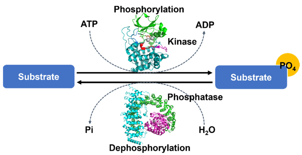
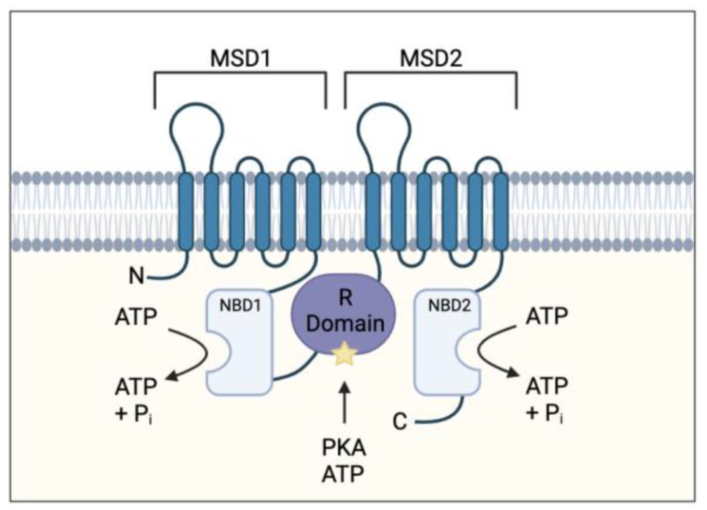
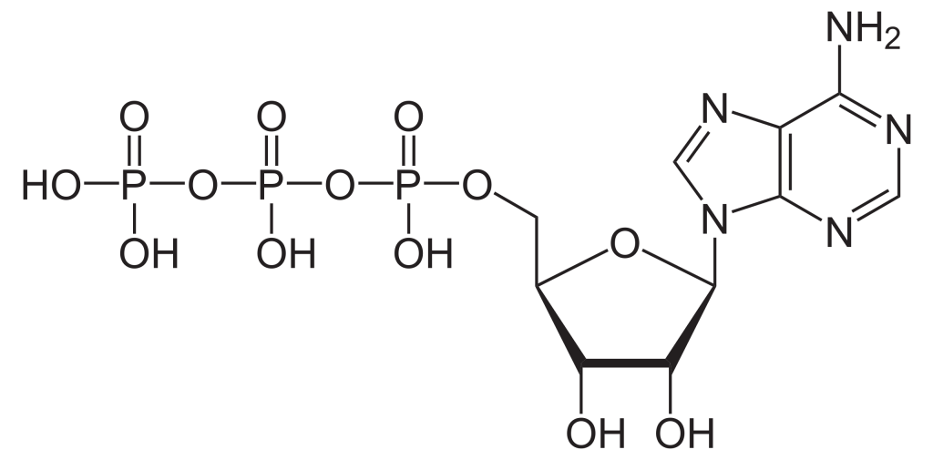
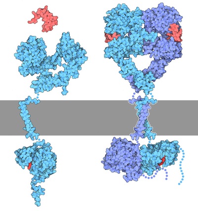
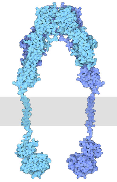
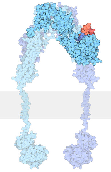
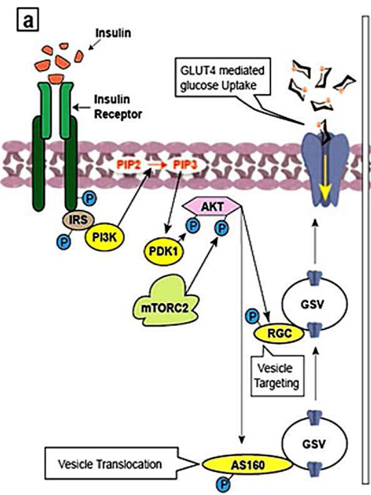
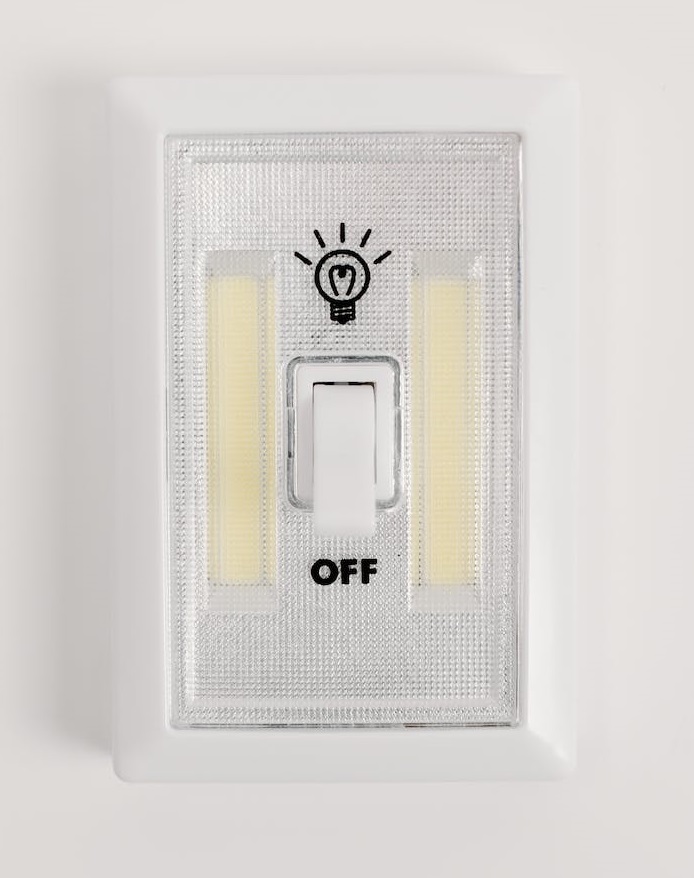
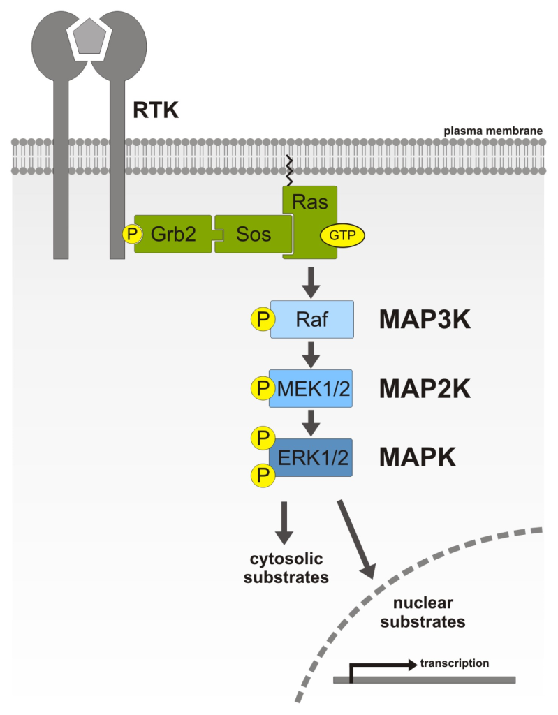
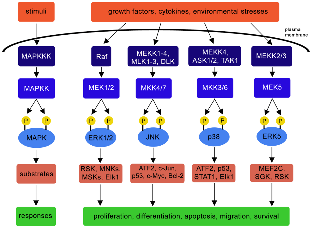
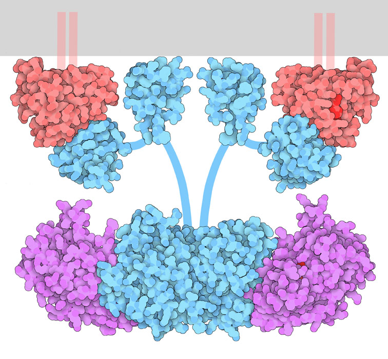
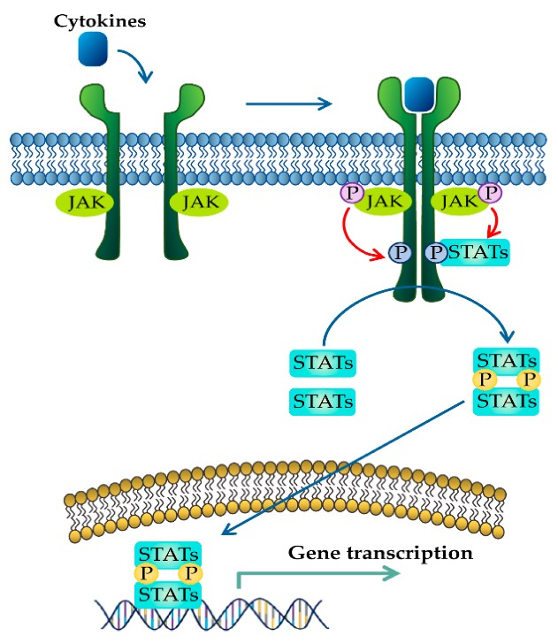
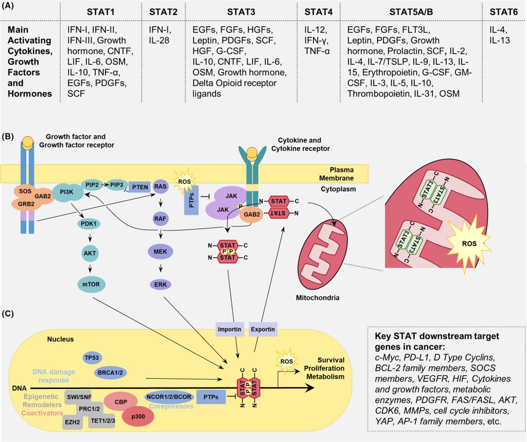
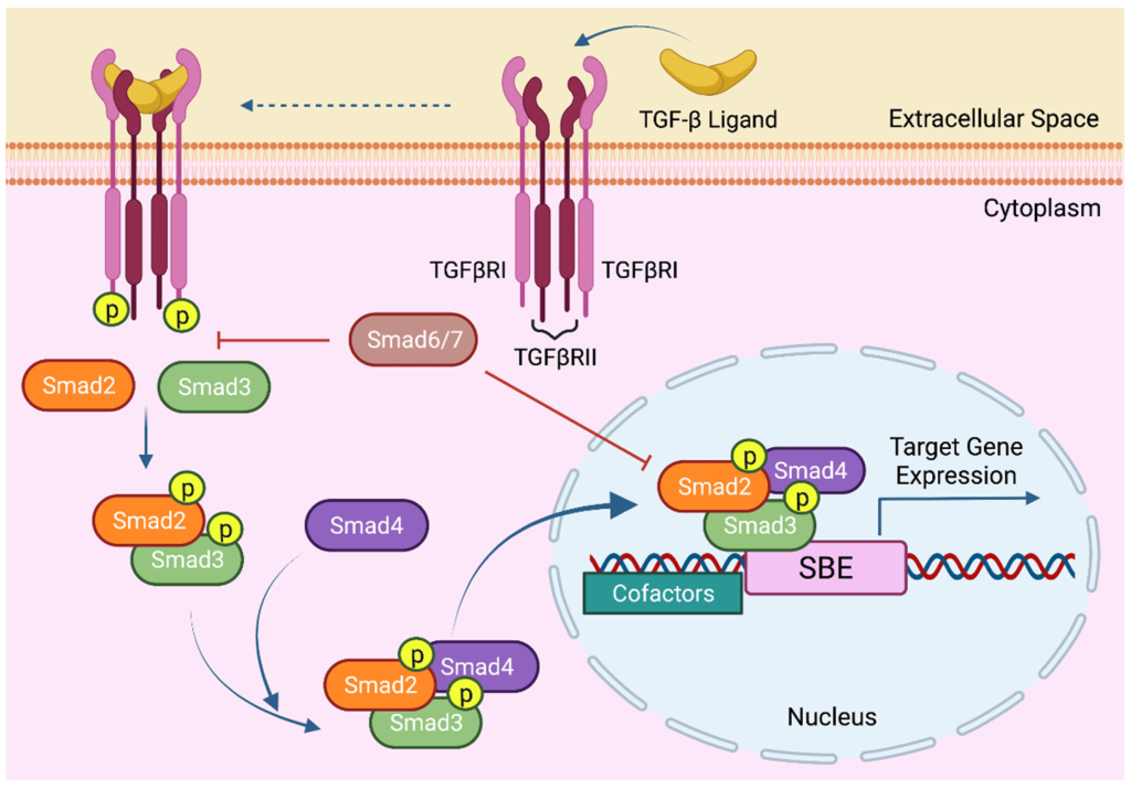
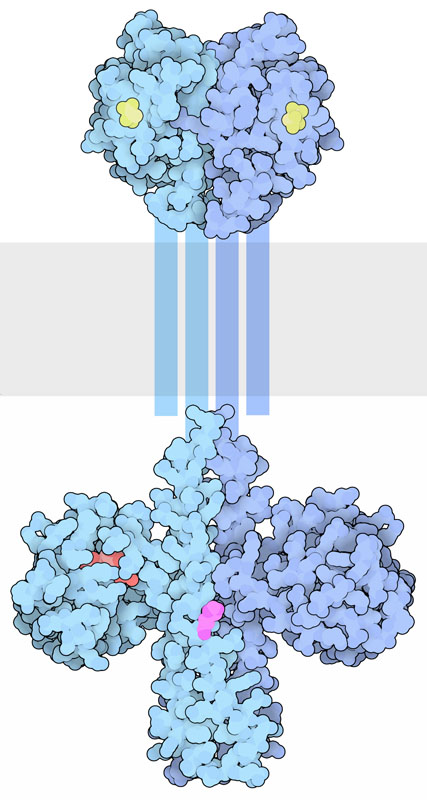
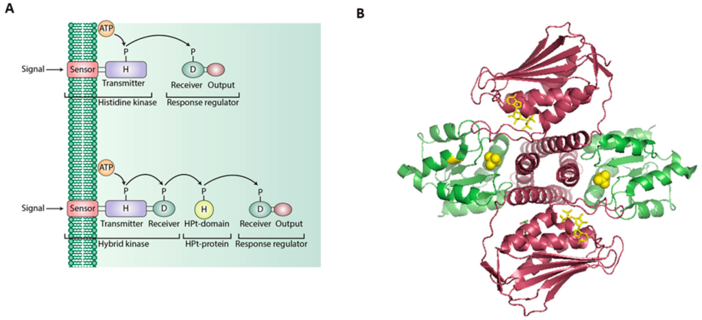
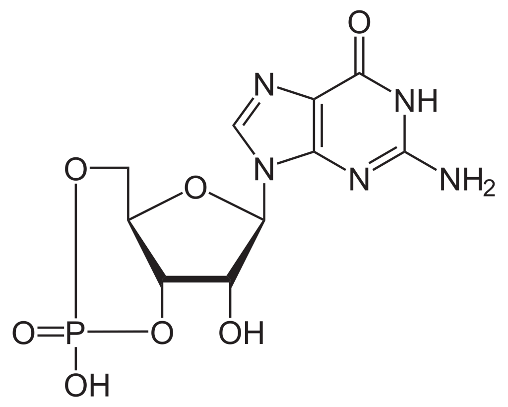
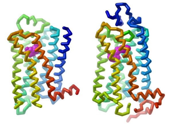

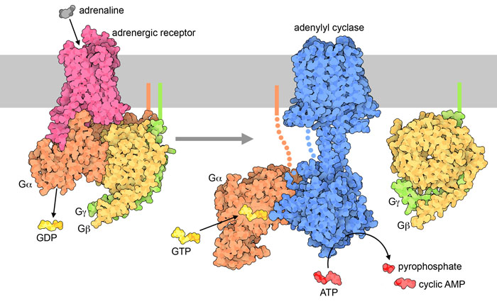
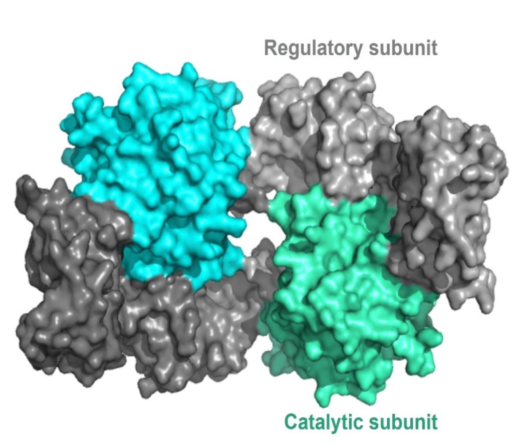
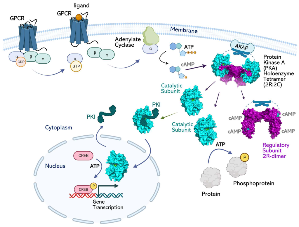
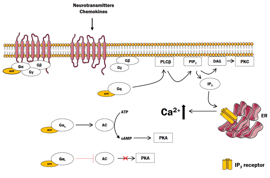
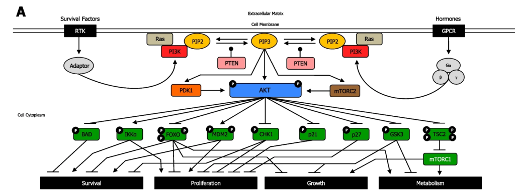

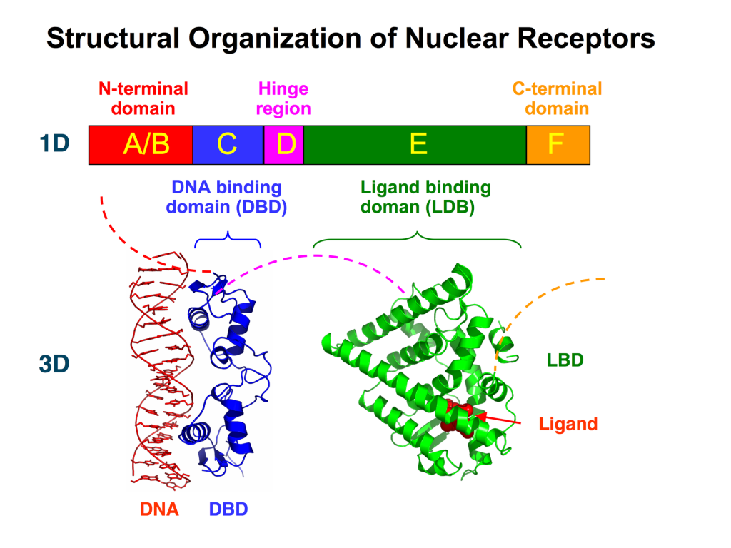
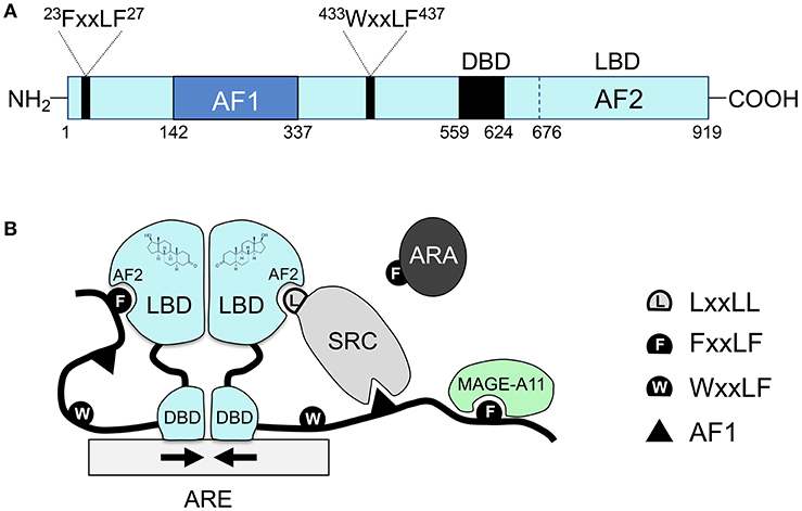
